Embed Size (px)
Citation preview

Journal of Non-Crystalline Solids 358 (2012) 921–924
Contents lists available at SciVerse ScienceDirect
Journal of Non-Crystalline Solids
j ourna l homepage: www.e lsev ie r .com/ locate / jnoncryso l
Lead sulfide quantum dots in glasses controlled by silver diffusion
Kai Xu, Jong Heo ⁎Division of Advanced Nuclear Engineering, Pohang University of Science and Technology (POSTECH), Pohang, Gyeongbuk 790-784, Republic of KoreaDepartment of Materials Science and Engineering, Pohang University of Science and Technology (POSTECH), Pohang, Gyeongbuk 790-784, Republic of Korea
⁎ Corresponding author. Tel.: +82 54 279 2147.E-mail address: [email protected] (J. Heo).
0022-3093/$ – see front matter © 2012 Elsevier B.V. Alldoi:10.1016/j.jnoncrysol.2012.01.007
a b s t r a c t
a r t i c l e i n f oArticle history:Received 4 November 2011Received in revised form 28 December 2011Available online 28 January 2012
Keywords:PbS;Quantum dots;Ag;Photoluminescence
Precipitation of PbS quantum dots (QDs) in silicate glasses controlled by Ag+ ion diffusion was investigated.Ag+ ions penetrated ~0.5 μm into the glass when the glass was immersed in the AgNO3 solution at 80 °C. Agnano-particles (NPs) and PbS QDs were formed after heat-treatment at temperatures of 420–460 °C for 10 h.PbS QDs in Ag+-diffused regions photoluminesced at longer wavelengths than did those in Ag+-free regions.This indicates that PbS QDs thus formed in regions containing Ag NPs were larger than those in Ag+-free re-gions and this size difference was confirmed from the transmission electron microscope images. PbS QDs cangrow at temperature as low as 420 °C in Ag+-diffused regions and this implies that PbS QDs form preferen-tially using Ag NPs as nucleating agents.
© 2012 Elsevier B.V. All rights reserved.
1. Introduction
Narrow-band lead sulfide (PbS, Eg=0.41 eV at 298 K) semicon-ductor with a large exciton Bohr radius (aB=18 nm) has a strongquantum confinement effect that is suitable for use in tunable infra-red active optoelectronic materials [1–3]. PbS quantum dots (QDs) in-corporated into glasses have possible applications as saturableabsorbers of ultrafast laser [4] and in amplifiers for fiber-optic tele-communication [5]. High-temperature fusion and subsequent ther-mal treatment of precursor glasses have been used to precipitateQDs, but this process normally results in an uncontrolled spatial dis-tribution of QDs in the glass matrix [6]. Ion-implantation [7] and fem-tosecond laser irradiation techniques [8] have been employed torealize space-selective formation of QDs for micro- and nano-photonic devices. For example, femtosecond laser irradiation can in-duce preferential precipitation of PbS QDs within the irradiated area[8]. However, this external radiation produces a large number of de-fects in QDs and causes the serious damages to the parent glasses.
Ag nano-particles (NPs) are well-known as nucleating agents forcontrolled crystallization of glasses [9]. A small amount of Ag2O inglasses can promote precipitation of PbS QDs in them. Heat-treatment induces precipitation of Ag NPs which facilitated formationof PbS QDs. As a result, intensities of the absorption and photolumi-nescence (PL) from QDs are significantly increased [10]. However,the solubility of Ag2O in precursor glasses was limited to less thanseveral tens of parts per million by weight that restricted the deep in-vestigation. One possible method of increasing Ag concentration inglasses is to use the ion-exchange technique to facilitate diffusion of
rights reserved.
Ag+ ions [11]. If feasible, this method would allow further investiga-tion of the potential of Ag NPs to control the size and spatial distribu-tions of QDs inside the glass matrix. In this work, Ag+ ions wereincorporated into sodium-alumino-silicate glasses by immersing theglasses in AgNO3 solution. After heat-treatment, considerable differ-ences were observed in the wavelengths (λPL) of PL from PbS QDsdue to the effect of Ag+ ion diffusion. PbS QDs can form the largersizes and grow faster in Ag+-diffused regions than those in Ag+-free regions.
2. Experimental procedures
Nominal composition (mol%) of the glass was 50SiO2–35Na2O–5Al2O3–8ZnO–2ZnS with an additional 1 mol% of PbO. Starting pow-der batches of ~25 g were mixed, then melted in an alumina crucibleat ~1350 °C for 45 min under ambient atmosphere. Melts werequenched by pouring onto a brass mold, then pressed with anotherbrass plate. The glasses were annealed at 350 °C for 3 h to releasethe thermal stress before cutting into pieces of ~1.0×1.0 cm with~1.5-mm thickness. Glasses were then optically polished and im-mersed for 10 h in a solution of AgNO3 in deionized water. Concentra-tion of Ag+ ions in the solution was 1 mol/L, and temperatures of themixture were held at 40, 60 or 80 °C in a water bath. Afterward, spec-imens were removed from the bath and further heat-treated at 420,440 or 460 °C for 10 h.
Depth to which Ag+ ions penetrated the glass wasmeasured usingenergy dispersive X-ray spectroscopy (EDX) after etching the glasssurface using a focused ion beam. Transmission electron microscope(TEM) under an accelerating voltage of 200 kV was used to measurethe size of PbS QDs. Specimens for the TEM measurement were pre-pared using the focused ion beam method. PL spectra were recordedat room temperature by exciting the specimen with the 800-nm
(a)
(b)
922 K. Xu, J. Heo / Journal of Non-Crystalline Solids 358 (2012) 921–924
radiation from a continuous-wave Ti-sapphire laser. Signals were col-lected and amplified using a combination of a mechanical chopper of50-Hz frequency, a 1/4-m monochromator, an InGaAs detector, and alock-in amplifier system. Optical absorption spectra were used toidentify the formation of Ag NPs. PL and absorption measurement un-certainties were known to be less than 1.5% including instrumentaland experimental errors. Uncertainties during the estimation of PbSQD sizes were approximately 15%.
3. Results
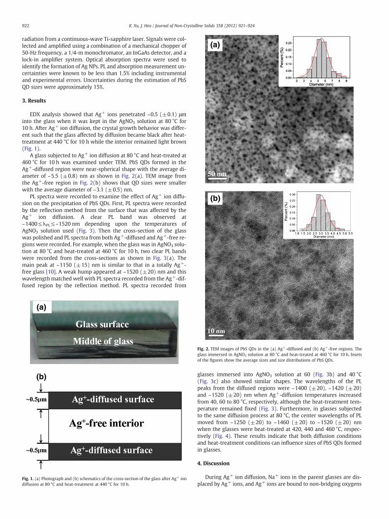
EDX analysis showed that Ag+ ions penetrated ~0.5 (±0.1) μminto the glass when it was kept in the AgNO3 solution at 80 °C for10 h. After Ag+ ion diffusion, the crystal growth behavior was differ-ent such that the glass affected by diffusion became black after heat-treatment at 440 °C for 10 h while the interior remained light brown(Fig. 1).
A glass subjected to Ag+ ion diffusion at 80 °C and heat-treated at460 °C for 10 h was examined under TEM. PbS QDs formed in theAg+-diffused region were near-spherical shape with the average di-ameter of ~5.5 (±0.8) nm as shown in Fig. 2(a). TEM image fromthe Ag+-free region in Fig. 2(b) shows that QD sizes were smallerwith the average diameter of ~3.1 (±0.5) nm.
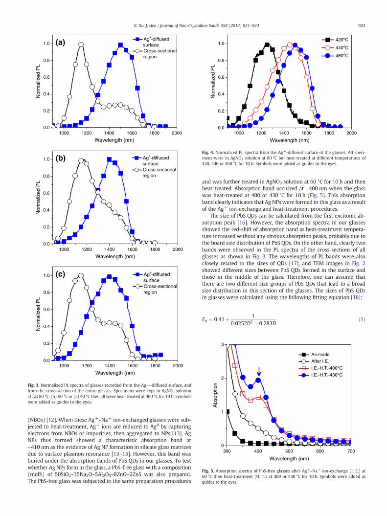
PL spectra were recorded to examine the effect of Ag+ ion diffu-sion on the precipitation of PbS QDs. First, PL spectra were recordedby the reflection method from the surface that was affected by theAg+ ion diffusion. A clear PL band was observed at~1400≤λPL≤~1520 nm depending upon the temperatures ofAgNO3 solution used (Fig. 3). Then the cross-section of the glasswas polished and PL spectra from both Ag+-diffused and Ag+-free re-gions were recorded. For example, when the glass was in AgNO3 solu-tion at 80 °C and heat-treated at 460 °C for 10 h, two clear PL bandswere recorded from the cross-sections as shown in Fig. 3(a). Themain peak at ~1150 (±15) nm is similar to that in a totally Ag+-free glass [10]. A weak hump appeared at ~1520 (±20) nm and thiswavelength matched well with PL spectra recorded from the Ag+-dif-fused region by the reflection method. PL spectra recorded from
(a)
(b)
Fig. 1. (a) Photograph and (b) schematics of the cross-section of the glass after Ag+ iondiffusion at 80 °C and heat-treatment at 440 °C for 10 h.
Fig. 2. TEM images of PbS QDs in the (a) Ag+-diffused and (b) Ag+-free regions. Theglass immersed in AgNO3 solution at 80 °C and heat-treated at 460 °C for 10 h. Insetsof the figures show the average sizes and size distributions of PbS QDs.
glasses immersed into AgNO3 solution at 60 (Fig. 3b) and 40 °C(Fig. 3c) also showed similar shapes. The wavelengths of the PLpeaks from the diffused regions were ~1400 (±20), ~1420 (±20)and ~1520 (±20) nm when Ag+-diffusion temperatures increasedfrom 40, 60 to 80 °C, respectively, although the heat-treatment tem-perature remained fixed (Fig. 3). Furthermore, in glasses subjectedto the same diffusion process at 80 °C, the center wavelengths of PLmoved from ~1250 (±20) to ~1460 (±20) to ~1520 (±20) nmwhen the glasses were heat-treated at 420, 440 and 460 °C, respec-tively (Fig. 4). These results indicate that both diffusion conditionsand heat-treatment conditions can influence sizes of PbS QDs formedin glasses.
4. Discussion
During Ag+ ion diffusion, Na+ ions in the parent glasses are dis-placed by Ag+ ions, and Ag+ ions are bound to non-bridging oxygens
1000 1200 1400 1600 1800 20000.0
0.2
0.4
0.6
0.8
1.0N
orm
aliz
ed P
L
Wavelength (nm)
Ag+-diffused surface
Cross-sectional region
(a)
1000 1200 1400 1600 1800 20000.0
0.2
0.4
0.6
0.8
1.0
Nor
mal
ized
PL
Wavelength (nm)
Ag+-diffused surface
Cross-sectional region
(b)
1000 1200 1400 1600 1800 20000.0
0.2
0.4
0.6
0.8
1.0 (c)
Nor
mal
ized
PL
Wavelength (nm)
Ag+-diffused surface
Cross-sectional region
Fig. 3. Normalized PL spectra of glasses recorded from the Ag+-diffused surface, andfrom the cross-section of the entire glasses. Specimens were kept in AgNO3 solutionat (a) 80 °C, (b) 60 °C or (c) 40 °C then all were heat-treated at 460 °C for 10 h. Symbolswere added as guides to the eyes.
1000 1200 1400 1600 1800 20000.0
0.2
0.4
0.6
0.8
1.0
Nor
mal
ized
PL
Wavelength (nm)
420oC
440oC
460oC
Fig. 4. Normalized PL spectra from the Ag+-diffused surface of the glasses. All speci-mens were in AgNO3 solution at 80 °C but heat-treated at different temperatures of420, 440 or 460 °C for 10 h. Symbols were added as guides to the eyes.
300 400 500 600 7000
1
2
3
Abs
orpt
ion
Wavelength (nm)
As-made
After I.E.
I.E.-H.T.-400oC
I.E.-H.T.-430oC
Fig. 5. Absorption spectra of PbS-free glasses after Ag+–Na+ ion-exchange (I. E.) at60 °C then heat-treatment (H. T.) at 400 or 430 °C for 10 h. Symbols were added asguides to the eyes.
923K. Xu, J. Heo / Journal of Non-Crystalline Solids 358 (2012) 921–924
(NBOs) [12]. When these Ag+–Na+ ion-exchanged glasses were sub-jected to heat-treatment, Ag+ ions are reduced to Ag0 by capturingelectrons from NBOs or impurities, then aggregated to NPs [13]. AgNPs thus formed showed a characteristic absorption band at~410 nm as the evidence of Ag NP formation in silicate glass matricesdue to surface plasmon resonance [13–15]. However, this band wasburied under the absorption bands of PbS QDs in our glasses. To testwhether Ag NPs form in the glass, a PbS-free glass with a composition(mol%) of 50SiO2–35Na2O–5Al2O3–8ZnO–2ZnS was also prepared.The PbS-free glass was subjected to the same preparation procedures
and was further treated in AgNO3 solution at 60 °C for 10 h and thenheat-treated. Absorption band occurred at ~400 nm when the glasswas heat-treated at 400 or 430 °C for 10 h (Fig. 5). This absorptionband clearly indicates that Ag NPs were formed in this glass as a resultof the Ag+ ion-exchange and heat-treatment procedures.
The size of PbS QDs can be calculated from the first excitonic ab-sorption peak [16]. However, the absorption spectra in our glassesshowed the red-shift of absorption band as heat-treatment tempera-ture increased without any obvious absorption peaks, probably due tothe board size distribution of PbS QDs. On the other hand, clearly twobands were observed in the PL spectra of the cross-sections of allglasses as shown in Fig. 3. The wavelengths of PL bands were alsoclosely related to the sizes of QDs [17], and TEM images in Fig. 2showed different sizes between PbS QDs formed in the surface andthose in the middle of the glass. Therefore, one can assume thatthere are two different size groups of PbS QDs that lead to a broadsize distribution in this section of the glasses. The sizes of PbS QDsin glasses were calculated using the following fitting equation [18]:
E0 ¼ 0:41þ 10:0252D2 þ 0:283D
ð1Þ

924 K. Xu, J. Heo / Journal of Non-Crystalline Solids 358 (2012) 921–924
where, D (nm) is the diameter of PbS QDs. E0 is the effective band gapenergy calculated from the first excitonic absorption peak. First,values of E0 were estimated for two special cases following the previ-ous work [10]. Stokes shift of PL band with the wavelength of~1520 nm in Fig. 3(a) was assumed to be ~50 nm while that of the~1150 nm PL band is ~130 nm. Then, diameters of PbS QDs responsi-ble for those two PL bands were ~5.5 (±0.8) nm and ~3.4 (±0.5) nm,respectively. These results were applicable only to this specific glassthat was Ag+ ion-exchanged at 80 °C and heat-treated at 460 °C for10 h, and were comparable to those estimated from the TEM images.
In addition, near-IR emission from PbS QDs was observed evenafter heat-treatment at temperature as low as 420 °C (Fig. 4). The for-mation of PbS QDs was not confirmed in the glasses without addedAg+ ions [10]. These results suggest that Ag NPs formed by the Ag+
ion-exchange can facilitate formation and growth of PbS QDs. Further-more, the wavelength of PL from the Ag+-diffused regions red-shiftedas the temperatures of AgNO3 solution and subsequent heat-treatmentincreased (Figs. 3 and 4). This trend provides further evidence on theproposition that the size of PbS QDs can be tuned by adjusting Ag+
ion-exchange conditions. Detailed relationships between PbS QDs andAg NPs are being investigated.
5. Conclusions
Ag+ ions were diffused into silicate glasses and the effect of Ag+
ions on the formation of PbS QDs was investigated. Penetrationdepth of Ag+ ions into the glass was ~0.5 μm. After heat-treatment,PbS QDs formed. TEM images and simple calculations showed thelarger size of PbS QDs in Ag+ ion-exchanged regions than those inAg+-free regions. λPL of Ag+-diffused regions was longer than thatfrom the Ag+-free regions. Because λPL increases with QD size, thisdifference also indicates that Ag+ ion diffusion led to formation oflarger PbS QDs. Optical absorption spectra from the PbS-free glass
confirmed that Ag NPs precipitated during heat-treatment. PbS QDscan grow and emit in the near-IR even after heat-treatment at tem-perature as low as 420 °C.
Acknowledgments
This work was supported by Basic Science Research (2010-0022407), Priority Research Centers (2011-0031405) and WorldClass University (WCU) (R31-30005) programs through the NationalResearch Foundation of Korea (NRF) funded by the Ministry of Educa-tion, Science and Technology.
References
[1] F.W. Wise, Acc. Chem. Res. 33 (2000) 773.[2] T. Okuno, A.A. Lipovskii, T. Ogawa, I. Amagai, Y. Masumoto, J. Lumin. 87–89
(2000) 491.[3] E.H. Sargent, Adv. Mater. 17 (2005) 515.[4] A.M. Malyarevich, K.V. Yumashev, A.A. Lipovskii, J. Appl. Phys. 103 (2008) 81301.[5] J. Heo, C. Liu, J. Mater. Sci. Mater. Electron. 18 (2007) S135.[6] N.F. Borrelli, D.W. Smith, J. Non-Cryst. Solids 180 (1994) 25.[7] R.E. de Lamaestre, J. Majimel, F. Jomard, H. Bernas, J. Phys. Chem. B 109 (2005)
19148.[8] C. Liu, Y.K. Kwon, J. Heo, B.H. Kim, I. Sohn, J. Am. Ceram. Soc. 93 (2010) 1221.[9] S.D. Stookey, Ind. Eng. Chem. 51 (1959) 805.
[10] K. Xu, C. Liu, S. Dai, X. Shen, X. Wang, J. Heo, J. Non-Cryst. Solids 357 (2011) 2428.[11] R.H. Doremus, J. Phys. Chem. 68 (1964) 2212.[12] P.W. Wang, Appl. Surf. Sci. 120 (1997) 291.[13] J. Zhang, W. Dong, J. Sheng, J. Zheng, J. Li, L. Qiao, L. Jiang, J. Cryst. Growth 310
(2008) 234.[14] M. Vollmer, U. Kreibig, Optical Properties of Metal Clusters, Springer, Berlin, 1995.[15] D. Manikandan, S. Mohan, K.G.M. Nair, Mater. Res. Bull. 38 (2003) 1545.[16] I. Kang, F.W. Wise, J. Opt. Soc. Am. B 14 (1997) 1632.[17] U. Woggon, Optical Properties of Semiconductor Quantum Dots, Springer, Berlin,
1997.[18] I. Moreals, K. Lambert, D. Smeets, D.D. Muynck, T. Nollet, J.C. Martins, F. Vanhaecke,
A. Vantomme, C. Delerue, G. Allan, Z. Hens, ACS Nano 3 (2009) 3023.


![Microsensor Measurements ofSulfate Reduction and Sulfide ...Jorgensen1992b.pdf · constants, respectively, of the sulfide equilibrium system, [S2-] is the sulfide concentration, and](https://img.pdfslide.us/doc/110x75/5e9a6d84dc840a57bc1baa83/microsensor-measurements-ofsulfate-reduction-and-sulfide-amp-constants.jpg)