Embed Size (px)
Citation preview
•
'l'C!!J:>R TISSUE llBGlSTRIC
LOS ANGELES OOUNTY llOSPITAL
~
PR01'000L
Fol'
MONTHLY SLIDES
August, 1960
Genito•urinary System
CASE NO, 1
ACCESSION NO, 10828
IW1E I AGE:
L, T, 65 SEX: MALE FACE: WHITE
CONTRIBUTOR: John P, Blanchard, M,D, Bakersfield, California.
TISSUE FROM: Mass, left kidney
CLINICAL ABSTFACT:
August, 1960
OUTSIDE NO, J60•37
History: The pa~ient noted onset of weight loss and anorexia 5 months prlor to admission on January 4, 1960, He had been previously in excellent health. There was an increase in size of the abdomen and a sense of fullness in the left upper quadrant, but no other symptoms.
Physical examination showed a large firm, fairly well circum• scribed mass in the left upper quadrant, He appeared acutely and chronically ill,
Laboratory examination: The hemoglobin was 9.1 grams; rbc 3,5 million, wbc 5,300; differential normal, The urine was negative,
SUroERY:
January 6, 1960, an apparently encapsulated retroperitoneal mass was attached to the left kidney, pushing adjacent organs medially and superiorly, It was removed from the spleen and stomach with some difficulty, The left kidney and adrenal ~~ere removed with the tumor, but the adrenal was not involved in the mass, There was no evidence of metastases,
Gross PATHOLOGY:
The tumor mass weighed 3550 grams and was .25 em, in its great• est diameter, It '~as coarsely bosselated extem!lllY and seemed to be delimited b7 a fairly smooth and definite capsule, The external surface was tan to grayish and on one surface there was some attached peritoneum. A subsidiary nodule 4,0 em, in diameter was loosely attached. The broad surface on one side showed a kidney embedded in the tumor mass, This kidney was of average size, having a diameter of 12 em, from pole to pole, The surface was pink to red and the capsule stripped with ease, leaving a smooth surface, It was attached to the mass at the lower pole, and the capsule of the kidney was continuous ~11th the capsule of the neo• plasm, The neoplasm could be stripped away from the kidney itself by blunt dissection and was not an integral part of the kidney, The lddney showed normal architecture; the pelvis and calyces were normal, On section, the neoplasm had a variegated su~ace, Centrally it was fairly dense, yellowish to gray and appeared hyalinized, almost cartilaginous, This had an irregular distribution. Peripherally it was softer, but still quite firm and rubbery and the cut surface had a vague, whorled arrangement, It was gray to white and in places slightly pinkish. The
- 2 - August, 1960
Accession No, 10828 Case No. 1
pink areas were softer and more fleshy. This was particularly true of a nodule which occurred just below the renal pelvis. The aaall attached nodule was white, moderately rubbery and well circumscribed. The cut surface bulged above the edge.
POST-oPERATIVE OOURSE AND FOLLOW•UP:
The patient gradually regained his strength and weight, In July, 1960 be entered the Hospital for " recurrence in the left abdomen, " He had felt poorly for one month, and has lost weight. Surgery on July 20, 1960 disclosed an egg shaped tumor in the left retroperitoneal area at the site of the previous neoplasm, It was discrete, not definitely encapsul~ted and was easily removed in its entirety. Post-operative course has been uneventful.
1'he cut eu·rface of the 15 x 10 x 12 em. mass was rubbery to soft with areas of liquefaction centrally, moist, semitransluscent, and fairly homogeneous,

CASE NO. 2
ACCESSlON NO. 10917
NAME: O. P. AGE: 54 SEX: Male RACE: Negro
CONTIUBU'l'OR: Suleiman K. Abul•Haj, M,D, Washington, D. c.
TISSUE FROM: Cystic mass • left testis
CLINICAL ABSTRACT:
August, 1960
OUTSIDE NO, S6059•59
History: The history given by the patient was unreliable .• He entered the hospital on December 12, 1959 complaining of a 11ainless large scrotal mass, which he noted two weeks before.
Physical examination revealed an obese middle-aged colored male in no apparent! distress with girdle and buffalo type obesity and bilateral moderate gynecomastia and sparsity of body hair. There was a large cystic scrotal mass located on the left side, It was estimated as of "grapefruit" size. It trans:-_'tlluminated, was non-tender, and obscured the left testis. Because of the history given, it was felt by the attending urologist that the mass represented a large hydrocele. The remainder of the Fhysical examination was essentially non-revealing.
Laboratory findings; Studies were within normal limits. An admission chest film was interpreted as within normal limits,
SURGERY:
On December 16, 1959 a left orcliiectomy was performed. At operation the large cystic mass was removed, but no testis could be identified, although the left vas defetens ended abruptly into the mass. (The surgeon was unaware that he was removing the testis).
GROSS PATHOLOGY:
The specimen consisted of a large ovoid multicystic mass measuring 12 x 20 em. in the greatest dimension. It had been previously bisected and the contents removed. The extemal surface was smooth and in s.ome areas composed of multiple compartments separated by and walled off with a grey-white soft tough tissue of varying thickness, The walls of the compartments contained many irregular areas of light yellow-tan, finely granular, soft tissue which in turn contained occasional small cysts. These areas varied from 1•3 em. in greatest dimension. The internal surface of the compartments was coarsely granular, light tan, and in others it was smooth and glistening. In a few localities testi• cular tissue could be recognized about the walls of the cystic compartments.
- 2 - August, 1960
Accession No. 10917 Case No. 2
POLLOW•UP:
The patient was seen in May, 1960 for his six months' post• operative checkup, and was free of disease and ~doing very well physi• cally. " A psychiatric evaluation reported him to be "rather feebleminded," but there was no implication of this developing after orchiectomy.
Q\SB t«>, 3
ACCESSION NO. 10571
NAME: AGE:
M. 1\. 65 SEX: Female RACE:
CONTRIBUTOR: B, F. Ducey, M, D. Ventura, Calif.
Clluc.
TISSUE F!Dl: Tumor of left kidney
CLINICAL ABSTRACT:
AUS'JSt, 1960
OUTSIDE NO, 59•913
Historyj The patient sought medical attention in April, 1959 because of "vague digestive difficulty," A routine medical survey dis• closed an enl arged left kidney, and an intravenous pyelogram confirmed the clinical impression of a mass involving the lower pole of that organ. There were no urinary symptoms, nor any history of previous disease invol ving the genito-urinary tract, The patient was taking "vitamins, iron and Bl2 for an anemia which bad been present for some time,"
Physical gvaminati~ revealed a palpable mass in the left ~pper quadrant, while cystoscopic examination revealed a markedly deformed left kidney which produced some urine, although the output of dye was retarded.
Laboratory examination: She had normal blood levels of urea and creatinine, and essentially negative urine, The hemoglobin was 10,3 grams, with 33~ packed cell vol ume,
SURGERY:
Nephrectomy was performed on May 5, 1959 with removal of the perirenal fat and a portion of attached ureter, A portion of the left adrenal gland was adherent t~ the upper renal pole, and was removed with the kidney.
GROSS PATHOLOGY:
The specimen we.ighed 580 grams and was a pear•shaped mass 19 em, in ita greatest l'4trgi18 and 15 em. in its great.est width by 12 em. in its greatest thickness. The smaller pole represented the major portion of a kidney 9 x 5 em., while the wide portion of the mass consisted of a spheroid tumor intimately attached to the lower renal pole, the latter being flattened and some1~hat hollowed out by the tumor mass. The upper renal pole was i ntact, and had a fragment of golden yellow, soft, ragged suprarenal adrenal, Hemi•section through the long axis of the specimen revealed the large lower pole to represent a spheroid tumor of symmetri• cal shape, compris ed of a central cavity, 7 em, in diameter, lined by
- 2 - August, 1960
Accession No. 10571 Case No. 3
fairly smooth, glistening membrane surrounded by a 3·4 em, thickness of highly cellular, light tan tissue showing a definite t rabecular pattern, the arrangement of the trabeculae being perpendicular to the external tumor surface, There was a di stinct external capsule which appeared to be complete, separating the tumor tissue from the adjacent kidney, although the two types of tissue shared a eoamon capsule over an extent of 4 em, The central tumor cyst was filled with ember, gelatinous fluid, The kidney proper appeared essentially nor.nal, ex• cept for distortion of the inferior calyces, and there was no sign of extension of neoplasm thro~gh the common capsule into the renal substance,
COURSE:
Post-operative convalescence was uneventful and the patient was discharged one week after the nephrectomy,
This patient had a "checkup" examination by her urologist on June 3, 1960, at which time sh~ had no complaints, and there were no findings suggestive of disease of the urinary tract. A chest X-ray film was. reported as being normal.
CASE NO, 4 August, 1960
ACCESSION NO. 10843 OutSIDE NO, A-59·127
NAME: K, H, AGE: 74 SEX: Male .RACE: White
CONTBIBUTORS: E, L, Benjamin and D, R. Dickson, M.D. Santa Barbara, California
TISSUE PliJM: Bladder (autopsy)
CLlNlCAL ABSTRACT:
History: Hematuria was first observed in December, 1957 and from the onset the urine was never free of blood.. Cystoscopy was per• formed several times, and bladder polyps were fulgurated. These were submitted in fragments measuring 2•3 an. in diameter, High voltage irradiation therapy was recommended in March, 1959,
Course: He was admitted on March 27, 1959 for cobalt irradia• Uon therapy. Cystoscopic examination showed coarse trabeculations and a large bleeding tumor involving the fundus and lateral walls. Bilateral hydroureteronephrosis was present, The second admission was on August 25, 1959 with fever and hematuria which responded to further irradiation and antibiotics, He was admitted the third time on September 5, 1959 with pyelonephritis which responded to chlOro!I!YCetin. The final admis• sion on November 27, 1959 was for terminal care. The patient expired on December 2, 1959.
GROSS PATHOLOGY:
Externally the urinary bladder was a .hard, ovoid structure measuring 15 by 8,5 by 7 em, and extending to the symphysi& pubis, Its serosa was smooth and opaque, On sectioning, the entire bladder wall, with the exception of the anterior portion, was replaced by lobulated to fasciculated friable, white neoplastic tissue measuring up to 3,5 em. in thiclmess, 'l'he lumen of the bladder was an irregularly compressed spa~e containing a few ml, of clotted blood and lined with shaggy, lobulated, and necrotic white neoplastic tissue. Metastatic neoplastic nodules up to 10 11111. in diameter occurred in both lungs. Metastases were also present !n the hypogastric, right COIIIDOn iliac and right parabroncbial lymph nodes.
CASE 00. 5 August, 1960
ACCESSION NO, 10461 OU'ISIDE !«), S - 1464-69
NAME: J. R. AGE: 40 SEX: Male RACE: Cauc.
CONTRIBUTOR: Suleiman K. Abul•Haj, M.D. Washington, D. c.
TISSUE FR:lM: Mass, left tesUs
CLINICAL ABSTRACT:
History: The patient became aware of a progressively enlarging scrotal ruass approximately 8 months prior to seeking medical attention. The mass was non-tender and painless.
Physical examination revealed a l arge orenge•sized, non-tender , rubbery, fil~, rounded intrascrotal mass, the relntion of which to the testes could not be ascertained. The reaminder of the physical examination was esscnti.ally within notmal l imits ,
Chest film and bone survey were unremarltable.
SURG!U~:
On March 13, 1959 the scrotal mass as well as the left testis and extemal vas defe reua were removed. At operation the mass was found to be discrete, enes:>&cit\f:ed, shelled with ease, and was eampletely independent of the adj acent left testis, epididymis, and cord structures. I t was outside the cord sheath and was loosely attached to the processus vaginalis.
GROSS PATHOLOGY:
The specimen consisted of a large, rounded, encapsulated, rubbery, soft mass measuring 8 x 6 x 5 em, in diameter and a small atrophic testis with a segment of vas deferens , The .testis was other• wise unremarkable. The mass possessed a glistening, smooth, slightly knobby surface. Cut surface was gelatinous , gray-white, slippery and displayed a semitranslucent ground glass appearance mottled with irregular gray-white fish•flesh areas and focal zones of recent hemorrhage,
FO~I-UP:
The p~tient was seen in the Tumor Foll ow-up Clinic of Brooke A~ Hospital (T~:as) in March, 1960, approximately one year after re• moval of the scrotal mass , He was doing well and was free of disease.

CASE NO. 6
ACCESSION NO, 7100
NAME: R. B. AGE: 66 SEX: Male RACB: Caue.
OONTRIBU'l'OR: W. K. Bullock, M.D. Los Angeles, Calif.
TISSUE Pial: Bladdet:
CLINICAL ABSTRACT;
August, 1960
OUTSIIS NO, 54./t551
History: The patient was admitted to LACGH in Aprtl, :t954 with the history of ft:equeney and buming on urlnation for four weeks. Gross hematuria, intermittent and uniformly mixed with udne, developed three weeks before entry and penh ted until two days prtor to entry, He bad lost six pounds of weight in on.e IIGllth,
Physical examination was normal. KUB and I-V urogram showed good function bilateJ:ally. Contraat cystogram revealed a filling de• feet in the left half of the bladder and the air contrast view showed a 2 x 4 em, soft tissue density projecting into the bladder on the left &ide,
alysis :
SURGBI\Y:
done,
Labontolil examinationi The hemoglobin was 11,0 gm, Udn• 20•30 WBC hdf pt:esent in microscopic examination,
On Apdl 20, 1954 a segmental resection of the bladder was
GR:>SS PATBJLOGY:
The specimen consisted of a segment of bladder 4,5 x 3.5 x 1 em. Attached to the central portion of the mucosal surface was a papillary growth which measured 2,5 em, in diameter, Its site of attachment to the mucosa measured 1,8 em, in diameter, The surface of the papillary growth was ~rtsed of numerous verrucous-like projections which gave the surface a slightly roughened appearance.
Repeated cystoscopic examinations wet:e done semiannually until May, 1956 and no evidence of tumor reeut:rence was noted. Follow-up letter from the Central Tumor Registry in June, 1958 reported that the pat!ient was having no difficulty with the bladdet:, The patient died at Good Samarttan Hospital on February 4, 1960, Autopsy showed no residual tumot: of the bladder nor any abdominal, pelvic or pertaortie lymphade• nopathy. Diagnoses included: bronchogenic carcinoma, undifferentiated left upper lobe bronchus, metastatic to hiler and mediastinal lymph nodes
- 2 - August, 1960
Accee~ion No. 7100 Case No. 6
and light adrenal; bronchopneumOnia; generali~ed arteriosclerosis; pulmonary emphysema, moderate; cerebral edema; chronic passive congestive liver with fatty metamorphosis; ·and focal myocardial fibrosis,

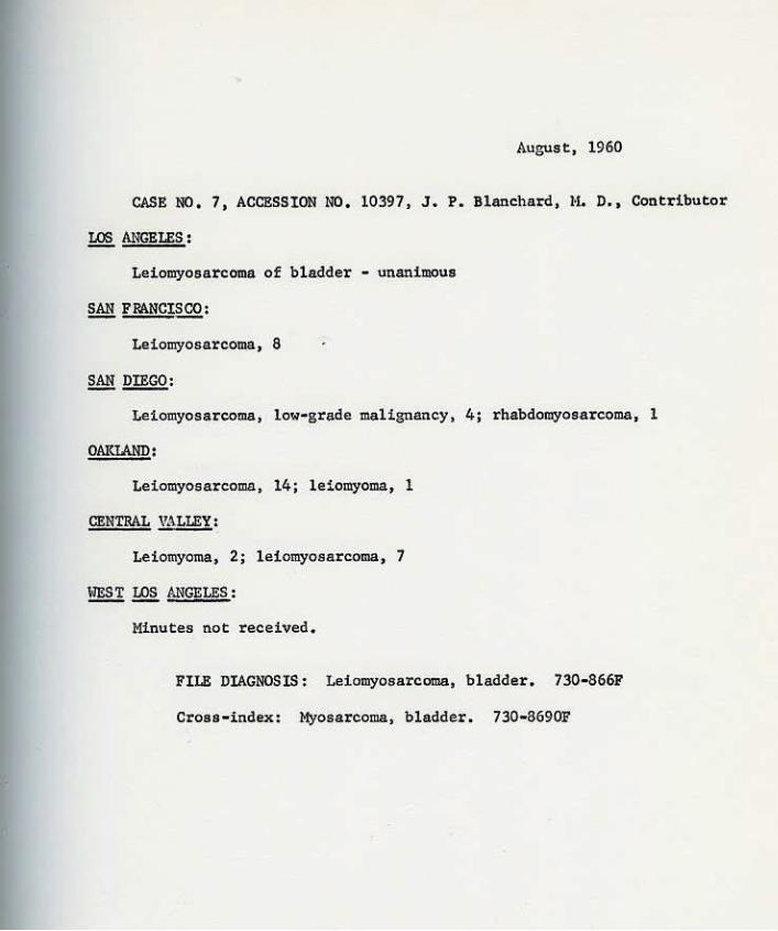
CASE NO. 7
ACCESSION NO. 10397
NAME: P. S . AGE: 24 SEX: Male RACE: White
CONtRIBUTOR: J. P . Blanchard, M.D. Bakersfield, Calif.
TISSUE PR:lM: Mass - right supra-pubic area
CLINICAL ABSTRACT:
August, 1960
OUTSIDE NO. J59•1011
History: The pati"ent bad pain in the lower abdomen and right groin IJince December 20, 1959 and weight loss of 20 pounds (he had been taking dexedrine and not eating well) • He had hematuria for 10 days prior to admission.
fjlvsical examination: An ill•defiuecl mass was palpable in the right supra·P'.Jbic area. At cystoscopy (March 3, 1959) a bleeding funoo gating mass, 2•3 em. in diameter was seen on the righ.t dome of the bladder.
Laboratory exsmination: Mild anemia of secondary type; hematuria.
SURJERlt:
Laparotomy was performed on March 9, 1959. An oval mass was located in the soft tissue superiorly and to th'! ri~t of the bladder. One end was embedded in the bladder wall and projected into the cavity as noted on cystoscopy. The mass was well circumscribed otherwise and was resected in toto with a portion of the dome of the bladder. Periwaortic lymph nodes were enlarged and film, extending to the diaphr&glll, One was resected for microscopic study.
Gro!lS PATHOlOGY:
The 2 x 5 x 4 em. tumor was circumscribed but not encapsulated. The cut section wall homogeneous, tan-gray, rather soft with zones c-f. soft, gray, almost watery tissue. The portion which projected into the bladder vall was hemorrhagic.
1'0LUJW•UP:
As of July, 1960 the patient is in good health. In January, 1960, cystoscopy was nesative and the chest x-ray was reported normal.
CASE NO. 8
ACCESSION NO. 10317
NAME: D, S, AGE: 3 yrs.. SEX: Female BACE: Cauc.
CONTRIBUTOR: Irving Madoff, M.D. Los Angeles, cali.f.
TISSUE FKIM: Right kidney
CLINICAL ABSTRACT;
August, 1960
OUTSIDE NO. HS-103•59
liistory: The patient was brought to the attending physician beeause of a mass in the right axilla of a few months duration,
Physical Examination: During routine physical examination, a non-tender, large, firm mass was palpated in the right mid-abdomen which was approximately 10 CID, in diameter. An intravenous pyelogram revealed a non-functioning right kidney; apparently the left side func• tioned normally. The child was g.iven radiaton therapy, ten treatments, the last one given five days before surgery, with a reduction in the size of the abdominal mass by approximately SO%,
Laboratory Findings: The hemoglobin was 14.9 grams, The urine had a specific gravity of 1.025, hazy, yellow, acid; it was nega• tive for glucose and albumin, and strongly positive for acetone, Microscopically the urine had 4 to 8 WBC per hpf; fine granular casts, 5 to 10 per hpf; few mucus threads·, and epithelial cells.
SURGERY:
The right kidney was removed with about 8 to 10 CID, of attached ureter,
GlOSS PATHOLOGY;
The kidney was approximately 9 x 6 x 4,5 em, The capsule was considerably thickened, particularly over the upper half and it was moderately adherent. The upper portion of the kidney was moderately bulbous and firm, On section a fairly discrete tumor mau was found occupying the entire upper half of the kidney, The mass had a pale grayish-yellow appearance with irregular 4reas of yellow mottling, fo• cal necrosis and a few small areas of hemon:bage around the periphery, There appeared to be a well defined eapsule around the mass separating it from the adjacent kidney parenchyma, A few of the calyces next to the tumor wet'e moderately dilated and some of the papillae were congested, The remaining kidney parenchyma was not remarkable, The pelvis and ureters showed no obvious gross change,
COURSE:
The post-operative course was uneventful,
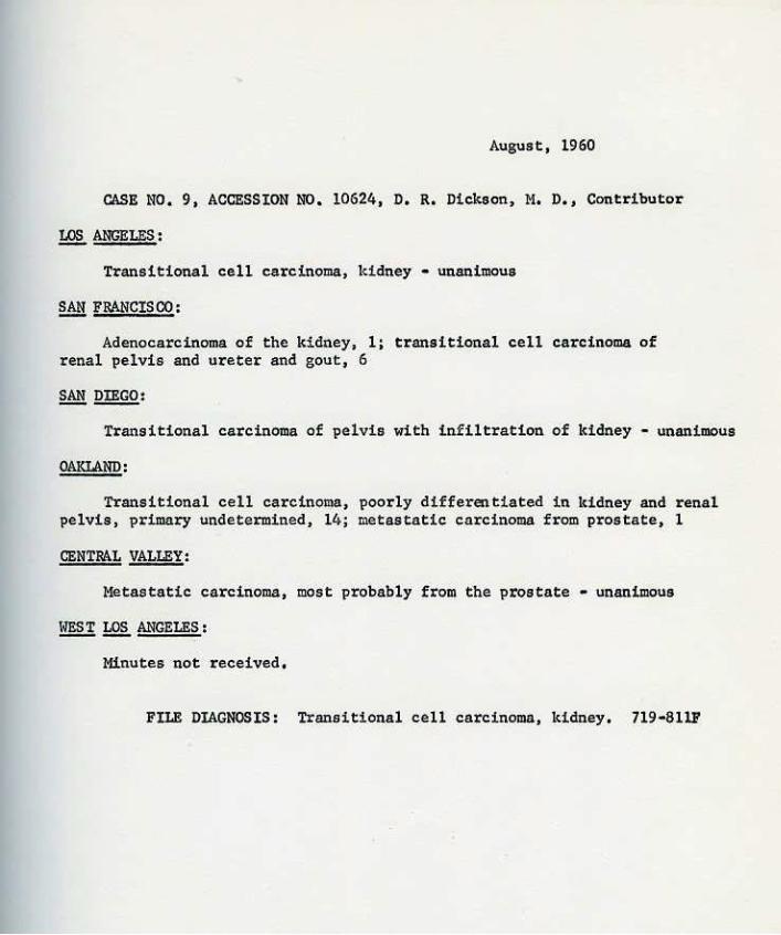
CASE 00, 9
ACCESSION NO, 10624
NAlofi>: F. D. AGE: 76 SEX: Male RACE: W.
CONTRIBUTOR: D, R, Dickson, M,D, santa Barbara, Calif,
TISSUE FroM: Autopsy • right kidney
CLINICAL ABSTRACT:
August, 1960
OUTSIDE NO, A55·65
This 76 year old rancher had an episode of massive melena during which the l:!gb, dropped to 5.3 grams !:.i 1952, The source of the bleeding was not demonstrated, He was then well until May, 1955 when he noted onset of constant dull, aching, mid-epigastric pain, which gradually increased in intensity and was associated with no other symptom except weight loss, He was admit.ted on July 11, 1955,
Physical examination was negative aside from a moderately enlarged prostate gland, IVP showed a non-functioning right kidney.
He deteriorated gradually, developing phlebothrombosis of the leg veins and congestive failure, and died on July 20, 1955,
AUTOPSY FINDING:
At autopsy the right kidney bad a normal contour with mild intemal hydronephrosis of the calyces only, The capsule was thickened, fibrous, and fixed to the cortex, The renal pelvis was contracted, and the pelvis and entire right uteter down to within S =· of the ureteral orifice had diffuse mural thickening and fixation to the adventitial tissues. The mucosa of the pelvis and ureter was smooth to faintly trabeculated, pale tan•gray. The superior 10 em. of the ureter was stenosed but the lumen was patent throughout. The periureteral tissues had a dif-fuse semi•rigid sheet•like fibrous consistency without local nodularity, Metastases were present in the mesenteric and retroperitoneal lymph nodes, liver, and lumbar vertebra, Incidental findings were a 6 em, diameter ulcerated, low grade leiomyosarcoma of the ileum and a latent adenocarcinoma of the prostate gland,

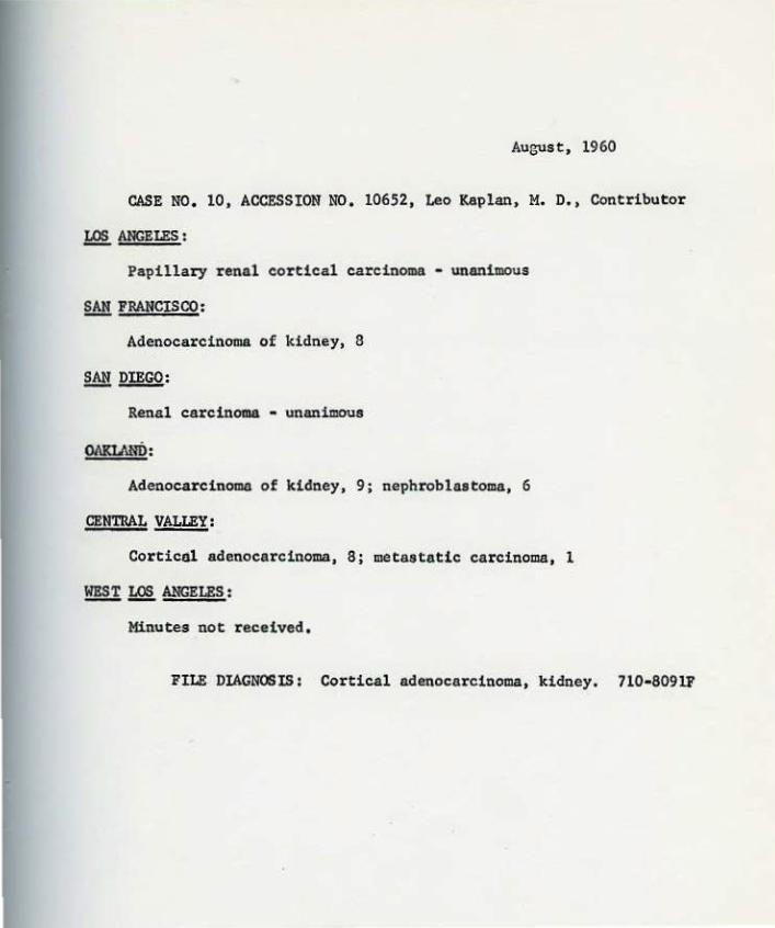
CASE NO, 10
ACCESSION NO, 10652
NAME: AGE:
R. J. 45 SEX: Male RACE: White
CONTRIBUTOR: Leo Kaplan, M.D. Los Angeles, calif.
nssUE FR:lM: Kidney mass
CLINICAL ABSTRACT:
August, 1960
OUTSIDE NO, ~ 2935•59B
History: The patieGt was admitted to Mt, Sinai Hospital on September 20, 1959 because of a previous clinical workup indicating the presence of an enlarg~d left kidney mass, identified by retrograde and intravenous pyelography· as a probable primary renal neoplasm. In addition there had been enlargement of the left axillary lymph nodes, There had been a history also of the removal of a number of tumor masses from the left lung and hilar area by pneumonectomy at another hospital.
Laboratot'Y;ilf!!fpatim:a: The admission blood count showed 11,8 grams of hemoglobi~tn 37% hematocrit and 12,350 WBC's with a slight shift to the left, The platelets were normal, The urinalysis showed no protein or glucose and only occasional :RBC' s, WBC' s and hyaline casts,
SURGERY:
The renal mass, left adrenal gland and spleen were removed on September 21, 1959.
GROSS PATHOWGY:
The specimens received included an adrenal mass measuring 8.5 ~ 5 x 4,8 em. with foci of still intact ribbon-like yellow cortex stretched over a large gray•white glistening inte.rnal neoplasm, It exhibited foci of red-pink cystic and noncystic zones of hemorrhagic necrosis. The kidney mass after extrusion of an abundance of bloody granular material, still weighed 2110 grams, and meaaured 20,5 x 19 x 14,5 em. It was covered by a bluish membranous capsule and on section consisted largely of brown-red necrotic cystic debris, To one side, it was attached to a kidney that measured 15 x 7 x 8 em, and was fused indistinguishably with its mid•third posteriorly. There was no extension of the tumor into hilar veins, The excised spleen weighing 128 grams showed only fibrinoid artedolar degenerative change and follicular fibrosis.
OOUliSB:
'Zt;e patient 1 s post-operative course was uneventful beyond the appearance of a 2 x 3 em, mass in the right mid biceps area, The patient was treated with nitrogen mustard and was discharged on 10·3·59,
- 2 - August, 1960
Accession No. 10652 Case No, 10
FOLLOW•UP:
The patient was readmitted January 23, 1960 and expired the next day, Autopsy disclosed metastatic tumor in lymph nodes (retroperitoneal, para•aortic, mediastinal, paratracheal, mesenteric, porta hepatis, tight ileo•femoral, axillary, cervical and inguinal), left pleura, liver, right adrenal gland, right parietal lobe of the brain, heart, pericardium, omentum, peritoneum, IDI.Iscle, and skin. Other findings included follicular adenomas of the thyroid, pulmonary atelectasis, pleeral effusion, pertcaxdial effusion, acute ulcerative esophagitis, chronic interstitial viral pneumonitis, DPH, focal pancreatic fibrosis, and cavernous hemangioma of the liver.
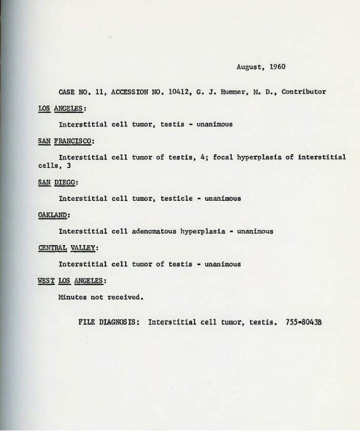
CASB IC, 11
ACCESSION NO, 10412
NAME: R. K . AGE : 45 SEX: Male BACB : \Jhit e
CONTRIBUTOR: Geo~e J, Hummer, M, D, Santa Monica, Califomia
TISSUE FlllM: TUmor, right testicle
CLINICAL ABSTRACT:
August , 1960
Oll'l'SIJE NO, 8•4938•58
History: About three months prior to admission, while taking a shower , the patient noticed a ha rd nodule in the right lower pole of the t es t icle. In the two weeks pr ior to admission, it increased in size and had taken on a "mushroom" shape. There was no pain except for a mild degree of tendemess which he bad not been ll".Jare of before.
Physical examination: The scrotal contents revealed both testes to be small. The left testis was normal in consist ency and non• tender, The right testis was not enla~ed but at the lower pole there was a firm, smooth and moderately tender nodule about 2 em, , in diameter. It seemed to be in the epididymis and yet was attached to and could not be divorced from the testicle , I t was the impression, however , that the nodul e represented part of the testicle , Further examination of the geni to•urinary tract was negative.
Laboratory findings: Non-diagnosti c, including a urine for quantitative Catechol Amine which was 3 ug/100 react ion.
Sti}IGE Rr:.
On October 23, 1958 , an orchidectomy was performed under general anesthesia, Upon l i f ting the testicle !rom the scrotum, the tunica was opened and showed a t umor mass occupying the lower one• fourth of the testicle proper, separate from the epididymis, The tumor was confined within the tunica and showed little, if any, infl ammatory reaction,
GR?SS PATIIOLOGY:
The specimen consisted of the right testicl e, incl uding the epididymis, with attached 10 em. of spermatic cord. 'l'he lower pole of the testis proper was the site of a 1 em,, in diamet er, small, demar· cated, uniform tan, neoplastic nodule,
COURSE:
The patient made an uneventful post-operative recovery and was dis~ed on October 25, 1958,
- 2 - August, 1960
Accession No. 10412 Case No. 11
FOLIIJW•UP:
'l'be patient 18 doing very well (July, 1960). Be is seen by his physician about once a 'IDOiltb. 'l'bere has been uo evidenc.e of recur• renee.
CASE NO. 12 August, 1960
ACCESSION NO. 10959 Otl'l'SlDE NO. 1161•60
NAME: P. G, AGE: 70 SEX: Female RACE: C4uc.
OONTBIBUTOR: Paul R. 'thompson, M,D, Pasadena, C&lifomia
TISSUE FIDM: Right kidney
CLINICAL ABSTRACT;
History; 'the patient bad pain in the right flank which lasted th~e to four days at a time, and tiredness and weakness for undetermined length of time. She noted frequency every two hours, nocturia three to four times, but no hematuria, hesitancy, or dysuria,
Phvsi,c,al findings: Negative except for very bard mass pal• pated in the right addabdomen,
Cystoscopy revealed the bladc!e!:' and ureteral orifices to be normal. 'there was moderate urethritis, f/5 ureteral catheter was passed to the right kidney and pyelograma ~vealed right calculus, lVP con• firmed the calculus, G,l, series revealed normal colon, right renal calculus.
Strn;ERY:
tight nephro-ureterectomy was performed on April 28, 1960,
GIDSS PA'l'II)IQGY;
The specimen was a total right kidney that weighed 254.2 grama and had the overall dimensions of 15.5 em, in length by 7,8 em. on the medio•lateral axis by 6.2 em. in the anterior posterior axis. There was a considerable amount of perirenal fat. 'the ureter had been a:mputated above the ureteropelvic junction in an area of neoplasm which completely distorted its orifice, 'the superior pole and hilus were deformed by a bulging neoplastic yellowish•wbite mass that occupied the whole euperior pole of the kidney practically, and on bisection this was seen to be comprised of a neoplasm taking origin from the renal pelv1s that pre• seated a variegated glistening gray•wbite to yellowish gritty surface, The superior calyx and the superior pole of the kidney had been converted into a hydronephrotic ue at1d the renal cortex in this area measured no more than 0,3 em. 'l'he renal pelvic lining in the uninvolved zone showed a roughened grayish-white granularity covered with a dirty yellowish• brown membrane, In the inferior calyces there was extensive lichenifi• cation, with opacification aud granularity of the mucosa, but no gross neoplasm. Again there was a marked degree of hydronephrosis, but the cortical medullary portion of the kidney measured up to 1.9 em, in tbidaleaa- 'l'he neoplasm largely appeared to occlude and occupy the
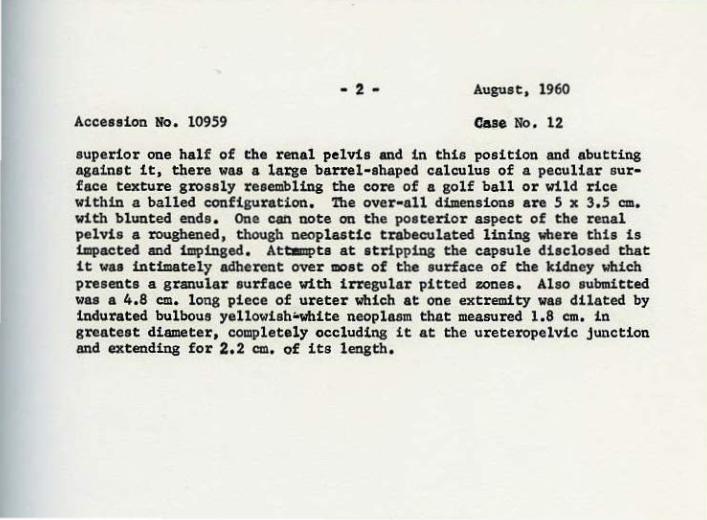
- 2- August, 1960
Accession No. 10959 CUe No. 12
superior one half of the renal pelvis and in this position and abutting against it, there was a large barrel-shaped calculus of a peculiar sur• face texture grossly resembling the core of a golf ball or wild rice within a balled configuration. The over•all dimensi ons a r e 5 x 3 . 5 em. with bl uuted ends. One cat1 note on the posterior aspect of the renal pel vis a roughened, though neopl astic trabeculated lining wbera this is impacted and impinged. Attllllpts at stripping the capsule disclosed that it was intimately adherent over most of the surface of the kidney which presents a gr anular surface with irregular pitted zones. Also submitted was a 4.8 em. l ong piece of ureter which at one extremity was dilated by indurated bulbous yellowieh~ite neoplasm that measured 1.8 em. in greatest diameter, completely occluding it at t he ureteropelvic junction and extending for 2.2 em. of its l ength.
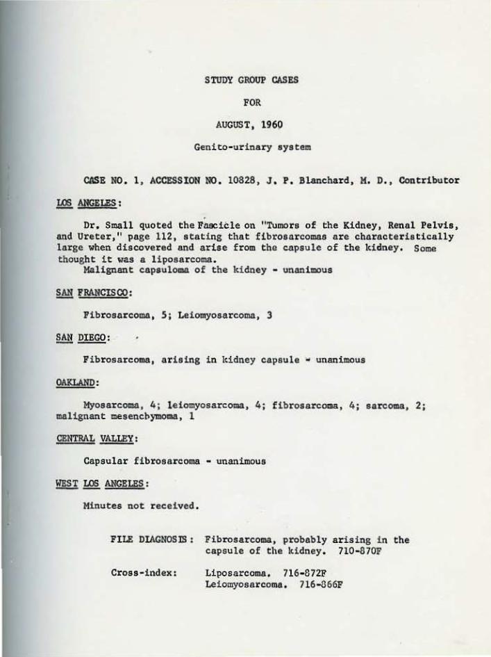
STUDY GROUP CASES
FOR
AUGUST, 1960
Genito-urinary system
CASE NO. 1, ACCESSI ON NO. 10828, J . P. Blanchard, M. D., Contributor
~ANGELES:
Dr. Small quoted the Fli9Cicle on "Tumor s of the Kidney, Renal Pelvis, and Ureter," page 112, stating that fibrosarcomas are characteristically large when discovered and arise from the capsule of the kidney. Some thought it was a liposarcoma.
Malignant capsuloma of the kidney - unanimous
~FRANCISCO:
Fibrosarcoma, 5; Leiomyosarcoma, 3
~DIEGO:
Fibrosarcoma, arising in kidney capsule • unanimous
OAKLAND:
Myosarcoma, 4; l.eiomyosarcoma, 4; fibrosarcoma, 4; sarcoma, 2; malignant mesencbymoma, 1
CENTRAL VALLEY:
Capsular fibrosarcoma • unanimous
WEST LOS ANGELES:
Minutes not received.
FILE DIAGNOSIS : Fibrosarcoma, probably arl.Smg in the capsule of the kidney. 7l0-870F
Cross-index: Liposarcoma. 716· 872F Leiomyosarcoma. 716-666F
August, 1960
CASE NO, 2, ACCESSION NO. 10917, S, K, Abul-Haj, M. D., Contributor
.!&§. ANGELES :
Androblastoma - unanimous.
It was pointed out that the microscopic features of this tumor paralleled Fig, 86 in the Fascicle of "Tumors of the Male Sex Organs," This tumor is consid~red analogous to ovarian arrhenoblastomas,
~ 'FRANCISOO:
Gyn-androblastoma (androblastoma), 8
§M! DIEGO:
Mixed androblastoma, 4; teratocarcinoma, with a predominance of arrhenoblastoma, 1
OAKlAND:
Androblastoma • unanimous
CENTRAL VALLEY:
Interstitial cell tumor, 1; angioendothelioma, 3; androblastoma, 5
!:!!§.! ~ ANGELES :
Minutes not received,
FILE DIAGNOSIS: Androblastoma, 755-8041A
August, 1960
CASE NO. 3, ACCESSION NO. 10571, E. F. Ducey, M. D. , Contributor
~ANGELES:
Renal cortical adenoma, 1; renal cortical carcinoma, 5
It was stated that Bell ' s book originally arbitrar ily set the criterion of size as the differentiating feature between renal cortical adenoma and carcinoma. In th~ beginning a 'diameter of over 2 em. was sufficient for classification in the carcinoma group, but more recently diameters of 3 and 4 em. t~ere still acceptable in the diagnosis of adenomas. Dr. Budd stated that these may be considered always as carcinoma, but often these tumors take a long time to develop histological features of malignancy,
Adenocarcinoma, kidney • 8
§.@. DIEGO:
Renal carcinoma • unanimous
OAKLAND:
Adenocarcinoma of kidney • unanimous
CENTRAL vALLEY:
Large cortical adenoma, 5; adenocarcinoma, 4
Those who used the latter term relied on the size, not the histology.
~ 1Q2. ANGELES:
Minutes not received.
FILE DIAGNOSIS : Ret111l cortical adenocarcinoma. 710· 8091F
Cross- index: Renal cortical adenoma. 710· 8091A
August, 1960
CASE NO, 4, ACCESSION NO, 10843, E. L, Benjamin, !1. D., and D. R. Dickson, t1. D. , Contributors
!:Q! ANGELES :
Spindle cell carcinoma of the bladder • unanimous
SAN FRANCISOO:
Mixed mesodermal tumor, 2; anaplastic carcinoma, 6.
~DIEGO:
Anaplastic carcinoma, bladder - unanimous
OAKLAND:
Poorly differentiated neoplasm (irradiated), probably bladder, 13; rhabdomyosarcoma, 2
CENTRAL VALLEY:
Adenocarcinoma, 2; transitional cell carcinoma, 2; myosarcoma, 4
~ 1Q! ANGELES:
Minutes not received,
FILE DIAGNOsiS: Spindle cell carcinoma, bladder, 730· 819,1F
Cross•index: Sarcoma, bladder. 730· 879F
August, 1960
CASE NO. 5, ACCESSION NO. 10461, S. K. Abul-Haj, M. D. , Contributor
LOS ANGELES :
Dr. Edmondson submitted the diagnosis of liposarcoma of spermatic cord . Dr. Budd thought this a tumor of mesenchyme and stated that these cells could be still benign; he suggested the term embryonal mesenchymoma, which received the unanimous vote of those present.
The contributor stated: "Special stains for acid mucopolysaccharide& on several representative sections from the mass revealed by far the bulk of the tumor to consist of a ground substance matrix, composed almost ex• exluaively of hyaluronic acid , Within this matrix, the cella are dominant• ly myxoma or myxosarcoma cella with only occasional lipoblast, Fat stains show these lipoblasts. The aff ini ty of mucopolysaccharides for fat (i. e . deposition of fat occurs wherever there is deposition of ground substance acid mucopolysaccarides) cause me to raise the question whether we should call such a tumor a liposarcoma or myxoid liposarcoma, just on the basis of the presence of fat in a tumor which is composed dominantly of myxoid connective tissue that is neoplastic. Deposition of fat in ground substances occur in human and experimental arteriosclerosis of the monkey, in Xanthomas, pretibial myxedema circumscripta tumor (exclusively hyalu· ronic acid) in the sex skin of the monkey (an almost exact duplication of myxedema circumscripta in man) in generalized myxedema in malignant exoph• thalmos (in the retroorbital tissue and ocular muscle where abundant muco• polysaccharides are deposited and are responsible for the exophthalmos) and in myxomas, chondromas, and chondrosarcoma and myxomatous areas of neurilemmomas. This is only citing a few examples, Virchow, Schultz, Antschow and Bjohrling' recognized such affinity many years ago.
''Also, it has been my experience in mY1tedema circumscripta and sex akin of the monkey that injection of the enzyme hyaluronidase rapid dis · solution of the mucoid tumors and mobilizes the fat from the site of de· position, as the tumor disappears .
"The behavior of such tumor clinically, like others which have been previously ~esignated myxoid liposarcoma, is similar to that of myxosarcoma rather than liposarcoma (inability to metastasize and repeated local recur· rence following incomplete excision). I have seen several such tumors arising about the G. U. tract, one within the labia minora, another at the bladder neck, and one of the spermatic cord."
~ FRANCISCO :
Adenomatoid tumor, 1; liposarcoma, 4; lipomyxoma, 3
Myxoid liposarcoma, lo1~·grade malignancy, 3; myxolipoma, 1, fibromyxoma, 1

Accession No. 10461 Case No. 5
OAKLAND:
- 2 - August, 1960
Liposar coma, 6; myxoma, 5; myosar coma, 2; mesenchymoma, 2
CENTRAL VAU.EY:
Adenomatoid tumor, S; fibromyxoma , 2; mesenchymoma, 1; embryonal carcinoma, 1.
~~ANGELES :
Minutes not received.
FILE DIAGNOSIS : Lit>Osarcoma, spermatic cord. 762-872F
Cross- index: Embryonal mesenchymoma, spermatic cord. 762-887B ~~oma. spermatic cord. 762· 871B Adenomatoid tumor. 762· 883SB

August, 1960
CASE NO. 6, ACCESSION NO. 7100, W. 1<. Bullock, M. D., Contributor
.!&§. ANGELES :
Pleomorphic carcinoma of bladder - unanimous
~ FRANCISCO:
Anaplastic carcinoma of bladder, 7; rhabdomyosarcoma, l
~DIEGO:
Anaplastic carcinoma of bladder, 3; myosarcoma of bladder, 2
OAKlAND:
Transitional cell carcinoma, bladder, poorly differentiated - unanimous
CENTRAL VALLEY:
l1yosarcoma, 5; anaplastic carcinoma, 4
~~ANGELES:
Minutes not received.
FILE DIAGNOSIS: Pleomorphic carcin.oma, bladder. 730-8191G
Cross-index: Myosarcoma, bladder. 730-8690F
August, 1960
CASE NO, 7, ACCESSION NO, 10397, J. P. Blanchard, H. D., Cont:ributor
LOS ANGELES :
Leiomyosarcoma of bladder - unanimous
~FRANC!~:
Leiomyosarcoma, 8
~DIEGO:
Leiomyosarcoma, low-grade malignancy, 4; rhabdomyosarcoma, l
OAKIAND:
Leiomyosarcoma, 14; leiomyoma, 1
CENTRAL VALLEY:
Leiomyoma, 2; leiomyosarcoma, 7
~~ANGELES:
Minutes not received.
FILE DIAGNOSIS: Leiomyosarcoma, bladder, 730-866F
Cross-index: Myosarcoma, bladder. 730-8690F

August, 1960
CASE NO. 8, ACCBSSION NO. 10317, Irving Madoff, M. D. , Contributor
~ANGELES:
Wilm' s tumor , i r radiated • unanimous
~FRANCISCO:
Wilm' a tumor , irradiated, 8
~DIEGO:
Embryoma, kidney • unanimous
OAKLAND:
Wilm's tumor (irradiated) - unanimous
CENTRAL VALLEY:
Hemangioma, 6; Wi.lm1a tumor, 3
~~ANGELES:
Minutes not received .
FILE DIAGNOSIS: ~lilm 1 s tumor, lddney, irr a diated . 710· 8834F
August, 1960
CASE NO, 9, ACCESSION NO, 10624, D. R. Dickson, M. D., Contributor
12§.. ANGELES :
Transitional cell carcinoma, kidney - unanimous
~ FRANCISCO :
Adenocarcinoma of the kidney, 1; transitional cell carcinoma of renal pelvis and. ureter and gout, 6
Transitiona.l carcinoma of pelvis with infiltration of kidney - unanimous
OAKLAND:
Transitional cell carcinoma, poorly differentiated in kidney and renal pelvis, primary undetermined, 14; metastatic carcinoma from prostate, 1
CENTRAL VALLEY:
Metastatic carcinoma, most probably from the prostate • unanimous
~~ANGELES:
Minutes not received,
FILE DIAGNOSIS: Transitional cell carcinoma, kidney, 719•811F
August, 1960
CASE NO, 10, ACCESSION NO, 10652, Leo Kaplan, M. D. , Contributor
1Q§. ANGELES :
Papillary renal cortical carcinoma - unanimous
§!!FRANCISCO:
Adenocarcinoma of kidney, 8
§M! DIEGO:
Renal carcinoma - unanimous
Adenocar cinoma of kidney, 9; nephroblastoma, 6
CENTRAL VALLEY:
Cor ticdl adenocarcinoma, 8; metastatic carcinoma, 1
~ 1Q§. ANGELES:
Minutes not received,
FILE DIAGNOSIS: Cortical adenocarcinoma, kidney. 710-8091F
August, 1960
CASE NO. 11, ACCESSION NO, 10412, G. J. Hummer, M. D., Contributor
~ANGELES:
Interstitial cell tumor, testis - unanimous
Interstitial cell tumor of testis, 4; focal hyperplasia of interstitial cells, 3
§.@:DIEGO:
Interstitial cell tumor, testicle • unanimous
OAKLAND:
Interstitial cell adenomatous hyperplasia • unanimous
CENTRAL VALLEY:
Interstitial cell tumor of test.is • unanimous
~~ANGELES:
t1inutes not received,
FILE DIAGNOOIS: Interstitial cell tumor, testis. 755•804JB
August, 1960
CASE NO. 12, ACCESSION NO, 10959, P. R. Thompson, M. D., Contributor
12§. ANGELES :
Epidermoid carcinoma of renal pelvis • unaniQous
It was stated that this entity is usually associated with stones .
SAN FRANCISCO:
Squamous carcinoma of the renal pelvis • unanimous
SAN DIEGO:
Squamous cell carc.inoma, kidney pel vis - unanimous
OAKLAND:
Cornified squamous cell carcinoma, renal pelvis - unanimous
CENTRAL VALlEY:
Epidermoid carcinoma - unanimous
BE! 12§. ANGELES :
Minutes not received,
FI LE DIAGNOSIS: Epidermoid carcinoma of renal pelvis , 719· 814F