Embed Size (px)
Citation preview
LCA Haemato-Oncology Clinical Guidelines Lymphoid Malignancies Part 5: Less Common Lymphoid Malignancies
August 2015
LCA HAEMATO-ONCOLOGY CLINICAL GUIDELINES
2
Contents
1. Introduction ............................................................................................................................................... 5
1.1. Types of B-cell lymphomas .............................................................................................................. 5
1.2. Types of T-cell lymphomas/leukaemias .......................................................................................... 5
2. Overview and Generic Guidance ................................................................................................................ 6
3. Service Configuration across the LCA......................................................................................................... 7
3.1. Children, teenagers and young adults ............................................................................................. 7
4. Patient Information and Support ............................................................................................................... 8
5. Management of Common Disease and Treatment-related Complications ............................................... 9
5.1. Superior vena cava obstruction ....................................................................................................... 9
5.2. Cord compression ............................................................................................................................ 9
5.3. CNS involvement ............................................................................................................................. 9
5.4. Febrile neutropenia ......................................................................................................................... 9
5.5. Nausea and vomiting ..................................................................................................................... 10
5.6. Tumour lysis syndrome (TLS) and hyperuricaemia ....................................................................... 10
6. Supportive Care ........................................................................................................................................ 11
6.1. Transfusions .................................................................................................................................. 11
6.2. Haemostasis and thrombosis ........................................................................................................ 11
6.3. Infection prophylaxis ..................................................................................................................... 11
6.4. Breathlessness ............................................................................................................................... 12
6.5. Weight loss .................................................................................................................................... 12
6.6. Pain ................................................................................................................................................ 12
6.7. Complex symptom management .................................................................................................. 12
7. End of Treatment Information ................................................................................................................. 13
7.1. Treatment summary and care plan ............................................................................................... 13
8. Follow-up Arrangements ......................................................................................................................... 14
9. Survivorship and Rehabilitation ............................................................................................................... 15
10. Research and Clinical Trials .................................................................................................................... 15
11. End-of-life Care ...................................................................................................................................... 16
12. Marginal Zone Lymphoma ..................................................................................................................... 17
12.1. Introduction ................................................................................................................................... 17
12.2. Investigation and diagnosis ........................................................................................................... 17
12.3. Treatment ...................................................................................................................................... 18
CONTENTS
3
12.4. Supportive care ............................................................................................................................. 20
13. Hairy Cell Leukaemia .............................................................................................................................. 21
13.1. Introduction ................................................................................................................................... 21
13.2. Investigation and diagnosis ........................................................................................................... 21
13.3. Service configuration ..................................................................................................................... 22
13.4. Management of disease-related complications ............................................................................ 22
13.5. Treatment ...................................................................................................................................... 22
13.6. Supportive care ............................................................................................................................. 23
14. Mantle Cell Lymphoma .......................................................................................................................... 24
14.1. Introduction ................................................................................................................................... 24
14.2. Investigation and diagnosis ........................................................................................................... 24
14.3. Treatment ...................................................................................................................................... 25
14.4. Supportive care ............................................................................................................................. 27
15. Burkitt Lymphoma ................................................................................................................................. 28
15.1. Introduction ................................................................................................................................... 28
15.2. Investigation and diagnosis ........................................................................................................... 28
15.3. Management of specific disease-related complications ............................................................... 30
15.4. Treatment ...................................................................................................................................... 30
15.5. End of treatment information ....................................................................................................... 31
15.6. Specific or miscellaneous considerations ...................................................................................... 31
16. Peripheral T-Cell Lymphomas and Leukaemias ..................................................................................... 32
16.1. Introduction ................................................................................................................................... 32
16.2. Clinical presentation and referral pathways ................................................................................. 32
16.3. Investigation and diagnosis ........................................................................................................... 32
16.4. Service configuration ..................................................................................................................... 33
16.5. Treatment ...................................................................................................................................... 34
17. T-cell Prolymphocytic Leukaemia (T-PLL) .............................................................................................. 38
17.1. Introduction ................................................................................................................................... 38
17.2. Presentation .................................................................................................................................. 38
17.3. Investigation and diagnosis ........................................................................................................... 38
17.4. Service configuration ..................................................................................................................... 38
17.5. Treatment ...................................................................................................................................... 39
17.6. Supportive care ............................................................................................................................. 39
17.7. Research and clinical trials ............................................................................................................ 39
LCA HAEMATO-ONCOLOGY CLINICAL GUIDELINES
4
18. T-cell Large Granular Lymphocyte Leukaemia and Chronic NK LPD ...................................................... 40
18.1. Introduction ................................................................................................................................... 40
18.2. Presentation .................................................................................................................................. 40
18.3. Investigations and diagnosis .......................................................................................................... 40
18.4. Service configuration ..................................................................................................................... 41
18.5. Treatment ...................................................................................................................................... 41
18.6. Management of specific disease-related complications ............................................................... 42
18.7. Follow-up arrangements ............................................................................................................... 42
Annex 1: Multidisciplinary Teams (MDTs) and Constituent Hospital Trusts ................................................... 43
Annex 2: SIHMDS or Current Diagnostic Services and Contacts ..................................................................... 44
Annex 3: Guideline for the Management of Tumour Lysis Syndrome (TLS) ................................................... 45
Annex 4: Guidelines for Use of Rasburicase in Adult Haematology and Oncology Patients ........................... 47
Appendices ...................................................................................................................................................... 49
INTRODUCTION
5
1. Introduction
This part of the LCA Haemato-Oncology Clinical Guidelines focuses on the less common tumours that fall
under the umbrella of non-Hodgkin lymphoma (NHL).
There are many different types of NHL. Lymphomas are often described as B-cell lymphomas or T-cell
lymphomas, according to whether they began in B-cell lymphocytes or T-cell lymphocytes.
1.1. Type of B-cell lymphomas
B-cell lymphomas are more common than T-cell lymphomas. About 9 out of 10 people diagnosed with NHL
have a B-cell lymphoma.
The most common types of B-cell lymphoma are:
diffuse large B-cell lymphoma (DLBCL)
follicular lymphoma (FL).
(Separate LCA guidelines exist for these most common B-cell lymphomas on the LCA website.)
Other less common types include:
marginal zone lymphomas
hairy cell leukaemia
mantle cell lymphoma
Burkitt lymphoma
lymphoplasmacytic lymphoma (also called Waldenström’s macroglobulinaemia (WM)). Guidelines
for WM are included in the LCA plasma cell disorders clinical guideline.
1.2. Types of T-cell lymphomas/leukaemias
1.2.1. Peripheral T-cell lymphomas
Peripheral T-cell lymphomas (PTCL) are a heterogeneous group of rare malignancies accounting for
approximately 10–12% of all lymphoid neoplasms. Most have an aggressive clinical behaviour and, apart
from ALK-positive anaplastic large cell lymphoma (ALCL), a poor response to conventional chemotherapy
with only 30% survival at 3 years. Patients often present with disease at extranodal sites and poor PS.
a) Nodal
Peripheral T-cell lymphoma, unspecified
Angioimmunoblastic T-cell lymphoma
Anaplastic large cell lymphoma (ALK+, ALK-, primary cutaneous)
Adult T-cell lymphoma/leukaemia.

LCA HAEMATO-ONCOLOGY CLINICAL GUIDELINES
6
b) Extranodal
Extranodal NK/T-cell lymphoma, nasal type
Enteropathy-associated T-cell lymphoma
Hepatosplenic T-cell lymphoma
Subcutaneous panniculitis-like T-cell lymphoma.
c) Cutaneous
Mycosis fungoides/Sézary syndrome
Lymphomatoid papulosis.
d) T-cell leukaemias
T-cell prolymphocytic leukaemia (T-PLL)
T-cell large granular lymphocyte leukaemia and chronic NK LPD.
2. Overview and Generic Guidance
For simplicity, and to avoid repetition in each tumour-specific guideline, general information which is
applicable to all rare lymphoid malignancies is covered in this section. It includes generic areas of guidance
such as service and multidisciplinary team (MDT) configuration across the LCA, the management of
children, teenagers and young adults with lymphomas, patient support and information, management of
disease and treatment-related complications, supportive care, treatment summary and care plan,
survivorship and rehabilitation, research and clinical trials and end of life care. Specific chapters will then
provide tumour-specific information for the individual lymphomas.
SERVICE CONFIGURATION ACROSS THE LCA
7
3. Service Configuration across the LCA
All new diagnoses and cases under consideration for treatment should be reviewed and discussed in the
local multidisciplinary team (MDT) meeting (see Annex 1).
For contact details of SIHMDS or current diagnostics services please see Annex 2.
It is recommended that cases requiring second line treatment or greater are similarly discussed in the MDT,
particularly in view of the wide range of potential management options in this context.
The MDT referral form should include full patient identifier details including NHS number, relevant
presenting history, associated symptoms including presence or absence of B symptoms, and co-morbidities.
The recorded MDT outcome should include histological confirmation of diagnosis including grade (1/2, 3a
or 3b as per WHO classification), stage and the relevant ICD code with recording of prognostic index/score
where applicable. A designated key worker and the agreed management approach, expectant or treatment
with specific modality and regimen indicated, should conclude the MDT outcome.
The completed MDT outcome form should be authorised by the MDT lead and distributed to the patient’s
GP within 24 hours.
3.1. Children, teenagers and young adults
Children below the age of 16 years with a diagnosis of lymphoma must be referred to the paediatric
oncology team at the principal treatment centre (PTC) and must not be managed exclusively by adult site-
specific teams.
The joint PTC for children aged below 16 years for South Thames is The Royal Marsden (Sutton)/St
George’s Hospital.
The PTC for North Thames (including North West London) is Great Ormond Street Hospital/University
College London Hospitals.
All patients <1 year from both North and South Thames should be referred to Great Ormond Street Hospital.
Please see Appendix 2 for contact information for the children’s PTCs.
Teenagers aged 16–18 should be managed at a PTC for teenage and young adult (TYA) cancers. Young
adults aged 19–24 should be given the choice of being managed at a PTC or TYC designated hospital.
The PTC for TYA for South Thames is The Royal Marsden (Sutton).
The PTC for North Thames (including North West London) is University College London Hospitals.
All patients within this age range, regardless of place of care, should be referred to the TYA MDT at the
relevant PTC. Please see Appendix 3 for information about how to make a referral and contact information
for the PTC and TYA designated centres in the LCA.
LCA HAEMATO-ONCOLOGY CLINICAL GUIDELINES
8
4. Patient Information and Support
If the diagnosis is confirmed, patients should be informed that they have a cancer of the blood, bone
marrow and immune system. Their prognosis should be discussed including reference to co-morbidities
that may influence management approach and prognostic indices as appropriate. It is particularly
important that this process is done sensitively, in a timely manner and with consideration of any specific
needs and feelings of the patient. Possible management options, including appropriate treatment options,
clinical trials and research studies, should be discussed.
All patients must have access to a key worker. This is usually (but not always) the clinical nurse specialist.
The LCA has produced a key worker policy (see Appendix 4: LCA Key Worker Policy) which sets out the
definition of a key worker and provides an overview of their role and responsibilities.
The key worker/clinical nurse specialist should ensure that all patients are offered a holistic needs
assessment (HNA) (see Appendix 5: LCA Holistic Needs Assessment Tool) at key pathway points, including
within 31 days of diagnosis; at the end of each treatment regime; and whenever a person requests one.
Following each HNA, every patient should be offered a written care plan. This plan should be developed
with the patient and communicated to all appropriate healthcare and allied healthcare professionals.
Written and verbal information is essential and the key worker/clinical nurse specialist plays a key role in
ensuring that patients have access to appropriate and relevant written information about their condition.
The Bloodwise or Macmillan Cancer Support information booklets and the NHS Information Prescription
are good sources of patient information at diagnosis: patient leaflets are available for all treatment options
and are also available for download on the following websites:
www.macmillan.org.uk/information-and-support/lymphoma/lymphoma-non-hodgkin/understanding-
cancer
https://bloodwise.org.uk/
www.nhs.uk/ipg/pages/ipstart.aspx
Particularly important aspects of communication and patient information may include:
treatment intent – whether the condition is curable/incurable
the concept of watch and wait
the range and types of therapy (including novel treatments and SCT)
clinical trials
fertility
treatment toxicity and late effects.
MANAGEMENT OF COMMON DISEASE AND TREATMENT-RELATED COMPLICATIONS
9
5. Management of Common Disease and Treatment-related Complications
5.1. Superior vena cava obstruction
Superior vena cava obstruction (SVCO) is nearly always associated with malignancy, usually lung cancer
(80% of cases) but sometimes lymphoma, breast cancer or germ cell tumours. It occurs most commonly in
patients with known cancer but can be the presenting feature of a new diagnosis.
5.1.1. Signs
Although the signs of SVCO are characteristic, they are often absent and so an index of suspicion is needed
based on tumour type and symptoms:
thoracic vein distension (65%)
neck vein distension (55%)
tachypnoea
facial/conjunctival oedema (55%)
central/peripheral cyanosis (15%)
arm oedema (10%)
plethora (15%)
vocal cord paresis (3%).
For more information see the LCA Acute Oncology Clinical Guidelines.
5.2. Cord compression
Spinal cord compression due to malignant infiltration or vertebral collapse requires immediate
management and referral. The LCA Acute Oncology Clinical Guidelines contain detailed information
regarding management and referral for spinal cord compression.
5.3. CNS involvement
If the patient presents with neurological symptoms or signs then a lumbar puncture and MRI brain/spine
looking for meningeal disease is mandatory.
Treatment of confirmed CNS disease is with methotrexate 12.5mg, cytarabine 50mg and hydrocortisone
50mg. Patients are treated twice weekly (for 4 weeks) until the CSF clears and then once weekly for a
further four weeks, then every two weeks until radiotherapy if indicated.
5.4. Febrile neutropenia
Suspected or proven infection in a neutropenic patient is a medical emergency and is an indication for
immediate assessment and prompt treatment with intravenous (IV) antibiotics within 1 hour of
presentation to anywhere within the hospital. Patients who are neutropenic following anti-cancer
treatment may initially appear well. However, infections may progress within hours to shock or death,
especially when due to gram-negative bacilli. The LCA Acute Oncology Clinical Guidelines provide guidance
LCA HAEMATO-ONCOLOGY CLINICAL GUIDELINES
10
to admitting clinicians when faced with a case of suspected infection and neutropenia in both solid tumour
oncology and haemato-oncology. If there is clinical suspicion of neutropenic sepsis in existing inpatients,
they should be treated within 1 hour of clinical onset, as defined by baseline observations, Early Warning
Score (EWS) or clinical suspicion. Local policy should be followed for antibiotic cover. Patients with
neutropenic pyrexia or sepsis should be treated according to local protocols for neutropenic sepsis
(and following National Institute for Health and Care Excellence (NICE) guidance).
In addition, for haematology oncology patients the following are mandatory:
urinalysis
midstream specimen of urine
chest X-ray
swabs: throat (bacterial and viral), CVAD site if present and any other focal lesions as appropriate
sputum and stool culture
CMV, EBV, adeno PCR if indicated.
For neutropenic sepsis, use G-CSF to encourage neutrophil recovery; G-CSF can be used prophylactically in
those patients with recurrent septicaemia.
5.5. Nausea and vomiting
Follow pan-London nausea and vomiting protocol/local policy and/or the LCA Acute Oncology Clinical
Guidelines.
5.6. Tumour lysis syndrome (TLS) and hyperuricaemia
See Annex 3 for information on tumour lysis syndrome and Annex 4 for guidance on the use of rasburicase.
Patients with aggressive disease may already be in tumour lysis prior to the initiation of chemotherapy.
Tumour lysis is indicated by a high LDH, uric acid, hyperkalaemia, hyperphosphataemia, hypocalcaemia and
renal failure. The mainstay of treatment is avoidance by aggressive IV hydration from diagnosis and
especially at the start of cytoreductive therapy, rasburicase as per protocol (if G6PD is normal) followed by
allopurinol. If TLS does occur, patients undergoing intensive therapy must be supported with appropriate
fluid and electrolyte management and, if necessary, ICU transfer with haemofiltration until TLS resolves and
renal function improves.
SUPPORTIVE CARE
11
6. Supportive Care
Supportive care is very important for all patients with haematological malignancies. There are many aspects
to consider and they are carefully documented in current clinical trial protocols. These protocols are
available for download and should be consulted for precise details of appropriate supportive care, even if
patients are not entering the clinical trial.
Patients should ideally be nursed in isolation rooms with appropriate protocols to prevent infections. Clean,
neutropenic diets should be instituted and appropriate infection control measures should be undertaken.
Prophylaxis and treatment of infection from presentation should be instituted based on local protocols with
antibiotic choice largely dependent on local microbiological flora. For patients who will undergo intensive
treatment schedules, a central venous access device should be inserted as soon as is safely possible.
6.1. Transfusions
Transfusion triggers should be chosen in advance for patients, depending on co-morbidities. For patients
with no co-morbidities or bleeding risk, and in those who do not lead active lifestyles, it would be
reasonable to aim for a target Hb>80g/dL.
Red cell transfusions should be avoided if there is any risk of leukostasis.
Platelets should be transfused when the platelet count is <10 x 109/L, or <20 x 109/L in the setting of sepsis.
All platelet products should be single donor collections in order to limit the risk of allo-sensitisation. HLA-typing
should be done prior to starting treatment in order to address donor status if transplantation is appropriate for
the patient, and in case HLA-matched platelets become necessary during treatment (as often occurs in women
who have had children). Irradiated blood products should be requested for patients on protocols containing
fludarabine, cladribine and clofarabine and for at least one month prior to a planned SCT.
6.2. Haemostasis and thrombosis
Ensure that patients have good control of blood pressure (if they are known to be hypertensive) and do not
suffer from constipation – if not appropriately managed, both conditions can increase the risk of severe
life-threatening haemorrhage.
Avoid aspirin/non-steroidal anti-inflammatory drugs (NSAIDs) and intramuscular injections (unless platelets
>50 x 109/L if IM L-asparaginase is to be used). Avoid arterial blood gases unless absolutely necessary –
ensure platelets >50 x 109/L.
A proton-pump inhibitor (PPI) should be administered during corticosteroid-containing treatment phases.
6.3. Infection prophylaxis
During intensive treatment regimens in induction, intensification and consolidation, patients should receive
routine prophylaxis for PCP (co-trimoxazole), HSV/VZV reactivation (acyclovir), bacterial and fungal
infections (with either an azole or, during regimens containing vincristine, with non-azole antifungals). For
low-risk regimens such as CHOP-21 fluconazole is appropriate. For salvage regimens including high-dose
cytarabine an extended triazole such as itraconazole may be considered (refer to local antifungal
guidelines, local protocols on neutropenic sepsis and NICE guidance).

LCA HAEMATO-ONCOLOGY CLINICAL GUIDELINES
12
G-CSF is used to hasten recovery of the neutrophil count, decrease risk of infection and reduce hospital
stay. However, evidence supporting improved survival with G-CSF is lacking.
A neutropenic diet should be followed until counts recover. Patients should be nursed in a neutral-pressure
or positive-pressure isolation room with appropriate air and water filtration systems during inpatient stays
and at least during phase 1 induction.
Co-trimoxazole must be stopped one week prior to, and during, high-dose methotrexate intensification.
Avoid co-trimoxazole on the day that methotrexate is given when the patient is on maintenance therapy.
In the event of allergy to co-trimoxazole, local policies should be followed with an alternative prophylactic
agent, such as nebulised pentamidine, oral dapsone or oral atovaquone.
6.4. Breathlessness
Any inpatient showing signs of respiratory distress should be assessed by a physician with knowledge of
treatment for patients with lymphoma and, if appropriate, referred for respiratory physiotherapy
assessment in accordance with local on-call guidelines, unless of overt metabolic cause.
Any patient showing signs of non-acute breathlessness should be assessed by a physician with
knowledge of treatment for patients with lymphoma. Referral for respiratory physiotherapy
assessment and intervention should always be considered.
Ongoing breathlessness management strategies can be provided by occupational therapy or
physiotherapy.
6.5. Weight loss
A screening tool for the assessment of dietary issues should be completed weekly for inpatients and,
if issues are identified, a referral should be made to a specialist dietitian.
Referral for specialist dietetic input should be made in the following instances:
Any patient with neutropenia should be provided with information and education on the
neutropenic diet and be referred to a specialist dietitian.
If artificial feeding is being considered, a referral to the specialist dietitian should be made.
Any patient with mucositis should be referred for dietetic assessment, as well as for specialist
speech and language assessment.
Weight loss/malnutrition should be identified through weekly screening of inpatients.
6.6. Pain
People reporting pain should be considered for non-pharmacological intervention including, but not limited
to, TENS (transcutaneous electrical nerve stimulation), complementary therapy and psychological
intervention such as mindfulness.
6.7. Complex symptom management
Discuss with the specialist palliative care team for advice on symptom management, e.g. pain, mucositis,
when there is no/poor response to standard interventions. If appropriate, referral can be made to the
specialist palliative care team using the LCA referral form for specialist palliative care (see Appendix 7).

END OF TREATMENT INFORMATION
13
7. End of Treatment Information
An end of treatment consultation should be offered to every patient. This should include an end of
treatment HNA and associated written care plan and should also include the discussion and provision of a
comprehensive treatment summary. On successful completion of treatment, both the patient and their GP
should be made aware of follow-up plans and potential future disease or treatment-related issues.
7.1. Treatment summary and care plan
There are two related but distinct documents which patients should be given at the end of their treatment.
A treatment summary provides a summary of the cancer treatments received by the end of the first
treatment, planned follow-ups (including mechanisms for these), and signs and symptoms of which to
be aware. Their aim is to provide information not only to the patient but also to the GP about possible
consequences of cancer and its treatment, signs of recurrence and other important information. The
treatment summary should be completed by the named clinical nurse specialist/key worker using the
LCA template with the patient and a copy sent to the GP and the patient (see Appendix 6: NCSI
Treatment Summary).
A care plan is generated as a result of an HNA and is the agreed plan between the patient and
healthcare professional about how the identified areas of concern will be addressed. This may cover
provision of information (e.g. through an information prescription), onward referral for specialist
assessment and intervention (e.g. breathlessness management), or things which the patient themselves
can do (e.g. contact their HR department about graduated return to work options).
Recommendation: An end of treatment consultation should be offered to every patient. This should
include an end of treatment HNA and associated written care plan, and should also include the discussion
and provision of a comprehensive treatment summary.
Lymphoma team, key worker and patient support group contact details should be reiterated and details of
future follow-up arrangements provided.
Patients should be educated regarding the potential symptoms and signs that might indicate disease
progression or recurrence and counselled on the need to re-present to the unit should these occur.
People should be offered access to a health and wellbeing clinic at the end of treatment. This should
provide information to enable a person to self-manage any expected consequences of their cancer and its
treatment, as well as general health promotion information, including diet and physical activity.
The MDT outcome form and clinic letters will serve to communicate diagnosis, treatment initiation and new
lines of treatment with the GP.
LCA HAEMATO-ONCOLOGY CLINICAL GUIDELINES
14
8. Follow-up Arrangements
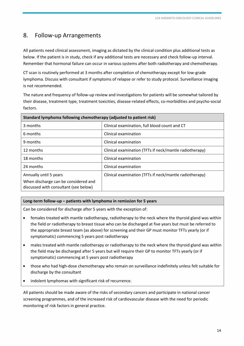
All patients need clinical assessment, imaging as dictated by the clinical condition plus additional tests as
below. If the patient is in study, check if any additional tests are necessary and check follow-up interval.
Remember that hormonal failure can occur in various systems after both radiotherapy and chemotherapy.
CT scan is routinely performed at 3 months after completion of chemotherapy except for low-grade
lymphoma. Discuss with consultant if symptoms of relapse or refer to study protocol. Surveillance imaging
is not recommended.
The nature and frequency of follow-up review and investigations for patients will be somewhat tailored by
their disease, treatment type, treatment toxicities, disease-related effects, co-morbidities and psycho-social
factors.
Standard lymphoma following chemotherapy (adjusted to patient risk)
3 months Clinical examination, full blood count and CT
6 months Clinical examination
9 months Clinical examination
12 months Clinical examination (TFTs if neck/mantle radiotherapy)
18 months Clinical examination
24 months Clinical examination
Annually until 5 years
When discharge can be considered and discussed with consultant (see below)
Clinical examination (TFTs if neck/mantle radiotherapy)
Long-term follow-up – patients with lymphoma in remission for 5 years
Can be considered for discharge after 5 years with the exception of:
females treated with mantle radiotherapy, radiotherapy to the neck where the thyroid gland was within
the field or radiotherapy to breast tissue who can be discharged at five years but must be referred to
the appropriate breast team (as above) for screening and their GP must monitor TFTs yearly (or if
symptomatic) commencing 5 years post radiotherapy
males treated with mantle radiotherapy or radiotherapy to the neck where the thyroid gland was within
the field may be discharged after 5 years but will require their GP to monitor TFTs yearly (or if
symptomatic) commencing at 5 years post radiotherapy
those who had high-dose chemotherapy who remain on surveillance indefinitely unless felt suitable for
discharge by the consultant
indolent lymphomas with significant risk of recurrence.
All patients should be made aware of the risks of secondary cancers and participate in national cancer
screening programmes, and of the increased risk of cardiovascular disease with the need for periodic
monitoring of risk factors in general practice.
SURVIVORSHIP AND REHABILITATION
15
9. Survivorship and Rehabilitation
Issues in relation to rehabilitation and the long-term consequences of treatment should be monitored
throughout the patient pathway and highlighted to the appropriate allied health professionals if required.
Survivorship issues can relate to the effects of the disease process and/or management of long-term
adverse effects of treatment. Please refer to LCA Survivorship Guidelines for more information.
Any patient experiencing or reporting reduced mobility and/or ability to perform activities of daily living
should be referred for occupational therapy and physiotherapy assessment.
People reporting ongoing consequences such as fatigue, anxiety and pain should be considered for
non-pharmacological intervention, including, but not limited to, TENS (transcutaneous electrical nerve
stimulation), complementary therapy and psychological intervention such as mindfulness.
Referrals include:
physiotherapist/occupational therapist (fatigue management, rehabilitation)
dietitian if symptoms are impacting on intake/nutritional status, especially in cases of GI disturbances
and mucositis, weight loss
speech and language therapist (weight loss, dysphagia or concerns regarding aspiration).
10. Research and Clinical Trials
Where possible, biobanking of all patient blood and tissue samples is encouraged in a certified facility within
the context of an ethically approved research framework and in compliance with the Human Tissue Act.
Eligible patients should always be offered the opportunity to consider clinical trials for any stage of disease
management. This may be either at the treating centre or within the LCA in another lymphoma centre.
Occasionally, it may be appropriate to refer patients to centres outside the LCA for clinical trials open
elsewhere and, in a reciprocal manner, centres within the LCA may occasionally receive referrals of patients
from other integrated cancer systems. Such collaboration within and across cancer systems should be
supported.
A list of clinical trials that are open across the LCA is available at www.londoncanceralliance.nhs.uk/trials.
LCA HAEMATO-ONCOLOGY CLINICAL GUIDELINES
16
11. End-of-life Care
Common causes of death include lymphoma (treatment-resistant disease progression or transformation),
therapy-related complications and infections.
Although predicting when death may occur is often inaccurate, it is important to consider and offer
discussions with patients, and partners/relatives/carers/friends as appropriate, when it is apparent that
disease or its complications are progressing and further treatment is futile.
Such matters require sensitivity and consideration of patients’ cultures, beliefs, wishes and
communications as well as those of their next of kin, in particular.
Where appropriate, patients should be asked about their preferred place of death, and local and national
guidance on resuscitation decisions should be followed.
The best interests of the patient must be at the forefront of discussions and decision-making regarding end-
of-life care while early involvement of local palliative care teams (hospital and/or community) will optimally
facilitate the formation of individualised end-of-life care packages. Care may be required from specialist
palliative care teams which are available in the cancer units and centres affiliated to the LCA.
To support consideration of referral to specialist palliative care, please refer to the LCA Referral Criteria for
Specialist Palliative Care (see Appendix 8). The LCA form for referral to specialist palliative care can be
found in Appendix 7: LCA Specialist Palliative Care Referral Form.

MARGINAL ZONE LYMPHOMA
17
12. Marginal Zone Lymphoma
12.1. Introduction
Marginal zone lymphoma (MZL) is an uncommon form of NHL associated with chronic infection and
inflammation (e.g. gastric MALT lymphoma and Helicobacter pylori infection). Other microbes include
Campylobacter jejuni, Borelia burgdoferi, Chlamydia psittaci and hepatitis C. MALT lymphomas of the
salivary and thyroid glands are associated with Sjögren’s syndrome and Hashimoto’s thyroiditis
respectively.
Marginal zone lymphoma is usually very indolent, growing slowly.
12.1.1. Classification
Extranodal MZL (MALT):
gastric (30%)
non-gastric (70%), e.g. pulmonary, cutaneous, ocular.
Nodal MZL
Splenic MZL
12.2. Investigation and diagnosis
12.2.1. General initial assessment
FBC, differential, DAT
Renal, liver and bone profile, LDH, Igs, paraprotein, B2M
BM aspirate and trephine with immunohistochemistry
CT-NCAP (MRI if orbital disease)
Careful attention to extranodal sites
Infection screens – hepatitis B and C, HIV, and H pylori, chlamydia etc as indicated.
12.2.2. Specific investigations – MALT
a) Histology
Cellular composition includes small lymphocytes, centrocyte-like cells, ‘monocytoid’ B-cells,
plasmacytoid cells
Invasion of epithelial structures and existing germinal centres
CD5-, CD10-, CD19+, CD20+, CD23-, sIgM+, sIgD+ or -
Disease localised to or centred on an extranodal site.
b) For gastric types EUS and OGD
At diagnostic endoscopy the lesion should be photographed to allow assessment of macroscopic
response in follow-up examinations. At least 8–10 biopsies should be taken from the area of
abnormality. This is to ensure sufficient diagnostic material is available and to exclude foci of

LCA HAEMATO-ONCOLOGY CLINICAL GUIDELINES
18
transformation. Two biopsies should be taken from other areas of the stomach to assess multifocal
lymphoma and any background disease. Biopsies from normal antral mucosa should be included to
assess Helicobacter status. EUS should be performed to assess depth of invasion of the gastric wall,
lateral extent of the primary lesion and regional lymph node status.
If histology or CLO test does not identify Helicobacter, stool samples for Helicobacter pylori antigen
should be sent to microbiology.
The presence of t(11;18) in 25–40% of cases is associated with failure of eradication therapy and
disseminated disease but lower risk of high grade transformation.
c) SMZL
Diagnosis is usually made on morphology and immunophenotype of PB/BM lymphocytes in a patient
presenting with splenomegaly.
Often identified in an elderly patient referred with suspected CLL or MCL.
Low level (<20g/l) serum paraprotein (IgG or IgM) is common.
Check DAT as AIHA rare but documented.
Cryoglobulins in HCV+ cases.
12.2.3. Poor prognostic features
a) Gastric MALT
H pylori negative
Tumour invasion beyond the mucosa
T(11;18)
Bcl 10 nuclear expression.
b) SMZL
Hb <120g/dl
High lymphocyte count (>18 x 109/L)
Albumin <35 g/l
LDH >ULN
TP53 del/mut
Abnormalities of chromosome 7 (del 7q 22–32)
High grade transformation (v rare).
12.3. Treatment
All patients should be considered for clinical trials where possible. For more details on clinical trials see
section 10: Research and Clinical Trials.
12.3.1. Indications for treatment
Symptomatic disease
GI bleeding
MARGINAL ZONE LYMPHOMA
19
Bulky disease
Cytopenias.
12.3.2. Gastric MALT
a) Primary treatment
Localised stage IE/IIE disease should receive Helicobacter pylori eradication as primary therapy
(remission in 70%).
Helicobacter pylori eradication therapy:
Lansoprazole 30 mg BD – 1 week.
Amoxicillin 1g BD – 1 week.
Clarithromycin 500 mg BD – 1 week.
For alternatives to this regimen dependent on previous attempts at Helicobacter pylori eradication
or allergies, please see BNF.
Breath test must be at least 4 weeks post treatment to confirm eradication.
NOTE: patients must stop taking antacids, PPIs, H2 antagonists or bismuth preparations for 2 weeks
prior to breath test.
Surgery is generally to be avoided except in emergency situations to control bleeding or repair a
perforation; these are very rare in gastric marginal zone lymphoma.
Follow-up with upper GI endoscopy and biopsy at 6, 12 and 24 months for the first 2 years.
b) Recurrent or resistant gastric MALT
Patients with recurrent disease after 1 year, stage greater than 1E or persistent disease after
Helicobacter eradication should be considered for treatment with chemotherapy (chlorambucil +
rituximab, R-CVP), single agent rituximab or low-dose IF radiotherapy.
Patients with persistent/recurrent asymptomatic disease after therapy can be followed up by repeat
endoscopy without further treatment.
Patients with persistent/recurrent symptomatic disease who have received treatment should be
re-discussed in lymphoma MDT for other relevant options.
For patients with large cell transformation, CHOP and rituximab chemotherapy is recommended.
12.3.3. Non-gastric MALT
Watch and wait in asymptomatic patients
IF radiotherapy for localised disease
Rituximab monotherapy
R+ chemotherapy (as per FL protocols)
Ocular MALT – consider doxycycline 100 mg BD x 3 weeks.
12.3.4. Splenic MZL
Watch and wait is a reasonable approach in asymptomatic patients (25% may never need treatment).
LCA HAEMATO-ONCOLOGY CLINICAL GUIDELINES
20
Treatment is usually indicated for bulky splenomegaly and cytopenias, and more rarely lymphocytosis.
Splenectomy (pre-surgical immunisations and post-surgery prophylaxis with penicillin V or
clarithromycin) can result in long-term remissions.
Splenic irradiation may be an alternative in patients not fit for surgery although morbidity is the same.
Alkylating agents (e.g. chlorambucil) are relatively ineffective.
Rituximab monotherapy.
FR, BR or FCR.
R-CVP is sometimes used but no clear evidence that this is superior to R alone and is less effective than
Purine Analog (PA) +R.
R-CHOP for high grade transformation.
Ribavarin/interferon in HCV+ patients.
12.3.5. Nodal MZL
Watch and wait if asymptomatic
Stage 1 – consider local RT
Systemic chemotherapy +R, as per FL protocol.
12.4. Supportive care
All patients who have received purine analogues or bendamustine should receive irradiated blood
products. See section 6 for general supportive care guidance.
12.4.1. Follow-up arrangements
See section 8 for general follow-up arrangements.
a) Gastric MALT
Eradication of Helicobacter should be confirmed by stool sample for Helicobacter antigen at least
4 weeks after completion of Helicobacter therapy.
Follow-up endoscopy should be performed at a minimum of 6, 12 and 24 months and thereafter at the
discretion of the clinician. If possible the procedures should be performed by the same endoscopist.
The area of previous abnormality should be photographed. Some 8–10 biopsies should be performed
from the area of previous abnormality and submitted for histological examination.
There are no uniform criteria for the definition of histological remission.
Complete histological remission can take >12 months.
EUS should be performed to exclude the possibility of sub-mucosal disease (which can exist under
apparently normal mucosa and may not be included in mucosal biopsies) 12 months after completion
of treatment.
b) Other MZL
Directed by individual clinical circumstances
Follow-up usually every 3 months for first year and then 6 monthly.
HAIRY CELL LEUKAEMIA
21
13. Hairy Cell Leukaemia
13.1. Introduction
Hairy cell leukaemia (HCL) is a rare disorder (2% of all haematological malignancies). Median age is late 50s
and it is four times more common in males than females. The clinical course is extremely indolent. Unusual
para-neoplastic phenomena can occur.
The majority of patients present with pancytopenia and splenomegaly. A monocytopenia is characteristic.
13.2. Investigation and diagnosis
13.2.1. Evaluation at presentation
History of infections, B symptoms: weight loss and night sweats
PS
Physical examination for splenomegaly
FBC + PBF
Sample sent to HMDS for diagnostic immunophenotype. See Annex 2 for details of SIHMDS
Further tests performed at diagnosis include: biochemistry, LDH, Igs, B2M, viral serology.
13.2.2. Diagnosis of HCL
Examination of the peripheral blood film normally shows characteristic hairy cells (but the count may
be low).
Four immunophenotypic markers relatively specific for hairy cells can be used: CD11c, CD25, CD103
and CD123. A score of 4/4 is diagnostic.
A definitive diagnosis requires a bone marrow biopsy. Confirmation of the nature of the infiltration is
obtained by immunocytochemistry using CD20 and/or DBA44.
Good quality trephine biopsies are required not only for diagnosis but also for monitoring response to
treatment.
BRAF is mutated in almost 100% of classical HCL cases. In cases where the diagnosis is in doubt this can
be confirmatory but does not otherwise influence management.
Abdominal CT scan or ultrasound is required at diagnosis to assess spleen size and identify patients
with large abdominal nodes.
13.2.3. Staging
There is no recognised staging system.
LCA HAEMATO-ONCOLOGY CLINICAL GUIDELINES
22
13.2.4. Prognosis
There are no currently recognised prognostic markers.
There is a suggestion that patients presenting with bulky abdominal nodes and those with more severe
anaemia and/or thrombocytopenia have slightly less good outcome.
Any cases which do not have BRAF mutations or which have IgVH 3-34 usage have poorer outcome.
HCL-v is an entirely separate disease (now included with the group of splenic lymphomas in WHO),
which presents with high WBC, has a different immunophenotype, is BRAF negative and has a poorer
outcome than HCL.
13.3. Service configuration
All newly diagnosed cases need to be registered through the MDT, even if only for W&W management
plan.
Any patient requiring therapy, either initial or subsequent treatments, should be discussed in the MDT
and considered for suitable clinical trials.
13.4. Management of disease-related complications
See the CLL guidelines as very similar.
13.5. Treatment
13.5.1. Indications for treatment
Anaemia (<100 g/l)
Thrombocytopenia (<100 x 109/L)
Neutropenia (<0.5)
Bulky lymphadenopathy/splenomegaly
Systemic symptoms.
13.5.2. Watch and wait
Rarely patients are asymptomatic and, in the absence of the above criteria, W&W is reasonable.
13.5.3. First-line therapy
Most patients will require therapy to correct the cytopenias and the associated problems of anaemia,
infections and bleeding.
The mainstay of the treatment of HCL comprises the two nucleoside analogues pentostatin and cladribine.
Both agents induce a high rate (>80%) of complete remissions which, in the majority of patients, are
prolonged; median DFS for patients treated with pentostatin is 15 years in our series (N=250), with OS at
10 years of 97%. The majority of relapsed patients achieve second remissions when re-treated. Survival is
better for patients achieving CR than in those reaching PR. Both pentostatin and cladribine are well
tolerated and the only long-term effect is lymphopenia.
HAIRY CELL LEUKAEMIA
23
Regimens
Pentostatin (deoxycoformycin) is given by injection every 2 weeks. Usual dose 4 mg/m2. Dose
reduce/omit if low GFR. Usually treat to max response plus 2. Most patients receive 8–10 injections.
Usually assess BM trephine after 8 and if still involved continue for a further 4 injections before
repeating.
2CDA (cladribine) is given by sub-cutaneous injection (daily 0.14 mg/kg x 5), BM trephine assessment
approximately 3 months after completing treatment. If residual disease is present then give a second
cycle of 2CDA.
Splenectomy is sometimes indicated in refractory cases with massive splenomegaly or for those unable
to receive standard chemotherapy.
Rituximab is not as effective as a single agent but may be useful in frail patients considered unfit for
PA therapy.
Interferon alpha (3MU 3x/w) is very rarely used but may be helpful in patients with profound
pancytopenia and infection to improve their counts prior to PA therapy.
Monitor response by blood counts and BM trephine biopsy with immunocytochemistry and abdominal
CT (if previously abnormal).
13.5.4. Treatment for relapsed/refractory patients
Most patients relapse after >5 years in remission and can be re-treated with the same or the
alternative PA; subsequent remissions tend to be less durable than the first.
Patients who fail to achieve a good initial remission or who relapse in <5 years should receive PA +
rituximab (375 mg/m2 x 6–8).
Very refractory patients should be entered into clinical trials where possible or referred to centres with
a specialist interest for advice. The BRAF inhibitor (vemurafenib) has been shown to be effective but
responses appear short-lived.
13.6. Supportive care
Patients receiving Purine analogue therapy require pneumocystis prophylaxis with co-trimoxazole
(480 mg tablet daily), or nebulised pentamidine if allergic to co-trimoxazole. Acyclovir prophylaxis
(200 mg BD) against herpes zoster is also recommended. Both should be given during therapy and for
up to 6 months afterwards.
Growth factors, e.g. G-CSF, could also be used to treat neutropenia (<0.5 x 10⁹/L) before, during and/or
after the use of either pentostatin or cladribine.
All patients who have received these drugs should have irradiated blood products indefinitely.
See section 6 for general supportive care guidance.
LCA HAEMATO-ONCOLOGY CLINICAL GUIDELINES
24
14. Mantle Cell Lymphoma
14.1. Introduction
Mantle cell lymphoma (MCL) is a rare B-NHL with an incidence of 0.5 per 100,000 in the Western world.
The median age at presentation is 70 with a male to female ratio of 2–3:1.
Most patients present with advanced stage disease with bone marrow involvement. The gastrointestinal
tract is a common site of involvement and approximately a third of patients present with circulating disease
in the peripheral blood. CNS involvement has been described in MCL (cumulative incidence approximately
4%) and is more frequent with blastoid morphology and advanced disease.
14.2. Investigation and diagnosis
14.2.1. Evaluation at presentation
B symptoms
PS (ECOG)
Physical examination for lymphadenopathy and organomegaly
FBC+PBF
Sample to HMDS for diagnostic immunophenotype
Further tests performed at diagnosis include:
Biochemistry
LDH
Igs
B2M
Viral serology
GI symptoms at presentation should be investigated with OGD or colonoscopy
MRI +/- lumbar puncture for CSF analysis if suspicion of CSF involvement.
14.2.2. Diagnosis of MCL
Morphologically, small to medium sized lymphocytes with irregular nuclear contours. Blastoid
morphology (about 20% of cases at diagnosis) is recognised by an increase in nuclear size and
chromatin dispersal with prominent nucleoli.
MCL cells express a mature B cell phenotype with expression of CD19, CD20, CD22 and CD79a. CD5 is
expressed in the vast majority of cases but, in contrast to CLL, CD20 expression is strong, FMC7 is
expressed and CD23 is usually absent.
The diagnostic hallmark is t(11;14) with over-expression of cyclin D1, but approximately 6% of cases are
negative for this translocation and over-express cyclin D2, D3 or E.
CT scan and bone marrow examination are required for staging at diagnosis. Although MCL is FDG avid, the role of PET-CT in MCL is not clearly established and is not recommended routinely.
MANTLE CELL LYMPHOMA
25
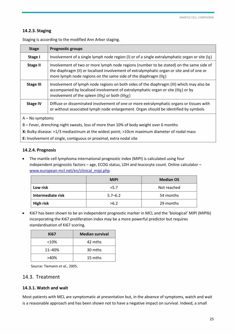
14.2.3. Staging
Staging is according to the modified Ann Arbor staging.
Stage Prognostic groups
Stage I Involvement of a single lymph node region (I) or of a single extralymphatic organ or site (IE)
Stage II Involvement of two or more lymph node regions (number to be stated) on the same side of
the diaphragm (II) or localised involvement of extralymphatic organ or site and of one or
more lymph node regions on the same side of the diaphragm (IIE)
Stage III Involvement of lymph node regions on both sides of the diaphragm (III) which may also be
accompanied by localised involvement of extralymphatic organ or site (IIIE) or by
involvement of the spleen (IIIS) or both (IIISE)
Stage IV Diffuse or disseminated involvement of one or more extralymphatic organs or tissues with
or without associated lymph node enlargement. Organ should be identified by symbols
A – No symptoms
B – Fever, drenching night sweats, loss of more than 10% of body weight over 6 months
X: Bulky disease: >1/3 mediastinum at the widest point; >10cm maximum diameter of nodal mass
E: Involvement of single, contiguous or proximal, extra nodal site
14.2.4. Prognosis
The mantle cell lymphoma international prognostic index (MIPI) is calculated using four
independent prognostic factors – age, ECOG status, LDH and leucocyte count. Online calculator –
www.european-mcl.net/en/clinical_mipi.php
MIPI Median OS
Low risk <5.7 Not reached
Intermediate risk 5.7–6.2 54 months
High risk >6.2 29 months
Ki67 has been shown to be an independent prognostic marker in MCL and the ‘biological’ MIPI (MIPIb)
incorporating the Ki67 proliferation index may be a more powerful predictor but requires
standardisation of Ki67 scoring.
Ki67 Median survival
<10% 42 mths
11–40% 30 mths
>40% 15 mths
Source: Tiemann et al., 2005.
14.3. Treatment
14.3.1. Watch and wait
Most patients with MCL are symptomatic at presentation but, in the absence of symptoms, watch and wait
is a reasonable approach and has been shown not to have a negative impact on survival. Indeed, a small
LCA HAEMATO-ONCOLOGY CLINICAL GUIDELINES
26
proportion of patients have ‘indolent’ MCL which can be managed expectantly, sometimes for years. These
patients generally present with isolated lymphocytosis with or without splenomegaly. Molecular markers
(e.g. SOX11, IGH mutation status) to identify this group of patients have been proposed but are not yet
established.
14.3.2. First line therapy
All patients should be considered for entry into a clinical trial.
Patients fit for high-dose therapy should be treated with one or two high dose Ara-c containing
induction regimens followed by consolidation with a high dose regimen such as BEAM or LEAM and an
autologous stem cell transplant (ASCT):
1. NORDIC MCL2 protocol – R-maxiCHOP alternating with R-HDAra-C
2. R-CHOP(21)x3 alternating with R-DHAPx3 (Delarue Blood 2013 and European ‘MCL younger’ trial).
Interim CT is performed after five cycles of treatment.
Patients not fit for high-dose therapy should be treated with R-CHOPx6 followed by rituximab
maintenance (Kluin-Nelemans, 2012). Alternative induction regimens that could be considered in less
fit patients or those unable to tolerate CHOP include R-CVP (which may be equally effective),
R-bendamustine, R-FC and R-chlorambucil. CNS prophylaxis may be justified in patients with blastoid,
advanced stage disease.
14.3.3. Treatment for relapsed/refractory patients
All patients should be considered for entry into a clinical trial. There is no standard second line regimen.
In younger/fitter patients consideration should be given to salvage treatment followed by LEAM ASCT
consolidation (if they did not receive this upfront) or by allogeneic SCT if there is a suitable donor.
Salvage treatment can be with R-Gem-P, R-DHAP, R-bendamustine or R-bortezomib. Patients relapsing
within 6 months of treatment with rituximab are unlikely to benefit from addition of this to their
salvage regimen.
In older patients suitable regimens include R-bendamustine, R-FC, R-bortezomib, R-chlorambucil and
temsirolimus (IFR). Patients relapsing within 6 months of treatment with rituximab are unlikely to
benefit from addition of this to their salvage regimen.
Novel agents: ibrutinib is approved in the US and EU for treatment of relapsed MCL. Ibrutinib is an option
if funding is available. Lenalidomide has also shown promising activity in relapsed MCL (but is not funded).
Idelalisib has less activity in MCL but broader PI3K inhibitors may be more promising.
Splenectomy may be useful in selected patients, especially those with leukaemic presentation and
splenomegaly with no nodal disease.
Radiotherapy can be useful in localised disease, usually for palliation.
Allogeneic HSCT should be considered for suitable patients.
MANTLE CELL LYMPHOMA
27
14.4. Supportive care
Acyclovir prophylaxis (200 mg BD) is recommended for all patients. Co-trimoxazole prophylaxis is
recommended in addition for patients receiving purine analogues. Both should be given during therapy
and for up to 6 months afterwards. See section 6 for general supportive care guidance.
LCA HAEMATO-ONCOLOGY CLINICAL GUIDELINES
28
15. Burkitt Lymphoma
15.1. Introduction
Adult Burkitt lymphoma (BL) can be endemic, sporadic or associated with immunodeficiency. Endemic BL is
invariably EBV-positive and occurs in equatorial Africa with a peak incidence in childhood (jaw, facial bones
and GI tract). Sporadic BL has a median age of 30 with a male preponderance (2–3:1). The incidence is
approximately 2.5 cases per million per annum.
15.2. Investigation and diagnosis
15.2.1. Clinical features
Rapidly progressive bulky nodal and/or extranodal disease with frequent bone marrow, intestinal tract
and leptomeningeal disease.
Investigations must be completed rapidly as treatment should commence within 48 hours of diagnosis.
FBC and differential, ESR, CRP.
Clotting screen.
U&Es, LFTs, uric acid, Ca, PO4, B2M, LDH.
Immunoglobulin profile, serum protein electrophoresis.
Full hepatitis B profile: Hep B S Ab, Hep B S Ag, Hep B c Ab, Hep C Ab.
HIV Ab with counselling and consent.
Lumbar puncture with CSF for cytology, flow cytometry ± PCR for IgH gene rearrangement if suspicious
cells seen.
MRI scan if spinal cord involvement/CNS suspected/may be used in pregnancy/patient allergic to iodine
contrast.
PET/CT (high sensitivity no false negatives) but do not delay treatment to obtain.
Preferably an excision lymph node biopsy by designated surgeons (or in some circumstance a core
biopsy of an inaccessible lymph node or extra lymphatic organ or in rare cases requiring urgent medical
treatment). Fine needle aspiration is not adequate for the diagnosis.
The biopsy should be examined by a haematopathologist. Diagnostic criteria for BL and its variants have
changed considerably over time. For details of SIHMDS services please see Annex 2.
BURKITT LYMPHOMA
29
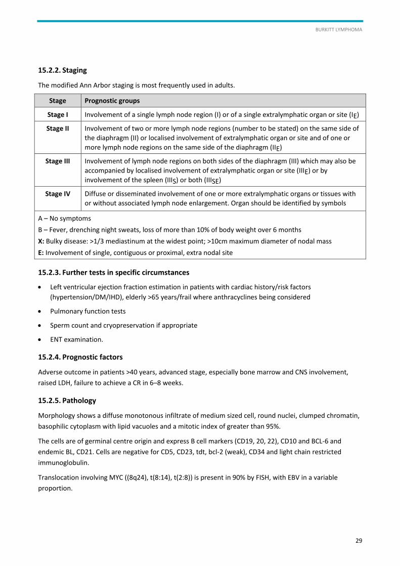
15.2.2. Staging
The modified Ann Arbor staging is most frequently used in adults.
Stage Prognostic groups
Stage I Involvement of a single lymph node region (I) or of a single extralymphatic organ or site (IE)
Stage II Involvement of two or more lymph node regions (number to be stated) on the same side of
the diaphragm (II) or localised involvement of extralymphatic organ or site and of one or
more lymph node regions on the same side of the diaphragm (IIE)
Stage III Involvement of lymph node regions on both sides of the diaphragm (III) which may also be
accompanied by localised involvement of extralymphatic organ or site (IIIE) or by
involvement of the spleen (IIIS) or both (IIISE)
Stage IV Diffuse or disseminated involvement of one or more extralymphatic organs or tissues with
or without associated lymph node enlargement. Organ should be identified by symbols
A – No symptoms
B – Fever, drenching night sweats, loss of more than 10% of body weight over 6 months
X: Bulky disease: >1/3 mediastinum at the widest point; >10cm maximum diameter of nodal mass
E: Involvement of single, contiguous or proximal, extra nodal site
15.2.3. Further tests in specific circumstances
Left ventricular ejection fraction estimation in patients with cardiac history/risk factors
(hypertension/DM/IHD), elderly >65 years/frail where anthracyclines being considered
Pulmonary function tests
Sperm count and cryopreservation if appropriate
ENT examination.
15.2.4. Prognostic factors
Adverse outcome in patients >40 years, advanced stage, especially bone marrow and CNS involvement,
raised LDH, failure to achieve a CR in 6–8 weeks.
15.2.5. Pathology
Morphology shows a diffuse monotonous infiltrate of medium sized cell, round nuclei, clumped chromatin,
basophilic cytoplasm with lipid vacuoles and a mitotic index of greater than 95%.
The cells are of germinal centre origin and express B cell markers (CD19, 20, 22), CD10 and BCL-6 and
endemic BL, CD21. Cells are negative for CD5, CD23, tdt, bcl-2 (weak), CD34 and light chain restricted
immunoglobulin.
Translocation involving MYC ((8q24), t(8:14), t(2:8)) is present in 90% by FISH, with EBV in a variable
proportion.
LCA HAEMATO-ONCOLOGY CLINICAL GUIDELINES
30
Importantly the presence of MYC is not diagnostic of BL and is seen in 10% of DLBCL and 30–50% of BL
unclassified (cases intermediate between BL and DLBCL). Frequently with breakpoints in BCL6 or t(14:18) as
well as MYC so called ‘double hit’ lymphomas.
Greater diagnostic precision can be achieved with gene profiling (high expression of GC B cell genes and low
expression of MHC 1 and NFkB pathway genes) but outside of clinical trials it is not yet routinely available.
15.2.6. Imaging
Baseline contrast-enhanced CT with PET if available. End of treatment PET/CT.
15.3. Management of specific disease-related complications
Specific disease-related complications such as leptomeningeal involvement and spinal cord compression
should be managed according to local treatment centre policy
15.4. Treatment
Treatment for BL and its variants should be prompt (<48 hours) with adequate supportive care.
Tumour lysis should be managed with rigorous hydration and rasburicase.
All new and relapsing cases should be discussed at the next local network MDT meeting with central review
by a specialist haematopathologist.
Treatment facilities to administer induction treatment should be BSCH level 3. Patients with less than a CR
or with chemosensitive relapse should be referred to a JACIE-accredited SCT units for consolidation
autograft.
Rapid access pathways should be in place to allow both primary care and specialist medical teams to
urgently refer suspected cases with transfer once stable to a level 3 centre.
Brief intense chemotherapy is the treatment of choice. Patients are managed according to risk groups.
The CNS is frequently involved.
Low risk patients should receive CODOX-M x 3 cycles – patients must have at least three of the following:
Normal LDH level
WHO performance status 0–1
Ann Arbor stage I–II
Number of extranodal sites (e.g. bone marrow, GI tract, CNS) 1.
All other patients are considered high risk/patients under 60 years should receive R-CODOX-M/R-IVAC x 2.
Tumour lysis is common and patients should receive rasburicase (see Annex 3 and Annex 4).
Patients who fail to achieve a CR or chemosensitive relapse should be referred for consolidation with an
autologous SCT. Allografting is not indicated.
Elderly patients should be treated with the most intensive treatment they can tolerate following careful
assessment and presentation and continual re-evaluation of co-morbidities, toxicities and functional capacity.
Patients over 60 years should be considered for the age-adjusted protocol of R-CODOX-M/R-IVAC.
Cases should be considered on an individual basis. Good results in the elderly have been reported with
BURKITT LYMPHOMA
31
DA-EPOCH-R and GMALL (omission of HD cytosine arabinoside and methotrexate reduced to 500 mg/m2) for
those not suitable for intensive therapy. Patients unable to tolerate these regimens consider standard RCHOP.
Intrathecal prophylaxis should be given when CNS penetrative drugs are not included in the regimen.
15.5. End of treatment information
Short duration, intense chemotherapy cures 90% of low-risk and 60–80% of high-risk patients. Relapse, if it
occurs, is usually within a year, with cure defined as remission of >2 years. Patients should be aware of
possible symptoms or relapse/progression and given details to contact the medical team urgently in these
occurrences. Information on possible symptoms and contact details should be included in the treatment
summary (see section 7: End of Treatment Information).
15.6. Specific or miscellaneous considerations
HIV-positive patients usually have good CD4 counts and should be managed with antiretroviral therapy
concurrently with aggressive short duration chemotherapy. More mucositis and more severe infections
are seen.
Patients in the TYA age group should be referred for discussion in a specialist TYA MDT with the option
of being treated in a TYAC. See section 3.1 for more details. Consideration should be given to
alternative age-appropriate/adolescent protocols, e.g. SFOP LMB.
Patients with Burkitt-like lymphoma and BL unclassifiable (feature intermediate between DLBCL and
Burkitts) do poorly with RCHOP and where possible should be treated as BL.
LCA HAEMATO-ONCOLOGY CLINICAL GUIDELINES
32
16. Peripheral T-Cell Lymphomas and Leukaemias
16.1. Introduction
Peripheral T-cell lymphoma (PTCL) is a heterogeneous group of rare malignancies accounting for
approximately 10–12% of all lymphoid neoplasms. Most have an aggressive clinical behaviour and, apart
from anaplastic large cell lymphoma (ALCL), a poor response to conventional chemotherapy with only 30%
survival at 3 years. Patients often present with disease at extranodal sites and poor PS.
16.1.1. WHO classification
a) Nodal
Peripheral T-cell lymphoma, unspecified
Angioimmunoblastic T-cell lymphoma
Anaplastic large cell lymphoma (ALCL) (ALK+, ALK-, primary cutaneous)
Adult T-cell lymphoma/leukaemia.
b) Extranodal
Extranodal NK/T-cell lymphoma, nasal type
Enteropathy-associated T-cell lymphoma
Hepatosplenic T-cell lymphoma
Subcutaneous panniculitis-like T-cell lymphoma.
c) Cutaneous
Mycosis fungoides/Sézary syndrome
Lymphomatoid papulosis.
16.2. Clinical presentation and referral pathways
16.2.1. Presentation
Patients usually present with palpable lymphadenopathy and systemic symptoms. Presentation at
extranodal sites is common and patients may therefore be under the care of other hospital specialties
before the histological diagnosis is confirmed.
16.2.2. Referral pathways
Referral from primary care should be made using the 2 week wait referral form to rapid access neck lump
diagnostic clinics. Presentation via other specialties can delay diagnosis.
16.3. Investigation and diagnosis
16.3.1. Diagnosis
Diagnosis is based on examination of adequate histological material from tissue biopsy supplemented by
detailed immunohistochemistry, flow cytometry, cytogenetics and molecular genetics. Expert
haematopathology review is essential for the correct classification of the different subtypes. For details of
PERIPHERAL T-CELL LYMPHOMAS AND LEUKAEMIAS
33
SIHMDS please see Annex 2. Unlike B-cell lymphomas, there is no simple test for clonality and this should
be established by polymerase chain reaction (PCR) for rearrangement of T-cell receptor genes.
16.3.2. Investigations
FBC, renal and liver profile, LDH, Igs, Beta2 microglobulin
Virology: hepatitis B&C, HIV, EBV (including EBV PCR), HTLV1
Strongyloides serology in ATLL
BM aspirate and trephine biopsy
CT-NCAP (PET)
Lumbar puncture/MRI of brain is not routinely required in the absence of CNS symptoms or signs.
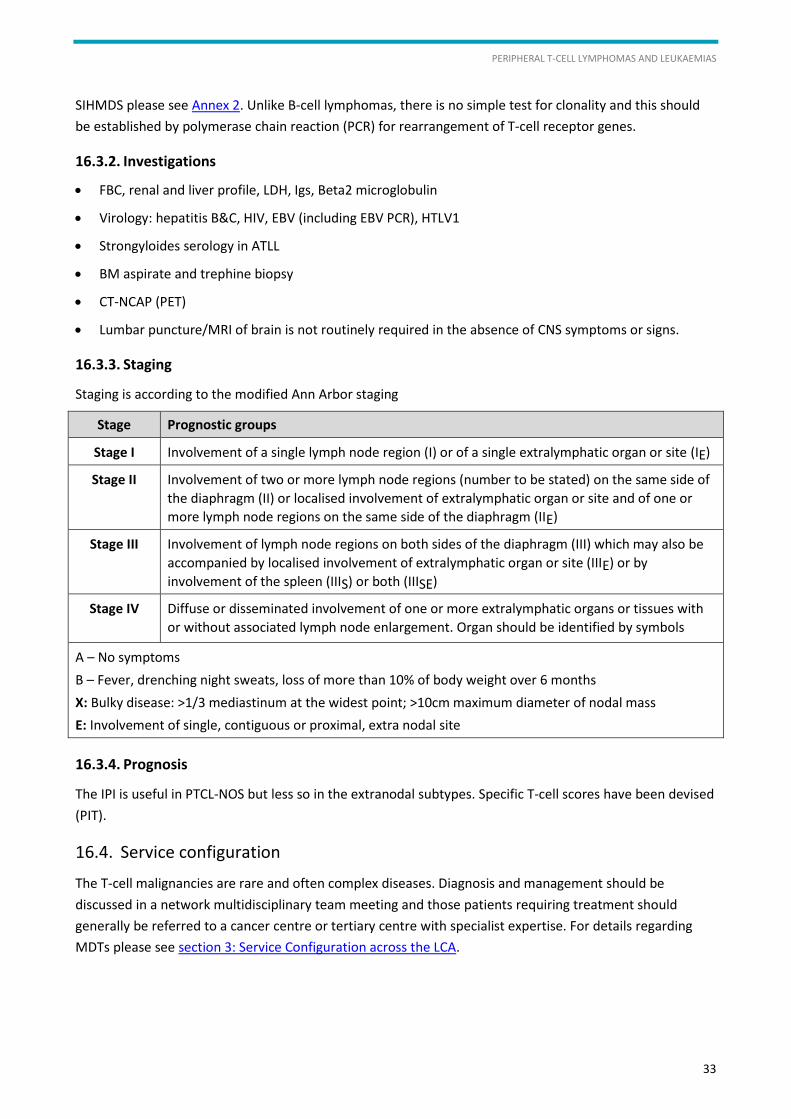
16.3.3. Staging
Staging is according to the modified Ann Arbor staging
Stage Prognostic groups
Stage I Involvement of a single lymph node region (I) or of a single extralymphatic organ or site (IE)
Stage II Involvement of two or more lymph node regions (number to be stated) on the same side of
the diaphragm (II) or localised involvement of extralymphatic organ or site and of one or
more lymph node regions on the same side of the diaphragm (IIE)
Stage III Involvement of lymph node regions on both sides of the diaphragm (III) which may also be
accompanied by localised involvement of extralymphatic organ or site (IIIE) or by
involvement of the spleen (IIIS) or both (IIISE)
Stage IV Diffuse or disseminated involvement of one or more extralymphatic organs or tissues with
or without associated lymph node enlargement. Organ should be identified by symbols
A – No symptoms
B – Fever, drenching night sweats, loss of more than 10% of body weight over 6 months
X: Bulky disease: >1/3 mediastinum at the widest point; >10cm maximum diameter of nodal mass
E: Involvement of single, contiguous or proximal, extra nodal site
16.3.4. Prognosis
The IPI is useful in PTCL-NOS but less so in the extranodal subtypes. Specific T-cell scores have been devised
(PIT).
16.4. Service configuration
The T-cell malignancies are rare and often complex diseases. Diagnosis and management should be
discussed in a network multidisciplinary team meeting and those patients requiring treatment should
generally be referred to a cancer centre or tertiary centre with specialist expertise. For details regarding
MDTs please see section 3: Service Configuration across the LCA.
LCA HAEMATO-ONCOLOGY CLINICAL GUIDELINES
34
16.5. Treatment
Where possible patients should be entered into clinical trials. Current NCRN first-line treatment trial is
CHEMO-T.
Patients often have very aggressive disease and treatment delays should be avoided (use of GCSF may
be indicated).
Infection risk is high because of immunosuppression.
16.5.1. Treatment of specific subtypes
a) PTCL-NOS, AITL, ALCL
Outside trial, CHOP (14 or 21) x 6 remains the standard first-line therapy. Phase 2 data suggest that
strong consideration should be given to consolidation with auto-HSCT in first remission for non-ALCL
subtypes (especially in AITL).
ALCL ALK+ has very good outcome and patients with limited stage disease can be considered for short
course chemotherapy and IFRT. There is some evidence to suggest that younger patients with this
subtype benefit from the addition of etoposide. ASCT not recommended in first CR.
Alternative induction therapies include: CHOEP, GEM-P, ICE, PMitCEBO.
Relapsed or refractory disease should be treated with relapse-schedule combination chemotherapy
(usually platinum-based) and considered for allo-HSCT with reduced intensity conditioning or
autologous stem cell transplantation or novel therapies within a trial setting. Outcome for patients with
relapsed/refractory disease is extremely poor.
Outside a trial a number of agents show promise, particularly gemcitabine, bendamustine,
pralaterexate and romidepsin, but the data are insufficient to recommend routine use.
Brentuximab vedotin has good activity in relapsed ALCL but is also effective in other CD30 + PTCL.
In AITL the timing and selection of therapy depend on clinical presentation and prognostic features.
Rare spontaneous regressions can occur. Alternative treatments with some efficacy include steroids,
purine analogues (FC), immunomodulatory drugs (thalidomide, lenalidomide, ciclosporin).
CNS prophylaxis should be considered using the same criteria as for diffuse large B-cell NHL.
Primary cutaneous ALCL (ALK-neg) should be managed with local excision +/- radiotherapy and
chemotherapy reserved for those patients with systemic disease. Lymphomatoid papulosis is a CD30+
cutaneous disease which is sometimes mistaken for primary cutaneous ALCL. It is radiosensitive and
both PUVA therapy and low-dose oral methotrexate are effective at preventing recurrent lesions. The
use of conventional chemotherapy is not indicated in the management of lymphomatoid papulosis.
b) ATLL
Hypercalcaemia is common at diagnosis and requires vigorous management (see management of
complications).
Exclude co-infection with strongyloides prior to commencing therapy. Appropriate antimicrobial
prophylaxis during therapy should be instituted for seropositive patients.
PERIPHERAL T-CELL LYMPHOMAS AND LEUKAEMIAS
35
Smouldering and chronic:
No benefit from early chemotherapy, therefore watch and wait. Zidovudine (ZDV) + interferon-
+/- monoclonal antibodies may be considered (especially in chronic ATL) in the context of a
clinical trial
Lymphoma type
Induction with CHOP or alternative multi-agent regimen plus G-CSF with concurrent ZDV +
interferon-
ZDV + interferon- maintenance +/- monoclonal antibodies (MoAbs)
OR allogeneic transplant in 1st CR for eligible patients
Leukaemia (acute) type
Induction with antiretoviral therapy alone (ZDV + interferon-)
OR induction with CHOP or alternative multi-agent regimen plus G-CSF + concurrent ZDV +
interferon-
Allo HSCT in 1st CR for eligible patients
OR ZDV + interferon- maintenance +/- MoAbs
OR consolidation with novel agents, e.g. arsenic trioxide, αIFN; proteasome inhibitor in clinical trials
CNS prophylaxis should be considered using the same criteria as for diffuse large B-cell NHL.
c) Extranodal NK/T cell lymphoma
Demonstration of EBV virus in the biopsy is important diagnostically and monitoring EBV PCR is useful
to assess response/relapse.
Assigning a conventional IPI score is of limited value as most cases are localised and have a low score,
yet the survival is still poor.
The distinction at diagnosis between localised disease and disseminated disease is important as the
latter has a dismal prognosis and might be considered for experimental therapy as first line if available.
Outcome is unsatisfactory with CHOP-like therapy and entering patients into relevant clinical trials if
available is recommended.
Patients with localised disease should receive radiation with 50–55 Gy.
The value of additional chemotherapy (CHOP, etoposide-based or asparaginase-based) for local disease
remains unclear but is considered conventional pending more information.
Asparaginase-containing regimens such as SMILE (Steroids, Methotrexate, Ifosfamide, L-asparaginase
and Etoposide, Yamaguchu et al JCO 2011, Kwang et al Blood 2012) should be considered in
disseminated first-line and in relapsed or refractory disease.
High-dose therapy is unproven and there is no basis to recommend it outside a trial.
d) Enteropathy-associated T-cell lymphoma (EATL)
It is important to liaise with an experienced gastroenterologist to assist with biopsy, staging and
follow-up, and to manage nutritional problems.
LCA HAEMATO-ONCOLOGY CLINICAL GUIDELINES
36
Assigning a conventional IPI score is of limited value as there is no good prognostic group and most
cases are stage I–IIE.
Successful surgical de-bulking is probably beneficial. Many patients present with acute
abdomen/perforation and require emergency operations. However, recovery from this can delay onset
of chemotherapy which may have an adverse impact.
If there are trials available at the time of diagnosis, entry should be considered as there is no
satisfactory standard therapy.
CHOP-like therapy, with or without an up-front autograft, remains a common approach outside a trial,
and adoption of a more intensive approach such as the NCRI/SNLG protocol is a reasonable option in
fitter patients.
Nutrition is a major issue in managing these patients and dietetic/gastroenterology advice is essential
at all stages of treatment and follow-up.
e) Hepato-splenic T-cell lymphoma
Often occurs in younger males, can be associated with a history of chronic inflammatory disorders and
follows an aggressive clinical course.
Outcomes are extremely poor with conventional treatment.
Autologous or allogeneic bone marrow transplantation could be considered in younger fit patients
achieving CR.
f) Subcutaneous panniculitis-like T-cell lymphoma (SPTCL)
Very rare.
This is not a universally aggressive disease and careful initial assessment and observation should be
undertaken before committing to treatment.
CHOP-like chemotherapy appears to be effective and produces survivors.
Relapsed disease may respond to dose intensification in some patients.
Local radiotherapy has a place for good prognosis localised symptomatic skin involvement which does
not resolve with topical steroids.
g) Cutaneous T cell lymphoma (CTCL)
Communication and collaboration with dermatology and clinical oncology are vital for optimal patient
management. It is therefore recommended that all patients are referred to a specialist centre such as St
John’s Institute of Dermatology with access to appropriate expertise and all therapeutic modalities.
In the early stages, mycosis fungoides (MF) may be controlled by skin directed therapy (SDT) alone, such as
PUVA, topical chemotherapy, bexarotene and electron beam irradiation. A variety of treatment modalities
have been used to treat more advanced CTCL, with varying success.
Initial assessment
Repeated skin biopsies classified according to the WHO classification.
Staging CT scans (except MF stage IA/IB and LyP).

PERIPHERAL T-CELL LYMPHOMAS AND LEUKAEMIAS
37
PB for WBC, lymphocyte and Sézary cell counts, serum LDH, liver and renal function, lymphocyte subsets,
CD4:CD8 ratios, HTLV-I serology and TCR gene analysis.
Bone marrow aspirate/trephine biopsies MF stage IIB or above and CTCL variants, not LyP.
Lymph node biopsy if rapidly enlarging node or advanced clinical stage and palpable adenopathy (>1.5 cm).
Staging: TNM, Bunn and Lambert.
Review in MDT stages IIA–IVB. Membership to include dermato-pathologist, dermatologist, clinical
oncologist, haemato-oncologist, palliative care.
Treatment algorithm for MF/SS
Prognostic group (stage)
1st line 2nd line Experimental
Low risk (IA–IIA)
Expectant or SDT SDT
Intermediate risk (IA–IIA)
SDT; PUVA+IFN; PUVA+Bex; TSEBT
HDACi; Ontak; trials
Stage III erythrodermic
MTX; ECP/IFN/Bex combinations; trials
Alemtuzumab; chemo TSEB; HDACi; Ontak; Trials
RIC-AlloSCT
High risk (IIB/IV)
Radiotherapy (including TSEBT); chemotherapy; trials
HDACi; Ontak; Trials; palliative therapy
RIC-AlloSCT
Abbreviations used in table:
SDT – skin directed therapy: topical therapy; phototherapy (TLO1/PUVA); radiotherapy; TSEBT – total skin electron
beam therapy; IFN – alpha interferon; Bex – bexarotene; HDACi – histone deacetylase inhibitors
(vorinostat/romidepsin)*; ECP – extracorporeal photopheresis; MTX – methotrexate; Ontak – Denileukin difititox; RIC-
AlloSCT – reduced intensity conditioned allogeneic stem cell transplant.
LCA HAEMATO-ONCOLOGY CLINICAL GUIDELINES
38
17. T-cell Prolymphocytic Leukaemia (T-PLL)
17.1. Introduction
T-cell prolymphocytic leukaemia (T-PLL) is a rare aggressive disease accounting for <2% of all leukaemias.
Usually the response to traditional chemotherapy is poor with a median survival of only 7 months.
17.2. Presentation
Patients usually present with rapid onset of symptoms with:
Splenomegaly (common)
Marked leucocytosis, usually >100 x 109/L
Lymphadenopathy is rare (it is more common with B-PLL)
Systemic disease including rashes (20%), pleural effusions, CNS involvement (-10%).
17.3. Investigation and diagnosis
17.3.1. Investigation
History of infections, B symptoms, weight loss and night sweats
PS
Physical examination for lymphandenopathy/splenomegaly/hepatomegaly/skin rash
FBC and PBF
Sample sent to HMDS for diagnostic immunophenotype, cytogenetics and molecular genetics (TCR
gene rearrangement). See Annex 2 for details of SIHMDS
Further tests performed at diagnosis include:
Biochemistry (including urate and LDH)
Serum Igs and B2M
Viral serology (hepatitis, EBV, CMV, HIV)
A BM aspirate and trephine biopsy is not necessary to make the diagnosis and can be misleading if PB
results are not reported. A BM should be performed pre- and post-treatment to evaluate response
CT scans are required for initial evaluation and to assess response to treatment
LN biopsy is only required if there is doubt about the diagnosis.
17.3.2. Staging and prognosis
There are no specific staging or prognostic systems for T-PLL.
17.4. Service configuration
All cases should be discussed at the MDT at presentation and at relapse.
Since this is a very rare disease, discussion with a centre with a specialist interest is advised.
Early referral should be made to a transplant centre for consideration of SCT in first remission for
eligible patients.

T-CELL PROLYMPHOCYTIC LEUKAEMIA (T-PLL)
39
17.5. Treatment
17.5.1. Watch and wait
A small percentage of patients (15%) may present with an isolated lymphocytosis which can remain stable
for a prolonged period. Progression is inevitable and may occur rapidly. Therefore patients should be
monitored closely (q1–2 monthly).
17.5.2. First-line therapy
Intravenous alemtuzumab (30 mg 3x/week) is the most effective treatment in T-PLL and administered
as a single agent can achieve response rates of about 90%. Patients should continue on treatment to
complete response (12–18 weeks).
Slow or incomplete responders may benefit from the addition of a purine analogue, e.g. pentostatin
4 mg/m2/week or FC.
Eligible patients should be referred for consideration of an SCT (allo or auto) in first remission.
During and after alemtuzumab treatment patients should be monitored weekly until recovery of CD4
counts >200 μL and CR and then 1–3 monthly depending on clinical status.
17.5.3. Relapsed/refractory disease
Previously good responders to alemtuzumab who retain expression of CD52 on the T-PLL cells at
relapse can be retreated with alemtuzumab.
Patients refractory to alemtuzumab or CD52-negative have few effective treatment options.
Possibilities include: FMC, pentostatin, nelarabine, bendamustine, intensive regimens (mini-BEAM, IVE).
Eligible patients should be referred for transplant in second remission.
17.6. Supportive care
Infusion reactions with alemtuzumab are common and should be managed with careful use of pre-
medication and pethidine to control rigors as needed.
Patients receiving alemtuzumab and/or purine-analogue containing chemotherapy should receive PCP
and herpes prophylaxis. Those receiving alemtuzumab require weekly monitoring for CMV reactivation
and pre-emptive treatment with oral valganciclovir or IV ganciclovir/foscarnet if CMV titres rise.
The use of G-CSF should be considered in patients with prolonged/severe neutropenia to support
delivery of therapy.
Patients who have received PAs or antibodies should have irradiated blood products.
See section 6 for general supportive care guidance.
17.7. Research and clinical trials
Where possible all eligible patients should be entered into an appropriate clinical trial and
consideration should be given to referring a patient to a specialist centre where a suitable trial may be
open. For more details see section 10: Research and Clinical Trials.
Given the rarity of this disorder biobanking of material should be attempted in all cases.
LCA HAEMATO-ONCOLOGY CLINICAL GUIDELINES
40
18. T-cell Large Granular Lymphocyte Leukaemia and Chronic NK LPD
18.1. Introduction
This is a rare disorder accounting for <3% of all cases of small lymphocytic leukaemias. Median age is
55 years and median survival is >10 years.
It consists of two separate entities: 80–90% are mature CD3+/CD8+ T-LGLs; 10–20% have an NK cell
phenotype. Both follow a chronic benign clinical course.
18.2. Presentation
Usually an incidental finding on an FBC.
Cytopenias are characteristic, particularly neutropenia with associated infections, mouth ulcers and
fatigue.
Autoimmune disorders including haemolytic anaemia, red cell aplasia and sometimes
thrombocytopenia.
About one third of patients have a history of rheumatoid arthritis.
Splenomegaly is common but the lymph nodes and other organs, including skin, are usually not involved.
An aggressive NK cell leukaemia occurs in young adult males and is characterised by neutropenia,
anaemia, and thrombocytopenia accompanied by significant marrow infiltration, splenomegaly and
hepatomegaly. It is associated with Epstein–Barr virus and usually follows a rapidly fatal clinical course.
18.3. Investigations and diagnosis
18.3.1. Investigations
History of infections, mouth ulcers, B symptoms: weight loss and night sweats.
PS.
Physical examination for lymphadenopathy/splenomegaly/hepatomegaly.
FBC + PBF.
Sample sent to HMDS for diagnostic immunophenotype and molecular genetics (TCR gene rearrangement). See Annex 2 for details of SIHMDS.
Further tests performed at diagnosis include: DAT, reticulocyte count, biochemistry (including urate and LDH), serum Igs and B2M, viral serology, auto-immune profile.
A BM aspirate and trephine biopsy is not always necessary at diagnosis. A BM should be performed pre- and post-treatment to evaluate response.
CT scans are not required for initial evaluation outside clinical trials but abdominal US may be useful to assess splenic enlargement.
LN biopsy is only required if there is doubt about the diagnosis.
T-CELL LARGE GRANULAR LYMPHOCYTE LEUKAEMIA AND CHRONIC NK LPD
41
18.3.2. Diagnosis
Persistent LGL (T or NK) lymphocytosis (2–20 x 109/L) for >6 months.
Distinction from normal reactive T-cell populations can be difficult and relies on demonstrating
clonality (PCR, cytogenetics, KIR antigens).
The finding of a clonal T-cell population should be interpreted with caution, and always in the clinical
context.
STAT3 mutations are present in about 40% of cases.
18.3.3. Staging and prognosis
No specific systems exist for staging and prognosis.
18.4. Service configuration
All newly diagnosed cases need to be registered through the MDT, even if only for W&W management
plan.
Any patient requiring therapy, either initial or subsequent treatments, should be discussed in the MDT
and considered for suitable clinical trials.
The majority of cases can be managed in local hospitals with facilities for OP monitoring and therapy.
18.5. Treatment
18.5.1. Watch and wait
Usually a very indolent disorder, and a third to a half of patients may not require treatment.
Indications for treatment: symptomatic anaemia (<9 g/dl) and/or the need for transfusion; severe
neutropenia (< 0.5 x 10⁹/L) associated with infection; severe thrombocytopenia (< 50 x 109/L); or any
combination of these.
18.5.2. First-line therapy
There is no well-established therapy.
Options include:
Weekly low-dose oral methotrexate (10 mg/m2) for at least 3–4 months before assessing response
Cyclosporin A 100 mg BD (to achieve serum trough of 100–150) for at least 3–4 months before
assessing response
Low dose daily oral cyclophosphamide (50 mg/day)
Prednisolone and/or growth factors may help to speed initial response but should not be given on
prolonged schedules.
18.5.3. Relapsed/refractory disease
Pentostatin 4 mg/m2 every 2 weeks
LCA HAEMATO-ONCOLOGY CLINICAL GUIDELINES
42
Alemtuzumab for relatively short course of therapy to obtain improvement in cytopenias (4–6 weeks)
Very rarely patients have aggressive disease/transformation requiring intensive regimens and
consideration of SCT.
18.6. Management of specific disease-related complications
Infections, autoimmune cytopenias (manage as for CLL, see LCA CLL guidelines).
18.7. Follow-up arrangements
Frequency of follow-up is determined by individual clinical circumstances.
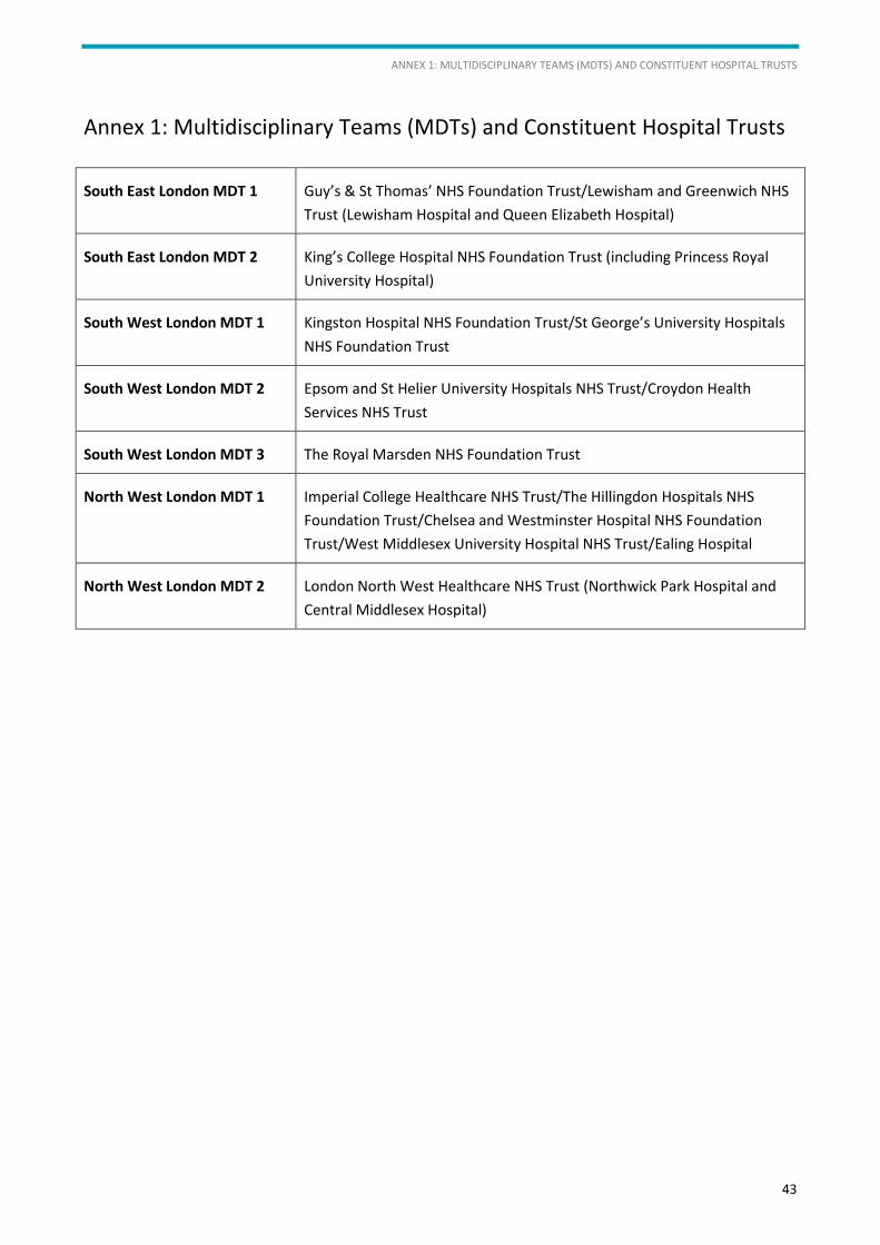
ANNEX 1: MULTIDISCIPLINARY TEAMS (MDTS) AND CONSTITUENT HOSPITAL TRUSTS
43
Annex 1: Multidisciplinary Teams (MDTs) and Constituent Hospital Trusts
South East London MDT 1 Guy’s & St Thomas’ NHS Foundation Trust/Lewisham and Greenwich NHS
Trust (Lewisham Hospital and Queen Elizabeth Hospital)
South East London MDT 2 King’s College Hospital NHS Foundation Trust (including Princess Royal
University Hospital)
South West London MDT 1 Kingston Hospital NHS Foundation Trust/St George’s University Hospitals
NHS Foundation Trust
South West London MDT 2 Epsom and St Helier University Hospitals NHS Trust/Croydon Health
Services NHS Trust
South West London MDT 3 The Royal Marsden NHS Foundation Trust
North West London MDT 1 Imperial College Healthcare NHS Trust/The Hillingdon Hospitals NHS
Foundation Trust/Chelsea and Westminster Hospital NHS Foundation
Trust/West Middlesex University Hospital NHS Trust/Ealing Hospital
North West London MDT 2 London North West Healthcare NHS Trust (Northwick Park Hospital and
Central Middlesex Hospital)
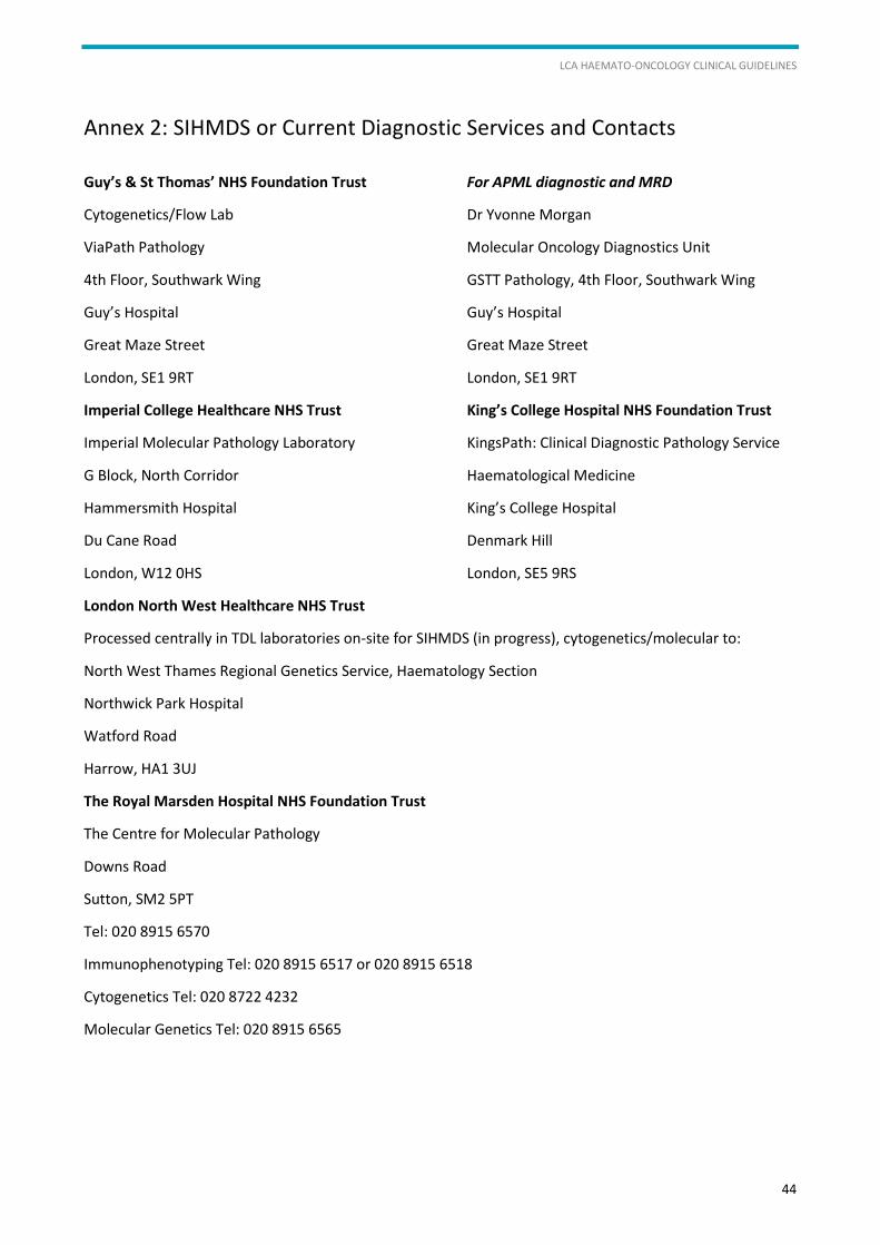
LCA HAEMATO-ONCOLOGY CLINICAL GUIDELINES
44
Annex 2: SIHMDS or Current Diagnostic Services and Contacts
Guy’s & St Thomas’ NHS Foundation Trust
Cytogenetics/Flow Lab
ViaPath Pathology
4th Floor, Southwark Wing
Guy’s Hospital
Great Maze Street
London, SE1 9RT
For APML diagnostic and MRD
Dr Yvonne Morgan
Molecular Oncology Diagnostics Unit
GSTT Pathology, 4th Floor, Southwark Wing
Guy’s Hospital
Great Maze Street
London, SE1 9RT
Imperial College Healthcare NHS Trust
Imperial Molecular Pathology Laboratory
G Block, North Corridor
Hammersmith Hospital
Du Cane Road
London, W12 0HS
King’s College Hospital NHS Foundation Trust
KingsPath: Clinical Diagnostic Pathology Service
Haematological Medicine
King’s College Hospital
Denmark Hill
London, SE5 9RS
London North West Healthcare NHS Trust
Processed centrally in TDL laboratories on-site for SIHMDS (in progress), cytogenetics/molecular to:
North West Thames Regional Genetics Service, Haematology Section
Northwick Park Hospital
Watford Road
Harrow, HA1 3UJ
The Royal Marsden Hospital NHS Foundation Trust
The Centre for Molecular Pathology
Downs Road
Sutton, SM2 5PT
Tel: 020 8915 6570
Immunophenotyping Tel: 020 8915 6517 or 020 8915 6518
Cytogenetics Tel: 020 8722 4232
Molecular Genetics Tel: 020 8915 6565
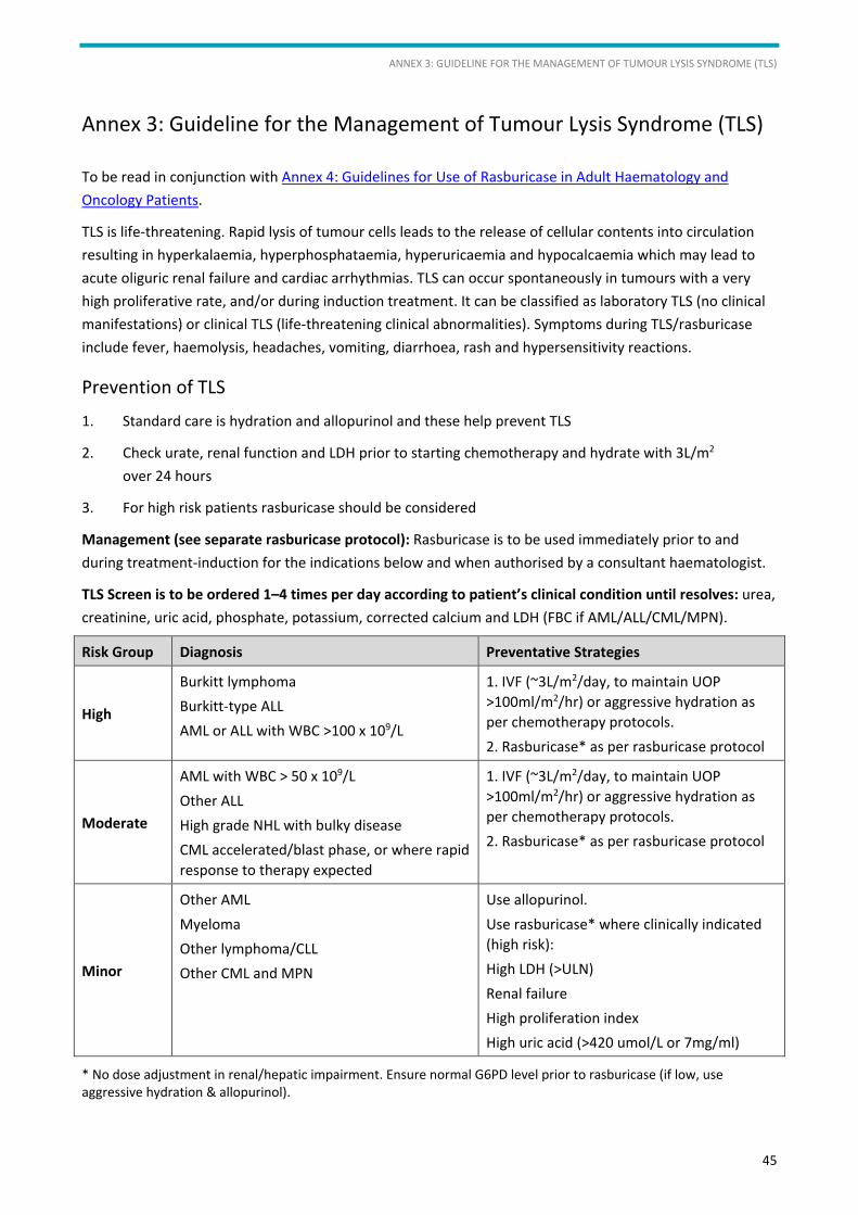
ANNEX 3: GUIDELINE FOR THE MANAGEMENT OF TUMOUR LYSIS SYNDROME (TLS)
45
Annex 3: Guideline for the Management of Tumour Lysis Syndrome (TLS)
To be read in conjunction with Annex 4: Guidelines for Use of Rasburicase in Adult Haematology and Oncology Patients.
TLS is life-threatening. Rapid lysis of tumour cells leads to the release of cellular contents into circulation resulting in hyperkalaemia, hyperphosphataemia, hyperuricaemia and hypocalcaemia which may lead to acute oliguric renal failure and cardiac arrhythmias. TLS can occur spontaneously in tumours with a very high proliferative rate, and/or during induction treatment. It can be classified as laboratory TLS (no clinical manifestations) or clinical TLS (life-threatening clinical abnormalities). Symptoms during TLS/rasburicase include fever, haemolysis, headaches, vomiting, diarrhoea, rash and hypersensitivity reactions.
Prevention of TLS
1. Standard care is hydration and allopurinol and these help prevent TLS
2. Check urate, renal function and LDH prior to starting chemotherapy and hydrate with 3L/m2 over 24 hours
3. For high risk patients rasburicase should be considered
Management (see separate rasburicase protocol): Rasburicase is to be used immediately prior to and during treatment-induction for the indications below and when authorised by a consultant haematologist.
TLS Screen is to be ordered 1–4 times per day according to patient’s clinical condition until resolves: urea, creatinine, uric acid, phosphate, potassium, corrected calcium and LDH (FBC if AML/ALL/CML/MPN).
Risk Group Diagnosis Preventative Strategies
High
Burkitt lymphoma Burkitt-type ALL AML or ALL with WBC >100 x 109/L
1. IVF (~3L/m2/day, to maintain UOP >100ml/m2/hr) or aggressive hydration as per chemotherapy protocols. 2. Rasburicase* as per rasburicase protocol
Moderate
AML with WBC > 50 x 109/L Other ALL High grade NHL with bulky disease CML accelerated/blast phase, or where rapid response to therapy expected
1. IVF (~3L/m2/day, to maintain UOP >100ml/m2/hr) or aggressive hydration as per chemotherapy protocols. 2. Rasburicase* as per rasburicase protocol
Minor
Other AML Myeloma Other lymphoma/CLL Other CML and MPN
Use allopurinol. Use rasburicase* where clinically indicated (high risk): High LDH (>ULN) Renal failure High proliferation index High uric acid (>420 umol/L or 7mg/ml)
* No dose adjustment in renal/hepatic impairment. Ensure normal G6PD level prior to rasburicase (if low, use aggressive hydration & allopurinol).

LCA HAEMATO-ONCOLOGY CLINICAL GUIDELINES
46
References:
Cairo MS, Bishop M. Tumour lysis syndrome: New therapeutic strategies and classification. Br J Haematology 2004; 127: 3–11.
Coiffier B, Altman A, Pui CH et al. Guidelines for the management of paediatric and adult tumour lysis syndrome: an evidence-based review. Journal of Clinical Oncology 2008; 26: 2767–78.
Howard SC, Jones DP and Pui CH. The tumor lysis syndrome. NEJM 2011; 364(19): 1844–54.
Hummel M, Reiter S, Adam K et al. Effective treatment and prophylaxis of hyperuricemia and impaired renal function in tumor lysis syndrome with low doses of rasburicase. Eur J Haematol 2008; 80: 331–6.
Jones GL, Will A, Jackson GH et al. Guidelines for the management of tumour lysis syndrome in adults and children with haematological malignancies on behalf of the British Committee for Standards in Haematology. British Journal of Haematology 169(5): 661–71.
NHS Economic Evaluation Database, NIHR Centre for Reviews and Dissemination. The University of York, 2012.
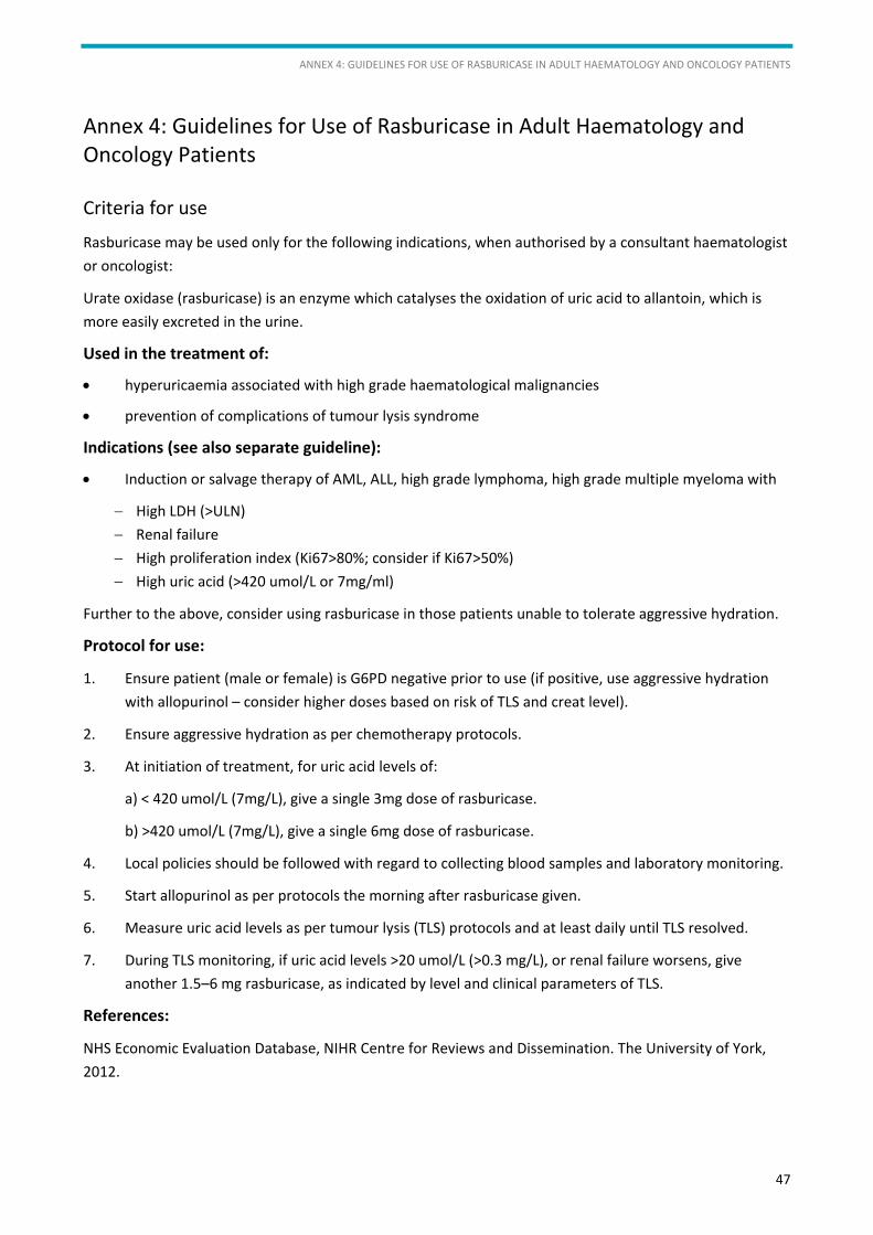
ANNEX 4: GUIDELINES FOR USE OF RASBURICASE IN ADULT HAEMATOLOGY AND ONCOLOGY PATIENTS
47
Annex 4: Guidelines for Use of Rasburicase in Adult Haematology and Oncology Patients
Criteria for use
Rasburicase may be used only for the following indications, when authorised by a consultant haematologist or oncologist:
Urate oxidase (rasburicase) is an enzyme which catalyses the oxidation of uric acid to allantoin, which is more easily excreted in the urine.
Used in the treatment of:
• hyperuricaemia associated with high grade haematological malignancies
• prevention of complications of tumour lysis syndrome
Indications (see also separate guideline):
• Induction or salvage therapy of AML, ALL, high grade lymphoma, high grade multiple myeloma with
− High LDH (>ULN) − Renal failure − High proliferation index (Ki67>80%; consider if Ki67>50%) − High uric acid (>420 umol/L or 7mg/ml)
Further to the above, consider using rasburicase in those patients unable to tolerate aggressive hydration.
Protocol for use:
1. Ensure patient (male or female) is G6PD negative prior to use (if positive, use aggressive hydration with allopurinol – consider higher doses based on risk of TLS and creat level).
2. Ensure aggressive hydration as per chemotherapy protocols.
3. At initiation of treatment, for uric acid levels of:
a) < 420 umol/L (7mg/L), give a single 3mg dose of rasburicase.
b) >420 umol/L (7mg/L), give a single 6mg dose of rasburicase.
4. Local policies should be followed with regard to collecting blood samples and laboratory monitoring.
5. Start allopurinol as per protocols the morning after rasburicase given.
6. Measure uric acid levels as per tumour lysis (TLS) protocols and at least daily until TLS resolved.
7. During TLS monitoring, if uric acid levels >20 umol/L (>0.3 mg/L), or renal failure worsens, give another 1.5–6 mg rasburicase, as indicated by level and clinical parameters of TLS.
References:
NHS Economic Evaluation Database, NIHR Centre for Reviews and Dissemination. The University of York, 2012.
LCA HAEMATO-ONCOLOGY CLINICAL GUIDELINES
48
Trifilio SM, Pi J, Zook J et al. Effectiveness of a single 3-mg rasburicase dose for the management of hyperuricemia in patients with haematological malignancies. Bone Marrow Transplantation 2011; 46: 800–805.
Hummel M, Reiter S, Adam K et al. Effective treatment and prophylaxis of hyperuricemia and impaired renal function in tumor lysis syndrome with low doses of rasburicase. Eur J Haematol 2008; 80: 331–6.
Trifilio S, Gordon L, Singhal S et al. Reduced-dose rasburicase (recombinant xanthine oxidase) in adult cancer patients with hyperuricemia. Bone Marrow Transplant 2006; 37: 997–1001.
Hutcherson DA, Gammon DC, Bhatt MS and Faneuf M. Reduced-dose rasburicase in the treatment of adults with hyperuricemia associated with malignancy. Pharmacotherapy 2006; 26: 242–7.
Special warnings and precautions for use
Allergic reactions may occur with this product, patients should be closely monitored and full resuscitation facilities should be at hand. Should any serious allergic or anaphylactic reaction occur treatment should be immediately discontinued and appropriate resuscitation given.
Caution should be exercised in patients with a history of atopic allergies.
Administration of rasburicase decreases serum uric acid to below normal levels, but has no direct effect in reversing hyperphosphataemia, hyperkalaemia and hypocalcaemia. If severe these abnormalities should be corrected following standard treatment guidelines.
There are limited data available to recommend the sequential use of rasburicase and allopurinol.
To ensure accurate measurement of uric acid plasma level during treatment with rasburicase, a strict sample handling procedure must be followed to minimise ex vivo degradation of the analyte. Local policies should be followed with regard to collecting blood samples and laboratory monitoring.
APPENDICES
49
Appendices
Appendix 1: 2 Week Wait Referral Forms
North West London
South East London
South West London
Appendix 2: Treatment of Children
Appendix 3: Treatment of Teenagers and Young Adults
Teenagers and Young Adults PTC Referrals
Teenagers and Young Adults MDT Proforma
Appendix 4: LCA Key Worker Policy
Appendix 5: LCA Holistic Needs Assessment Tool
Appendix 6: NCSI Treatment Summary
Appendix 7: LCA Specialist Palliative Care Referral Form
Appendix 8: LCA Referral Criteria to Specialist Palliative Care

© London Cancer Alliance 2015
Published by London Cancer Alliance
London Cancer Alliance
5th Floor Alliance House
12 Caxton Street
London SW1H 0QS
www.londoncanceralliance.nhs.uk


![Hematological malignancies - БГМУHematological malignancies Leukemia is a malignant proliferation of white blood cells (lymphoid cells [lymphocytes] or myeloid cells [granulocytes](https://img.pdfslide.us/doc/110x75/5f0624c37e708231d416825d/hematological-malignancies-oe-hematological-malignancies-leukemia-is-a-malignant.jpg)