Embed Size (px)
Citation preview
Cell Tissue Res (2005) 319: 267–278DOI 10.1007/s00441-004-0981-8
REGULAR ARTICLE
Maria Giovanna Sabbieti . Luigi Marchetti .Maria Gabriella Gabrielli . Maura Menghi .Stefano Materazzi . Giovanna Menghi .Lawrence G. Raisz . Marja M. Hurley
Prostaglandins differently regulate FGF-2 and FGF receptorexpression and induce nuclear translocation in osteoblasts viaMAPK kinaseReceived: 18 November 2003 / Accepted: 24 June 2004 / Published online: 10 December 2004# Springer-Verlag 2004
Abstract We have previously reported that prostaglandinF2α (PGF2α) and its selective agonist fluprostenol increasebasic fibroblast growth factor (FGF-2) mRNA and proteinproduction in osteoblastic Py1a cells. The present reportextends our previous studies by showing that Py1a cellsexpress FGF receptor-2 (FGFR2) and that treatment withPGF2α or fluprostenol decreases FGFR2 mRNA. We haveused confocal and electron microscopy to show that, underPGF2α stimulation, FGF-2 and FGFR2 proteins accumu-late near the nuclear envelope and colocalize in thenucleus of Py1a cells. Pre-treatment with cycloheximideblocks nuclear labelling for FGF-2 in response to PGF2α.Treatment with SU5402 does not block prostaglandin-mediated nuclear internalization of FGF-2 or FGFR2.Various effectors have been used to investigate the signaltransduction pathway. In particular, pre-treatment withphorbol 12-myristate 13-acetate (PMA) prevents thenuclear accumulation of FGF-2 and FGFR2 in responseto PGF2α. Similar results are obtained by pre-treatmentwith the protein kinase C (PKC) inhibitor H-7. In addition,
cells treated with PGF2α exhibit increased nuclearlabelling for the mitogen-activated protein kinase(MAPK), p44/ERK2. Pre-treatment with PMA blocksprostaglandin-induced ERK2 nuclear labelling, as con-firmed by Western blot analysis. We conclude that PGF2αstimulates nuclear translocation of FGF-2 and FGFR2 by aPKC-dependent pathway; we also suggest an involvementof MAPK/ERK2 in this process.
Keywords Fibroblast growth factor-2/FGF receptor-2 .Mitogen-activated protein kinases/ERK2 . ProstaglandinF2α . Confocal laser scanning microscopy . Transmissionelectron microscopy . Rat osteoblasts
Introduction
Prostaglandins (PGs) have both stimulatory and inhibitoryeffects on bone formation in cell and organ culture (Raiszand Koolemans-Beynen 1974; Raisz and Fall 1990; Raiszet al. 1993). Data concerning structure-activity relationsand signal transduction mechanism for the anabolic effectsof PGs are conflicting. The stimulation of cyclic adenosinemonophosphate (cAMP) production is probably importantwith respect to the ability of these compounds to increasebone formation (Nagata et al. 1994; Scutt et al. 1995;Woodiel et al. 1996), whereas the activation of proteinkinase C (PKC) has been implicated in their mitogeniceffect on osteoblasts and their inhibitory effect on collagensynthesis (Quarles et al. 1993; Fall et al. 1994).
The effects of PGs on bone are similar to those of basicfibroblast growth factor-2 (FGF-2). FGF-2 is a member ofa protein family of which there are 23 members; theseproteins function in an autocrine or paracrine manner tomodulate the proliferation and differentiation of cell typesof epithelial, neuroectodermal and mesenchymal origin(Hurley et al. 2002). FGF-2 is produced by cells of theosteoblast lineage and can act as a local regulator of boneremodelling; like PGs, FGF-2 stimulates bone cell repli-cation (Hurley et al. 2002; Globus et al. 1988, 1989),
This research was supported by grants from University of Camerinoand Fondazione Carima Italy and by National Institutes of HealthGrant AR-46025 (to M.M.H)
M. G. Sabbieti (*) . L. Marchetti . M. G. Gabrielli .M. Menghi . G. MenghiDepartment of Comparative Morphology and Biochemistry,Via Gentile III da Varano,62032 Camerino (MC), Italye-mail: [email protected].: +39-737-402713Fax: +39-737-402708
S. MaterazziDepartment of Chemistry, University “La Sapienza”,Piazzale Aldo Moro,10 Rome, Italy
L. G. Raisz . M. M. HurleyDivision of Endocrinology and Metabolism, University ofConnecticut School of Medicine,263 Farmington Avenue,Farmington, CT 06030-1850, USA
increases bone resorption (Shen et al. 1989), reducesalkaline phosphatase activity (Rodan et al. 1989) andinhibits type I collagen synthesis in osteoblasts (McCarthyet al. 1989; Hurley et al. 1993). Intermittent FGF-2treatment stimulates bone formation in vitro (Canalis et al.1988) and in vivo (Norrdin et al. 1990; Aspenberg et al.1991; Nagata et al. 1994). We have previously reportedthat prostaglandin F2α (PGF2α) and its selective agonistfluprostenol (Flup) regulate the expression of FGF-2 inimmortalized rat osteoblastic Py1a cells and that Flup isable to induce nuclear translocation of FGF-2 (Sabbieti etal. 1999). The signalling pathway for FGF-2 is complexand involves the binding and activation of one or morehigh affinity tyrosine kinase receptors at the cell surfaceresulting in ligand/receptor activation of downstreamgenes.
Some authors have demonstrated a role for extracellularsignal-regulated kinase (ERK) in the biological responsesto both PGs and FGF-2 in osteoblasts. ERK is a memberof the family of mitogen-activated protein kinases(MAPKs) involved in the regulation of cell growth,differentiation and apoptosis (Nishida and Gotoh 1993;Matsuda et al. 1998). Activation of the ERK signallingpathway has recently been shown to be involved in theinduction of cyclo-oxygenase (Cox-2), the rate-limitingenzyme in the production of PGs (Wadhwa et al. 2002),and in anabolic responses to growth factors in osteoblast-like cells (Chaudhary and Avioli 1998). Since earlierstudies have demonstrated that acidic FGF and phorbolesters preferentially activate ERKs in COS cells (Klingen-berg et al. 2000), we have examined whether the activationof ERK is important in the PG regulation of the expressionof FGF-2 and FGFR2.
In addition to the activation of intracellular signallingproteins, recent studies have shown that certain FGFslacking nuclear localization sequences can be transportedto the nucleus following internalization from the cellsurface (Mehta et al. 1998). In this report, we haveextended our studies to examine whether PGs also regulateFGFR mRNA expression and the signalling pathwayinvolved. Confocal laser scanning and electron microsco-py have been utilized to demonstrate that PG induces thenuclear translocation of FGF-2/FGFR2 via the MAPK,p44/ERK2 signalling pathway in rat osteoblastic Py1acells.
Materials and methods
Cell cultures
Immortalized rat osteoblastic Py1a cells were plated at5,000 cells/cm2 in 6-well culture dishes in F-12 culturemedium with 5% non-heat inactivated fetal calf serum(FCS; Invitrogen, Milan, Italy), penicillin and streptomy-cin (Sigma-Aldrich, Milan, Italy). Cells were grown for 6days to approximately 80% confluence. These cells werethen pre-cultured for another 24 h in serum-free F-12 withantibiotics and 1 mg/ml BSA before treatment with PGF2α
(10−5 M; Sigma-Aldrich) and Flup (10−8 M; BiomolResearch Laboratories, Plymouth). Control cultures weretreated with appropriate vehicles (Sabbieti et al. 1999).
mRNA levels
Total RNA was extracted from cells by the method ofChomczynski and Sacchi (1987). Cells were scraped into4 M guanidinium thiocyanate and extracted with phenol/chloroform isoamyl alcohol (24:1) and total RNA wasprecipitated with isopropanol. For Northern analysis,20 μg total RNA was denaturated and fractionated on a1% agarose/2.2 M formaldehyde gel, transferred to nylonmembrane by positive pressure, and fixed to the filter byUV irradiation (Sambrook et al. 1989; Towbin et al. 1979).After 4 h pre-hybridization, filters were hybridizedovernight with a random primer [32P]dCTP-labelledcDNA probe for the mRNA of interest (Mansukani et al.1990). Filters were washed in 1× SSC (150 mM NaCl,15 mM sodium citrate, pH 7.0), 1% SDS solution at roomtemperature and then three times at 65°C in 0.1% SDS andfinally exposed to XAR-5 film at −70°C. Signals werequantified by densitometry and normalized to thecorresponding values for glyceraldehyde 3-phosphatedehydrogenase (G3PDH).
Cell culture for Western blotting
Py1a cells were plated at 5,000 cells/cm2 in 100-mmculture dishes, in Ham’s F-12 culture medium with 5%non-heat inactivated FCS, penicillin and streptomycin.Cells were grown for 5 days to confluence. The cells weresubsequently serum-deprived 24 h before addition ofPGF2α (10−5 M).
Protein levels
Proteins were extracted with Cytobuster Protein extractionreagent (Calbiochem-Inalco, Milan, Italy), resolved bySDS-polyacrylamide gel electrophoresis (12%) and trans-ferred onto polyvinylidene fluoride (Hybond-P) membrane(Amersham Biosciences). The subsequent steps wereperformed with the ECL Advance Western BlottingDetection Kit (Amersham Biosciences); in brief, mem-branes were blocked with Advance Blocking agent inPBS-T (phosphate-buffered saline containing 0.1% Tween20) for 1 h at room temperature. The membranes were thenincubated with a polyclonal rabbit anti-ERK2 antibody(Calbiochem-Inalco) diluted 1:500 in blocking solution for1 h at room temperature. After being washed with PBS-T,the blots were incubated with horseradish peroxidase(HRP)-conjugated goat anti-rabbit IgG (Amersham Bio-sciences) diluted 1:100,000 in blocking solution for 1 h atroom temperature. After further washes with PBS-T,immunoreactive bands were visualized by using luminol
268
reagents and Hyperfilm-ECL film (Amersham Bioscienc-es) according to the manufacturer’s instructions.
Confocal laser scanning microscopy
FGF-2 and FGFR2 protein immunodetection
Py1a cells were plated at 3,500 cells/cm2 in 6-well culturedishes containing coverslips (previously cleaned andsterilized) in F-12 culture medium with 5% FCS (Sabbietiet al. 2000). Cells were grown for 4 days to approximately80% confluence and then pre-cultured for another 24 h inserum-free F-12 with antibiotics and 1 mg/ml BSA beforetreatment with PGF2α (10−5 M) and Flup (10−8 M).Control cultures were treated with appropriate vehicles.After stimulation, cells were briefly rinsed with PBS0.1 M, pH 7.4 and fixed in 4% paraformaldehyde (PFA)diluted in PBS for 25 min at room temperature. Cells werewashed three times with PBS, permeabilized with 0.3%Triton X-100 for 30 min and incubated with 0.5% BSAdiluted in PBS for 20 min at room temperature. Cells wereincubated with a 1:100 dilution of a polyclonal rabbit anti-FGF-2 antibody (Sigma-Aldrich) in PBS for 2 h at roomtemperature. After being rinsed, cells were incubated witha 1:75 dilution of goat anti-rabbit IgG conjugated withfluorescein isothiocyanate (FITC; Sigma-Aldrich) in PBSfor 90 min at room temperature. To detect FGFR2, fixedand permeabilized cells were incubated with a 1:14dilution of a polyclonal goat anti-C-term-FGFR2 antibody(Bek C-17; Santa Cruz Biotechnology, Santa Cruz, Calif.,USA) in PBS for 2 h at room temperature. After beingwashed, cells were incubated with a 1:150 dilution ofAlexa Fluor 594 donkey anti-goat IgG conjugate (Molec-ular Probes, USA) diluted in PBS for 90 min at roomtemperature. Control experiments were performed byusing a non-immune goat IgG, by complexing the primaryantibody with a relative blocking peptide or by omittingthe primary antibody. After a washing step, coverslipswere mounted on slides with PBS/glycerol (1:1).
Double-labelling for FGF-2 and FGFR2
Fixed and permeabilized cells were incubated sequentiallywith anti-FGF-2 antibody, anti-FGFR2 antibody, AlexaFluor 594 donkey anti-goat conjugate and goat anti-rabbitIgG FITC conjugate to prevent cross-reactivity of thesecondary antibodies. Control experiments for double-labelling included additional tests; in order to test thecross-reactivity of the secondary antibodies, incubationswith only the Alexa Fluor 594 donkey anti-goat IgGconjugate and the goat anti-rabbit IgG-FITC wereperformed.
ERK2 immunodetection
After fixation and permeabilization as above, cells wereincubated with 1:60 dilution of a polyclonal rabbit anti-ERK2 (Calbiochem-Inalco) in PBS for 2 h at roomtemperature. After being washed, cells were incubated in a1:75 dilution of goat anti-rabbit IgG conjugate with FITCin PBS for 90 min at room temperature. Controlexperiments were performed by using a non-immunerabbit IgG and by omitting the primary antibody. After awash step, coverslips were mounted on slides with PBS/glycerol (1:1).
Confocal analysis
FGF-2-binding sites were visualized by means of a NikonDiaphot-TMD-EF inverted microscope with a 60× oilimmersion lens with a numerical aperture 1.4 Plan Apoobjective. The microscope was attached to a Bio-RadMRC 600 confocal laser imaging system (Bio-Rad,Hertfordshire, UK) equipped with a krypton/argon laser.Black level, gain and laser intensity, Kalman averaging,excitation intensity, pinhole aperture and Z-series analysisof cells were carried out as previously detailed (Sabbieti etal. 2000). Sections were examined and original imageswere stored as a PIC format file and then printed with anEpson Stylus Photo 890 on Epson glossy photo paper.
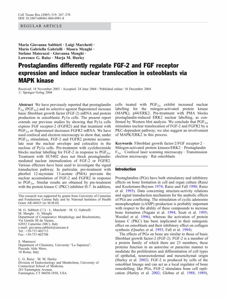
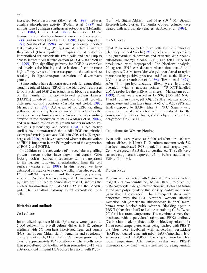
Fig. 1 Time-course effect of PGF2α (a) and Flup (b) on FGFR2mRNA expression in Py1a cells. Cells were treated with effectorsfor the indicated times. Total RNA was extracted from cells and asample of 20 μg was utilized for Northern analysis. Filters wereprobed for FGFR2 and bands were quantified by densitometry andnormalized to G3PDH
269
Transmission electron microscopy
Cell cultures for immunoelectron microscopy
Py1a cells were plated at 3,500 cells/cm2 on 100-mmculture dishes and grown for 6 days in F-12 medium with5% FCS, penicillin and streptomycin. At confluence, cellswere pre-cultured in serum-free F-12 containing antibio-tics and 1 mg/ml BSA and then treated with vehicle orPGF2α (10−5 M) for 6 h and 24 h. After two rinses in F-12medium and one rapid wash in 0.1 M cacodylate buffer,pH 7.4, cells were fixed on plates with 4% PFA and 0.5%glutaraldehyde in 0.1 M cacodylate buffer, pH 7.4, for 3 hat 4°C. The cells were rinsed several times for a total of30 min in 0.1 M PBS, pH 7.4, containing 0.1% BSA and7% sucrose, at 4°C and subsequently scraped off the platesand collected in Falcon tubes. The centrifuged cells(1,300 rpm for 4 min) were placed on 2.6% agarose and,after centrifugation, cells pre-embedded in agarose weredehydrated in methanol from 50% up to 90% andembedded in Unicryl resin (British Bio Cell International,Cardiff, UK) for 72 h at 4°C under UV lamp. Ultrathinsections (about 60 nm in thickness) of the plastic-
embedded cells were cut by means of an LKB UltrotomeV and collected on uncoated 400-mesh nickel grids.
Immunogold labelling for FGF-2 and FGFR2 andtransmission electron-microscopy analysis
Floating grids were rehydrated with 0.05 M TRIS-bufferedsaline (TBS), pH 7.6, and pre-incubated with 1% BSA in0.05 M TBS, pH 7.6, for 30 min at room temperature.Grids were then incubated with polyclonal rabbit anti-FGF-2 antibody diluted 1:600 in 1% BSA in 0.05 M TBS,pH 7.6, for 3 h at 4°C in a humid chamber or withpolyclonal goat anti-C-term-FGFR2 antibody diluted 1:10in 1% BSA in 0.05 M TBS, pH 7.6, overnight at 4°C in ahumid chamber. After being rinsed in 0.05 M TBS, pH7.6, and pre-incubation with 1% BSA in 0.05 M TBS, pH7.6, for 10 min, grids were incubated with Auroprobe EMgoat anti-rabbit G-10 (10-nm gold labelled IgG; Amer-sham Life Science, Buckinghamshire, England) diluted1:15 in 0.05 M TBS, pH 7.6, containing 0.05% Tween 20or with rabbit anti-goat IgG conjugated to 10-nm golddiluted 1:15 in 0.05 M TBS, pH 7.6, containing 0.05%
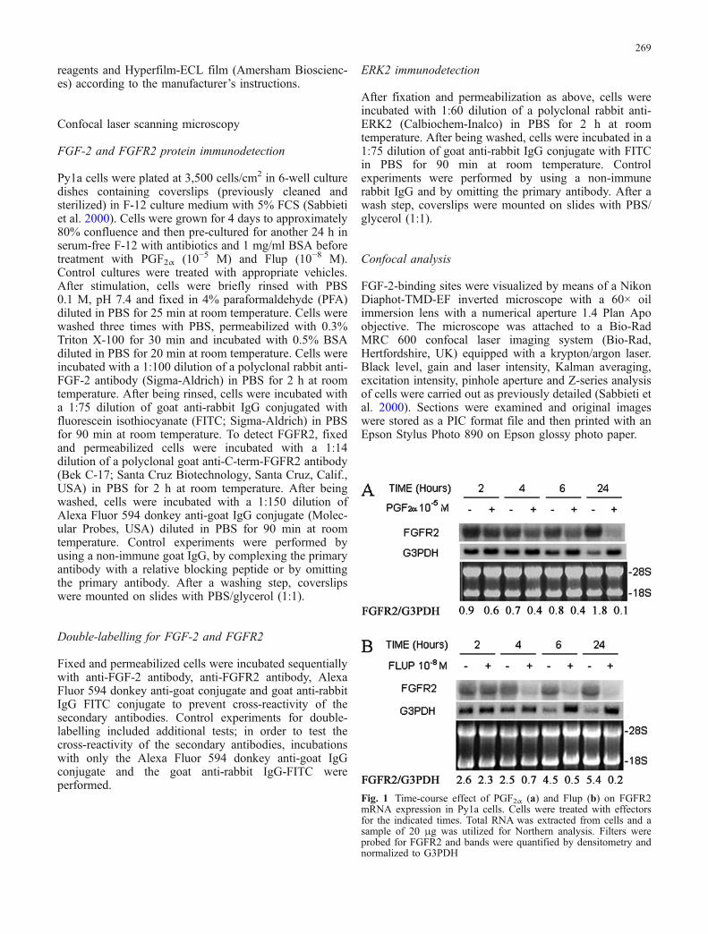
Fig. 2 Untreated Py1a osteoblasts (left column) or Py1a osteoblaststreated with PGF2α for 6 h (middle column) and 24 h (right column).Optical sections obtained on a Bio-Rad MRC-600 confocal laserscanning microscope (CLSM). Micrographs showing cells double-stained with FGF-2 (green pseudo colour) and FGFR2 (red pseudocolour) in the same area. Colocalization of the two signalscorresponding to FGF-2 (green FITC staining) and FGFR2 (red
Alexa Fluor 594 staining), assessed by confocal analysis of singleoptical sections, is shown as a yellow pseudo colour in thecomposite merged images. The intensity-coded scales, with whitebeing the most intense, are shown right. Note that the basal labellingfor FGF-2 and FGFR2 changes after treatment with PGF2α for 6 hand 24 h when both proteins can also be detected in some cells.×400
270
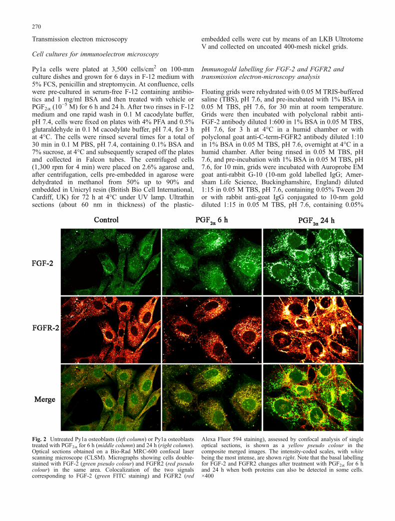
Fig. 3 Py1a cells treated for24 h with PGF2α. CLSM-scanned optical sections ob-tained from the base (top left) tothe apex (bottom right) of re-presentative cells labelled forFGF-2 (a) and FGFR2 (b). Thenuclear translocation of bothproteins seems to involve arestricted region of the nuclearenvelope. ×750
271
Tween 20 and 5% fetal bovine serum for 90 min at roomtemperature in a humid chamber. Sections were thenwashed several times in 0.05 M TBS, pH 7.6, and distilledwater. Control experiments were carried out by omittingthe primary antibody or by using a non-immune rabbitIgG. Sections were finally counterstained with uranylacetate (5 min) and lead citrate (2 min) at roomtemperature. All specimens were analysed by means of aPhilips EM 201C electron microscope at an acceleratingvoltage of 60 kV.
Results
Expression of FGFR2 mRNA in osteoblastic Py1acells
Time-course studies of the effects of PGs on FGFR2mRNA expression in osteoblastic Py1a cells weredetermined by Northern analysis. Representative experi-ments are shown in Fig. 1a, b). In the absence of serum,Py1a cells expressed a 4-kb FGFR2 mRNA transcript.PGF2α (10−5 M) and Flup (10−8 M) caused a reduction inFGFR2 mRNA levels at 4 h and both effectors caused amaximal down-regulation at 24 h.
Immunodetection of FGF-2 and FGFR2 proteins
Double-labelling of Py1a cells (Fig. 2, left column) thathad been serum-deprived for 24 h showed a prominentbasal cytoplasmic staining for both FGF-2 and FGFR2.Treatment with PGF2α (10−5 M) or Flup (10−8 M) for 6 h
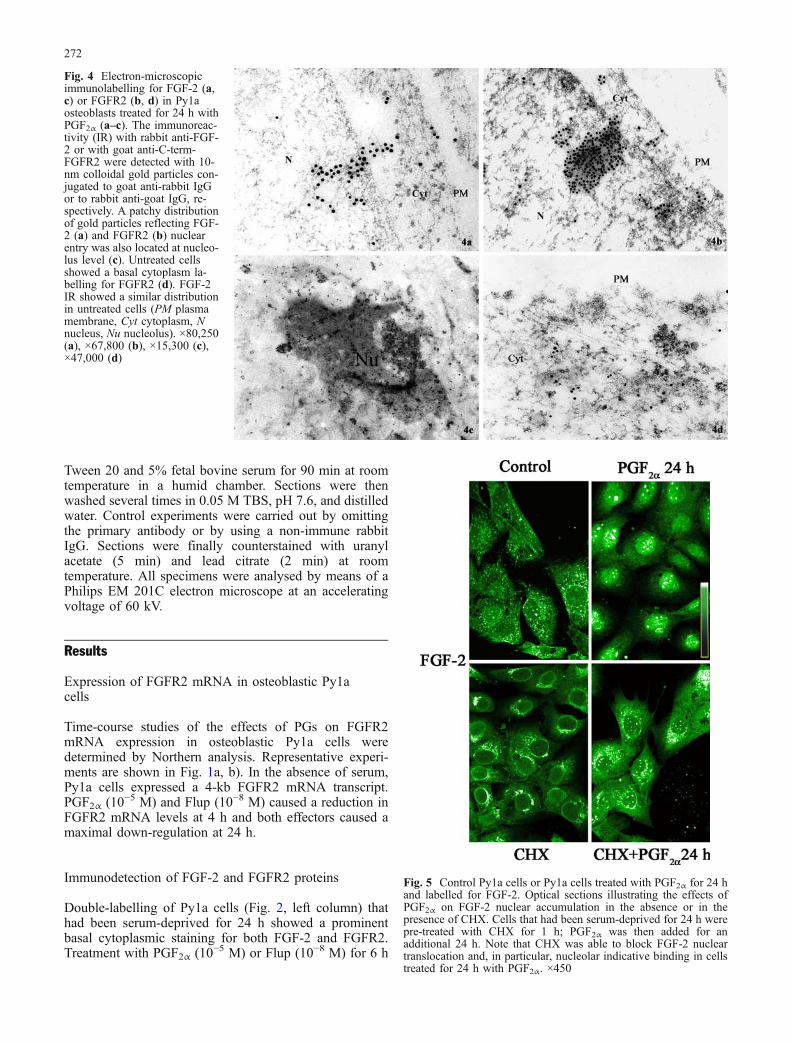
Fig. 4 Electron-microscopicimmunolabelling for FGF-2 (a,c) or FGFR2 (b, d) in Py1aosteoblasts treated for 24 h withPGF2α (a–c). The immunoreac-tivity (IR) with rabbit anti-FGF-2 or with goat anti-C-term-FGFR2 were detected with 10-nm colloidal gold particles con-jugated to goat anti-rabbit IgGor to rabbit anti-goat IgG, re-spectively. A patchy distributionof gold particles reflecting FGF-2 (a) and FGFR2 (b) nuclearentry was also located at nucleo-lus level (c). Untreated cellsshowed a basal cytoplasm la-belling for FGFR2 (d). FGF-2IR showed a similar distributionin untreated cells (PM plasmamembrane, Cyt cytoplasm, Nnucleus, Nu nucleolus). ×80,250(a), ×67,800 (b), ×15,300 (c),×47,000 (d)
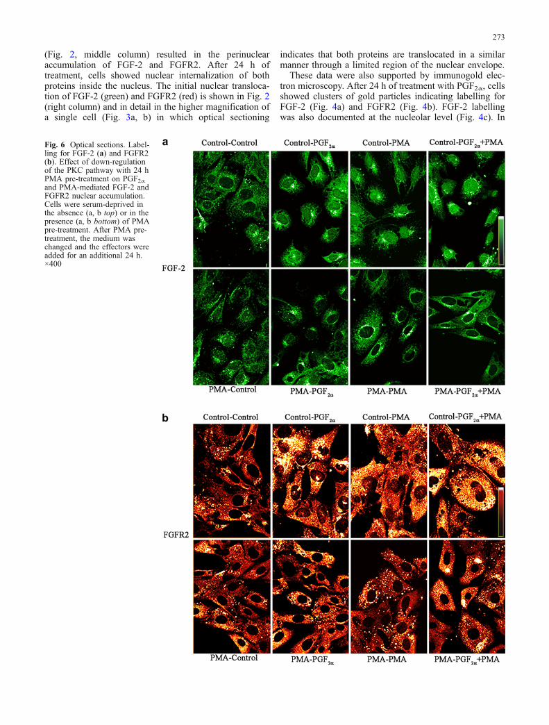
Fig. 5 Control Py1a cells or Py1a cells treated with PGF2α for 24 hand labelled for FGF-2. Optical sections illustrating the effects ofPGF2α on FGF-2 nuclear accumulation in the absence or in thepresence of CHX. Cells that had been serum-deprived for 24 h werepre-treated with CHX for 1 h; PGF2α was then added for anadditional 24 h. Note that CHX was able to block FGF-2 nucleartranslocation and, in particular, nucleolar indicative binding in cellstreated for 24 h with PGF2α. ×450
272
(Fig. 2, middle column) resulted in the perinuclearaccumulation of FGF-2 and FGFR2. After 24 h oftreatment, cells showed nuclear internalization of bothproteins inside the nucleus. The initial nuclear transloca-tion of FGF-2 (green) and FGFR2 (red) is shown in Fig. 2(right column) and in detail in the higher magnification ofa single cell (Fig. 3a, b) in which optical sectioning
indicates that both proteins are translocated in a similarmanner through a limited region of the nuclear envelope.
These data were also supported by immunogold elec-tron microscopy. After 24 h of treatment with PGF2α, cellsshowed clusters of gold particles indicating labelling forFGF-2 (Fig. 4a) and FGFR2 (Fig. 4b). FGF-2 labellingwas also documented at the nucleolar level (Fig. 4c). In
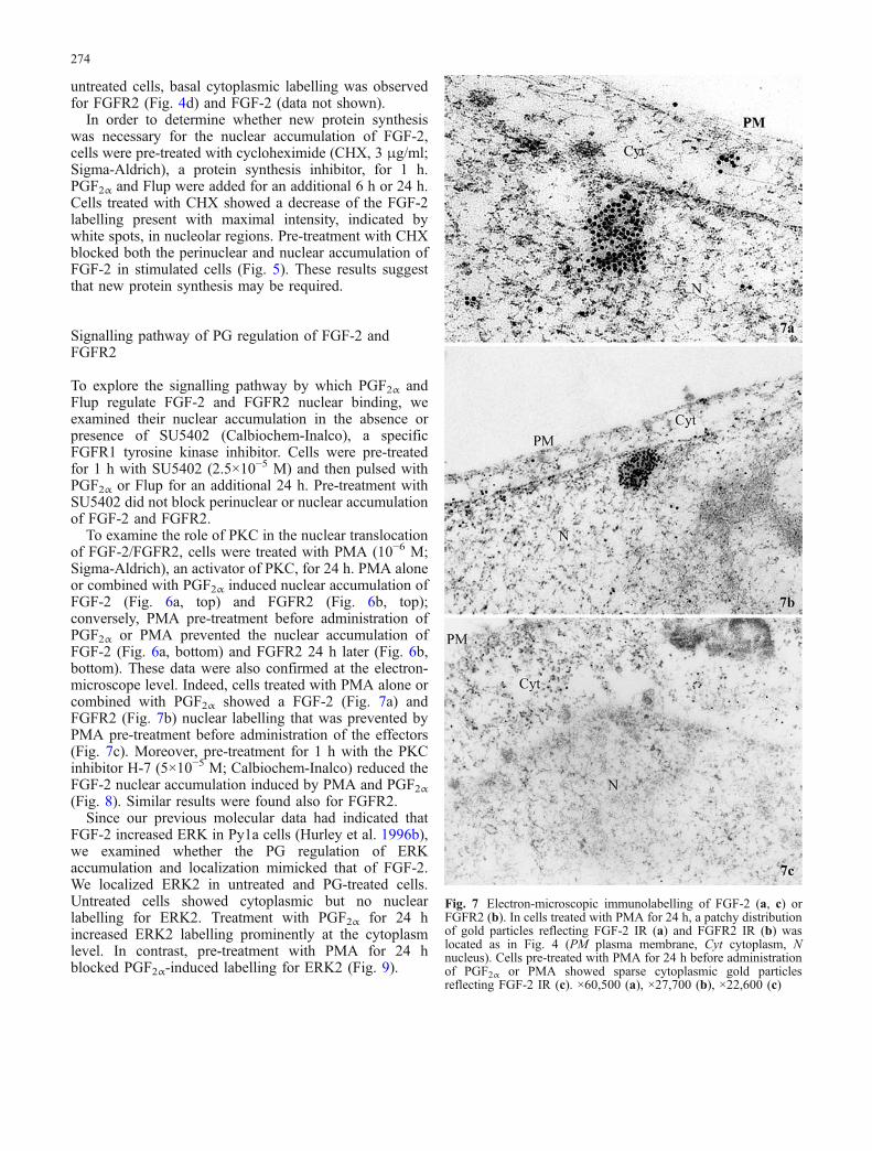
Fig. 6 Optical sections. Label-ling for FGF-2 (a) and FGFR2(b). Effect of down-regulationof the PKC pathway with 24 hPMA pre-treatment on PGF2αand PMA-mediated FGF-2 andFGFR2 nuclear accumulation.Cells were serum-deprived inthe absence (a, b top) or in thepresence (a, b bottom) of PMApre-treatment. After PMA pre-treatment, the medium waschanged and the effectors wereadded for an additional 24 h.×400
273
untreated cells, basal cytoplasmic labelling was observedfor FGFR2 (Fig. 4d) and FGF-2 (data not shown).
In order to determine whether new protein synthesiswas necessary for the nuclear accumulation of FGF-2,cells were pre-treated with cycloheximide (CHX, 3 μg/ml;Sigma-Aldrich), a protein synthesis inhibitor, for 1 h.PGF2α and Flup were added for an additional 6 h or 24 h.Cells treated with CHX showed a decrease of the FGF-2labelling present with maximal intensity, indicated bywhite spots, in nucleolar regions. Pre-treatment with CHXblocked both the perinuclear and nuclear accumulation ofFGF-2 in stimulated cells (Fig. 5). These results suggestthat new protein synthesis may be required.
Signalling pathway of PG regulation of FGF-2 andFGFR2
To explore the signalling pathway by which PGF2α andFlup regulate FGF-2 and FGFR2 nuclear binding, weexamined their nuclear accumulation in the absence orpresence of SU5402 (Calbiochem-Inalco), a specificFGFR1 tyrosine kinase inhibitor. Cells were pre-treatedfor 1 h with SU5402 (2.5×10⁻⁵ M) and then pulsed withPGF2α or Flup for an additional 24 h. Pre-treatment withSU5402 did not block perinuclear or nuclear accumulationof FGF-2 and FGFR2.
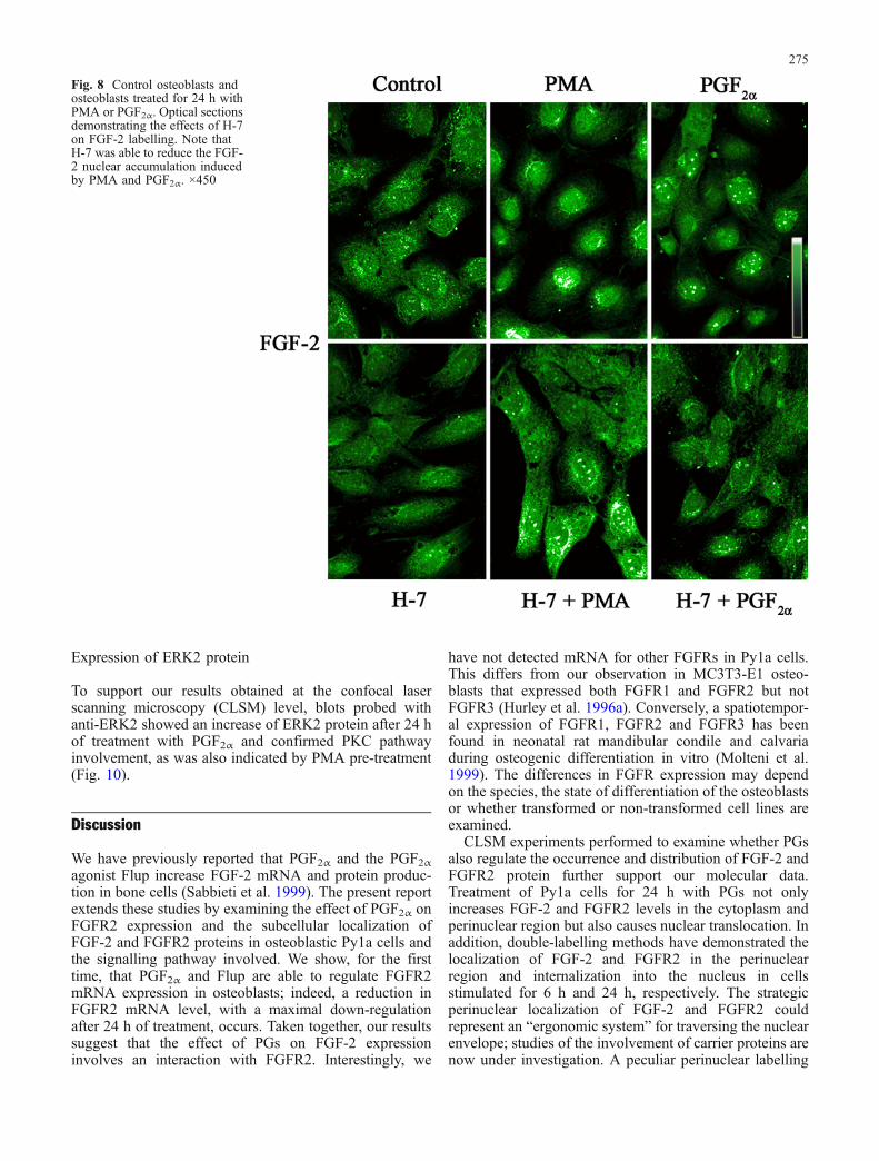
To examine the role of PKC in the nuclear translocationof FGF-2/FGFR2, cells were treated with PMA (10−6 M;Sigma-Aldrich), an activator of PKC, for 24 h. PMA aloneor combined with PGF2α induced nuclear accumulation ofFGF-2 (Fig. 6a, top) and FGFR2 (Fig. 6b, top);conversely, PMA pre-treatment before administration ofPGF2α or PMA prevented the nuclear accumulation ofFGF-2 (Fig. 6a, bottom) and FGFR2 24 h later (Fig. 6b,bottom). These data were also confirmed at the electron-microscope level. Indeed, cells treated with PMA alone orcombined with PGF2α showed a FGF-2 (Fig. 7a) andFGFR2 (Fig. 7b) nuclear labelling that was prevented byPMA pre-treatment before administration of the effectors(Fig. 7c). Moreover, pre-treatment for 1 h with the PKCinhibitor H-7 (5×10⁻⁵ M; Calbiochem-Inalco) reduced theFGF-2 nuclear accumulation induced by PMA and PGF2α(Fig. 8). Similar results were found also for FGFR2.
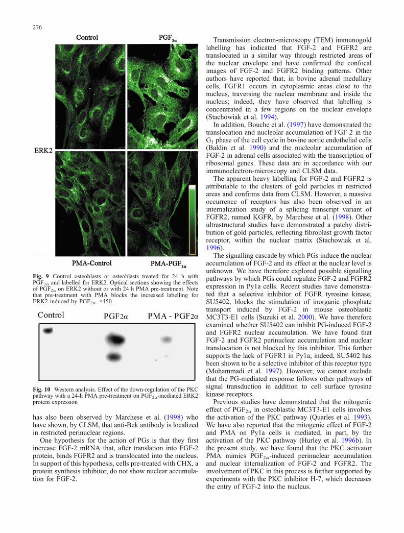
Since our previous molecular data had indicated thatFGF-2 increased ERK in Py1a cells (Hurley et al. 1996b),we examined whether the PG regulation of ERKaccumulation and localization mimicked that of FGF-2.We localized ERK2 in untreated and PG-treated cells.Untreated cells showed cytoplasmic but no nuclearlabelling for ERK2. Treatment with PGF2α for 24 hincreased ERK2 labelling prominently at the cytoplasmlevel. In contrast, pre-treatment with PMA for 24 hblocked PGF2α-induced labelling for ERK2 (Fig. 9).
Fig. 7 Electron-microscopic immunolabelling of FGF-2 (a, c) orFGFR2 (b). In cells treated with PMA for 24 h, a patchy distributionof gold particles reflecting FGF-2 IR (a) and FGFR2 IR (b) waslocated as in Fig. 4 (PM plasma membrane, Cyt cytoplasm, Nnucleus). Cells pre-treated with PMA for 24 h before administrationof PGF2α or PMA showed sparse cytoplasmic gold particlesreflecting FGF-2 IR (c). ×60,500 (a), ×27,700 (b), ×22,600 (c)
274
Expression of ERK2 protein
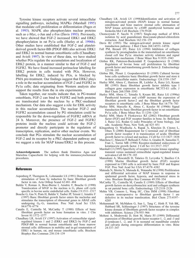
To support our results obtained at the confocal laserscanning microscopy (CLSM) level, blots probed withanti-ERK2 showed an increase of ERK2 protein after 24 hof treatment with PGF2α and confirmed PKC pathwayinvolvement, as was also indicated by PMA pre-treatment(Fig. 10).
Discussion
We have previously reported that PGF2α and the PGF2αagonist Flup increase FGF-2 mRNA and protein produc-tion in bone cells (Sabbieti et al. 1999). The present reportextends these studies by examining the effect of PGF2α onFGFR2 expression and the subcellular localization ofFGF-2 and FGFR2 proteins in osteoblastic Py1a cells andthe signalling pathway involved. We show, for the firsttime, that PGF2α and Flup are able to regulate FGFR2mRNA expression in osteoblasts; indeed, a reduction inFGFR2 mRNA level, with a maximal down-regulationafter 24 h of treatment, occurs. Taken together, our resultssuggest that the effect of PGs on FGF-2 expressioninvolves an interaction with FGFR2. Interestingly, we
have not detected mRNA for other FGFRs in Py1a cells.This differs from our observation in MC3T3-E1 osteo-blasts that expressed both FGFR1 and FGFR2 but notFGFR3 (Hurley et al. 1996a). Conversely, a spatiotempor-al expression of FGFR1, FGFR2 and FGFR3 has beenfound in neonatal rat mandibular condile and calvariaduring osteogenic differentiation in vitro (Molteni et al.1999). The differences in FGFR expression may dependon the species, the state of differentiation of the osteoblastsor whether transformed or non-transformed cell lines areexamined.
CLSM experiments performed to examine whether PGsalso regulate the occurrence and distribution of FGF-2 andFGFR2 protein further support our molecular data.Treatment of Py1a cells for 24 h with PGs not onlyincreases FGF-2 and FGFR2 levels in the cytoplasm andperinuclear region but also causes nuclear translocation. Inaddition, double-labelling methods have demonstrated thelocalization of FGF-2 and FGFR2 in the perinuclearregion and internalization into the nucleus in cellsstimulated for 6 h and 24 h, respectively. The strategicperinuclear localization of FGF-2 and FGFR2 couldrepresent an “ergonomic system” for traversing the nuclearenvelope; studies of the involvement of carrier proteins arenow under investigation. A peculiar perinuclear labelling
Fig. 8 Control osteoblasts andosteoblasts treated for 24 h withPMA or PGF2α. Optical sectionsdemonstrating the effects of H-7on FGF-2 labelling. Note thatH-7 was able to reduce the FGF-2 nuclear accumulation inducedby PMA and PGF2α. ×450
275
has also been observed by Marchese et al. (1998) whohave shown, by CLSM, that anti-Bek antibody is localizedin restricted perinuclear regions.
One hypothesis for the action of PGs is that they firstincrease FGF-2 mRNA that, after translation into FGF-2protein, binds FGFR2 and is translocated into the nucleus.In support of this hypothesis, cells pre-treated with CHX, aprotein synthesis inhibitor, do not show nuclear accumula-tion for FGF-2.
Transmission electron-microscopy (TEM) immunogoldlabelling has indicated that FGF-2 and FGFR2 aretranslocated in a similar way through restricted areas ofthe nuclear envelope and have confirmed the confocalimages of FGF-2 and FGFR2 binding patterns. Otherauthors have reported that, in bovine adrenal medullarycells, FGFR1 occurs in cytoplasmic areas close to thenucleus, traversing the nuclear membrane and inside thenucleus; indeed, they have observed that labelling isconcentrated in a few regions on the nuclear envelope(Stachowiak et al. 1994).
In addition, Bouche et al. (1997) have demonstrated thetranslocation and nucleolar accumulation of FGF-2 in theG1 phase of the cell cycle in bovine aortic endothelial cells(Baldin et al. 1990) and the nucleolar accumulation ofFGF-2 in adrenal cells associated with the transcription ofribosomal genes. These data are in accordance with ourimmunoelectron-microscopy and CLSM data.
The apparent heavy labelling for FGF-2 and FGFR2 isattributable to the clusters of gold particles in restrictedareas and confirms data from CLSM. However, a massiveoccurrence of receptors has also been observed in aninternalization study of a splicing transcript variant ofFGFR2, named KGFR, by Marchese et al. (1998). Otherultrastructural studies have demonstrated a patchy distri-bution of gold particles, reflecting fibroblast growth factorreceptor, within the nuclear matrix (Stachowiak et al.1996).
The signalling cascade by which PGs induce the nuclearaccumulation of FGF-2 and its effect at the nuclear level isunknown. We have therefore explored possible signallingpathways by which PGs could regulate FGF-2 and FGFR2expression in Py1a cells. Recent studies have demonstra-ted that a selective inhibitor of FGFR tyrosine kinase,SU5402, blocks the stimulation of inorganic phosphatetransport induced by FGF-2 in mouse osteoblasticMC3T3-E1 cells (Suzuki et al. 2000). We have thereforeexamined whether SU5402 can inhibit PG-induced FGF-2and FGFR2 nuclear accumulation. We have found thatFGF-2 and FGFR2 perinuclear accumulation and nucleartranslocation is not blocked by this inhibitor. This furthersupports the lack of FGFR1 in Py1a; indeed, SU5402 hasbeen shown to be a selective inhibitor of this receptor type(Mohammadi et al. 1997). However, we cannot excludethat the PG-mediated response follows other pathways ofsignal transduction in addition to cell surface tyrosinekinase receptors.
Previous studies have demonstrated that the mitogeniceffect of PGF2α in osteoblastic MC3T3-E1 cells involvesthe activation of the PKC pathway (Quarles et al. 1993).We have also reported that the mitogenic effect of FGF-2and PMA on Py1a cells is mediated, in part, by theactivation of the PKC pathway (Hurley et al. 1996b). Inthe present study, we have found that the PKC activatorPMA mimics PGF2α-induced perinuclear accumulationand nuclear internalization of FGF-2 and FGFR2. Theinvolvement of PKC in this process is further supported byexperiments with the PKC inhibitor H-7, which decreasesthe entry of FGF-2 into the nucleus.
Fig. 9 Control osteoblasts or osteoblasts treated for 24 h withPGF2α and labelled for ERK2. Optical sections showing the effectsof PGF2α on ERK2 without or with 24 h PMA pre-treatment. Notethat pre-treatment with PMA blocks the increased labelling forERK2 induced by PGF2α. ×450
Fig. 10 Western analysis. Effect of the down-regulation of the PKCpathway with a 24-h PMA pre-treatment on PGF2α-mediated ERK2protein expression
276
Tyrosine kinase receptors activate several intracellularsignalling pathways, including MAPKs (Marshall 1995)that modulate cell proliferation or differentiation (Pages etal. 1993). MAPK also phosphorylates nuclear proteinssuch as c-Myc, c-Jun and c-Fos (Davis 1993). Previously,we have showed that FGF-2 increased ERK1 and ERK2phosphorylation in rat Py1a cells (Hurley et al. 1996b).Other studies have established that FGF-2 and platelet-derived growth factor-BB (PDGF-BB) also activate ERK1and ERK2 in normal human osteoblastic cells (Chaudharyand Avioli 1997). In view of these data, we have studiedwhether PGs regulate the accumulation and localization ofERK2 protein, in a manner similar to that of FGF-2 andFGFR2. We have found increased perinuclear labelling forERK2 protein in cells treated with PGs. However,labelling for ERK2, induced by PGs, is blocked byPMA pre-treatment. Our findings suggest that ERK2 playsa role in the nuclear accumulation of FGF-2 and FGFR2 inPy1a cells; data originating from Western analysis alsosupport the results from the in situ experiments.
Taken together, our results indicate that, in PG-treatedosteoblasts, FGF-2 can bind to FGFR2 and both proteinsare translocated into the nucleus by a PKC-mediatedmechanism. Our data also suggest a role for ERK activityin this nuclear accumulation. The nuclear FGF-2 andFGFR2 accumulation caused by PG stimulation could beresponsible for the down-regulation of FGFR2 mRNA at24 h. Moreover, the presence of FGF-2 and FGFR2proteins inside the nucleus could activate the FGF-2promoter and directly participate in the regulation oftranscription, replication, and/or other nuclear events. Weconclude that PGs stimulate the nuclear accumulation ofFGF-2 and its receptor by a PKC-dependent pathway andwe suggest a role for MAP kinase/ERK2 in this process.
Acknowledgements The authors thank Dimitrios Agas andMariolina Capacchietti for helping with the immunocytochemicalprocedures.
References
Aspenberg P, Thorngren K, Lohmander LS (1991) Dose dependentstimulation of bone by induction by basic fibroblast growthfactor in rats. Acta Orthop Scand 62:481–484
Baldin V, Roman A, Bosc-Bierne I, Amalric F, Bouche G (1990)Translocation of bFGF to the nucleus is G1 phase cell cyclespecific in bovine aortic endothelial cells. Embo J 9:1511–1517
Bouche G, Gas N, Prats H, Baldin V, Tauber JP, Teissié J, Amalric F(1997) Basic fibroblast growth factor enters the nucleolus andstimulates the transcription of ribosomal genes in ABAE cellsundergoing G0–G1 transition. Proc Natl Acad Sci USA84:6770–6774
Canalis E, Centrella M, McCarthy T (1988) Effects of basicfibroblast growth factor on bone formation in vitro. J ClinInvest 81:1572–1577
Chaudhary LR, Avioli LV (1997) Activation of extracellular signal-regulated kinases 1 and 2 (ERK1 and ERK2) by FGF-2 andPDGF-BB in normal human osteoblastic and bone marrowstromal cells: differences in mobility and in-gel renaturation ofERK1 in human, rat, and mouse osteoblastic cells. BiochemBiophys Res Commun 238:134–139
Chaudhary LR, Avioli LV (1998)Identification and activation ofmitogen-activated protein (MAP) kinase in normal humanosteoblastic and bone marrow stromal cells: attenuation ofMAP kinase activation by cAMP, parathyroid hormone andforskolin.Mol Cell Biochem 178:59-68
Chomczynski P, Sacchi N (1987) Single-step method of RNAisolation by acid guanidinium thiocyanate-phenol-chloroformextraction. Anal Biochem 162:156–159
Davis RJ (1993) The mitogen-activated protein kinase signaltransduction pathway. J Biol Chem 268:14553–14556
Fall PM, Breault DT, Raisz LG (1994) Inhibition of collagensynthesis by prostaglandins in the immortalized rat osteoblasticclonal cell line Py1a: structure activity relations and signaltransduction mechanism. J Bone Miner Res 9:1935–1943
Globus RK, Patterson-Buckendahl P, Gospodarowicz D (1988)Regulation of bovine bone cell proliferation by fibroblastgrowth factor and transforming growth factor β. Endocrinology123:98–105
Globus RK, Plouet J, Gospodarowicz D (1989) Cultured bovinebone cells synthesize basic fibroblast growth factor and store itin their extracellular matrix. Endocrinology 124:1539–1547
Hurley MM, Abreu C, Harrison JR, Lichtler A, Raisz LG, KreamBE (1993) Basic fibroblast growth factor inhibits type Icollagen gene expression in osteoblastic MC3T3-E1 cells. JBiol Chem 268:5588–5593
Hurley MM, Abreu C, Tetradis S, Kream BE, Raisz LG (1996a)Parathyroid hormone and cAMP increase the expression offibroblast growth factor-2 and the fibroblast growth factorreceptors in osteoblastic cells. J Bone Miner Res 14:776–783
Hurley MM, Marcello K, Abreu C, Kessler M (1996b) Signaltransduction by basic fibroblast growth factor in rat osteoblasticPy1a cells. J Bone Miner Res 11:1256–1263
Hurley MM, Marie P, Florkiewicz RZ (2002) Fibroblast growthfactor (FGF) and FGF receptor families in bone. In: BelizikianJ, Raisz LG, Rodan G (eds) Principles of bone biology, 2ndedn. Academic Press, New York, pp 825–851
Klingenberg O, Wiedlocha A, Rapak A, Khnykin D, Citores L,Olnes S (2000) Requirement for C-terminal end of fibroblastgrowth factor receptor 4 in translocation of acidic fibroblastgrowth factor to cytosol and nucleus. J Cell Sci 113:1827–1838
Marchese C, Mancini P, Belleudi F, Felici A, Gradini R, Sansolini T,Frati L, Torrisi MR (1998) Receptor-mediated endocytosis ofkeratinocyte growth factor. J Cell Sci 111:3517–3527
Marshall CJ (1995) Specificity of receptor tyrosine kinase signaling:transient versus sustained extracellular signal-regulated kinaseactivation.Cell 80:179–185
Mansukani A, Moscatelli D, Talarico D, Levystka V, Basilico CA(1990) Murine fibroblast growth factor (FGF) receptorexpressed in CHO cells is activated by basic FGF and KaposiFGF. Proc Natl Acad Sci USA 87:4378–4382
Matsuda N, Morita N, Matsuda K, Watanabe M (1998) Proliferationand differential activation of MAP kinases in response toepidermal growth factor, hypoxia, and mechanical stress invitro. Biochem Biophys Res Commun 49:350–354
McCarthy TL, Centrella M, Canalis E (1989) Effects of fibroblastgrowth factors on deoxyribonucleic acid and collagen synthesisin rat parietal bone cells. Endocrinology 125:2118–2126
Mehta VB, Connors L, Wang HC, Chiu IMM (1998) Fibroblastvariants nonresponsive to fibroblast growth factor 1 aredefective in its nuclear translocation. Biol Chem 273:4197–4205
Mohammadi M, McMahon G, Sun L, Tang C, Hirth P, Yeh BK,Hubbard SR, Schlessinger J (1997) Structures of the tyrosinekinase domain of fibroblasts growth factor receptor in complexwith inhibitors. Science 276:955–966
Molteni A, Modrowski D, Hott M, Marie PJ (1999) Differentialexpression of fibroblast growth factor receptor-1, -2, and -3 andβ-syndecan-1, -2, and -4 in neonatal rat mandibular condyleand calvaria during osteogenic differentiation in vitro. Bone24:337–347
277
Nagata T, Kaho K, Nishikawa S, Shindrara H, Wakano Y, Ishida H(1994) Effect of prostaglandin E2 on mineralization of bonenodules formed by fetal rat calvarial cells. Calcif Tissue Int55:451–457
Nishida E, Gotoh Y (1993) The MAP kinase cascade is essential fordiverse signal transduction pathways. Trends Biochem Sci18:128–131
Norrdin RW, Jee WSS, High WB (1990) The role of prostaglandinsin bone in vivo. Prostaglandins Leukot Fatty Acids 41:139–150
Pages G, Lenormand P, L’Allemain G, Chambard JC, Pouyssegur J(1993) Mitogen-activated protein kinases p42mapk and p44mapk
are required for fibroblast proliferation. Proc Natl Acad SciUSA 90:8319–8323
Quarles CD, Haupt DM, Davidai G, Middleton JP (1993) Prosta-glandin F2α induced mitogenesis in MC3T3-E1 osteoblasts:role of protein kinase-C-mediated tyrosine phosphorylation.Endocrinology 132:1505–1513
Raisz LG, Fall PM (1990) Biphasic effects of prostaglandin E2 onbone formation in cultured fetal rat calvariae: interaction withcortisol. Endocrinology 126:1654–1659
Raisz LG, Koolemans-Beynen AR (1974) Inhibition of bonecollagen synthesis by prostaglandin E2 in organ culture.Prostaglandins 8:377–385
Raisz LG, Fall PM, Petersen DN, Lichtler A, Kream BE (1993)Prostaglandin E2 inhibits α1 (I) procollagen gene transcriptionand promoter activity in the immortalized rat osteoblastic clonalcell line Py1a. Mol Endocrinol 7:17–22
Rodan SB, Wesolowski G, Yoon K, Rodan GA (1989) Opposingeffects of fibroblast growth factor and pertussis toxin onalkaline phosphatase, osteopontin, osteocalcin and type Icollagen mRNA levels in ROS 17/2.8 cells. J Biol Chem264:19934–19941
Sabbieti MG, Marchetti L, Abreu C, Montero A, Hand AR, RaiszLG, Hurley MM (1999) Prostaglandins regulate the expressionof fibroblast growth factor-2 in bone. Endocrinology 140:434–444
Sabbieti MG, Marchetti L, Hurley MM, Menghi G (2000) Nuclearand cytoplasmic lectin receptor sites in rat Py1a osteoblasts.Histol Histopathol 15:1107–1117
Sambrook J, Fritsch EF, Maniatis T (eds) (1989) Molecular cloning,a laboratory manual. Cold Spring Harbor Laboratory Press,Cold Spring Harbor, NY
Scutt A, Zeschnigk M, Bertram P (1995) PGE2 induces thetransition from nonadherent to adherent bone marrow mesen-chymal precursor cells via a cAMP/EP2-mediated mechanism.Prostaglandins 49:383–395
Shen V, Kohler G, Huang J, Huang SS, Peck WA (1989) An acidicfibroblast growth factor stimulates DNA synthesis, inhibitscollagen and alkaline phosphatase synthesis and inducesresorption in bone. Bone Miner 7:205–219
Stachowiak M, Moffett J, Joy A, Puchacz E, Florkiewicz R,Stachowiak E (1994) Regulation of bFGF gene expression andsubcellular distribution of bFGF protein in adrenal medullarycells. J Cell Biol 127:203–223
Stachowiak MK, Maher PA, Joy A, Mordechai E, Stachowiak EK(1996) Nuclear accumulation of fibroblast growth factorreceptors is regulated by multiple signals in adrenal medullarycells. Mol Biol Cell 7:1299–1317
Suzuki A, Palmer G, Bonjour JP, Caverzasio J (2000) Stimulation ofsodium-dependent phosphate transport and signaling mecha-nism induced by basic fibroblast growth factor in MC3T3-E1osteoblast-like cells. J Bone Miner Res 15:95–102
Towbin H, Staehelin T, Gordon J (1979) Electrophoretic transfer ofproteins from polyacrylamide gels to nitrocellulose sheets:procedure and some applications. Proc Natl Acad Sci USA76:4350–4354
Wadhwa S, Godwin SL, Peterson DR, Epstein MA, Raisz LG,Pilbeam CC (2002) Fluid flow induction of cyclo-oxygenase 2gene expression in osteoblasts is dependent on an extracellularsignal-regulated kinase signaling pathway. J Bone Miner Res17:266–274
Woodiel FN, Fall PM, Raisz LG (1996) Anabolic effects ofprostaglandins in cultured fetal rat calvariae: structure-activityrelations and signal transduction pathway. J Bone Miner Res11:1249–1255
278