Embed Size (px)
Citation preview

Laureti et al.
Supporting Information SI Materials and Methods Bacterial strains, plasmids and growth conditions. Streptomyces strains were cultivated on SFM, in YEME and on or in HT media and manipulated as described previously (1). Growth curves, RNA extractions, LC-MS and HPLC analysis were obtained from cultures grown in or on MP5 medium (2). Escherichia coli strains were cultivated in LB and SOB liquid medium (3). When needed these antibiotics were added into the cultures: ampicillin, apramycin, kanamycin, spectinomycin at 50µg/ml, chloramphenicol and nalidixic acid at 25µg/ml. For λ red genes induction, 10mM of filtered L-arabinose was added to the culture. DNA manipulation. Plasmid and BAC DNA were extracted from E. coli by alkaline lysis method (3). Genomic DNA extraction of Streptomyces and pulsed-field gel electrophoresis were performed as already described (4). Southern blots were performed with a Hybond-N nylon membrane (Amersham-Pharmacia) and a vacuum transfer system (BioRad), as previously described (5). Amplification of DNA fragments by PCR was performed with Taq DNA polymerase (NEB) or Takara polymerase (Fermentas), according to the manufacturer’s instructions. PCR products and restriction fragments were purified from agarose gel with the High Pure PCR product purification kit (Roche). Overexpression of the TCS. The two component system-encoding sequence was amplified by PCR from S. ambofaciens ATCC23877 genomic DNA with the primers OE468-F and OE469-R (see Table S10 for sequences). The PCR conditions, the cloning steps into pIB139 and the integration of the recombinant plasmid (pOE-4689, Table S9) in S. ambofaciens ATCC23877 were as described for the overexpression of the LAL regulator (see Materials and Methods). Construction of the glycosyltransferase mutant strain. The REDIRECT system was used to make an in-frame deletion of samR0481 in S. ambofaciens ATCC/OE484 as already described for the deletion of samR0467 (see Materials and Methods). However, in this case the aac(3)IV + oriT mutagenesis cassette was used as template in the PCR reaction with the primers D481-F and D481-R (Table S10) and the BAC of interest used was BBC (Table S9). Antimicrobial and antiproliferative tests. The antibacterial activities of the purified stambomycin compounds were first analysed by loading a drop of compounds (mixture of stambomycin A and B or stambomycin C and D; from 1 µg up to 12.5 µg) onto a lawn of Bacillus subtilis ATCC6633 and Micrococcus luteus used as indicator strains. The IC50 and IC90 values were then determined only for

Laureti et al.
the mixture of stambomycin C/D (the most active fraction) against Bacillus subtilis BGSC 1A72, Mycobacterium smegmatis ATCC700084 and against clinical isolates of Enterococcus faecalis and Staphylococcus aureus (strains from Libragen, Toulouse, France). Analyses were also performed against Gram-negative bacteria (Escherichia coli and Pseudomonas aeruginosa, two clinical isolates from Libragen), against yeast (Candida albicans IHEM8060) and filamentous fungi (Aspergillus fumigatus GASP4707 and Fusarium oxysporum DSM2018). For the antiproliferative activities and cytotoxity, the IC50 values were determined for stambomycin A/B and stambomycin C/D using human adenocarcinoma (HT29), breast (MCF7), lung (H460) and prostate (PC3) cancer cell lines and ovary sane cell line from an adult Chinese hamster (CHO-K1), respectively. The cell viability was determined by measuring the cellular concentration of ATP using the CellTiter-Glo® Luminescent Cell Viability Assay (Promega, G7573) according to the recommendation of the manufacturer.
Identification of stambomycins by LC-MS comparative metabolic profiling. Streptomyces ambofaciens ATCC23877, ATCC/OE484 and ATCC/OE484/∆467 spores were stored in 25% (v/v) aqueous glycerol at -20 °C. 10µl of spores from the stocks of the ATCC23877, ATCC/OE484 and ATCC/OE484/∆467 strains were used separately to inoculate MP5 liquid medium and the resulting cultures were grown for 3-4 days at 30 °C and 180 rpm. The cultures were centrifuged for 10 minutes at 4000 rpm and 4°C. The mycelia were collected, extracted with 5 ml of methanol and sonicated for 10 minutes. The resulting suspension was filtered through a spin filter (0.4 µm) and the filtrate was analysed by LC-ESI-TOF-MS. A Sigma Ascentis C18 column (100 x 2.1mm, 2.7 µm) connected to Dionex 3000 RS-HPLC coupled with a Bruker MaXis UHR-Q-TOF mass spectrometer was used. The flow rate was 0.2 ml/min. Water/0.1% formic acid (solvent A) and methanol/0.1% formic acid (solvent B) were used as mobile phases. The elution profile was: 80% solvent A/20% solvent B for 5 minutes, gradient to 100% solvent B for 10 minutes, 100% solvent B for 5 minutes, equilibrate back to 80% solvent A/20% solvent B. The mass spectrometer was calibrated at the beginning of each run with 10 mM sodium formate and the following settings in positive ESI mode were used. Full scan: 50-2000 m/z, End plate offset: -500 V, Capillary: -4500 V, Nebulizer gas (N2): 1.6 bar, Dry gas (N2): 8 L/min, Dry Temperature: 180 °C.

Laureti et al.
ORF Product size (aa)
% identity / similarity
Species Putative function Proposed role
samR0465 8154 Type I modular PKS Polyketide biosynthesis
samR0466 3661 Type I modular PKS Polyketide biosynthesis
samR0467 5771 Type I modular PKS Polyketide biosynthesis
samR0468 217 84/88 S. griseus (SGR874) Response regulator Regulation
samR0469 442 77/84 S. griseus (SGR875) Histidine kinase Regulation
samR0470 261 87/93 S. griseus (SGR876) Putative permease protein Resistance
samR0471 312 88/93 S. griseus (SGR877) Putative ABC transporter ATP-
binding protein Resistance
samR0472 244 55/68 S. erythraea N-dimethyltransferase D-Mycaminose biosynthesis
samR0473 185 46/59 S. fradiae Isomerase D-Mycaminose biosynthesis
samR0474 6333 Type I modular PKS Polyketide biosynthesis
samRCDS1 3556 Type I modular PKS Polyketide biosynthesis
samR0475 3157 Type I modular PKS Polyketide biosynthesis
samR0476 3565 Type I modular PKS Polyketide biosynthesis
samRCDS2 1569 Type I modular PKS Polyketide biosynthesis
samR0477 5447 Type I modular PKS Polyketide biosynthesis
samR0478 414 43/62 S. fradiae Cytochrome P450 Polyketide hydroxylation
samR0479 401 41/56 R. castenholzii Cytochrome P450 Polyketide hydroxylation
samR0480 369 74/82 Streptomyces sp. TP-
A0274 Aminotransferase D-Mycaminose biosynthesis
samR0481 418 48/63 M. griseorubida Glycosyltransferase D-Mycaminose attachment
samR0482 595 64/75 S. hygroscopicus Acyl-CoA synthetase Unknown
samR0483 532 62/75 Micromonopora sp.
ATCC39149 Carboxyl transferase Precursor biosynthesis
samR0484 958 35/47 S. venezuelae (PikD) Transcriptional activator (LAL) Regulation
samR0485 255 87/94 S. griseus (SGR200) Type II thioesterase PKS editing
samR0486 329 77/84 S. tenebrarius (AprE) dTDP-glucose-4,6- dehydratase D-Mycaminose biosynthesis
samR0487 290 74/85 S. avermitilis (AveBIII) Glucose-1-phosphate thymidyltransferase
D-Mycaminose biosynthesis
Table S1. Properties of genes within the stambomycin biosynthetic cluster. Putative functions of the encoded proteins were deduced from analyses with the BlastP program (http://blast.ncbi.nlm.nih.gov/Blast.cgi). The % identity/similarity for the protein in the database with the highest end-to-end similarity is indicated. The stambomycin cluster is located in the right terminal region of the chromosome which has been fully sequenced (AM238664).

Laureti et al.
* * ***** * * * * AT1 AMIFSGQGS-53-VVQPA-22-VAGHSQGEI-18-VALRS-70-PVDYASHSAHVE-45-LRSTVEFSA AT2 GFLFTGQGS-50-YAQAG-22-LVGHSIGEL-18-VSARG-66-AVSHAFHSRLME-40-IREPVRFAD AT3 GFLFTGQGS-50-YAQAG-22-LVGHSVGEI-18-VSARG-66-AVSHAFHSRLME-40-VREPVRFAD AT4 ALLFSGQGS-50-YAQAG-22-LVGHSIGEL-18-VSARG-66-AVSHAF HSRLME-40-VREPVRFAD AT5 AFLFSGQGA-50-YTQPA-22-LVGHSIGEL-18-VSARA-66-AVSHAF HSRLME-40-IVAPVRFAD AT6 VFVFPGQGS-52-VVQPV-22-VVGHSQGEI-18-VALRS-71-PVDYAS HSVQVE-45-LRSTVRFEE AT7 GFLFTGQGA-51-WTQAG-22-LLGHSIGEV-18-VEARG-68-TVSHAF HSALME-46-VRQAVRFAD AT8 VFVFPGQGS-52-VVQPV-22-VVGHSQGEI-18-VALRS-71-PVDYAS HSVQVE-45-LRSTVRFEE AT9 AFLFTGQGA-50-WTQAG-22-LLGHSIGEI-18-VAARG-68-TVSHAF HSALME-41-VRETVRFAD AT10 AFLFTGQGA-50-WAQAG-22-LLGHSIGEI-18-VAQRG-68-TVSHAF HSALME-41-VRETVRFGD AT11 AFLFTGQGA-50-WAQAG-22-LLGHSVGEL-18-VAARG-68-TVSHAFHSVLME-46-VREAVRFAD AT12 ALLFSGQGS-50-YAQAG-22-LVGHSIGEL-18-VSARG-66-AVSHAF HSRLME-40-VREPVRFAD AT13 AFVLPGQGS-53-VIQPV-22-VVGHSQGEI-18-VTHRS-71-RIKGAA HSAVVE-45-MRQTVQFAP AT14 AFVFPGQGG-53-VTPVV-22-VLGHSQGEI-18-VALRG-71-RVDFSS HCAQVE-45-LVTPVDLDR AT15 AFLFSGQGS-50-YAQAG-22-LVGHSVGEL-18-VSARG-66-AVSHAFHSRLME-40-VREPVRFAD AT16 AFLFSGQGS-50-YAQAG-22-LVGHSIGEL-18-VSARG-66—AVSHAFHSRLME-40-VREPVRFAD AT17 ALLFSGQGS-50-YAQAG-22-LVGHSVGEL-18-VSARG-66-AVSHAFHSRRMD-40-VREPVRFAD AT18 VFVFPGQGS-53-VVQPV-22-VVGHSQGEI-18-VALRA-71-PVDYAS HCAQVE-45-LRNTVRFEE AT19 ALLFSGQGS-50-YAQAG-22-LVGHSIGEL-18-VSARG-66-AVSHAF HSHLME-40-VREPVRFAD AT20 AFLFTGQGA-49-HTQPA-22-LAGHSIGEL-18-VAARG-66-AVSHAF HSHLME-42-VRSTVRFAG AT21 VFVFPGQGS-51-VVQPV-22-VVGHSQGEI-18-VALRA-71-PVDYAS HCAQVE-45-LRNTVRFEE AT22 VFVFPGQGS-51-VVQPV-22-VVGHSQGEI-18-VALRA-71-PVDYAS HCAQVE-45-LRNTVRFEE AT23 ALLFSGQGS-50-YAQAG-22-LVGHSQGEL-18-VSARG-66-AVSHAFHSRLME-40-VREPVRFAD AT24 VFVFPGQGS-51-VVQPV-22-VVGHSQGEI-18-VALRA-71-PVDYAS HSAHVE-45-LRATVRFED AT25 AFVFSGQGA-51-WTQLG-22-LAGHSVGEV-18-VAARG-71-DVSHAFHSPRVD-45-IRATVRFAD Q Q GH[LVIFAM]G R [FP]H V malonyl-CoA Q Q GH[QMI]G R SH V methylmalonyl-CoA
Fig. S1. Sequence alignment of the PKS AT domains. Highly conserved active site residues are marked with an asterisk (6). The boxed residues are diagnostic of substrate specificity, in particular the amino acid underlined in green. The arginine residue underlined in yellow interacts with the carboxyl group of the substrates. The catalytic serine residue is underlined in blue. The consensus sequences for malonyl-CoA and methylmalonyl-CoA-specific AT domains are shown below the alignment. The substrate for AT13 could not be predicted because the circled specificity-conferring residue is different from that found in malonyl- and methylmalonyl-CoA-specific AT domains.

Laureti et al.
Fig. S2. Sequence alignment of the PKS KR domains. The GXGXXGXXXA consensus sequence in the N-termini is proposed to bind NAD(P)H (7). The residues belonging to the catalytic triad are marked with asterisks. The Y to R and N to S mutations of the catalytic residues in KR25 indicate that it is not active. The arrows indicate the residue predictive of B and A-type alcohol stereochemistry, respectively. The predicted configurations of the α- and β-stereocenters generated by each KR, according to the model of Keatinge-Clay (8), are indicated to the right of the alignment. A1 = 2R, 3S; A2 = 2S, 3S; B1 = 2R, 3R; B2 = 2S, 3R; C1 = 2R.
ER11 VNFRDVLNVLGMYPG-EVLVGGEAAGVV-67-SAGESVLVHAAAGGVGMAAVQVARHLG ER16 MNFRDVLNVLGMYPG-EVELGGEAAGVV-67-AAGESVLVHAAAGGVGMAAVQIARHVG ER4 VNFRDVLIALGQYPDPTALMGSEAAGVV-67-SAGESVLVHAAAGGVGMAAVQVARHLG ER7 VNFRDVLIALGMYPD-RAQMGAEAAGVV-67-RPGESVLVHSAAGGVGMAAVQLGRHLG :****** .** **. . :*.****** .*******:** *********:.**:*
Fig. S3. Sequence alignment of the PKS ER domains. The highly conserved putative NADPH binding motif is highlighted in yellow. The residue that is predictive of stereochemistry is highlighted in red. A tyrosine residue in this position predicts the 2S configuration. Another residue (usually Val or Ala) in this position predicts the 2R stereochemistry (9).
KR2 PHDTVLITGGTGALGARVARHLVCA-57-VVHTAGVLDDGLLTSLTPE-26-FVLFSSVAASFGTAGQASYAAANAFLD B1 KR3 -DGTVLVTGGTGALGAQVAR-LLAA-61-VVHAAGVLDDGVIDGLTPE-26-FVLFSSFTGAVGTAGQANYAAANAHLD B1 KR4 --GTVLVTGGTGALGAHTARLLARR-60-VVHAAGTVDDGVIGSLTPG-26-FVLYTSFAGVVGNLGQAAYAAGNAALD B1 KR5 PQGTVLITGGTGTLGSLLARHLVEH-62-VVHAAGVADDGVIEALTPE-26-FTVYASASSAFGSPGQANYAAANAFLE B1 KR6 --GTVLVTGGTGAVGAEVARWLAGR-60-VLHAAGVDGVTALDEVDAD-26-FVVFSSGAAVWGGGGQGAYAAGNAFLD A1 KR7 --GTVLVTGGTGGLGGEVARWLARR-60-VVHAAGVGTPGRLLDTDET-24-FVVFSSIAATWGSGGQGAYAAGNAFLD A1 KR8 PDGTVLITGGTGTLGGLLARHLVTE-62-VIHAAGVLDDGVFESMTPE-26-FVLYSSASATLGTGGQANYAAANSFLD B1 KR9 --GTVLVTGGTGALGKRVARWLAER-60-VVHAAGFGQAVPLADTDEA-26-FVVFSSIAATWGSGGQGVYAAANAHLD A1 KR10 -PGAVLVTGGTGALGAVVARWLADR-60-VVHAAGVLDDGTLDALTPE-26-FVAFSSLAGTVGSAGQGNYAAANAFVD B1 KR11 -PGTVLVTGGTGALGASVARWLAER-60-VVHAAGVAQSGPVETTRLA-26-FVLFSSIAATWGSGGQALYAAGNAYLD A1 KR12 AEGTVLVTGGTGALGALTARHLVVE-62-VVHAAGILDDGLVESLTED-25-FVMYSSMSGTFGSPGQGNYAAANAYLD B1 KR13 --STVLITGGTGGIGRHLAHHMAAR-56-VIHAAGVAQATALADCGES-24-FVLFSSGAGVWGGAGQAAYAAGNAVLD A1 KR14 ADGTVLVTGATGTLGSALARHLVRH-61-VVHTAAVLDDGVLAQMTDR-26-FALFSSAAGVLGGAGQANYAAANVFLD B1 KR15 --GTVLITGGTGALGSRVARWAALA-56-VVHAAGVGGLGRLAELTEE-24-FVLFGSVAAVWGGAGQAAYAAANARLE A1 KR16 --GTTLVTGGTGALGAHVARWLADR-56-VVHAAGSGGFGTLDDASEA-26-FVLFSSVSGIWGSGGQAAYGAANAALD A1 KR17 PGGTVLITGGTGALGALVARYLVDR-44-VFHLAGVLDDGVATALTPE-24-FVLFSSVSATLGSPGQASYAAANAYLD B1 KR18 --EAVLITGGTGALGAETARMLARR-56-VVHAAGTDPALPLDSTSVP-24-FVVFSSIAGVWGSGGQAAYAAANAHLD A1 KR19 --GTVLVTGGTGAIGGHVARWLATE-62-VMHTAGLGVLAPLADTGVA-26-VVHFSSIAAMWGVGQHGGYAAGNAYLD A2 KR20 AHGTVLVTGGTGVLGGRVARHLAAR-62-VVHAAGIVDDGVVTSLTPD-24-FVLFSSASATFGSAGQAGYAAANAVLD B1 KR21 PVGTVLVTGGTGVLGGLVARHLVTA-57-VVHAAGVLDDGVFESMTPK-23-FVFFSSAGGTFGPAGQANYAAANATLD B1 KR22 --GTVLVTGGTGGIGAHVARWLAAS-61-VFHAAGIVDSSILDSLTPD-26-FVLFSSLAGVFGSAGEGNYAPGNAFLD B2 KR23 -P--VLLTGGTGALGGKVARLLAER-56-VVHAAGIVDDGVLDALTPE-24-FVVFSSVAGVIGSAGQGPYAAANAHLD B1 KR24 --GPVLVTGGTGALGREVARWLARR-56-VVHTAGISTTAPLAGTSPA-25-FVLFSSIAGVWGGGGQAAYAAANAHLD A1 KR25 --GTVLITGGTGRRGRALATALAAN-55-VVHAVGAGEDTPWTELSPG-26-FVLVSSVTGVWGGTGAAVRAAASARMD C1 Cons GXGXXGXXXA * * *

Laureti et al.
Fig. S4. TDP-D-mycaminose biosynthetic pathway. Proposed pathway for biosynthesis of TDP-D-mycaminose from α-D-glucose-1-phosphate by putative aminodeoxysugar biosynthetic enzymes encoded by genes within the cluster. For each enzymatic step, the gene product is indicated in bold. The identity/similarity with the corresponding enzymes involved the biosynthesis of the β-D-mycaminose residue of spiramycin are indicated in brackets (10). TTP, thymidine triphosphate; TDP, thymidine diphosphate; NAD+, nicotinamide adenine dinucleotide; PMP, pyridoxamine 5´-phosphate; SAM, S-adenosyl-L-methionine.
TDP-D-mycaminose
OHOHO
HOOPO3=
HOOHO
HOHOOTDP
HOO
HOHOOTDP
O
O
HOOTDPO
HO O
HOOTDP
HOH2N
O
HOOTDP
HOMe2N
thymidyltransferase
TTP
SAMR0487 (71/82% orf4)
4,6-dehydratase
NAD+
SAMR0486 (68/78% orf5)
3,4-ketoisomerase
SAMR0473 (46/59% orf2)
aminotransferase
PMP
SAMR0480 (44/57% orf3)
N,N-dimethyltransferase
SAM
SAMR0472 (47/64% orf9c)
TDP-D-mycaminoseTDP-D-mycaminose
OHOHO
HOOPO3=
HOOHO
HOHOOPO3
=
HOOHO
HOHOOTDP
HOOHO
HOHOOTDP
HOO
HOHOOTDP
OO
HOHOOTDP
O
O
HOOTDPO
HO O
HOOTDPO
HO O
HOOTDP
HOH2N
O
HOOTDP
HOH2N
O
HOOTDP
HOMe2N
O
HOOTDP
HOMe2N
thymidyltransferase
TTP
SAMR0487 (71/82% orf4)
thymidyltransferase
thymidyltransferase
TTP
SAMR0487 (71/82% orf4)
4,6-dehydratase
NAD+
SAMR0486 (68/78% orf5)
4,6-dehydratase4,6-dehydratase
NAD+
SAMR0486 (68/78% orf5)
3,4-ketoisomerase
SAMR0473 (46/59% orf2)
3,4-ketoisomerase3,4-keto
isomerase
SAMR0473 (46/59% orf2)
aminotransferase
PMP
SAMR0480 (44/57% orf3)
aminotransferase
aminotransferase
PMP
SAMR0480 (44/57% orf3)
N,N-dimethyltransferase
SAM
SAMR0472 (47/64% orf9c)
N,N-dimethyltransferase
N,N-dimethyltransferase
SAM
SAMR0472 (47/64% orf9c)

Laureti et al.
Fig. S5. Transcriptional analysis of the strain overexpressing the two component system (mutant strain ATCC/OE468-9) and grown in MP5 medium. The expression of four biosynthetic genes (samR0467, samR0465, samR0477, samR0474), together with the expression of the regulatory genes samR0468, samR0469 and samR0484, were analyzed by RT-PCR using 4µg of total RNA. The constitutively-expressed hrdB gene, coding for the major sigma factor, was used as a positive control. Experiments carried out on three separate occasions gave the same result. T1= exponential phase; T2= transition phase; T3= stationary phase.

Laureti et al.
1376.9391
1377.9428
1378.9461
1379.9494
+MS, 4.2-4.4min #(247-261)
1376.9392
1377.9426
1378.9459
1379.9492
C 73 H 134 N O 22 ,1376.940.0
0.5
1.0
4x10Intens.
0.0
0.5
1.0
1.5
4x10
1376 1377 1378 1379 1380 1381 m/z
Fig. S6. High resolution mass spectrometry analysis of stambomycins A/B. Measured spectrum of stambomycins A/B (top panel) and simulated spectrum for C73H134N022
+ (bottom panel).
1362.9232
1363.9268
1364.9312
1365.9342
+MS, 4.2-4.4min #(247-261)
1362.9236
1363.9269
1364.9302
1365.9336
C 72 H 132 N O 22 ,1362.920.0
0.5
1.0
4x10Intens.
0.0
0.5
1.0
1.54x10
1362 1363 1364 1365 1366 1367 m/z
Fig. S7. High resolution mass spectrometry analysis of stambomycins C/D. Measured spectrum of stambomycins C/D (top panel) and simulated spectrum for C72H132N022
+ (bottom panel).

Laureti et al.
1203.8344
1204.8381
1205.8389
1206.8343 1207.8084 1208.8127
+MS, 14.5-14.6min #(859-863)
1203.8340
1204.8374
1205.8407
1206.8440
C 65 H 119 O 19 ,1203.830.0
0.5
1.0
1.5
4x10Intens.
0.0
0.5
1.0
1.5
4x10
1203 1204 1205 1206 1207 1208 1209 m/z
Fig. S8. High resolution mass spectrometry analysis of stambomycin A/B aglycones. Measured spectrum of stambomycin A/B aglycones (top panel) and simulated spectrum for C65H119019
+ (bottom panel).
1189.8183
1190.8229
1191.82251192.8143
+MS, 14.2-14.3min #(842-846)
1189.8184
1190.8217
1191.8250
1192.8284
C 64 H 117 O 19 ,1189.820
1000
2000
3000
Intens.
0
1000
2000
3000
1189 1190 1191 1192 1193 1194 1195 m/z
Fig. S9. High resolution mass spectrometry analysis of stambomycin C/D aglycones. Measured spectrum of stambomycin C/D aglycones (top panel) and simulated spectrum for C64H117019
+ (bottom panel).
Laureti et al.
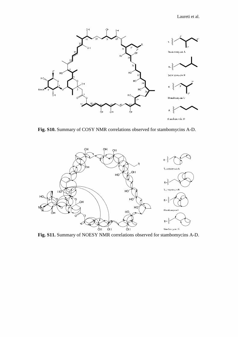
Fig. S10. Summary of COSY NMR correlations observed for stambomycins A-D.
Fig. S11. Summary of NOESY NMR correlations observed for stambomycins A-D.
Laureti et al.
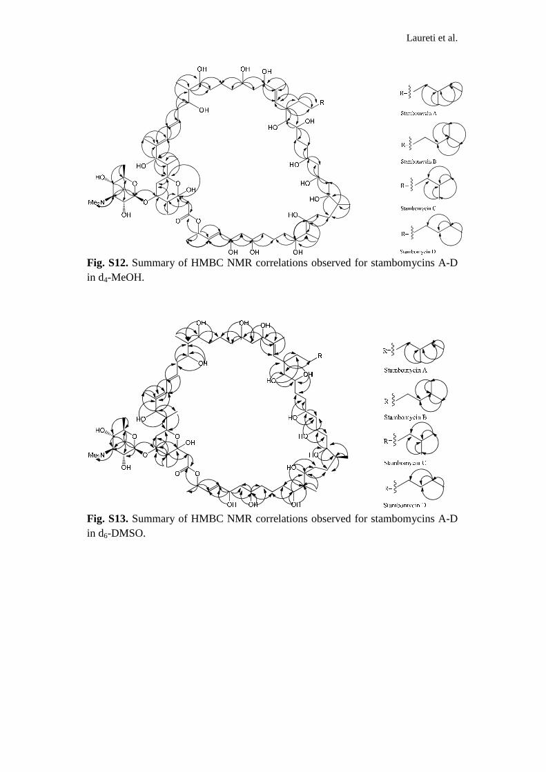
Fig. S12. Summary of HMBC NMR correlations observed for stambomycins A-D in d4-MeOH.
Fig. S13. Summary of HMBC NMR correlations observed for stambomycins A-D in d6-DMSO.
Laureti et al.
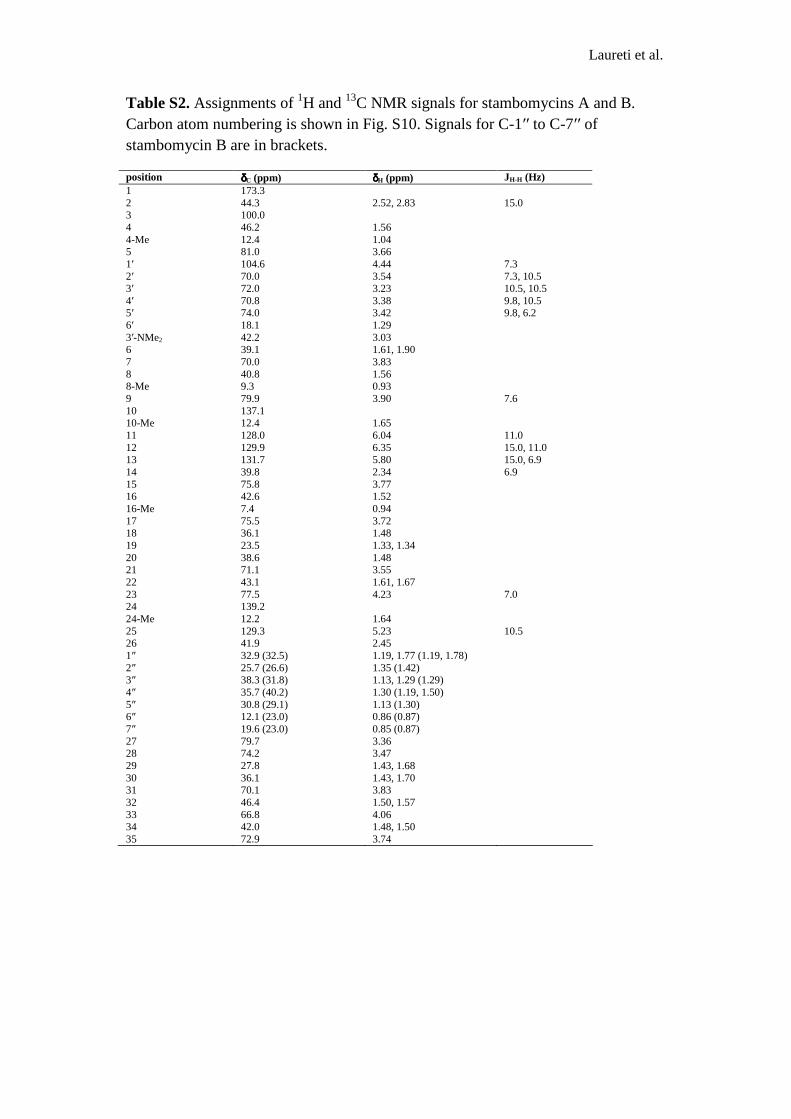
Table S2. Assignments of 1H and 13C NMR signals for stambomycins A and B. Carbon atom numbering is shown in Fig. S10. Signals for C-1′′ to C-7′′ of stambomycin B are in brackets.
position δδδδC (ppm) δδδδH (ppm) JH-H (Hz) 1 173.3 2 44.3 2.52, 2.83 15.0 3 100.0 4 46.2 1.56 4-Me 12.4 1.04 5 81.0 3.66 1′ 104.6 4.44 7.3 2′ 70.0 3.54 7.3, 10.5 3′ 72.0 3.23 10.5, 10.5 4′ 70.8 3.38 9.8, 10.5 5′ 74.0 3.42 9.8, 6.2 6′ 18.1 1.29 3′-NMe2 42.2 3.03 6 39.1 1.61, 1.90 7 70.0 3.83 8 40.8 1.56 8-Me 9.3 0.93 9 79.9 3.90 7.6 10 137.1 10-Me 12.4 1.65 11 128.0 6.04 11.0 12 129.9 6.35 15.0, 11.0 13 131.7 5.80 15.0, 6.9 14 39.8 2.34 6.9 15 75.8 3.77 16 42.6 1.52 16-Me 7.4 0.94 17 75.5 3.72 18 36.1 1.48 19 23.5 1.33, 1.34 20 38.6 1.48 21 71.1 3.55 22 43.1 1.61, 1.67 23 77.5 4.23 7.0 24 139.2 24-Me 12.2 1.64 25 129.3 5.23 10.5 26 41.9 2.45 1″ 32.9 (32.5) 1.19, 1.77 (1.19, 1.78) 2″ 25.7 (26.6) 1.35 (1.42) 3″ 38.3 (31.8) 1.13, 1.29 (1.29) 4″ 35.7 (40.2) 1.30 (1.19, 1.50) 5″ 30.8 (29.1) 1.13 (1.30) 6″ 12.1 (23.0) 0.86 (0.87) 7″ 19.6 (23.0) 0.85 (0.87) 27 79.7 3.36 28 74.2 3.47 29 27.8 1.43, 1.68 30 36.1 1.43, 1.70 31 70.1 3.83 32 46.4 1.50, 1.57 33 66.8 4.06 34 42.0 1.48, 1.50 35 72.9 3.74

Laureti et al.
36 40.8 1.48 36-Me 15.6 0.92 37 29.8 1.28, 1.11 38 36.0 1.50 39 76.3 3.72 40 42.3 1.51 40-Me 7.0 0.91 41 75.9 3.71 42 33.8 1.43, 1.62 43 22.9 1.43 44 38.8 1.45, 1.46 45 70.6 3.67 46 44.8 1.60, 1.65 47 71.1 4.28 6.3, 12.9 48 136.7 5.80 15.3, 6.3 49 130.8 5.72 6.3, 15.3 50 72.8 5.43 6.3, 51 20.8 1.33 6.3
Fig. S14. 1H NMR spectrum (700 MHz, d4-MeOH) of stambomycins A/B.

Laureti et al.
Fig. S15. 13C PENDANT NMR spectrum (125 MHz, d4-MeOH) of stambomycins A/B.
Fig. S16. DQFCOSY NMR spectrum (700 MHz, d4-MeOH) of stambomycins A/B.

Laureti et al.
Fig. S17. Expansion of methyl/methylene region of DQFCOSY spectrum of stambomycins A/B.
Fig. S18. HSQC NMR spectrum (700 MHz/175 MHz, d4-MeOH) of stambomycins A/B.

Laureti et al.
Fig. S19. HMBC NMR spectrum (700 MHz/175 MHz, d4-MeOH) of stambomycins A/B.
Fig. S20. Expansion of methyl region of HMBC spectrum of stambomycins A/B.

Laureti et al.
Fig. S21. NOESY NMR spectrum (700 MHz, d4-MeOH) of stambomycins A/B.
Fig. S22. TOCSY NMR spectrum (700 MHz, d4-MeOH) of stambomycins A/B.

Laureti et al.
Table S3. Assignments of 1H and 13C NMR signals for stambomycins C/D. Carbon atom numbering is shown in Fig. S10. Signals for C-1′′ to C-6′′ of stambomycin D are in brackets.
position δδδδC (ppm) δδδδH (ppm) JH-H (Hz)
1 173.3 2 44.3 2.52, 2.83 15.0 3 100.0 4 46.2 1.56 4-Me 12.4 1.04 5 81.0 3.66 1′ 104.6 4.44 7.3 2′ 70.0 3.54 7.3, 10.5 3′ 72.0 3.23 10.5, 10.5 4′ 70.8 3.38 9.8, 10.5 5′ 74.0 3.42 9.8, 6.2 6′ 18.1 1.29 3′-NMe2 42.2 3.03 6 39.1 1.61, 1.90 7 70.0 3.83 8 40.8 1.56 8-Me 9.3 0.93 9 79.9 3.90 7.6 10 137.1 10-Me 12.4 1.65 11 128.0 6.06 11.0 12 129.9 6.35 15.0, 11.0 13 131.7 5.81 15.0, 6.9 14 39.8 2.34 6.9 15 75.8 3.77 16 42.6 1.52 16-Me 7.4 0.95 17 75.5 3.72 18 36.1 1.48 19 23.5 1.32, 1.34 20 38.5 1.47 21 71.1 3.55 22 43.1 1.61, 1.67 23 77.5 4.23 6.9 24 139.4 24-Me 12.2 1.64 25 129.3 5.23 6.5 26 41.9 2.45 1′′ 32.5 (32.4) 1.18, 1.79 (1.18, 1.79) 2′′ 25.8 (26.6) 1.38 (1.37) 3′′ 40.3 (35.8) 1.13, 1.29 (1.31, 1.44) 4′′ 29.9 (34.0) 1.30 (1.43, 1.49) 5′′ 23.1 (29.2) 0.87 (1.29) 6′′ 23.1 (11.5) 0.87 (0.87) 27 79.7 3.36 28 74.2 3.47 29 27.8 1.43, 1.68 30 36.1 1.43, 1.70 31 70.1 3.83 32 46.4 1.50, 1.57 33 66.8 4.06 34 42.0 1.48, 1.50

Laureti et al.
35 72.9 3.74 36 40.8 1.48 36-Me 15.6 0.92 37 29.8 1.28, 1.12 38 36.0 1.50 39 76.3 3.72 40 42.3 1.51 40-Me 7.0 0.91 41 75.9 3.71 42 33.8 1.43, 1.62 43 22.9 1.43 44 38.8 1.45, 1.46 45 70.6 3.67 46 44.8 1.60, 1.65 47 71.1 4.28 6.2, 12.9 48 136.7 5.80 15.0, 6.2 49 130.8 5.72 6.2, 15.0 50 72.8 5.43 6.2 51 20.8 1.33 6.2
Fig. S23. 1H NMR spectrum (700 MHz, d4-MeOH) of stambomycins C/D.

Laureti et al.
Fig. S24. 13C PENDANT NMR spectrum (175 MHz, d4-MeOH) of stambomycins C/D.
Fig. S25. DQFCOSY NMR spectrum (700 MHz, d4-MeOH) of stambomycins C/D.

Laureti et al.
Fig. S26. Expansion of methyl/methylene region of DQFCOSY spectrum of stambomycins C/D.
Fig. S27. HSQC NMR spectrum (700 MHz/175 MHz, d4-MeOH) of stambomycins C/D.

Laureti et al.
Fig. S28. HMBC NMR spectrum (700 MHz/175 MHz, d4-MeOH) of stambomycins C/D.
Fig. S29. Expansion of methyl region of HMBC spectrum of stambomycins C/D.

Laureti et al.
Fig. S30. NOESY NMR spectrum (700 MHz, d4-MeOH) of stambomycins C/D.
Fig. S31. Summary of key COSY (bold lines) HMBC (arrows) and coupling
constant data observed for stambomycin aglycones.

Laureti et al.
Table S4. Assignments of 1H and 13C NMR signals for the aglycones of stambomycins A/B. Carbon atom numbering is shown in Fig. S10. Signals for C-1′′ to C-7′′ of stambomycin B are in brackets.
position δδδδC (ppm) δδδδH (ppm) JH-H (Hz)
1 173.3 2 44.2 2.51, 2.75 14.6 3 99.9 4 47.2 1.36 4-Me 12.3 1.06 5 70.1 3.60 6 39.3 1.45, 1.73 7 70.1 3.81 8 40.9 1.60 8-Me 9.3 0.93 9 79.6 3.92 7.4 10 136.9 10-Me 11.9 1.65 11 127.8 6.05 11.2 12 129.5 6.36 15.1, 11.2 13 131.2 5.79 15.1, 6.8 14 39.8 2.34 6.8 15 75.6 3.78 16 42.7 1.53 16-Me 7.4 0.95 17 75.2 3.72 18 35.9 1.48 19 23.3 1.33 20 38.4 1.47 21 70.8 3.54 22 42.9 1.61, 1.67 23 77.2 4.23 7.3 24 138.7 24-Me 12.2 1.67 25 129.6 5.22 10.2 26 41.8 2.45 1” 32.7 (32.6) 1.19, 1.77 (1.17, 1.81) 2” 25.4 (26.4) 1.37 (1.36) 3” 38.1 (32.6) 1.14, 1.29 (1.33) 4” 35.6 (40.1) 1.30 (1.19, 1.48) 5” 30.6 (29.6) 1.13 (1.32) 6” 11.6 (22.9) 0.86 (0.88) 7” 19.5 (22.9) 0.85 (0.88) 27 79.4 3.36 28 73.8 3.48 29 27.6 1.44, 1.67 30 35.8 1.45, 1.70 31 69.6 3.83 32 46.0 1.50, 1.57

Laureti et al.
33 66.6 4.07 34 41.6 1.48, 1.50 35 72.5 3.75 36 41.2 1.48 36-Me 15.4 0.92 37 30.4 1.14, 1.29 38 35.9 1.51 39 76.1 3.71 40 42.2 1.51 40-Me 6.8 0.92 41 75.6 3.71 42 33.4 1.43, 1.63 43 22.7 1.44 44 38.9 1.45, 1.48 45 70.5 3.68 46 44.7 1.59, 1.64 47 70.9 4.28 6.3, 12.8 48 136.5 5.78 15.0, 6.3 49 130.8 5.73 6.4, 15.0 50 72.9 5.43 6.4, 51 20.6 1.36 6.4
Fig. S32. 1H NMR spectrum (700 MHz, d4-MeOH) of stambomycin A/B aglycones.

Laureti et al.
Fig. S33. DQFCOSY NMR spectrum (700 MHz, d4-MeOH) of stambomycin A/B aglycones.
Fig. S34. HSQC NMR spectrum (700 MHz/175 MHz, d4-MeOH) of stambomycin A/B aglycones.

Laureti et al.
Fig. S35. HMBC NMR spectrum (700 MHz/175 MHz, d4-MeOH) of stambomycin A/B aglycones.
Fig. S36. TOCSY NMR spectrum (700 MHz, d4-MeOH) of stambomycin A/B aglycones.

Laureti et al.
Table S5. Assignments of 1H and 13C NMR signals for the aglycones of stambomycins C/D. Carbon atom numbering is shown in Fig. S10. Signals for C-1′′ to C-6′′ of stambomycin D are in brackets.
position δδδδC (ppm) δδδδH (ppm) JH-H (Hz)
1 172.9 2 44.2 2.51, 2.75 14.6 3 99.9 4 47.2 1.36 4-Me 12.3 1.06 5 70.1 3.59 6 39.4 1.45, 1.71 7 70.0 3.81 8 40.8 1.61 8-Me 9.2 0.93 9 79.6 3.92 7.5 10 136.7 10-Me 11.9 1.66 11 127.5 6.05 11.1 12 129.3 6.36 15.0, 11.1 13 131.2 5.79 15.0, 6.8 14 39.6 2.34 6.8 15 75.4 3.78 16 42.7 1.53 16-Me 7.3 0.95 17 75.3 3.72 18 36.0 1.48 19 23.1 1.32 20 38.4 1.45 21 70.6 3.54 22 42.8 1.61, 1.67 23 77.1 4.23 7.3 24 138.6 24-Me 12.1 1.66 25 129.4 5.22 10.0 26 41.6 2.44 1” 32.5 (32.4) 1.19, 1.77 (1.19, 1.80) 2” 25.6 (27.1) 1.39 (1.41) 3” 30.6 (32.9) 1.29, 1.29 (1.45) 4” 40.4 (32.4) 1.21 (1.44) 5” 22.9 (29.5) 0.88 (1.26) 6” 22.9 (11.6) 0.88 (0.87) 27 79.4 3.36 28 73.7 3.47 29 27.6 1.44, 1.67 30 35.7 1.43, 1.71 31 69.5 3.83 32 46.0 1.51, 1.57 33 66.4 4.07

Laureti et al.
34 41.6 1.47, 1.51 35 72.6 3.75 36 41.6 1.49 36-Me 15.6 0.93 37 30.5 1.13, 1.29 38 35.9 1.50 39 76.0 3.71 40 42.1 1.51 40-Me 6.8 0.91 41 75.5 3.71 42 33.4 1.44, 1.62 43 22.7 1.45 44 39.1 1.47, 1.51 45 70.4 3.68 46 44.7 1.60, 1.63 47 70.7 4.28 6.4, 12.5 48 136.5 5.78 15.0, 6.4 49 130.7 5.75 6.4, 15.0 50 72.6 5.43 6.4, 51 20.6 1.35 6.4
Fig. S37. 1H NMR spectrum (700 MHz, d4-MeOH) of stambomycin C/D aglycones.

Laureti et al.
Fig. S38. DQFCOSY NMR spectrum (700 MHz, d4-MeOH) of stambomycin C/D aglycones.
Fig. S39. HSQC NMR spectrum (700 MHz/175 MHz, d4-MeOH) of stambomycin C/D aglycones.

Laureti et al.
Fig. S40. HMBC NMR spectrum (700 MHz/175 MHz, d4-MeOH) of stambomycins C/D aglycones.

Laureti et al.
Fig. S41. Model for stambomycin biosynthesis.
The proposed structures of the intermediates assembled by each PKS module are shown attached to the respective ACP domains. The timing of hydroxylation of the propionyl starter unit is unknown and could occur on any of the ACP-bound intermediates, but the hydroxyl group introduced is required for release of the fully-assembled polyketide chain via macrolactonzation.

Laureti et al.
ACPKS AT
KR+
ACPKS AT
KR+
ACPKS AT
ERS
KR*DH
Module 14 Module 15 Module 16
ACPKS AT
KR+
ACPKS AT
KR#
ACPKS AT
KR*DH
ACPKS AT
KR*DH
ACPKS AT
KR^
ACPKS AT
KR*
ACPKS AT
KR+
ACPKS AT
KR
M
o
d
u
l
e
1
7
Module 18 Module 19 Module 20 Module 21 Module 22 Module 23 Module 24
TE
SamR0476 SamR0475 SamRCDS2 SamR0474
X
SamRCDS1
S
O
O
HO
HO
HO
HO
HO
HO
HO
R
HO
HO
HO
HO
HO
HO
HO
HO
S
O
HO
HO
HO
HO
HO
HO
HO
R
HO
HO
HO
HO
HO
HO
HO
HO
S
O
HO
HO
HO
HO
HO
HO
R
HO
HO
HO
HO
HO
HO
HO
HO
S
O
HO
HO
HO
HO
HO
R
HO
HO
HO
HO
HO
HO
HO
HO
S
O
HO
HO
HO
HO
R
HO
HO
HO
HO
HO
HO
HO
HO
S
HO
HO
HO
HO
R
HO
HO
HO
HO
HO
HO
HO
HO
O
S
HO
HO
HO
HO
R
HO
HO
HO
HO
HO
HO
HO
HO
O
S
O
HO
HO
HO
R
HO
HO
HO
HO
HO
HO
HO
HO
S
O
HO
HO
R
HO
HO
HO
HO
HO
HO
HO
HO
S
HO
HO
R
HO
HO
HO
HO
HO
HO
HO
HO
O
S
O
HO
R
HO
HO
HO
HO
HO
HO
HO
HO
X
X
X
X
X
X
X
X
X
X
X
O
O
HO
OH
OH OH
OH
HO
O
OH OH OH
OH
R
HO
HO
HO
HO
HO
O
O
HO
OH
OH OH
OH
O
OHO
OH
Me2N
O
OH OH OH
OH
R
HOOH
HO
HO
HO
HO
X=OH
NDP-D-mycaminoseO2, NAD(P)H
SamR0481SamR0478
orSamr0479
Fig. S41 cont.

Laureti et al.
Organism stambomycins
A/B C/D vancomycin
Compounds
of reference
Antibacterial activity (IC90, µg/ml): Bacillus subtilis BGSC 1A72 ND 33.53 +/- 0.86 0.425+/-0.008
Enterococcus faecalis LG40 ND 8.65 +/- 0.49 0.970+/-0.003
Staphylococcus aureus LG21 ND 33.95 +/- 0.16 0.952+/-0.007
Antiproliferative activity (IC50, µM):
HT29 1.77 +/- 0.04 1.74 +/- 0.04
1.32 +/- 0.08a
H460 1.49 +/- 0.03 1.30 +/- 0.13 1.51 +/- 0.12b; 5.21 +/- 0.22c
MCF7 >5.34 3.51+/- 0.16 3.69 +/- 0.11d; 0.40 +/- 0.03e
PC3 3.39 +/- 0.16 2.79 +/- 0.18 5.64 +/- 0.53d; 15.44 +/- 0.77f
Cytotoxicity (IC50, µM):
CHO-K1 8.47 +/- 0.67 8.46 +/- 0.52
1.99 +/-0.25a
Table S6. Biological activities of stambomycins A/B and C/D. IC90 and IC50 indicate the concentrations needed to inhibit the growth of 90% and 50% of cells in the population, respectively. HT29: human colon adenocarcinoma cell line; H460: lung cancer cell line; MCF7: breast cancer cell line; PC3: prostate cancer cell line; CHO-K1: adult Chinese hamster ovary sane cell line. ND: the IC90 values for stambomycins A/B were not determined, but these compounds were shown to possess antibacterial activity. Vancomycin was used as positive control in the antibacterial assays. The compounds used as reference for antiproliferative activity or cytotoxicity tests are doxorubicina for HT29 and CHO-K1, mycophenolic acidb and 5-fluorouracylc for H460, mitoxantroned and CD437e for MCF7 and mitoxantrone and vinorelbinf for PC3.

Laureti et al.
Species Protein Function Reference
S. venezuelae PikD* Pikromycin biosynthesis (11)
S. hygroscopicus RapH* Rapamycin biosynthesis (12)
S. hygroscopicus Orf6 Type I PKS cluster (13)
S. noursei NysRI, NysRII, NysRIII* Nystatin biosynthesis (14)
S. cinnamonensis MonH Monensis biosynthesis (15)
S. avermitilis AveR* Avermectin biosynthesis (16)
Streptomyces sp. CK4412 TmcN* Tautomycetin biosynthesis (17)
S. albus SalRI, SalRII Salinomycin biosynthesis (18)
S. natalensis PimR* Pimaricin biosynthesis (19)
S. hygroscopicus 17997 GdmRI, GdmRII* Geldanamycin biosynthesis (20)
S. hygroscopicus var. FkbN FK520 biosynthesis (21)
Streptomyces sp. FR-008 FscRII, FscRIII, FscRIV FR-008/candicidin (22)
S. nodosus AmphRI, AmphRII, Amphotericin biosynthesis (23)
S. platensis Mer-11107 PldR Pladienolide biosynthesis (24)
S. ambofaciens SAMR0484* Stambomycin biosynthesis This work
S. narbonensis NbmM Narbomycin biosynthesis AAM88362.1
S. avermitilis OlmRII, OlmRI Oligomycin biosynthesis (25)
Streptomyces sp307-9 TamH# Tirandamycin biosynthesis (26)
S. nanchangensis NlmRI Type I PKS cluster AAS46340.1
S. aizunensis Orf30 ECO-02301 biosynthesis (27)
S. nanchangensis MeiR Meilingmycin biosynthesis (28)
S. sp MP39-85 MlaH ML-449 biosynthesis (29)
Streptomyces sp BecH BE-14106 biosynthesis (30)
Nonomuraea Dbv3 A40926 biosynthesis (31)
S. hygroscopicus HbmRII, HbmRI Herbymicin biosynthesis (32)
S. lydicus SlgR2# Streptolydigin biosynthesis (33)
S. neyagawaensis Orf3 Concanamycin A biosynthesis
(34)
Amycolatopsis orientalis Orf4 ECO-0501 biosynthesis (35)
S. carzinostaticus Orf2 Neocarzilin biosynthesis (36)
S. griseus Sgr6177 Type I PKS cluster (37)
S. cyaneogriseus NemR Nemadectin biosynthesis BAF85834.1
S. hygroscopicus FkbN FK520 biosynthesis (21)
S. antibioticus IdmG Indanomycin biosynthesis (38)
S. lasaliensis Lsd8 Lasalocid biosynthesis (39)
Table S7. List of 44 LAL proteins from actinomycetes species found in literature involved in known secondary metabolite biosynthesis (of modular PKS). *LAL regulators that have been characterized experimentally. #LAL regulators of PKS/NRPS clusters.

Laureti et al.
Species Genes
S. albus J1074 fscRII, fscRIII, fscRIV
S. roseosporus NRRL 15998 SSGG_00417; SSGG_05502
S. hygroscopicus ATCC 53653 SSOG_00531; SSOG_00533; SSOG_05729; SSOG_07879
Micromonospora carbonacea ATCC 39149 MCAG_00565; MCAG_05134
Streptomyces sp. AA4 SSMG_05933
S. clavuligerus ATCC 27064 SSCG_03496
Streptomyces sp. C SSNG_07480
S. viridochromogenes DSM 40736 SSQG_00945; SSQG_00966
Kutzneria sp. 744 KUTG_07746; KUTG_08953; KUTG_09296
S. roseosporus NRRL 11379 (v4) SSIG_05739
Micromonospora sp. M42 MCBG_00014; MCBG_00015
Streptomyces sp. Mg1 SSAG_08104
Streptomyces sp. E14 SSTG_03938
Table S8. List of putative LAL regulatory genes involved in the regulation of a secondary metabolite gene cluster. The Blastp analysis was obtained using SamR0484 as bait and using the actinomycetes genome database of the Broad Institute (http://www.broadinstitute.org/annotation/genome/streptomyces_group/MultiHome.html). The genes retained were found nearside a modular PKS (except for SSTG_03938 located within a hybrid PKS-NRPS cluster) and the encoding proteins contain the typical domains of a LAL regulator.

Laureti et al.
Strains, BAC or plasmid Principal characteristicsa Reference
Strains:
S. ambofaciens
ATCC23877 wildtype (40)
ATCC/pIB139 empty vector integrated in the attB site This work
ATCC/OE484 Overexpression of the LAL regulator This work
ATCC/OE468-9 Overexpression of the two component system This work
ATCC/OE484/∆467 Overexpression of the LAL regulator and the gene samR0467 replaced by a kanamycin resistance cassette
This work
ATCC/OE484/∆481 Overexpression of the LAL regulator and the gene samR0481 replaced by an apramycin resistance cassette
This work
E.coli
DH5α General cloning strain (41)
ET12567/pUZ8002 Nonmethylating strain with mobilization plasmid for conjugation with Streptomyces
(42)
BW25113/pKD20 Strain used for the PCR-targeting mutagenesis (gam, bet, exo, bla) (43)
BAC or plasmids:
BBB BAC from the genomic library of S. ambofaciens (cat) (44)
BBC BAC from the genomic library of S. ambofaciens (cat) (44)
BBB/∆467::neo+oriT samR0467 replaced by a neomycin cassette in BBB (cat, neo) This work
BBBspec/∆467::neo+oriT cat of BBB/∆467::neo+oriT replaced by a aadA cassette This work
BBC/∆481::aac(3)IV+oriT samR0481 replaced by an apramycin cassette in BBC (cat, neo) This work
BBCspec/∆481::aac(3)IV +oriT
cat BBC/∆481::aac(3)IV+oriT replaced by a aadA cassette This work
pIJ776 oriT, neo (45)
pIJ778 oriT, aadA (45)
pGEMT-easy PCR cloning vector, bla Promega
pGEMT-0484 pGEMTeasy + samR0484 without promoter region This work
pIB139 Conjugative and integrative plasmid (oriT attPφC31 int φC31 aac(3)IV ermEp*) (46)
pOE-0484 pIB139 + samR0484 This work
pOE-4689 pIB139 + samR0468-9 This work
Table S9. List of strains, plasmids and BACs used in this work. a bla, ampicillin resistance gene; neo, kanamycin resistance gene; aac(3)IV, apramycin resistance gene; oriT, origin of transfer; aadA, spectinomycin/streptomycin resistance gene; gam, inhibitor of the host exonuclease V; bet, single-stranded DNA binding protein; exo, exonuclease promoting recombination along with bet; cat, chloramphenicol resistance gene; attPφC31, φC31 attachment site from the φC31 phage; intφC31, integrase gene of φC31.

Laureti et al.
Primers Nucleotide sequence (5′′′′→→→→3′′′′)
Overexpression:
OE484-F CATATGCTGGTCCATCGAGACGAAC
OE484-R TCTAGACTCTGCTCTCTCCAAGGCT
OE468-F AGGTCTAGAGTCAGCCGAGGAAAC
OE469-R CATATGACGAACGTGTCACGCGCGC
Deletion:
D467-F GGCGATCTCGCGCCGTCAAGTGATTTCGGGGCATTCATGTGTAGGCTGGAGCTGCTTC D467-R CAGAGCGACTCCCAGCGGGCAGGACCGTACATGGCGTCAATTCCGGGGATCCGTCGACC
D481-F GGCGGGCTCCGGGCCGCGCGGGGCCGCGACCCTCCCGTCTGCCTCTTCGTCCCGAAGCA
D481-R ACTGTTTCCGTCGGCGGGCCCGCCTGGAGGACGACAATGGCGCGCGCTTCGTTCGGGAC
Transcriptional analysis:
RT-467-F GTCGCCGGATCACCGAGGAA
RT-467-R AGGTCGCGGAACGCCTTGTC
RT-465-F TGCCTGCGGTGCTCCACCAA
RT-465-R CGTCGTCTTCTCCTCCATCG
RT-477-F GGAACAGCTCGCCGTACTCC
RT-477-R CCGAACTCGTCGGCGTATGG
RT-474-F ACCGCGCCGGAGGTGAGACA
RT-474-R GCTGCTCGCCTGCGTGGACA
RT-484-F CTGGAGACCTTCGGGGAGTG
RT-484-R TGCCCGAGCACTCCGAAATG
RT-468-F GTGGGTCAGGTGCGTCTTGAC
RT-468-R ACTGCAAGTGCTGGAGCACG
RT-469-F GAGGACGGGAGGGCTGAAGC
RT-469-R GCGTGGCATCCGACGCGACCC
HrdB-F CGCGGCATGCTCTTCCT
HrdB-R AGGTGGCGTACGTGGAGAAC
Table S10. Oligonucleotide primers used in this work. The underlined nucleotides are NdeI and XbaI restriction sites. The bold nucleotides are identical to the sequences at the extremities of the samR0467 (D467-F, -R) and samR0481 (D481-F, -R) genes, which were deleted.

Laureti et al.
References
1. Kieser T, Bibb MJ, Buttner MJ, Chater KF, & Hopwood DA (2000) Practical Streptomyces genetics. John Innes Fundation, Norwich, United Kingdom.
2. Pernodet JL, Alegre MT, Blondelet-Rouault MH, & Guerineau M (1993) Resistance to spiramycin in Streptomyces ambofaciens, the producer organism, involves at least two different mechanisms. J Gen Microbiol 139(5):1003-1011.
3. Sambrook J, Fritsch EF, & Maniatis T (1989) Molecular cloning: a laboratory manual, 2nd ed. Cold Spring Harbor Laboratory Press, Cold Spring Harbor, N.Y.
4. Leblond P, et al. (1996) The unstable region of Streptomyces ambofaciens includes 210 kb terminal inverted repeats flanking the extremities of the linear chromosomal DNA. Mol Microbiol 19(2):261-271.
5. Pang X, et al. (2004) Functional angucycline-like antibiotic gene cluster in the terminal inverted repeats of the Streptomyces ambofaciens linear chromosome. Antimicrob Agents Chemother 48(2):575-588.
6. Yadav G, Gokhale RS, & Mohanty D (2003) Computational approach for prediction of domain organization and substrate specificity of modular polyketide synthases. J Mol Biol 328(2):335-363.
7. Aparicio JF, et al. (1996) Organization of the biosynthetic gene cluster for rapamycin in Streptomyces hygroscopicus: analysis of the enzymatic domains in the modular polyketide synthase. Gene 169(1):9-16.
8. Keatinge-Clay AT (2007) A tylosin ketoreductase reveals how chirality is determined in polyketides. Chem Biol 14(8):898-908.
9. Kwan DH, et al. (2008) Prediction and manipulation of the stereochemistry of enoylreduction in modular polyketide synthases. Chem Biol 15(11):1231-1240.
10. Karray F, et al. (2007) Organization of the biosynthetic gene cluster for the macrolide antibiotic spiramycin in Streptomyces ambofaciens. Microbiology 153(Pt 12):4111-4122.
11. Wilson DJ, Xue Y, Reynolds KA, & Sherman DH (2001) Characterization and analysis of the PikD regulatory factor in the pikromycin biosynthetic pathway of Streptomyces venezuelae. J Bacteriol 183(11):3468-3475.
12. Kuscer E, et al. (2007) Roles of rapH and rapG in positive regulation of rapamycin biosynthesis in Streptomyces hygroscopicus. J Bacteriol 189(13):4756-4763.
13. Ruan X, Stassi D, Lax SA, & Katz L (1997) A second type-I PKS gene cluster isolated from Streptomyces hygroscopicus ATCC 29253, a rapamycin-producing strain. Gene 203(1):1-9.
14. Sekurova ON, et al. (2004) In vivo analysis of the regulatory genes in the nystatin biosynthetic gene cluster of Streptomyces noursei ATCC 11455 reveals their differential control over antibiotic biosynthesis. J Bacteriol 186(5):1345-1354.
15. Oliynyk M, et al. (2003) Analysis of the biosynthetic gene cluster for the polyether antibiotic monensin in Streptomyces cinnamonensis and evidence for the role of monB and monC genes in oxidative cyclization. Mol Microbiol 49(5):1179-1190.
16. Kitani S, Ikeda H, Sakamoto T, Noguchi S, & Nihira T (2009) Characterization of a regulatory gene, aveR, for the biosynthesis of avermectin in Streptomyces avermitilis. Appl Microbiol Biotechnol 82(6):1089-1096.
Laureti et al.
17. Hur YA, Choi SS, Sherman DH, & Kim ES (2008) Identification of TmcN as a pathway-specific positive regulator of tautomycetin biosynthesis in Streptomyces sp. CK4412. Microbiology 154(Pt 10):2912-2919.
18. Knirschova R, et al. (2007) Multiple regulatory genes in the salinomycin biosynthetic gene cluster of Streptomyces albus CCM 4719. Folia Microbiol (Praha) 52(4):359-365.
19. Anton N, Mendes MV, Martin JF, & Aparicio JF (2004) Identification of PimR as a positive regulator of pimaricin biosynthesis in Streptomyces natalensis. J Bacteriol 186(9):2567-2575.
20. He W, Lei J, Liu Y, & Wang Y (2008) The LuxR family members GdmRI and GdmRII are positive regulators of geldanamycin biosynthesis in Streptomyces hygroscopicus 17997. Arch Microbiol 189(5):501-510.
21. Wu K, Chung L, Revill WP, Katz L, & Reeves CD (2000) The FK520 gene cluster of Streptomyces hygroscopicus var. ascomyceticus (ATCC 14891) contains genes for biosynthesis of unusual polyketide extender units. Gene 251(1):81-90.
22. Chen S, et al. (2003) Organizational and mutational analysis of a complete FR-008/candicidin gene cluster encoding a structurally related polyene complex. Chem Biol 10(11):1065-1076.
23. Carmody M, et al. (2004) Analysis and manipulation of amphotericin biosynthetic genes by means of modified phage KC515 transduction techniques. Gene 343(1):107-115.
24. Machida K, et al. (2008) Organization of the biosynthetic gene cluster for the polyketide antitumor macrolide, pladienolide, in Streptomyces platensis Mer-11107. Biosci Biotechnol Biochem 72(11):2946-2952.
25. Guo J, et al. (2010) The pathway-specific regulator AveR from Streptomyces avermitilis positively regulates avermectin production while it negatively affects oligomycin biosynthesis. Mol Genet Genomics 283(2):123-133.
26. Carlson JC, et al. (2010) Identification of the tirandamycin biosynthetic gene cluster from Streptomyces sp. 307-9. Chembiochem 11(4):564-572.
27. McAlpine JB, et al. (2005) Microbial genomics as a guide to drug discovery and structural elucidation: ECO-02301, a novel antifungal agent, as an example. J Nat Prod 68(4):493-496.
28. Sun Y, Zhou X, Tu G, & Deng Z (2003) Identification of a gene cluster encoding meilingmycin biosynthesis among multiple polyketide synthase contigs isolated from Streptomyces nanchangensis NS3226. Arch Microbiol 180(2):101-107.
29. Jorgensen H, et al. (2010) Insights into the evolution of macrolactam biosynthesis through cloning and comparative analysis of the biosynthetic gene cluster for a novel macrocyclic lactam, ML-449. Appl Environ Microbiol 76(1):283-293.
30. Jorgensen H, et al. (2009) Biosynthesis of macrolactam BE-14106 involves two distinct PKS systems and amino acid processing enzymes for generation of the aminoacyl starter unit. Chem Biol 16(10):1109-1121.
31. Sosio M, Stinchi S, Beltrametti F, Lazzarini A, & Donadio S (2003) The gene cluster for the biosynthesis of the glycopeptide antibiotic A40926 by nonomuraea species. Chem Biol 10(6):541-549.

Laureti et al.
32. Rascher A, Hu Z, Buchanan GO, Reid R, & Hutchinson CR (2005) Insights into the biosynthesis of the benzoquinone ansamycins geldanamycin and herbimycin, obtained by gene sequencing and disruption. Appl Environ Microbiol 71(8):4862-4871.
33. Olano C, et al. (2009) Deciphering biosynthesis of the RNA polymerase inhibitor streptolydigin and generation of glycosylated derivatives. Chem Biol 16(10):1031-1044.
34. Haydock SF, et al. (2005) Organization of the biosynthetic gene cluster for the macrolide concanamycin A in Streptomyces neyagawaensis ATCC 27449. Microbiology 151(Pt 10):3161-3169.
35. Banskota AH, et al. (2006) Genomic analyses lead to novel secondary metabolites. Part 3. ECO-0501, a novel antibacterial of a new class. J Antibiot (Tokyo) 59(9):533-542.
36. Otsuka M, Ichinose K, Fujii I, & Ebizuka Y (2004) Cloning, sequencing, and functional analysis of an iterative type I polyketide synthase gene cluster for biosynthesis of the antitumor chlorinated polyenone neocarzilin in "Streptomyces carzinostaticus". Antimicrob Agents Chemother 48(9):3468-3476.
37. Ohnishi Y, et al. (2008) Genome sequence of the streptomycin-producing microorganism Streptomyces griseus IFO 13350. J Bacteriol 190(11):4050-4060.
38. Li C, Roege KE, & Kelly WL (2009) Analysis of the indanomycin biosynthetic gene cluster from Streptomyces antibioticus NRRL 8167. Chembiochem 10(6):1064-1072.
39. Migita A, et al. (2009) Identification of a gene cluster of polyether antibiotic lasalocid from Streptomyces lasaliensis. Biosci Biotechnol Biochem 73(1):169-176.
40. Pinnert-Sindico S (1954) Une nouvelle espèce de Streptomyces productrice d'antibiotiques : Streptomyces ambofaciens n. sp. caractères culturaux. [A new species of Streptomyces productive of antibiotics: Streptomyces ambofaciens. Culture characteristics]. Ann Inst Pasteur (Paris) 87(6):702-707.
41. Hanahan D (1983) Studies on transformation of Escherichia coli with plasmids. Journal of Molecular Biology 166:557-580.
42. MacNeil DJ, et al. (1992) Analysis of Streptomyces avermitilis genes required for avermectin biosynthesis utilizing a novel integration vector. Gene 111(1):61-68.
43. Datsenko KA & Wanner BL (2000) One-step inactivation of chromosomal genes in Escherichia coli K-12 using PCR products. Proc Natl Acad Sci U S A 97(12):6640-6645.
44. Choulet F, et al. (2006) Evolution of the terminal regions of the Streptomyces linear chromosome. Mol Biol Evol 23(12):2361-2369.
45. Gust B, Challis GL, Fowler K, Kieser T, & Chater KF (2003) PCR-targeted Streptomyces gene replacement identifies a protein domain needed for biosynthesis of the sesquiterpene soil odor geosmin. Proc Natl Acad Sci U S A 100(4):1541-1546.
46. Wilkinson CJ, et al. (2002) Increasing the efficiency of heterologous promoters in actinomycetes. J Mol Microbiol Biotechnol 4(4):417-426.