Embed Size (px)
Citation preview

1
Running head: Role of autophagy during oxidative stress
Author for correspondence: Diane C. Bassham, Department of Genetics, Development
and Cell Biology, 253 Bessey Hall, Iowa State University, Ames, IA 50011. Tel.
515-294-7461. Fax 515-294-1337. E-mail [email protected].
Research area: Environmental Stress and Adaptation
Plant Physiology Preview. Published on November 10, 2006, as DOI:10.1104/pp.106.092106
Copyright 2006 by the American Society of Plant Biologists
www.plantphysiol.orgon April 4, 2019 - Published by Downloaded from Copyright © 2006 American Society of Plant Biologists. All rights reserved.
2
DEGRADATION OF OXIDIZED PROTEINS BY AUTOPHAGY
DURING OXIDATIVE STRESS IN ARABIDOPSIS
Yan Xiong1,2, Anthony L. Contento1, Phan Quang Nguyen1,4 and Diane C. Bassham1,2,3,4
1Department of Genetics, Development and Cell Biology, 2Interdepartmental Plant
Physiology Program, 3Plant Sciences Institute and 4Interdepartmental Genetics Program,
253 Bessey Hall, Iowa State University, Ames, IA 50011, USA
www.plantphysiol.orgon April 4, 2019 - Published by Downloaded from Copyright © 2006 American Society of Plant Biologists. All rights reserved.

3
Footnotes
This research was supported by grants to D.C.B. from the Plant Responses to the
Environment Program of the National Research Initiative Competitive Grants Program,
US Department of Agriculture (grant no. 2002-35100-12034), and the National Science
Foundation (grant no. IOB-0515998).
Corresponding author: Diane C. Bassham. E-mail: [email protected]. Fax:
515-294-1337.
The author responsible for distribution of materials integral to the findings presented in
this article in accordance with the Journal policy described in the Instructions for Authors
(http://www.plantphysiol.org) is: D.C. Bassham ([email protected]).
Abbreviations: MV, methyl viologen; MDC, monodansylcadaverine; ROS, reactive
oxidative species.
www.plantphysiol.orgon April 4, 2019 - Published by Downloaded from Copyright © 2006 American Society of Plant Biologists. All rights reserved.

4
Abstract
Upon encountering oxidative stress, proteins are oxidized extensively by highly reactive
and toxic reactive oxidative species (ROS), and these damaged, oxidized proteins need to
be degraded rapidly and effectively. There are two major proteolytic systems for bulk
degradation in eukaryotes, the proteasome and vacuolar autophagy. In mammalian cells,
the 20S proteasome and a specific type of vacuolar autophagy, chaperone-mediated
autophagy, are involved in the degradation of oxidized proteins in mild oxidative stress.
However, little is known about how cells remove oxidized proteins when under severe
oxidative stress. Using two macroautophagy markers, monodansylcadaverine (MDC) and
GFP-AtATG8e, we here show that application of hydrogen peroxide or the ROS inducer
methyl viologen (MV) can induce macroautophagy in Arabidopsis thaliana plants.
Macroautophagy-defective RNAi-AtATG18a transgenic plants are more sensitive to MV
treatment than wild-type (WT) plants and accumulate a higher level of oxidized proteins
due to a lower degradation rate. In the presence of a vacuolar H+-ATPase inhibitor,
concanamycin A, oxidized proteins were detected in the vacuole of WT root cells but not
RNAi-AtATG18a root cells. Together, our results indicate that autophagy is involved in
degrading oxidized proteins under oxidative stress conditions in Arabidopsis.
www.plantphysiol.orgon April 4, 2019 - Published by Downloaded from Copyright © 2006 American Society of Plant Biologists. All rights reserved.

5
Introduction
Reactive oxidative species (ROS), the partially reduced or activated derivatives of oxygen,
are highly reactive and toxic and can lead to cell death by causing damage to proteins,
lipids, carbohydrates and DNA (Mittler et al., 2004). There are many potential sources of
ROS in plants. Under normal physiological conditions, ROS are continuously produced in
mitochondria, chloroplasts and peroxisomes as the byproducts of aerobic metabolic
processes such as respiration and photosynthesis. ROS production can be enhanced by
many abiotic stresses, such as drought stress, salt stress, heat shock, low temperature,
nutrient deprivation and high light (Malan et al., 1990; Prasad et al., 1994; Tsugane et al.,
1999; Desikan et al., 2001; Mittler, 2002). ROS can increase during some developmental
stages, for example senescence (Woo et al., 2004). In addition, some biotic stresses, such
as pathogen infection and wounding, also trigger an ROS burst produced by NADPH
oxidase-, amine oxidase- or cell wall bound peroxidase-dependent pathways. These ROS
then act as signaling molecules to activate stress response and defense pathways (Chen
and Schopfer, 1999; Orozco-Cardenas and Ryan,1999; Torres et al., 2002).
Due to the dual roles of ROS in toxicity and as signal molecules, plant cells have
developed sophisticated strategies to regulate their intracellular ROS concentration and to
detoxify excess ROS. These strategies can be divided into two categories: avoidance
mechanisms and scavenging mechanisms (for review, see Mittler, 2002). Avoidance
strategies, such as leaf movement and curling and D1 protein degradation leading to
Photosystem II photoinactivation in high light conditions, enable plants to avoid excess
ROS production (Okada et al., 1996; Mullineaux and Karpinski, 2002). Scavenging
strategies use numerous scavenging enzymes such as superoxide dismutase (SOD),
ascorbate peroxidase (APX), glutathione peroxidase (GPX) and catalase (CAT) or use
non-enzymatic antioxidants such as ascorbate and glutathione to detoxify the excess ROS
(Noctor and Foyer, 1998; Apel and Hirt, 2004). Most attention so far has been paid to
www.plantphysiol.orgon April 4, 2019 - Published by Downloaded from Copyright © 2006 American Society of Plant Biologists. All rights reserved.

6
these scavenging strategies. Studies of knockout, over-expression and antisense plants of
many scavenging enzymes have strongly indicated that these scavenging enzymes are
involved in plant growth, development and different biotic and abiotic stress responses by
regulating intracellular ROS concentration. Reduced expression of CAT1 in tobacco, for
example, causes hydrogen peroxide accumulation and induces cell death in palisade
parenchyma cells in high light conditions (Dat et al., 2003). Overexpression of thylakoidal
APX in Arabidopsis increases resistance to paraquat-induced photooxidative stress
(Murgia et al., 2004).
Once the generation of ROS exceeds these avoidance and scavenging mechanisms,
oxidative stress occurs and causes damage to cellular components such as proteins. How
plant cells degrade these damaged, oxidized proteins is unclear. The
proteasome-dependent proteolytic system, because of its selectivity, has been considered
to be a possible candidate for removing oxidized proteins. In mammalian cells the 20S
proteasome, the proteolytic core of the 26S proteasome complex, is involved in the
degradation of oxidatively modified proteins, and this degradation is ATP and
ubiquitin-independent (Grune et al., 1995; Ullrich et al., 1999; Dunlop et al., 2002;
Shringarpure et al., 2003). In plants, the 20S proteasome had been isolated from maize
roots and can be activated by mild oxidative conditions, such as sugar starvation (Basset et
al., 2002), although a direct role in degradation of oxidized proteins was not shown.
Another major proteolytic system that could potentially degrade oxidized proteins is
autophagy. Autophagy is a process in which cytoplasmic components are taken up into the
vacuole or lysosome for degradation. There are three major forms of autophagy,
macroautophagy, microautophagy and chaperone-mediated autophagy (CMA; Cuervo,
2004). In microautophagy, the cytosolic components are engulfed directly by the vacuolar
membrane. In macroautophagy, cytoplasmic components are surrounded by a double
www.plantphysiol.orgon April 4, 2019 - Published by Downloaded from Copyright © 2006 American Society of Plant Biologists. All rights reserved.

7
membrane structure to form an autophagosome. The outer membrane of the
autophagosome then fuses with the vacuole and the inner membrane and its contents are
degraded by vacuolar hydrolases. In some conditions, whole organelles, such as
mitochondria and peroxisomes, and parts of organelles, such as regions of Golgi and
endoplasmic reticulum, are removed by the macroautophagy pathway (Cuervo, 2004).
Macro- and microautophagy are generally thought to have no selectivity. In contrast, CMA
can selectively degrade cytosolic proteins by using a cytosolic chaperone, cyt-hsc70, to
recognize substrate proteins containing the signal motif KFERQ (Massey et al., 2004).
Recently, CMA has been found to participate in removing oxidized proteins in rat liver and
cultured mouse fibroblasts under oxidative stress (Kiffin et al., 2004). Whether
macroautophagy or microautophagy contribute to the degradation of oxidized proteins is
unclear. Although some yeast autophagy mutants, such as atg3, are more sensitive to
oxidative stress compared to WT (Thorpe et al., 2004), there is still no direct evidence to
support a role for macroautophagy in the removal of oxidized proteins during oxidative
stress.
In plants, only macroautophagy (referred to hereafter as autophagy) and microautophagy
have been identified and it is unclear whether CMA is present in plants (Thompson and
Vierstra, 2005). By sequence comparison, autophagy genes (ATGs) are well conserved
among yeasts, animals and plants, indicating that the basic molecular mechanism of
autophagy is also likely to be conserved in higher eukaryotes (Mizushima et al., 1998;
Liang et al., 1999; Doelling et al., 2002; Hanaoka et al., 2002). Phenotypic analysis of
Arabidopsis knockout and RNAi silenced lines of several homologs of yeast autophagy
genes have suggested that autophagy is involved in nutrient deprivation responses and in
regulating the senescence process (Doelling et al., 2002; Hanaoka et al., 2002; Xiong et al.,
2005). Furthermore, two useful autophagosome markers, monodansylcadaverine (MDC)
and GFP-AtATG8, have been developed for use in Arabidopsis (Yoshimoto et al., 2004;
www.plantphysiol.orgon April 4, 2019 - Published by Downloaded from Copyright © 2006 American Society of Plant Biologists. All rights reserved.

8
Contento et al., 2005; Thompson et al., 2005). MDC is a fluorescent dye that specifically
stains autophagosomes, whereas AtATG8 proteins are localized to autophagosomes after
being modified with the lipid phosphatidylethanolamine (PE) by an ubiquitination-like
reaction. These two markers now allow us to monitor the autophagic process in
Arabidopsis. Here, using these two autophagosome markers, we show that autophagy is
induced after treatment with hydrogen peroxide (H2O2) or methyl viologen (MV).
Autophagy-defective RNAi-AtATG18a transgenic plants are more sensitive to MV
treatment and accumulate a higher level of oxidized proteins compared to WT. Our data
suggest that autophagy is involved in degrading oxidized proteins during oxidative stress
in Arabidopsis.
www.plantphysiol.orgon April 4, 2019 - Published by Downloaded from Copyright © 2006 American Society of Plant Biologists. All rights reserved.

9
Results
Autophagy is induced by MV or H2O2 treatment
Although autophagy has been suggested to be involved in resistance to oxidative stress in
yeast (Thorpe et al., 2004), the function of autophagy during oxidative stress and whether
plant cells also use autophagy as a defense against oxidative stress is unclear. In a previous
study, we characterized one marker for autophagy in Arabidopsis, the fluorescent dye
MDC, which specifically stains autophagosomes (Contento et al., 2005). To investigate
whether oxidative stress can induce autophagy in Arabidopsis, we first examined the
response of WT plants to two well-known oxidative stress inducers, H2O2 and MV
(paraquat). Whereas H2O2 can diffuse across membranes and cause damage directly to
cellular components, MV acts by accepting electrons from Photosystem I in chloroplasts
and then reacting with oxygen to produce superoxide. 7-day-old WT seedlings grown on
nutrient Murashige and Skoog (MS) solid medium were transferred to the same medium
plus 10µM MV for 2 days or MS solid medium plus 10mM H2O2 for 6 hours, then stained
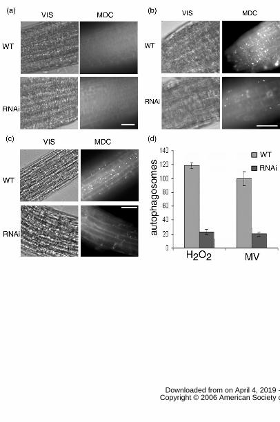
with MDC. Numerous motile MDC-stained autophagosomes were detected in root cells
after MV or H2O2 treatment, observed by fluorescence microscopy (Figure 1b, c). These
MDC-stained autophagosomes were absent from seedlings grown on control medium
(Figure 1a). These observations imply that autophagy can be induced by oxidative stress.
AtATG8 proteins have been shown to associate with autophagosomes and GFP-AtATG8
fusion proteins have been used as markers of autophagosomes in Arabidopsis (Yoshimoto
et al., 2004; Contento et al., 2005; Thompson et al., 2005). To further confirm the MDC
staining results, we introduced a transgene expressing a GFP-AtATG8e into WT Col-0
plants. When grown on nutrient MS solid medium, GFP-AtATG8e was dispersed
throughout the cytoplasm and punctate autophagosome-like structures were not visible
(Contento et al., 2005 and Figure S1). When these transgenic GFP-AtATG8e plants were
transferred for 2 days to solid MS medium with 10µM MV, numerous punctate structures
www.plantphysiol.orgon April 4, 2019 - Published by Downloaded from Copyright © 2006 American Society of Plant Biologists. All rights reserved.

10
were observed in root cells, similar to the MDC-stained punctate structures (Figure S1),
indicating the presence of autophagosomes. Together, these results strongly suggest that
autophagy is induced by oxidative stress.
RNAi-AtATG18a plants are more sensitive to MV treatment
The AtATG18a protein is required for autophagosome formation in Arabidopsis and its
transcript is up-regulated both during sucrose and nitrogen starvation and during
senescence. RNAi-AtATG18a transgenic lines with reduced AtATG18a expression show
hypersensitivity to sucrose and nitrogen starvation and are unable to produce
autophagosomes under these conditions (Xiong et al., 2005). To analyze the expression
pattern of AtATG18a under oxidative stress conditions, total RNA was isolated from WT
and RNAi-AtATG18a seedlings after incubation with 10µM MV for up to 72 hours, or in
the absence of MV as a control, and RT-PCR was performed using primers specific for
AtATG18a. As shown in figure 2, the transcript level of AtATG18a increased after MV
treatment in WT seedlings. Although this expression increase was also seen in
RNAi-AtATG18a seedlings, the level of AtATG18a transcript is much lower than in WT
seedlings (Figure 2). To determine whether autophagosome formation is disrupted in
RNAi-AtATG18a root cells under oxidative stress, 7-day-old RNAi seedlings were
transferred to solid MS medium plus 10µM MV for 2 days or plus 10mM H2O2 for 6 hours,
then stained with MDC (Figure 1b, c). Compared to WT seedlings, fewer MDC-stained
autophagosomes were detected in the root tips of these RNAi seedlings (Figure 1d). Both
H2O2 and MV treatment caused an increase in ROS in WT and in RNAi seedlings, as
measured by Amplex Red measurement of H2O2 concentration, indicating that the lack of
autophagosome production in the RNAi plants is not due to decreased production of ROS
in these plants (Figure S2). Overall, RNAi-AtATG18a seedlings had higher ROS levels
than WT, possibly due to an increase in damaged organelles caused by the autophagy
defect in these lines. These data suggest that AtATG18a is not only required in starvation-
www.plantphysiol.orgon April 4, 2019 - Published by Downloaded from Copyright © 2006 American Society of Plant Biologists. All rights reserved.

11
and senescence- induced autophagy, but also in oxidative stress-triggered autophagy.
To determine whether autophagy is required for the response to oxidative stress conditions,
growth of the RNAi-AtATG18a lines was compared with WT plants under oxidative stress.
7-day-old seedlings grown on nutrient MS solid medium (Figure 3a) were transferred to
medium with 10µM MV (Figure 3b). MV strongly inhibited growth of both WT and RNAi
plants (compare with Xiong et al., 2005). However, after 15 days, RNAi-AtATG18a plants
were already chlorotic, whereas WT seedlings were still green (Figure 3). In contrast,
silencing of an unrelated gene by RNAi had no effect on response to oxidative stress (data
not shown). These data imply that autophagy is necessary for plants to survive oxidative
stress conditions.
To assess whether the RNAi-AtATG18a lines are likely to be under increased oxidative
stress, the expression patterns of several ROS scavenging enzymes were compared
between WT and RNAi-AtATG18a seedlings. RNA was extracted from seedlings after
incubation in the presence (Figure 2a) or absence (Figure 2b) of MV for 72 hrs followed
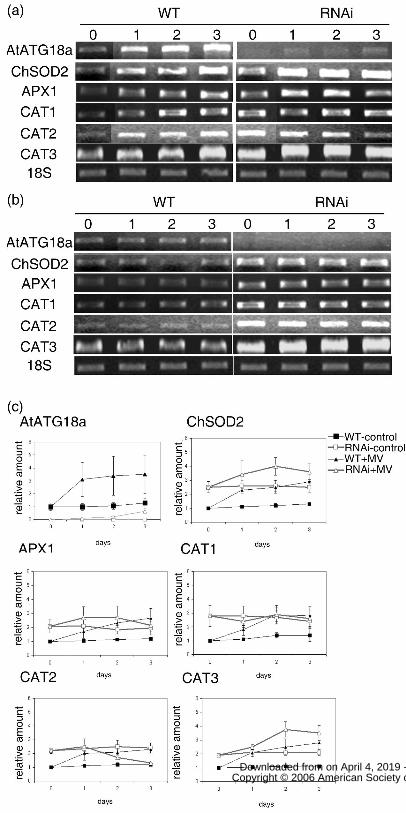
by RT-PCR analysis and quantification by densitometry (Figure 2c). As expected, in WT
seedlings, transcript abundance increased in the presence of MV for each gene, whereas it
remained constant in control samples. In RNAi-AtATG18a seedlings, the transcript level
was higher in each case than in WT plants, even in the absence of MV, possibly suggesting
that the RNAi seedlings are under increased or constitutive stress compared with WT. In
the presence of MV, the mRNA levels for catalase 3, ascorbate peroxidase and superoxide
dismutase increased still further, demonstrating that the RNAi seedlings are still able to
respond to increased ROS concentrations. The catalase 1 mRNA level remained constant
in the RNAi lines, and catalase 2 transcript level decreased at later time points, possibly
reflecting different roles or differential regulation of catalase isoforms in response to
oxidative stress.
www.plantphysiol.orgon April 4, 2019 - Published by Downloaded from Copyright © 2006 American Society of Plant Biologists. All rights reserved.

12
Autophagy is responsible for degradation of oxidized proteins during oxidative
stress
One result of oxidative stress is increased protein oxidation. To better understand the role
of autophagy in oxidative stress, the level of oxidized proteins was compared between WT
and RNAi-AtATG18a seedlings after MV treatment. Derivatization with
2,4-dinitrophenolhydrazine (DNPH) was used as this assay is well established for use in
plant systems (for example, Romero Puertas et al., 2002; Davletova et al., 2005; Guo and
Crawford, 2005). 7-day-old seedlings were transferred to medium containing 10µM MV
for up to 12 hours, followed by isolation of total proteins. After derivatization of the
carbonyl group of oxidized proteins with DNPH, immunoblotting was performed using
DNP antibodies to detect oxidized proteins. The signal on the blot was quantified by
densitometry of the entire lane of the gel. As shown in figure 4, RNAi plants accumulated
approximately 3.5-fold more oxidized proteins compared with WT seedlings after MV
treatment (compare WT+MV with RNAi+MV in figure 4a and c; P=0.0085). Similar
results were obtained by quantifying either the entire lane or individual prominent bands
(data not shown), with no evidence for selective increases in specific proteins. No signal
was seen in parallel control experiments in which the DNP derivatization step was omitted
(data not shown). Considering that RNAi-AtATG18a seedlings are defective in autophagy,
these data indicate that inhibition of autophagy leads to increased accumulation of
oxidized proteins during oxidative stress.
We considered two possibilities to explain the increased accumulation of oxidized proteins
in RNAi-AtATG18a seedlings during MV treatment, either an increased production of
oxidized proteins or a decrease in their degradation. To distinguish between these
possibilities, we analyzed the effect of concanamycin A on the level of oxidized proteins
after MV treatment. Concanamycin A is a vacuolar H+-ATPase inhibitor, which inhibits
www.plantphysiol.orgon April 4, 2019 - Published by Downloaded from Copyright © 2006 American Society of Plant Biologists. All rights reserved.

13
vacuolar enzyme activities by increasing the internal vacuolar pH and therefore prevents
vacuolar protein degradation (Drose et al., 1993). When treated with concanamycin A,
autophagosomes accumulate in the vacuole in Arabidopsis roots as they can no longer be
degraded (Yoshimoto et al., 2004). 7-day-old WT seedlings were transferred to medium
containing 10µM MV, or 1µM concanamycin A, or both 10µM MV and 1µM
concanamycin A for up to 12 hours, followed by protein isolation and immunoblotting
using DNP antibodies. MV and concanamycin A treatment caused a significant increase in
accumulation of oxidized proteins compared to MV treatment alone in WT seedlings
(approximately 3-fold; Figure 4a and c), and this accumulation level is similar to that in
RNAi-AtATG18a lines when treated with MV. Treatment of RNAi-AtATG18a seedlings
with MV and concanamycin A had no effect compared to MV alone (Figure S3),
indicating that the RNAi and concanamycin A most likely inhibit the same protein
degradation pathway. These data indicate that the increased accumulation of oxidized
proteins in RNAi-AtATG18a seedlings during MV treatment is most likely caused by the
decrease in their degradation, not by the increased production of oxidized proteins.
To further confirm that the increased accumulation of oxidized proteins in RNAi seedlings
is caused primarily by reduced degradation of these proteins by autophagy, the rate of
degradation of oxidized proteins was compared between WT and RNAi plants. Seedlings
were treated with 10µM MV for 2 days, by which time oxidized proteins had accumulated
to high levels in both WT and RNAi-AtATG18a seedlings. The seedlings were then
transferred back to solid medium lacking MV and the degradation rate of oxidized
proteins in WT and RNAi plants was compared. Initially, the amount of oxidized protein
detected remained constant or increased slightly (Figure 5), most likely due to the
continued presence of MV in the seedlings even after transfer back to control medium. As
shown in figure 5, 3 or 4 days after transfer back to control medium, the amount of
oxidized proteins in WT seedlings had decreased to a low level. In contrast,
www.plantphysiol.orgon April 4, 2019 - Published by Downloaded from Copyright © 2006 American Society of Plant Biologists. All rights reserved.

14
RNAi-AtATG18a plants retained a significantly higher level of oxidized proteins at this
time point after transfer (2-fold increase; P=0.002 for the 3 day time point). Together,
these data suggest that oxidized proteins can be transported to the vacuole for degradation
by the autophagy pathway. In this way, cells can eliminate oxidized proteins to increase
their survival under severe oxidative stress.
Oxidized proteins are delivered to the vacuole for degradation
To further confirm that during oxidative stress, oxidized proteins can be sent to the
vacuole for degradation by the autophagy pathway, the cellular localization of oxidized
proteins caused by MV treatment was compared between WT and RNAi- AtATG18a root
cells. 3-day-old WT and RNAi-AtATG18a seedlings were transferred to medium
containing 1µM concanamycin A to inhibit degradation in the vacuole, or both 1µM
concanamycin A and 10µM MV, for 12 hours. The oxidized proteins were then derivatized
in situ with DNP and detected using DNP antibody followed by immunofluorescence
confocal microscopy. In the expanded root cells shown, the vacuole occupies most of the
cell volume, and the cytoplasm is present as a thin layer adjacent to the plasma membrane.
After treatment with 1µM concanamycin A and 10µM MV for 12 hours, a strong DNP
signal was detected inside the vacuole of WT root cells (Figure 6). In contrast, this DNP
signal was absent from the vacuole of RNAi-AtATG18a root cells and instead seen in the
cytoplasm (see Figure S4 for 3D image). This provides direct evidence that autophagy is
responsible for transporting oxidized proteins to the vacuole for degradation under
oxidative stress conditions.
Discussion
As a non-specific protein degradation pathway, autophagy is activated in response to some
environmental stresses such as nutrient deficiency, and during certain stages of
development, for example senescence (Levine and Klionsky, 2004). In this study, several
www.plantphysiol.orgon April 4, 2019 - Published by Downloaded from Copyright © 2006 American Society of Plant Biologists. All rights reserved.

15
lines of evidence support the hypothesis that autophagy can also be induced for removal of
oxidized proteins during oxidative stress in Arabidopsis. (1) Using two autophagosome
markers, MDC and GFP-AtATG8e, numerous autophagosomes were detected in WT root
cells after 2 days of MV treatment but only a few autophagosomes were observed in
autophagy-defective RNAi-AtATG18a transgenic plants (Figure 1b), implying that
autophagy is induced by oxidative stress. (2) RNAi-AtATG18a seedlings were more
sensitive to oxidative stress compared to WT (Figure 3), implying that autophagy is
necessary for plants to survive such oxidative stress conditions. (3) After treatment with
MV for 12 hours, more oxidized proteins accumulated in RNAi-AtATG18a seedlings than
in WT seedlings (Figure 4), and in RNAi-AtATG18a seedlings, the degradation rate of
oxidized proteins was also slower than in WT (Figure 5), implying that autophagy is
responsible for degrading these oxidized proteins. (4) In WT seedlings, more oxidized
proteins accumulated after treatment with MV and concanamycin A together compared to
treatment with MV only (Figure 4), suggesting a role for the vacuole in degradation of
oxidized proteins. (5) Under oxidative stress, when treated with concanamycin A,
oxidized proteins can be detected in the vacuole of WT root cells but not RNAi-AtATG18a
roots cells (Figure 6 and S2). These data provide direct evidence that oxidized proteins are
sent to the vacuole for degradation by autophagy. In addition, two different sources of
ROS (direct addition of H2O2 and generation in the chloroplast by MV) both cause
autophagy, suggesting that it is general oxidative stress that induces autophagy, rather than
localized ROS production in a specific organelle. Based on these observations, we
hypothesize that one of the physiological roles of autophagy during oxidative stress is to
remove oxidized proteins.
In mammalian cells, the 20S proteasome and CMA pathway were found to degrade
oxidized proteins selectively: the 20S proteasome recognizes hydrophobic regions on the
surface of target proteins and is ATP and ubiquitin-independent (Dunlop et al., 2002),
www.plantphysiol.orgon April 4, 2019 - Published by Downloaded from Copyright © 2006 American Society of Plant Biologists. All rights reserved.

16
whereas the CMA pathway uses a pentapeptide KFERQ in proteins as a signal for
translocation into the lumen of the lysosome after being unfolded (Kiffin et al., 2004).
Furthermore, the efficiency of the 20S proteasome and CMA pathway in removal of
oxidized proteins changes with the duration and degree of oxidative damage. Mild
oxidative damage causes exposure of hydrophobic regions and partial protein unfolding,
facilitating the degradation of the oxidized proteins by the 20S proteasome and CMA
pathways. In contrast, under severe oxidative stress oxidized proteins easily aggregate and
cross-link together, decreasing the efficiency of the 20S proteasome and CMA pathways
(Dunlop et al., 2002; Kiffin et al., 2004). How mammalian cells remove oxidized proteins
rapidly and effectively under severe oxidative stress is unknown, and whether
macroautophagy contributes to their degradation is unclear. In plants, a CMA pathway has
not been demonstrated. The 20S proteasome has been isolated from maize roots and is
activated by carbonylation modification in mild oxidative conditions such as carbon
starvation, although inactivated by strong oxidative treatments such as a high
concentration of H2O2. However, no direct evidence was provided as to whether this
enhanced proteasome activity is related to degradation of oxidized proteins (Basset et al.,
2002). These data imply that the 20S proteasome plays an important role in removal of
oxidized proteins under mild oxidative stress but not under severe oxidative stress.
However, plants frequently suffer severe oxidative stress since they cannot escape from
unfavorable environments (Millar et al., 2001). Considering that a large population of
proteins is oxidized and that these proteins aggregate and are cross-linked during severe
oxidative stress, it is plausible that plant cells use autophagy to remove these damaged
proteins rather than the selective 20S proteasome pathway. Since autophagy is
non-selective and does not require a targeting signal that could be inaccessible in
aggregated and cross-linked proteins, it could effectively transfer oxidized proteins to the
vacuole for degradation.
www.plantphysiol.orgon April 4, 2019 - Published by Downloaded from Copyright © 2006 American Society of Plant Biologists. All rights reserved.

17
Previously, we showed that AtATG18a is required for autophagosome formation, and
autophagosomes were absent in RNAi-AtATG18a seedlings under nutrient deprivation and
senescence conditions (Xiong et al., 2005). When treated with MV, however, a few
MDC-stained autophagosomes can still be observed in RNAi-AtATG18a roots (Figure 1b).
This difference may be due to (1) the nature of RNAi technology: there is still a very low
level of mRNA remaining in the RNAi seedlings, and under MV treatment the AtATG18a
transcript level increased somewhat in the RNAi plants (Figure 2). This suggests that the
expression of AtATG18a in these RNAi plants is not completely silenced; and (2) the
differing extent of autophagy induced by nutrient deprivation, senescence and oxidative
stress. More autophagosomes were seen after MV or H2O2 treatment compared to during
nutrient deprivation conditions (unpublished data), implying that the extent of autophagy
during oxidative stress is higher than under nutrient deprivation and senescence conditions.
This may allow the detection of a few autophagosomes in RNAi plants during oxidative
stress but not in nutrient deprivation and senescence conditions.
In yeast, most of the autophagy related genes (ATG) are single copy. In contrast, some
Arabidopsis homologs of yeast ATGs exist as a small gene family. For example, there are
eight homologs for ATG18 (Xiong et al., 2005) and nine homologs for ATG8 (Hanaoka et
al., 2002). One hypothesis is that autophagy induced by different stresses or different
developmental stages may require different AtATG homologs. However, the same ATG18
homolog, AtATG18a, is required for nutrient depletion-, senescence- and oxidative
stress-induced autophagy. This implies that the molecular mechanism of autophagy is the
same in senescence, nutrient deprivation and oxidative stress. We have isolated T-DNA
knockout mutants for 4 additional ATG18 homologs, AtATG18c, AtATG18e, AtATG18f
and AtATG18g. Autophagosome formation detected by MDC staining was not affected in
these knockout mutants during senescence, nutrient deprivation and oxidative stress
conditions (Xiong et al., 2005 and unpublished data), implying that these genes are not
www.plantphysiol.orgon April 4, 2019 - Published by Downloaded from Copyright © 2006 American Society of Plant Biologists. All rights reserved.

18
required for already known autophagy functions. Of course, care must be taken in drawing
this conclusion as there may be some redundant functions between these AtATG18
homologs and knockout of one gene may not be enough to observe a clear phenotype.
Generating double or triple mutants will help to address this question.
www.plantphysiol.orgon April 4, 2019 - Published by Downloaded from Copyright © 2006 American Society of Plant Biologists. All rights reserved.

19
Materials and Methods
Plant materials and growth conditions
Arabidopsis thaliana plants were grown on nutrient solid MS medium as described
previously (Xiong et al., 2005) under long day conditions (16 hrs light) at 22�.
Arabidopsis thaliana seeds were surface-sterilized in 33% bleach and 0.1% Triton X-100
solution for 20 minutes followed by cold treatment for at least 2 days. For MV, H2O2 or
concanamycin A treatment, 7-day-old seedlings grown on nutrient solid MS medium were
transferred to the same medium containing 10µM MV, 10mM H2O2 or 1µM
concanamycin A and incubated for the indicated times.
Staining of seedlings with MDC and microscopy
Seedlings were stained with MDC as described (Contento et al., 2005) and observed using
fluorescence microscopy using a DAPI-specific filter. For counting of autophagosomes,
pictures were taken of equivalent regions of each root for multiple seedlings after MDC
staining. The number of autophagosomes visible in each image was counted and the
average determined for 10 seedlings per genotype and per treatment.
Hydrogen peroxide measurement
H2O2 was measured in extracts from Arabidopsis seedlings according to Rao et al. (2000).
Leaves were frozen, ground to a powder and stored at −80°C. Leaf powder (50-100 mg)
was extracted with 0.1 mL of 0.2 M HClO4, incubated on ice for 5 min, and then
centrifuged at 14,000g for 10 min at 4°C. The supernatant was neutralized with 0.2 M
NH4OH, pH 9.5, and was centrifuged again at 3,000g. The extracts were passed through
1× 1-cm columns of AG 1X-8 resin (Bio-Rad, Hercules, CA) and were eluted with 1mL
water.
The quantification of H2O2 in the extracts was performed using the Amplex Red Hydrogen
www.plantphysiol.orgon April 4, 2019 - Published by Downloaded from Copyright © 2006 American Society of Plant Biologists. All rights reserved.
20
Peroxide-Peroxidase Assay kit (Invitrogen-Molecular Probes, Carlsbad, CA), following
the manufacturer's directions. Fluorescence was measured with a Synergy HT
fluorescence plate reader (Bio-Tek, Winooski, VT) using excitation at 530nm and
fluorescence detection at 590nm. The concentration of H2O2 in each sample was
calculated using a standard curve.
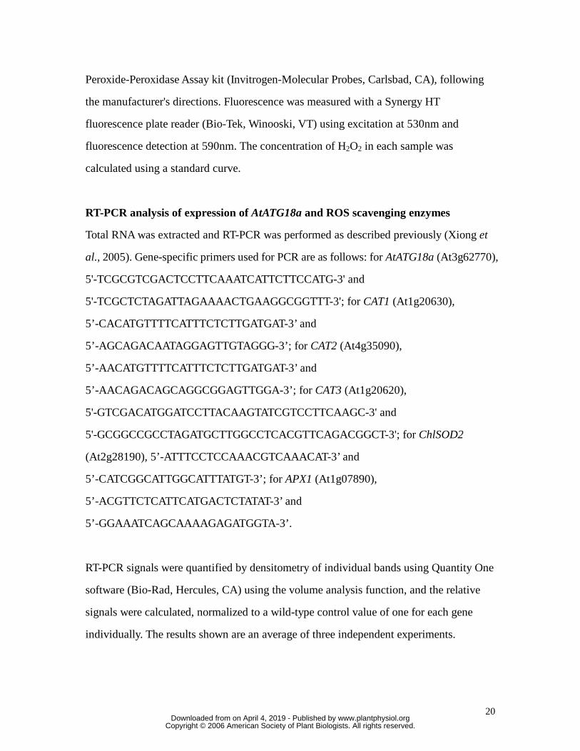
RT-PCR analysis of expression of AtATG18a and ROS scavenging enzymes
Total RNA was extracted and RT-PCR was performed as described previously (Xiong et
al., 2005). Gene-specific primers used for PCR are as follows: for AtATG18a (At3g62770),
5'-TCGCGTCGACTCCTTCAAATCATTCTTCCATG-3' and
5'-TCGCTCTAGATTAGAAAACTGAAGGCGGTTT-3'; for CAT1 (At1g20630),
5’-CACATGTTTTCATTTCTCTTGATGAT-3’ and
5’-AGCAGACAATAGGAGTTGTAGGG-3’; for CAT2 (At4g35090),
5’-AACATGTTTTCATTTCTCTTGATGAT-3’ and
5’-AACAGACAGCAGGCGGAGTTGGA-3’; for CAT3 (At1g20620),
5'-GTCGACATGGATCCTTACAAGTATCGTCCTTCAAGC-3' and
5'-GCGGCCGCCTAGATGCTTGGCCTCACGTTCAGACGGCT-3'; for ChlSOD2
(At2g28190), 5’-ATTTCCTCCAAACGTCAAACAT-3’ and
5’-CATCGGCATTGGCATTTATGT-3’; for APX1 (At1g07890),
5’-ACGTTCTCATTCATGACTCTATAT-3’ and
5’-GGAAATCAGCAAAAGAGATGGTA-3’.
RT-PCR signals were quantified by densitometry of individual bands using Quantity One
software (Bio-Rad, Hercules, CA) using the volume analysis function, and the relative
signals were calculated, normalized to a wild-type control value of one for each gene
individually. The results shown are an average of three independent experiments.
www.plantphysiol.orgon April 4, 2019 - Published by Downloaded from Copyright © 2006 American Society of Plant Biologists. All rights reserved.

21
Generation of GFP-AtATG8e transgenic plants
A DNA fragment encoding GFP-AtATG8e was obtained by digestion of plasmid
pJ4GFP-AtATG8e (Contento et al., 2005) with restriction enzymes SalI and SacI, and
ligated into the plant T-DNA binary vector pCAMBIA1300 (Cambia GPO, Canberra,
Australia). This construct was introduced into Agrobacterium tumefaciens strain GV3101
by electroporation (Merereau et al., 1990). pCAMBIA1300: GFP-AtATG8e was
introduced into Arabidopsis thaliana Columbia-0 plants by Agrobacterium-mediated
transformation using the floral dipping method (Clough and Bent, 1998). Transgenic
plants were screened using hygromycin resistance and expression confirmed by
immunoblotting using GFP antibody (Invitrogen, Carlsbad, CA). Homozygous T2
transformant seeds were used for further studies.
Oxidized protein analysis
For oxidized protein analysis, total proteins were isolated from seedlings after treatment
with MV for the indicated times by grinding in cold extraction buffer (0.1M Tris-HCl pH
7.5, 0.3M sucrose, 1mM EDTA, 0.1mM phenylmethylsulphonylfluoride and 1%
β-mercaptoethanol) and centrifugation at 1000g for 10 minutes. Oxidized proteins were
detected using an OxyBlot protein oxidation detection kit (Chemicon International,
Temecula, CA) according to the manufacturer’s instructions. DNP signals were quantified
by densitometry using Quantity One software (Bio-Rad, Hercules, CA) using the volume
analysis function, and the relative signals were calculated. In figure 4, signals were
normalized to a wild-type 0-hour control value of one. Signals were determined both for
the entire lane of gel and for individual prominent protein bands, with essentially
equivalent results for both methods. The results shown are an average of three independent
experiments for quantification of the entire gel lane. In figure 5, signals were normalized
to the WT +MV signal, which was set as 100%.
www.plantphysiol.orgon April 4, 2019 - Published by Downloaded from Copyright © 2006 American Society of Plant Biologists. All rights reserved.

22
Unpaired t-tests were conducted on the protein oxidation data to determine statistical
significance. Significant difference was defined by treatment comparisons with observed
P-values of less than 0.05 (P<0.05), allowing for a 95% confidence interval.
Immunolocalization and confocal microscopy
Three-day-old Arabidopsis seedlings grown on MS solid medium were transferred to the
same medium containing 1µM concanamycin A or 1µM concanamycin A plus 10µM MV
for 12-16 hours. Immunofluorescence staining of treated Arabidopsis seedling was
performed according to Muller et al. (1998) with the following modifications: Seedlings
were fixed with Carnoy’s solution (6 parts absolute ethanol, 1 part glacial acetic acid, 3
parts chloroform) for 15 minutes followed by successive washes with 50% ethanol, 25%
ethanol and 50mM PIPES, 5mM EGTA, 5mM MgSO4 (MTSB), then mounted on slides.
Prior to addition of block buffer (3% bovine serum albumin/MTSB), permeabilized
seedlings were treated with 40µl 1X 2,4-dinitrophenylhydrazine (DNPH, Chemicon
International) for 30 minutes and neutralized with 40µl neutralization solution (OxyBlot
protein oxidation kit, Chemicon International). Immunolabeling was done using anti-DNP
(OxyBlot protein oxidation kit, Chemicon International; 1:100) for 15-18 hours at 4ºC
followed by Alexa Fluor® 594-conjugated goat anti-rabbit secondary antibodies
(Molecular Probes, Eugene, OR, USA; 1:250) for 1 hour at room temperature. Samples
were washed five times with MTSB after each antibody. Images were collected using a
Leica TCS/NT confocal microscope (Leica Microsystems, Exton, PA, USA) and 3D
reconstruction was performed using MetaMorph (Molecular Devices Corporation,
Downingtown, PA) and Adobe Photoshop (Adobe Systems, Mountain View, CA, USA).
Upon request, all novel materials described in this publication will be made available in a
timely manner for non-commercial research purposes, subject to the requisite permission
from any third-party owners of all or parts of the material. Obtaining any permissions will
www.plantphysiol.orgon April 4, 2019 - Published by Downloaded from Copyright © 2006 American Society of Plant Biologists. All rights reserved.

23
be the responsibility of the requestor.
www.plantphysiol.orgon April 4, 2019 - Published by Downloaded from Copyright © 2006 American Society of Plant Biologists. All rights reserved.

24
Acknowledgements
We thank Dr David Oliver for helpful comments on the manuscript.
www.plantphysiol.orgon April 4, 2019 - Published by Downloaded from Copyright © 2006 American Society of Plant Biologists. All rights reserved.
25
Literature Cited
Apel K, Hirt H (2004) Reactive oxygen species: metabolism, oxidative stress, and signal
transduction. Annu Rev Plant Biol 55: 373-399
Basset G, Raymond P, Malek L, Brouquisse R (2002) Changes in the expression and the
enzymic properties of the 20S proteasome in sugar-starved maize roots. Evidence for an in
vivo oxidation of the proteasome. Plant Physiol 128: 1149-1162
Chen SX, Schopfer P (1999) Hydroxyl-radical production in physiological reactions. A
novel function of peroxidase. Eur J Biochem 260: 726-735
Contento AL, Xiong Y, Bassham DC (2005) Visualization of autophagy in Arabidopsis
using the fluorescent dye monodansylcadaverine and a GFP-AtATG8e fusion protein.
Plant J 42: 598-608
Clough SJ, Bent AF (1998) Floral dip: a simplified method for Agrobacterium-mediated
transformation of Arabidopsis thaliana. Plant J 16: 735-743
Cuervo AM (2004) Autophagy: in sickness and in health. Trends Cell Biol 14: 70-77
Dat JF, Pellinen R, Beeckman T, Van De Cotte B, Langebartels C, Kangasjarvi J, Inze D,
Van Breusegem F (2003) Changes in hydrogen peroxide homeostasis trigger an active cell
death process in tobacco. Plant J 33: 621-632
Davletova S, Rizhsky L, Liang HJ, Zhong SQ, Oliver DJ, Coutu J, Shulaev V, Schlauch K,
Mittler R (2005) Cytosolic ascorbate peroxidase 1 is a central component of the reactive
oxygen gene network of Arabidopsis. Plant Cell 17: 268-281
Desikan R, A-H Mackerness S, Hancock JT, Neill SJ (2001) Regulation of the
Arabidopsis transcriptome by oxidative stress. Plant Physiol 127: 159-172
www.plantphysiol.orgon April 4, 2019 - Published by Downloaded from Copyright © 2006 American Society of Plant Biologists. All rights reserved.

26
Doelling JH, Walker JM, Friedman EM, Thompson AR, Vierstra RD (2002) The
APG8/12-activating enzyme APG7 is required for proper nutrient recycling and
senescence in Arabidopsis thaliana. J Biol Chem 277: 33105-33114
Drose S, Bindseil KU, Bowmana EJ, Siebers A, Zeeck A, Altendorf K (1993) Inhibitory
effect of modified bafilomycins and concanamycins and P- and V-type
adenosinetriphosphatases. Biochemistry 32: 3902-3906
Dunlop RA, Rodgers KJ, Dean RT (2002) Recent developments in the intracellular
degradation of oxidized proteins. Free Radic Biol Med 33: 894-906
Grune T, Reinheckel T, Joshi M, Davies KJ (1995) Proteolysis in cultured liver epithelial
cells during oxidative stress. Role of the multicatalytic proteinase complex, proteasome. J
Biol Chem 270: 2344-2351
Guo FQ, Crawford NM (2005) Arabidopsis nitric oxide synthase1 is targeted to
mitochondria and protects against oxidative damage and dark-induced senescence. Plant
Cell 17: 3436-3450
Hanaoka H, Noda T, Shirano Y, Kato T, Hayashi H, Shibata D, Tabata S, Ohsumi Y (2002)
Leaf senescence and starvation-induced chlorosis are accelerated by the disruption of an
Arabidopsis autophagy gene. Plant Physiol 129: 1181-1193
Kiffin R, Christian C, Knecht E, Cuervo AM (2004) Activation of chaperone-mediated
autophagy during oxidative stress. Mol Biol Cell 15: 4829-4840
Liang XH, Jackson S, Seaman M, Brown K, Kempkes B, Hibshoosh H, Levine B (1999)
Induction of autophagy and inhibition of tumorigenesis by beclin 1. Nature 402: 672-676
Levine B, Klionsky DJ (2004) Development by self-digestion: molecular mechanisms and
www.plantphysiol.orgon April 4, 2019 - Published by Downloaded from Copyright © 2006 American Society of Plant Biologists. All rights reserved.

27
biological functions of autophagy. Dev Cell 6: 463-477
Malan C, Gregling MM, Gressel J (1990) Correlation between CuZn superoxide
dismutase and glutathione reductase and environmental and xenobiotic stress tolerance in
maize inbreds. Plant Sci 69: 157-166
Massey A, Kiffin R, Cuervo AM (2004) Pathophysiology of chaperone-mediate
autophagy. Int J Biochem Cell Biol 36: 2420-2434
Merereau M, Pazour GJ, Das A (1990) Efficient transformation of Agrobacterium
tumefaciens by electroporation. Gene 90: 149-151
Millar H, Considine MJ, Day DA, Whelan J (2001) Unraveling the role of mitochondria
during oxidative stress in plants. IUBMB Life 51: 201-205
Mittler R, Vanderauwera S, Gollery M, Breusegem F (2004) Reactive oxygen gene
network of plants. Trends Plant Sci 9: 490-498
Mittler R (2002) Oxidative stress, antioxidants and stress tolerance. Trends Plant Sci 7:
405-410
Mizushima N, Sugita H, Yoshimori T, Ohsumi Y (1998) A new protein conjugation system
in human. The counterpart of the yeast Apg12p conjugation system essential for
autophagy. J Biol Chem 273: 33889-33892
Muller A, Guan C, Galweiler L, Tanzler P, Huijser P, Marchant A, Parry G, Bennett M,
Wisman E (1998) AtPIN2 defines a locus of Arabidopsis for root gravitropism control.
EMBO J 17: 6903–6911
Mullineaux P, Karpinski S (2002) Signal transduction in response to excess light: getting
out of the chloroplast. Curr Opin Plant Biol 5: 43-48
www.plantphysiol.orgon April 4, 2019 - Published by Downloaded from Copyright © 2006 American Society of Plant Biologists. All rights reserved.

28
Murgia I, Tarantino D, Vannini C, Bracale M, Carravieri S, Soave C (2004) Arabidopsis
thaliana plants overexpressing thylakoidal ascorbate peroxidase show increased resistance
to paraquat-induced photooxidative stress and to nitric oxide-induced cell death. Plant J 38:
940-953
Noctor G, Foyer C (1998) Ascorbate and glutathione: keeping active oxygen under control.
Annu Rev Plant Physiol Plant Mol Biol 49: 249-279
Okada K, Ikeuchi M, Yamamoto N, Ono T, Miyao M (1996) Selective and specific
cleavage of the D1 and D2 proteins of photosystem II by exposure to singlet oxygen:
factors responsible for the susceptibility to cleavage of the proteins. Biochim Biophys
Acta 1274: 73-79
Orozco-Cardenas ML, Ryan C (1999) Hydrogen peroxide is generated systemically in
plant leaves by wounding and systemin via the octadecanoid pathway. Proc Natl Acad Sci
USA 96: 6553-6557
Prasad TK, Anderson MD, Martin BA, Stewart CR (1994) Evidence for chilling-induced
oxidative stress in maize seedlings and a regulatory role for hydrogen peroxide. Plant Cell
6: 65-74
Rao MV, Lee H, Creelman RA, Mullet JE, Davis KR. (2000) Jasmonic acid signaling
modulates ozone-induced hypersensitive cell death. Plant Cell 12: 1633–1646.
Romero-Puertas MC, Palma JM, Gomez M, Del Rio LA, Sandalio LM (2002) Cadmium
causes the oxidative modification of proteins in pea plants. Plant Cell Env 25: 677-686
Shringarpure R, Grune T, Mehlhase J, Davies KJ (2003) Ubiquitin conjugation is not
required for the degradation of oxidized proteins by proteasome. J Biol Chem 278:
311-318
www.plantphysiol.orgon April 4, 2019 - Published by Downloaded from Copyright © 2006 American Society of Plant Biologists. All rights reserved.

29
Thompson AR, Doelling JH, Suttangkakul A, Vierstra RD (2005) Autophagic nutrient
recycling in Arabidopsis directed by the ATG8 and ATG12 conjugation pathways. Plant
Physiol 138: 2097-2110
Thompson AR, Vierstra RD (2005) Autophagic recycling: lessons from yeast help define
the process in plants. Curr Opin Plant Biol 8: 165-173
Thorpe GW, Fong CS, Alic N, Higgins V, Dawes LW (2004) Cells have distinct
mechanisms to maintain protection against different reactive oxygen species:
oxidative-stress-response genes. Proc Natl Acad Sci USA 101: 6564-6569
Torres MA, Dangl JL, Jones JDG (2002) Arabidopsis gp91phox homologues AtrbohD and
AtrbohF are required for accumulation of reactive oxygen intermediates in the plant
defense response. Proc Natl Acad Sci USA 99: 517-522
Tsugane K, Kobayashi K, Niwa Y, Ohba Y, Wada K, Kobayashi H (1999) A recessive
Arabidopsis mutant that grows photoautotrophically under salt stress shows enhanced
active oxygen detoxification. Plant Cell 11: 1195-1206
Ullrich O, Reinheckel T, Sitte N, Hass R, Grune T, Davies KJ (1999) Poly-ADP ribose
polymerase activates nuclear proteasome to degrade oxidatively damaged histones. Proc
Natl Acad Sci USA 96: 6223-6228
Woo HR, Kim JH, Nam HG, Lim PO (2004) The delayed leaf senescence mutants of
Arabidopsis, ore1, ore3, and ore9 are tolerant to oxidative stress. Plant Cell Physiol 45:
923-932
Xiong Y, Contento AL, Bassham DC (2005) AtATG18a is required for the formation of
autophagosomes during nutrient stress and senescence in Arabidopsis thaliana. Plant J 42:
535-546
www.plantphysiol.orgon April 4, 2019 - Published by Downloaded from Copyright © 2006 American Society of Plant Biologists. All rights reserved.

30
Yoshimoto K, Hanaoka H, Sato S, Kato T, Tabata S, Noda T, Ohsumi Y (2004) Processing
of ATG8s, ubiquitin-like proteins, and their deconjugation by ATG4s are essential for
plant autophagy. Plant Cell 16: 2967-2983
www.plantphysiol.orgon April 4, 2019 - Published by Downloaded from Copyright © 2006 American Society of Plant Biologists. All rights reserved.

31
Figure legends
Figure 1. MDC staining of root tips. 7-day-old WT and RNAi-AtATG18a seedlings were
transferred to nutrient control solid MS medium for 2 days (a), to nutrient solid MS
medium containing 10µM MV for 2 days (b), or to nutrient solid MS medium containing
10mM H2O2 for 6 hours (c), followed by staining with MDC, and observed by
fluorescence microscopy. Scale bar = 50µm. (d) Number of autophagosomes per root
section were counted after MV or H2O2 treatment as above and the average number
determined for 10 seedlings per genotype and treatment. Error bars indicate SE.
Figure 2. Gene expression analysis upon MV treatment. 7-day-old WT and
RNAi-AtATG18a seedlings were transferred either to solid MS medium containing 10µM
MV (a) or to control medium (b) for the indicated times. Total RNA was isolated and
RT-PCR was performed using gene specific primers. 18S RNA was used as a loading
control. Cat1, catalase 1; Cat2, catalase 2; Cat 3, catalase 3; APX1, ascorbate peroxidase 1;
ChlSOD2, chloroplast superoxide dismutase. (c) The relative amounts of RT-PCR product
were quantified by densitometry from three independent repeats, with the wild-type
control value set as 1. Error bars indicate SE.
Figure 3. Phenotype of RNAi-AtATG18a plants under oxidative stress conditions.
7-day-old seedlings of WT and RNAi-AtATG18a plants (a) were transferred to MS
medium containing 10µM MV for 15 days (b).
Figure 4. Protein oxidation analysis. (a) 7-day-old seedlings of WT and RNAi-AtATG18a
plants were transferred to solid medium containing 10µM MV for the indicated hours. For
concanamycin A (conA) treatment, WT seedlings were transferred to solid medium
containing 1µM conA for the indicated times. Total proteins were isolated and derivatized
by DNP followed by immunoblotting using DNP antibody. Molecular size markers are
www.plantphysiol.orgon April 4, 2019 - Published by Downloaded from Copyright © 2006 American Society of Plant Biologists. All rights reserved.

32
indicated at the left. (b) Coomassie Blue-stained gel of samples from part (a) to
demonstrate equal loading. Molecular size markers are indicated at the left. (c) DNP
signals from part (a) were quantified by densitometry from three independent repeats with
the WT control value set as 1; error bars indicate SE.
Figure 5. Degradation of oxidized proteins. (a) 7-day-old seedlings of WT and
RNAi-AtATG18a plants were transferred to solid medium containing 10µM MV for 2
days (lane 1 and 5) then transferred back to normal solid MS medium and incubated for 2
days (lane 2 and 6), 3 days (lane 3 and 7) or 4 days (lane 4 and 8). Total proteins were
isolated and derivatized by DNP followed by immunoblotting using DNP antibody.
Molecular size markers are indicated at the left. (b) Coomassie Blue-stained gel of
samples from part (a) to demonstrate equal loading. Molecular size markers are indicated
at the left. (c) DNP signals from part (a) were quantified from three independent repeats,
with the zero-time WT signal (lane 1) set as 100%; error bars indicate SE.
Figure 6. Immunofluorescence analysis of the cellular localization of oxidized proteins.
3-day-old WT and RNAi-AtATG18a seedlings grown vertically on solid medium were
transferred to solid medium containing 1µM concanamycin (conA) or both 1µM
concanamycin and 10µM MV (MV+conA) and incubated for 12 hours. The oxidized
proteins were derivatized with DNP and detected using DNP antibodies, followed by
confocal microscopy. Scale bar = 20µm.
Supplemental figure legends
Figure S1. Subcellular localization of GFP-AtATG8e. 7-day-old transgenic
GFP-AtATG8e seedlings were transferred to nutrient control solid MS medium or to solid
medium containing 10µM MV for 2 days, then observed by fluorescence microscopy.
Scale bar = 20µm.
www.plantphysiol.orgon April 4, 2019 - Published by Downloaded from Copyright © 2006 American Society of Plant Biologists. All rights reserved.

33
Figure S2. Hydrogen peroxide measurement in seedlings. Seven-day post-germination
wild type and RNAi-AtATG18a seedlings were treated with hydrogen peroxide or methyl
viologen (MV). Hydrogen peroxide levels after 0-hour or 6-hour hydrogen peroxide
treatment, and 48-hour MV-treatment were determined using the Amplex Red Assay
(Invitrogen, Carlsbad, CA). Error bars represent Standard Error. An unpaired t-test was
used to determine the statistical significance of difference between each data set.
Figure S3. ConA has no effect on levels of oxidized proteins in RNAi-AtATG18a plants.
(a) 7-day-old RNAi-AtATG18a seedlings were transferred to solid medium containing
10µM MV or 10µM MV plus 1µM conA for the indicated hours. Total proteins were
isolated and derivatized by DNP followed by immunoblotting using DNP antibody.
Molecular size markers are indicated at the left. (b) Coomassie Blue-stained gel of
samples from part (a) to demonstrate equal loading. Molecular size markers are indicated
at the left. (c) DNP signals from part (a) were quantified by densitometry from three
independent repeats; error bars indicate SE.
Figure S4. 3D image of immunofluorescence analysis of the cellular localization of
oxidized proteins. 3-day-old WT and RNAi-AtATG18a seedlings grown vertically on solid
medium were transferred to solid medium containing 1µM concanamycin (conA) or both
1µM concanamycin and 10µM MV (MV+conA) and incubated for 12 hours. The oxidized
proteins were derivatized with DNP and detected using DNP antibodies, followed by
confocal microscopy.
www.plantphysiol.orgon April 4, 2019 - Published by Downloaded from Copyright © 2006 American Society of Plant Biologists. All rights reserved.
www.plantphysiol.orgon April 4, 2019 - Published by Downloaded from Copyright © 2006 American Society of Plant Biologists. All rights reserved.
www.plantphysiol.orgon April 4, 2019 - Published by Downloaded from Copyright © 2006 American Society of Plant Biologists. All rights reserved.

www.plantphysiol.orgon April 4, 2019 - Published by Downloaded from Copyright © 2006 American Society of Plant Biologists. All rights reserved.

www.plantphysiol.orgon April 4, 2019 - Published by Downloaded from Copyright © 2006 American Society of Plant Biologists. All rights reserved.

www.plantphysiol.orgon April 4, 2019 - Published by Downloaded from Copyright © 2006 American Society of Plant Biologists. All rights reserved.

www.plantphysiol.orgon April 4, 2019 - Published by Downloaded from Copyright © 2006 American Society of Plant Biologists. All rights reserved.