Embed Size (px)
Citation preview
Contents lists available at ScienceDirect
Neuroscience Letters
journal homepage: www.elsevier.com/locate/neulet
Research article
Lateral habenula lesions disrupt appetitive extinction, but do not affectvoluntary alcohol consumption
Rocio Donairea, Ignacio Morónb, Santos Blancoc, Alvaro Villatoroc, Fernando Gámizd,Mauricio R. Papinie, Carmen Torresa,⁎
a Department of Psychology, University of Jaén, 23071, SpainbDepartment of Psychobiology, Research Center for Mind, Brain, and Behavior (CIMCYC), University of Granada, Spainc Department of Experimental Biology, University of Jaén, SpaindDepartment of Psychobiology, Biomedic Research Center (CIBM), Neuroscience Institute “Federico Olóriz”, University of Granada, Spaine Department of Psychology, Texas Christian University, Fort Worth, USA
A R T I C L E I N F O
Keywords:Lateral habenulaReward lossConsummatory extinctionInstrumental extinctionVoluntary alcohol consumption
A B S T R A C T
This study analyzed the effects of LHb lesions on appetitive extinction and alcohol consumption. Eighteen maleWistar rats received neurochemical lesions of the LHb (quinolinic acid) and 12 received a vehicle infusion (PBS).In a runway instrumental task, rats received acquisition (12 pellets/trial, 6 trials/session, 10 sessions) and ex-tinction training (5 sessions). In a consummatory task, rats had daily access to 32% sucrose (5min, 10 sessions)followed by access to water (5 sessions). Then, animals received 2 h preference tests with escalating alcoholconcentrations (2%–24%), followed by two 24 h preference tests with 24% alcohol. Relative to Shams, LHblesions delayed extinction, as indicated by lower response latencies (instrumental task) and higher fluid con-sumption (consummatory task). LHb lesions did not affect alcohol consumption regardless of alcohol con-centration or test duration. The LHb modulates appetitive extinction and needs to be considered as part of thebrain circuit underlying reward loss.
1. Introduction
In rodents, the lateral habenula (LHb) receives afferents, via thestria medullaris and the inferior thalamic peduncle, from the en-topeduncular nucleus, lateral hypothalamus, lateral preoptic area,medial prefrontal cortex, lateral septum, ventral pallidum, raphe nuclei,locus coeruleus, bed nucleus of the stria terminalis, and ventral teg-mental area (VTA). LHb outputs join the fasciculus retroflexus to sendprojections to the rostromedial tegmental nucleus (RMTg), VTA, sub-stantia nigra pars compacta, raphe nuclei, laterodorsal tegmentum,locus coreuleus, hypothalamus, several thalamic nuclei, and the nucleusaccumbens [1–4]. This complex connectivity enables the LHb to in-tegrate motivational and emotional states crucial for survival [5].Several studies highlight a role of the LHb in reward loss (rewardswhose magnitude or quality is worse than expected). Single-cell re-cordings in macaque monkeys found that LHb neurons were activatedby stimuli predicting a small reward and by the small reward itself, aslong as it was unexpected [6]. One of the consequences of this activa-tion is the inhibition of the dopaminergic neurons in the brain rewardsystem, an action that depends on the connections between the LHb and
the RMTg (an inhibitory nucleus that projects on VTA [3]). The in-volvement of LHb in non-reward processing has also been observed inlesion studies showing delayed extinction after cocaine [7] or sucroseself-administration [8]. Human neuroimaging studies reveal increasedLHb activity in tasks involving response errors, missing rewards andnegative feedback [9–12]. However, the specific function of the LHb inreward loss situations is largely unknown.
Recent studies suggest a connection between reward loss and drugintake [13]. Rats exposed to unexpected reward omission or devalua-tion exhibit increased consumption of anxiolytics (alcohol, benzodia-zepines [14–16]). This loss-induced increase in anxiolytics intake (re-ferred to as emotional self-medication, ESM) could also be mediated bythe LHb, since its functional manipulation affects alcohol consumption.Rats with LHb lesions given intermittent access to 20% alcohol in-creased voluntary intake more rapidly than sham animals [17]. Thiseffect was dependent on lateral hypothalamus projections to the LHb[18]. Inhibition of LHb activity by high-frequency stimulation reducedvoluntary alcohol consumption under similar conditions [19]. Lesionsof the LHb also induced high rates of responding for alcohol in an op-erant self-administration task and blocked yohimbine-induced alcohol-
https://doi.org/10.1016/j.neulet.2019.03.044Received 24 December 2018; Received in revised form 14 March 2019; Accepted 25 March 2019
⁎ Corresponding author.E-mail address: [email protected] (C. Torres).
Neuroscience Letters 703 (2019) 184–190
Available online 27 March 20190304-3940/ © 2019 Elsevier B.V. All rights reserved.
T
seeking reinstatement [17]. Although these results suggest that the LHbregulates voluntary alcohol consumption, whether this influence de-pends on alcohol concentration has not been explored. This questionwould clarify whether LHb modulates the rewarding vs. the aversiveeffects of alcohol [20], since these are dose-dependent effects [21].
This study explored the role of the LHb in appetitive extinction andalcohol intake. Animals were exposed to two appetitive tasks, con-summatory and instrumental (counterbalanced), and subsequentlygiven extinction training. Two tasks were included to assess the gen-erality of the lesion effects and also because consummatory and in-strumental tasks not always produce the same results [22]. Aftercompleting these tasks, animals underwent preference testing with es-calating concentrations of alcohol (from 2 to 24%). Based on previousstudies, increased resistance to extinction and increased consumption ofhigh alcohol concentrations were expected in LHb-lesioned animalscompared with sham controls.
2. Method
2.1. Subjects
Thirty 90 day-old, male, Wistar rats, weighing 388.9 ± 4.3 g,served as subjects (Harlan Laboratories, Barcelona, Spain). Rats werehoused individually in polycarbonate cages with water continuouslyavailable, in a room with constant temperature (18–22 °C) and hu-midity (50–60%), and lights on between 08:00 and 20:00 h. Animalswere food deprived and maintained within 82–85% of their ad libweight. The experiment followed the European Union directive guide-lines for the use of animals in research (2010/63/EU) and Spanish Law(6/2013; R.D.53/2013).
2.2. Apparatus
Consummatory training involved 3 Plexiglas boxes(30× 15×30 cm, LxWxH). The sipper tube of a graduated cylinderwas inserted through a hole located in the front wall. The 32% sucrosesolution was prepared w/w by mixing 32 g of sucrose for every 68 g ofdistilled water. A magnetic mixer (Nahita, 680-9, Beriain, Spain) wasused to dissolve the sucrose. Session length was measured with amanual stop-watch (Extech, 365510, Madrid, Spain).
For instrumental extinction training, a straight runway was used(245×12×12 cm, LxWxH), divided into three sections by twoPlexiglas guillotine doors. Two sections (start, goal) were 20-cm long,the running section was 205-cm long. The walls and floor of the run-ways were made of black Plexiglas (0.7-mm thick). The entire length ofthe runway was covered by clear Plexiglas lids. Food pellets (45mg,formula P; Research Diets, Lancaster, NH, USA) were used as the re-ward. Time to run through the runway was manually registered with amanual chronometer (see above). Trials began when the start door wasraised and ends when the rat entered the goal section with its fourpaws.
Access to alcohol and water in the preference tests was provided inhome cages (32×15×30 cm, LxWxH) with the floor covered withsaw dust and containing two 150-ml plastic bottles. Fluid consumptionwas measured by weighing the bottles before and after each preferencesession (Cobos, JT-300C Digital Scale, Barcelona, Spain). Alcohol (96%,Panreac, Castellar del Vallés, Spain) was diluted in tap water on a v/vbasis. Animals were weighed daily (Baxtran, BS3, Girona, Spain).
2.3. Procedure
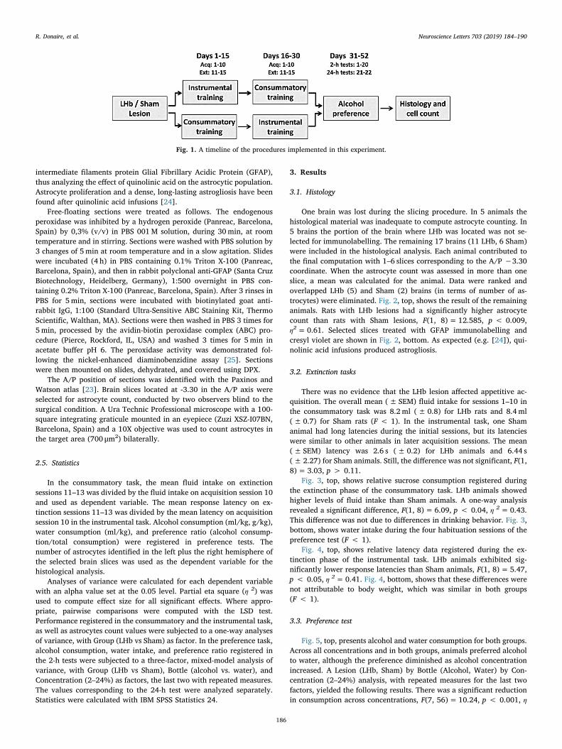
Fig. 1 shows a timeline of the general procedure.
2.3.1. SurgeryAnimals were randomly assigned to the LHb (n=18) or sham
(n=12) condition, and anesthetized with ketamine (150mg/kg, ip)
and xylazine (5mg/kg, ip). Once anesthetized, the rat’s head wasshaved and cleaned with betadine, and then set in a stereotaxic appa-ratus (Digital Lab Standard Stereotaxic, Stoelting, Dublin, Ireland). Amidline incision was made, the skull was scrapped clean of connectivetissue, and bregma was located. Quinolinic acid (Sigma Aldrich,Madrid, Spain), 0.12M, dissolved in a 10% phosphate buffered saline(PBS) solution, titrated to pH 7.4 with sodium hydroxide, was used asneurotoxin and administered with an infusion pump (HarvardApparatus, 11 Elite, Holliston, MA, USA). The neurotoxin was infusedin a volume of 0.175 μL, at a rate of 0.1 μL/min, over a period of 1:40 s.The injection cannula was kept at the lesion site for 1:30min to facil-itate the flow of the toxin. Four infusions were made, two in eachhemisphere: AP: −3.1, ML:± 0.7, DV: −4.7, and AP: -3.6, ML:± 0.75,DV: −5 [23]. Once the infusion procedure was concluded, the incisionwas closed with stitches, the suture was cleaned, and the animal wasplaced in a polycarbonate box under a light providing heat until theanesthesia wore off. Behavioral tests began when all animals were re-covered and completed their food deprivation schedule.
2.3.2. BehaviorTwo (counterbalanced) appetitive extinction tasks and an alcohol
preference test were conducted. Animals were fed at least 30min afterthe end of the corresponding test.
For the consummatory task, animals were placed in the box for a 5-min habituation session without fluids. On Days 1–10 (acquisition)animals had free access to 32% sucrose. On Days 11–15 (extinction),animals received water. Each session lasted 5min starting from the firstcontact with the sipper tube. Rats were transported in squads of 3 an-imals, all from the same group, with the order of squads randomizedacross days.
For the instrumental task, animals were transported in squads of 5animals, with the squad order randomized across days. Rats receivedthree habituation sessions (see [14] for details). Training began on Day4: animals were placed in the start box, the start door was opened andthe rat was allowed to run down the runway to obtain the reward (12pellets). A maximum time of 40 s was allowed to complete the trial. Assoon as the rat finished eating or 30 s had elapsed, it was placed back inits home cage for a 10-min intertrial interval. Each rat received 6 trialsper day during 10 acquisition sessions. In extinction (5 sessions), nofood was present and rats were enclosed in the goal box for 30 s.
The preference test started after completing extinction training.Animals were habituated on Days 1–4 to the two-bottle procedure withboth bottles containing tap water [15]. On Days 5–20, animals receivedincreasing concentrations of alcohol in one bottle and water in theother. The concentrations of alcohol were 2, 4, 6, 8, 10, 12, 16, and24%. Each concentration was presented for two consecutive days. OnDays 1–20, alcohol preference tests were 2-h long. This testing proce-dure was used before in our lab [14,15]. On Days 21–22, animals hadaccess to the highest concentration (24%) during 24 h, a procedure alsoused before in our lab [21]. The position of the bottles was changeddaily to minimize the effects of side preference.
2.4. Histology and astrocyte count
Rats were anaesthetized (sodium pentobarbital 5 mg diluted in10ml 0,9% physiologic serum; 0.1ml/100 g animal weight) and per-fused with 0.01M phosphate-buffered saline (PBS; pH 7.4), and thenwith 300ml of 4% paraformaldehyde in 0.1M phosphate buffer (PB).The brains were removed and then post-fixed for a further 4 h in thesame fixative at room temperature. Samples were then cryoprotected byimmersion overnight at 4 °C in 0.1 M PB containing 30% sucrose. Afterthis, they were embedded in OCT (Optimal Cutting Temperature;Sakura, Alphen aan den Rijn,The Netherlands) and the brain was letsolidify with Peltier system. Serial 40 μm coronal sections were pre-pared using a cryostat (Leica Microsystems CM1950, Barcelona, Spain)and stained with immunohistochemistry to label the astrocytic
R. Donaire, et al. Neuroscience Letters 703 (2019) 184–190
185
intermediate filaments protein Glial Fibrillary Acidic Protein (GFAP),thus analyzing the effect of quinolinic acid on the astrocytic population.Astrocyte proliferation and a dense, long-lasting astrogliosis have beenfound after quinolinic acid infusions [24].
Free-floating sections were treated as follows. The endogenousperoxidase was inhibited by a hydrogen peroxide (Panreac, Barcelona,Spain) by 0,3% (v/v) in PBS 001M solution, during 30min, at roomtemperature and in stirring. Sections were washed with PBS solution by3 changes of 5min at room temperature and in a slow agitation. Slideswere incubated (4 h) in PBS containing 0.1% Triton X-100 (Panreac,Barcelona, Spain), and then in rabbit polyclonal anti-GFAP (Santa CruzBiotechnology, Heidelberg, Germany), 1:500 overnight in PBS con-taining 0.2% Triton X-100 (Panreac, Barcelona, Spain). After 3 rinses inPBS for 5min, sections were incubated with biotinylated goat anti-rabbit IgG, 1:100 (Standard Ultra-Sensitive ABC Staining Kit, ThermoScientific, Walthan, MA). Sections were then washed in PBS 3 times for5min, processed by the avidin-biotin peroxidase complex (ABC) pro-cedure (Pierce, Rockford, IL, USA) and washed 3 times for 5min inacetate buffer pH 6. The peroxidase activity was demonstrated fol-lowing the nickel-enhanced diaminobenzidine assay [25]. Sectionswere then mounted on slides, dehydrated, and covered using DPX.
The A/P position of sections was identified with the Paxinos andWatson atlas [23]. Brain slices located at -3.30 in the A/P axis wereselected for astrocyte count, conducted by two observers blind to thesurgical condition. A Ura Technic Professional microscope with a 100-square integrating graticule mounted in an eyepiece (Zuzi XSZ-I07BN,Barcelona, Spain) and a 10X objective was used to count astrocytes inthe target area (700 μm2) bilaterally.
2.5. Statistics
In the consummatory task, the mean fluid intake on extinctionsessions 11–13 was divided by the fluid intake on acquisition session 10and used as dependent variable. The mean response latency on ex-tinction sessions 11–13 was divided by the mean latency on acquisitionsession 10 in the instrumental task. Alcohol consumption (ml/kg, g/kg),water consumption (ml/kg), and preference ratio (alcohol consump-tion/total consumption) were registered in preference tests. Thenumber of astrocytes identified in the left plus the right hemisphere ofthe selected brain slices was used as the dependent variable for thehistological analysis.
Analyses of variance were calculated for each dependent variablewith an alpha value set at the 0.05 level. Partial eta square (η 2) wasused to compute effect size for all significant effects. Where appro-priate, pairwise comparisons were computed with the LSD test.Performance registered in the consummatory and the instrumental task,as well as astrocytes count values were subjected to a one-way analysesof variance, with Group (LHb vs Sham) as factor. In the preference task,alcohol consumption, water intake, and preference ratio registered inthe 2-h tests were subjected to a three-factor, mixed-model analysis ofvariance, with Group (LHb vs Sham), Bottle (alcohol vs. water), andConcentration (2–24%) as factors, the last two with repeated measures.The values corresponding to the 24-h test were analyzed separately.Statistics were calculated with IBM SPSS Statistics 24.
3. Results
3.1. Histology
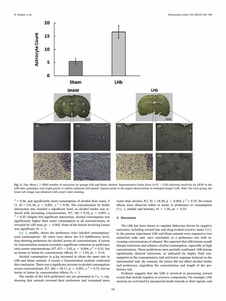
One brain was lost during the slicing procedure. In 5 animals thehistological material was inadequate to compute astrocyte counting. In5 brains the portion of the brain where LHb was located was not se-lected for immunolabelling. The remaining 17 brains (11 LHb, 6 Sham)were included in the histological analysis. Each animal contributed tothe final computation with 1–6 slices corresponding to the A/P −3.30coordinate. When the astrocyte count was assessed in more than oneslice, a mean was calculated for the animal. Data were ranked andoverlapped LHb (5) and Sham (2) brains (in terms of number of as-trocytes) were eliminated. Fig. 2, top, shows the result of the remaininganimals. Rats with LHb lesions had a significantly higher astrocytecount than rats with Sham lesions, F(1, 8)= 12.585, p < 0.009,η2=0.61. Selected slices treated with GFAP immunolabelling andcresyl violet are shown in Fig. 2, bottom. As expected (e.g. [24]), qui-nolinic acid infusions produced astrogliosis.
3.2. Extinction tasks
There was no evidence that the LHb lesion affected appetitive ac-quisition. The overall mean (± SEM) fluid intake for sessions 1–10 inthe consummatory task was 8.2 ml (± 0.8) for LHb rats and 8.4ml(± 0.7) for Sham rats (F < 1). In the instrumental task, one Shamanimal had long latencies during the initial sessions, but its latencieswere similar to other animals in later acquisition sessions. The mean(± SEM) latency was 2.6 s (± 0.2) for LHb animals and 6.44 s(± 2.27) for Sham animals. Still, the difference was not significant, F(1,8)= 3.03, p > 0.11.
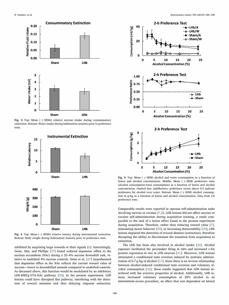
Fig. 3, top, shows relative sucrose consumption registered duringthe extinction phase of the consummatory task. LHb animals showedhigher levels of fluid intake than Sham animals. A one-way analysisrevealed a significant difference, F(1, 8)= 6.09, p < 0.04, η 2=0.43.This difference was not due to differences in drinking behavior. Fig. 3,bottom, shows water intake during the four habituation sessions of thepreference test (F < 1).
Fig. 4, top, shows relative latency data registered during the ex-tinction phase of the instrumental task. LHb animals exhibited sig-nificantly lower response latencies than Sham animals, F(1, 8)= 5.47,p < 0.05, η 2=0.41. Fig. 4, bottom, shows that these differences werenot attributable to body weight, which was similar in both groups(F < 1).
3.3. Preference test
Fig. 5, top, presents alcohol and water consumption for both groups.Across all concentrations and in both groups, animals preferred alcoholto water, although the preference diminished as alcohol concentrationincreased. A Lesion (LHb, Sham) by Bottle (Alcohol, Water) by Con-centration (2–24%) analysis, with repeated measures for the last twofactors, yielded the following results. There was a significant reductionin consumption across concentrations, F(7, 56)= 10.24, p < 0.001, η
Fig. 1. A timeline of the procedures implemented in this experiment.
R. Donaire, et al. Neuroscience Letters 703 (2019) 184–190
186
2=0.56, and significantly more consumption of alcohol than water, F(1, 8)= 131.36, p < 0.001, η 2=0.94. The concentration by bottleinteraction also reached a significant level, as alcohol intake was re-duced with increasing concentrations, F(7, 56)= 9.16, p < 0.001, η2=0.53. Despite this significant interaction, alcohol consumption wassignificantly higher than water consumption at all concentrations, asrevealed by LSD tests, ps < 0.002. None of the factors involving Lesionwas significant, Fs < 1.
Fig. 5, middle, shows the preference ratio (alcohol consumption/total consumption). All ratios were above the 0.5 indifference level,thus showing preference for alcohol across all concentrations. A Lesionby Concentration analysis revealed a significant reduction in preferenceratio across concentrations, F(7, 67)= 3.62, p < 0.004, η 2=0.31, butno lesion or lesion by concentration effects, Fs < 1.54, ps > 0.16.
Alcohol consumption in g/kg increased at about the same rate inLHb and Sham animals. A Lesion x Concentration analysis confirmedthis conclusion. There was a significant increase in alcohol consumptionacross concentrations, F(7, 56)= 20.11, p < 0.001, η 2=0.72, but nolesion or lesion by concentration effects, Fs < 1.
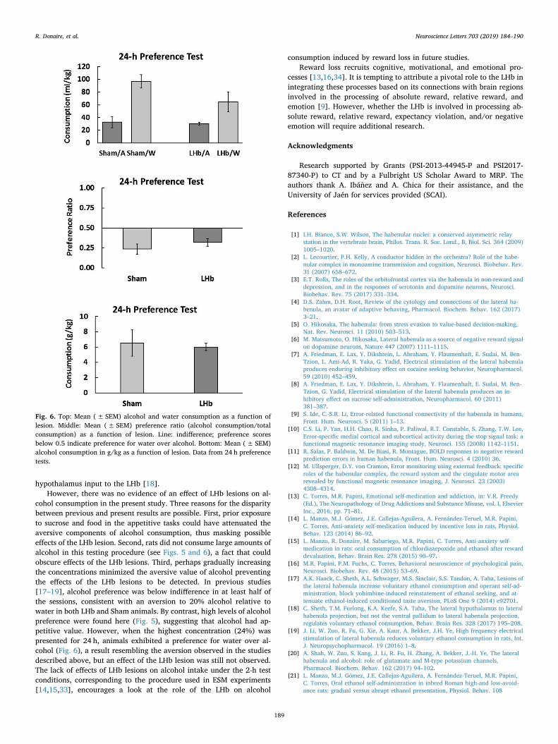
The results of the 24-h preference test are presented in Fig. 6, top,showing that animals reversed their preference and consumed more
water than alcohol, F(1, 8)= 18.30, p < 0.004, η 2=0.70. No Lesioneffects were observed either in terms of preference or consumption(Fig. 6, middle and bottom), Fs < 1.05, ps > 0.33.
4. Discussion
The LHb has been shown to regulate behaviors driven by negativeoutcomes, including reward loss and drug-evoked aversive states [26].In the present experiment LHb and Sham animals were exposed to twoextinction tasks and, once concluded, to a preference test with in-creasing concentrations of ethanol. We expected that LHb lesions woulddisrupt extinction and enhance alcohol consumption, especially at highconcentrations. These predictions were partially confirmed: LHb lesionssignificantly delayed extinction, as indicated by higher fluid con-sumption in the consummatory task and lower response latencies in theinstrumental task. By contrast, the lesion did not affect alcohol intakeand preference, regardless the concentration and length of the pre-ference test.
Evidence suggests that the LHb is involved in processing rewardevents that include negative or aversive components. For example, LHbneurons are activated by unexpected small rewards or their signals, and
Fig. 2. Top: Mean (± SEM) number of astrocytes for groups LHb and Sham. Bottom: Representative brain slices (A/P: −3.30) showing reactivity for GFAP in theLHb after quinolinic acid (right panel) or vehicle infusions (left panel). Squares point to the region shown below in enlarged images (10X, 40X). For each group, thelower left image was obtained with cresyl violet staining.
R. Donaire, et al. Neuroscience Letters 703 (2019) 184–190
187
inhibited by surprising large rewards or their signals [6]. Interestingly,Genn, Ahn, and Phillips [27] found reduced dopamine efflux in thenucleus accumbens (NAc) during a 32–4% sucrose downshift task, re-lative to unshifted 4% sucrose controls. Genn et al. [27] hypothesizedthat dopamine efflux in the NAc reflects the current reward value ofsucrose—lower in downshifted animals compared to unshifted controls.As discussed above, this function would be modulated by an inhibitoryLHb-RMTg-VTA-NAc pathway [28]. In the present experiment, LHblesions could have disrupted this pathway, interfering with the detec-tion of reward omission and thus delaying response extinction.
Comparable results were reported in operant self-administration tasksinvolving sucrose or cocaine [7,8]. LHb lesions did not affect sucrose orcocaine self-administration during acquisition training, a result com-parable to the lack of a lesion effect found in the present experimentduring acquisition. Therefore, rather than reducing reward value [7],stimulating motor behavior [29], or increasing distractability [30], LHblesions impaired the detection of reward absence (extinction), thereforedisrupting the ability to discriminate the transition from acquisition toextinction.
The LHb has been also involved in alcohol intake [20]. Alcoholexposure facilitated the pacemaker firing in vitro and increased c-fosprotein expression in vivo in LHb neurons [31]. Moreover, LHb lesionsattenuated a conditioned taste aversion induced by systemic adminis-tration of 0.7 g/kg of alcohol [17]. Since there is an inverse relationshipbetween alcohol-induced conditioned taste aversion and voluntary al-cohol consumption [32], these results suggested that LHb lesions in-terfered with the aversive properties of alcohol. Additionally, LHb le-sions increased voluntary consumption of 20% alcohol in anintermittent-access procedure, an effect that was dependent on lateral
Fig. 3. Top: Mean (± SEMs) relative sucrose intake during consummatoryextinction. Bottom: Water intake during habituation sessions prior to preferencetests.
Fig. 4. Top: Mean (± SEMs) relative latency during instrumental extinction.Bottom: Body weight during habituation sessions prior to preference tests.
Fig. 5. Top: Mean (± SEM) alcohol and water consumption as a function oflesion and alcohol concentration. Middle: Mean (± SEM) preference ratio(alcohol consumption/total consumption) as a function of lesion and alcoholconcentration. Dashed line: indifference; preference scores above 0.5 indicatepreference for alcohol over water. Bottom: Mean (± SEM) alcohol consump-tion in g/kg as a function of lesion and alcohol concentration. Data from 2 hpreference tests.
R. Donaire, et al. Neuroscience Letters 703 (2019) 184–190
188
hypothalamus input to the LHb [18].However, there was no evidence of an effect of LHb lesions on al-
cohol consumption in the present study. Three reasons for the disparitybetween previous and present results are possible. First, prior exposureto sucrose and food in the appetitive tasks could have attenuated theaversive components of alcohol consumption, thus masking possibleeffects of the LHb lesion. Second, rats did not consume large amounts ofalcohol in this testing procedure (see Figs. 5 and 6), a fact that couldobscure effects of the LHb lesions. Third, perhaps gradually increasingthe concentrations minimized the aversive value of alcohol preventingthe effects of the LHb lesions to be detected. In previous studies[17–19], alcohol preference was below indifference in at least half ofthe sessions, consistent with an aversion to 20% alcohol relative towater in both LHb and Sham animals. By contrast, high levels of alcoholpreference were found here (Fig. 5), suggesting that alcohol had ap-petitive value. However, when the highest concentration (24%) waspresented for 24 h, animals exhibited a preference for water over al-cohol (Fig. 6), a result resembling the aversion observed in the studiesdescribed above, but an effect of the LHb lesion was still not observed.The lack of effects of LHb lesions on alcohol intake under the 2-h testconditions, corresponding to the procedure used in ESM experiments[14,15,33], encourages a look at the role of the LHb on alcohol
consumption induced by reward loss in future studies.Reward loss recruits cognitive, motivational, and emotional pro-
cesses [13,16,34]. It is tempting to attribute a pivotal role to the LHb inintegrating these processes based on its connections with brain regionsinvolved in the processing of absolute reward, relative reward, andemotion [9]. However, whether the LHb is involved in processing ab-solute reward, relative reward, expectancy violation, and/or negativeemotion will require additional research.
Acknowledgments
Research supported by Grants (PSI-2013-44945-P and PSI2017-87340-P) to CT and by a Fulbright US Scholar Award to MRP. Theauthors thank A. Ibáñez and A. Chica for their assistance, and theUniversity of Jaén for services provided (SCAI).
References
[1] I.H. Bianco, S.W. Wilson, The habenular nuclei: a conserved asymmetric relaystation in the vertebrate brain, Philos. Trans. R. Soc. Lond., B, Biol. Sci. 364 (2009)1005–1020.
[2] L. Lecourtier, P.H. Kelly, A conductor hidden in the orchestra? Role of the habe-nular complex in monoamine transmission and cognition, Neurosci. Biobehav. Rev.31 (2007) 658–672.
[3] E.T. Rolls, The roles of the orbitofrontal cortex via the habenula in non-reward anddepression, and in the responses of serotonin and dopamine neurons, Neurosci.Biobehav. Rev. 75 (2017) 331–334.
[4] D.S. Zahm, D.H. Root, Review of the cytology and connections of the lateral ha-benula, an avatar of adaptive behaving, Pharmacol. Biochem. Behav. 162 (2017)3–21.
[5] O. Hikosaka, The habenula: from stress evasion to value-based decision-making,Nat. Rev. Neurosci. 11 (2010) 503–513.
[6] M. Matsumoto, O. Hikosaka, Lateral habenula as a source of negative reward signalon dopamine neurons, Nature 447 (2007) 1111–1115.
[7] A. Friedman, E. Lax, Y. Dikshtein, L. Abraham, Y. Flaumenhaft, E. Sudai, M. Ben-Tzion, L. Ami-Ad, R. Yaka, G. Yadid, Electrical stimulation of the lateral habenulaproduces enduring inhibitory effect on cocaine seeking behavior, Neuropharmacol.59 (2010) 452–459.
[8] A. Friedman, E. Lax, Y. Dikshtein, L. Abraham, Y. Flaumenhaft, E. Sudai, M. Ben-Tzion, G. Yadid, Electrical stimulation of the lateral habenula produces an in-hibitory effect on sucrose self-administration, Neuropharmacol. 60 (2011)381–387.
[9] S. Ide, C.-S.R. Li, Error-related functional connectivity of the habenula in humans,Front. Hum. Neurosci. 5 (2011) 1–13.
[10] C.S. Li, P. Yan, H.H. Chao, R. Sinha, P. Paliwal, R.T. Constable, S. Zhang, T.W. Lee,Error-specific medial cortical and subcortical activity during the stop signal task: afunctional magnetic resonance imaging study, Neurosci. 155 (2008) 1142–1151.
[11] R. Salas, P. Baldwin, M. De Biasi, R. Montague, BOLD responses to negative rewardprediction errors in human habenula, Front. Hum. Neurosci. 4 (2010) 36.
[12] M. Ullsperger, D.Y. von Cramon, Error monitoring using external feedback: specificroles of the habenular complex, the reward system and the cingulate motor arearevealed by functional magnetic resonance imaging, J. Neurosci. 23 (2003)4308–4314.
[13] C. Torres, M.R. Papini, Emotional self-medication and addiction, in: V.R. Preedy(Ed.), The Neuropathology of Drug Addictions and Substance Misuse, vol. I, ElsevierInc., 2016, pp. 71–81.
[14] L. Manzo, M.J. Gómez, J.E. Callejas-Aguilera, A. Fernández-Teruel, M.R. Papini,C. Torres, Anti-anxiety self-medication induced by incentive loss in rats, Physiol.Behav. 123 (2014) 86–92.
[15] L. Manzo, R. Donaire, M. Sabariego, M.R. Papini, C. Torres, Anti-anxiety self-medication in rats: oral consumption of chlordiazepoxide and ethanol after rewarddevaluation, Behav. Brain Res. 278 (2015) 90–97.
[16] M.R. Papini, P.M. Fuchs, C. Torres, Behavioral neuroscience of psychological pain,Neurosci. Biobehav. Rev. 48 (2015) 53–69.
[17] A.K. Haack, C. Sheth, A.L. Schwager, M.S. Sinclair, S.S. Tandon, A. Taha, Lesions ofthe lateral habenula increase voluntary ethanol consumption and operant self-ad-ministration, block yohimbine-induced reinstatement of ethanol seeking, and at-tenuate ethanol-induced conditioned taste aversion, PLoS One 9 (2014) e92701.
[18] C. Sheth, T.M. Furlong, K.A. Keefe, S.A. Taha, The lateral hypothalamus to lateralhabenula projection, but not the ventral pallidum to lateral habenula projection,regulates voluntary ethanol consumption, Behav. Brain Res. 328 (2017) 195–208.
[19] J. Li, W. Zuo, R. Fu, G. Xie, A. Kaur, A. Bekker, J.H. Ye, High frequency electricalstimulation of lateral habenula reduces voluntary ethanol consumption in rats, Int.J. Neuropsychopharmacol. 19 (2016) 1–8.
[20] A. Shah, W. Zuo, S. Kang, J. Li, R. Fu, H. Zhang, A. Bekker, J.-H. Ye, The lateralhabenula and alcohol: role of glutamate and M-type potassium channels,Pharmacol. Biochem. Behav. 162 (2017) 94–102.
[21] L. Manzo, M.J. Gómez, J.E. Callejas-Aguilera, A. Fernández-Teruel, M.R. Papini,C. Torres, Oral ethanol self-administration in inbred Roman high-and low-avoid-ance rats: gradual versus abrupt ethanol presentation, Physiol. Behav. 108
Fig. 6. Top: Mean (± SEM) alcohol and water consumption as a function oflesion. Middle: Mean (± SEM) preference ratio (alcohol consumption/totalconsumption) as a function of lesion. Line: indifference; preference scoresbelow 0.5 indicate preference for water over alcohol. Bottom: Mean (± SEM)alcohol consumption in g/kg as a function of lesion. Data from 24 h preferencetests.
R. Donaire, et al. Neuroscience Letters 703 (2019) 184–190
189
(2012) 1–5.[22] A.E. Mustaca, M. Bentosela, E. Ruetti, G. Kamenetzky, L. Cuenya, N. Justel, F. Lopez
Seal, S. Fosacheca, M.R. Papini, Similitudes y discrepancias en dos modelos ani-males de frustración, in: M.C. Richaud, E. Moreno (Eds.), Recientes Avances EnInvestigación En Ciencias Del Comportamiento, vol. 2, CIIPME-CONICET, BuenosAires, Argentina, 2009, pp. 921–940.
[23] G. Paxinos, C. Watson, The Rat Brain in Stereotaxic Coordinates, Academic press,New York, 2013.
[24] M. Dihné, F. Block, H. Korr, R. Töpper, Time course of glial proliferation and glialapoptosis following excitotoxic CNS injury, Brain Res. 902 (2001) 178–189.
[25] S.Y. Shu, G. Ju, L.Z. Fan, The glucose oxidase-DAB-nickel method in peroxidasehistochemistry of the nervous system, Neurosci. Lett. 85 (1988) 169–171.
[26] S. Lecca, F.J. Meye, M. Mameli, The lateral habenula in addiction and depression:an anatomical, synaptic and behavioral overview, Eur. J. Neurosci. 39 (2014)1170–1178.
[27] R.F. Genn, S. Ahn, A.G. Phillips, Attenuated dopamine efflux in the rat nucleusaccumbens during successive negative contrast, Behav. Neurosci. 118 (2004) 869.
[28] S.G. Nair, N.S. Strand, J.F. Neumairer, DREADDing the lateral habenula: a review of
methodological approaches for studying lateral habenula function, Brain Res. 1511(2013) 93–101.
[29] E.H. Lee, S.L. Huang, Role of lateral habenula in the regulation of exploratory be-havior and its relationship to stress in rats, Behav. Brain Res. 30 (1988) 265–271.
[30] L. Lecourtier, P.H. Kelly, Bilateral lesions of the habenula induce attentional dis-turbances in rats, Neuropsychopharmacol. 30 (2005) 484–496.
[31] W. Zuo, R. Fu, F.W. Hopf, G. Xie, K. Krnjević, J. Li, J.-H. Ye, Ethanol drives aversiveconditioning through dopamine I receptor and glutamate receptor-mediated acti-vation of lateral habenula neurons, Addict. Biol. 22 (2015) 103–116.
[32] A.S. Green, N.J. Grahame, Ethanol drinking in rodents: is free-choice drinking re-lated to the reinforcing effects of ethanol? Alcohol 42 (2008) 1–11.
[33] R. Donaire, S.E. Conrad, J.B. Thompson, M.R. Papini, C. Torres, Augmented vo-luntary consumption of ethanol induced by reward downshift increases locomotoractivity of male Wistar rats in the elevated plus maze, Behav. Proc. 150 (2018)59–65.
[34] L.A. Ortega, J.L. Solano, C. Torres, M.R. Papini, Reward loss and addiction: op-portunities for cross-pollination, Pharmacol. Biochem. Behav. 154 (2017) 39–52.
R. Donaire, et al. Neuroscience Letters 703 (2019) 184–190
190