Embed Size (px)
Citation preview

LASIODIPLODIA SPECIES ASSOCIATED WITH DYING EUPHORBIA INGENS IN
SOUTH AFRICA.
J.A. van der Linde1, D.L. Six2, M.J. Wingfield1, and J. Roux1
1Department of Microbiology and Plant Pathology, DST/NRF Centre of Excellence in Tree
Health Biotechnology (CTHB), Forestry and Agricultural Biotechnology Institute (FABI),
University of Pretoria, PBag X20, Hatfield, Pretoria, 0083, South Africa.
2College of Forestry and Conservation, Department of Ecosystem and Conservation Sciences,
The University of Montana, Missoula, MT 59812, United States of America.
Corresponding author, e-mail: [email protected]
Various species of Euphorbia occur in South Africa, including herbaceous, succulent and
woody types. The largest of the succulent Euphorbia spp. in South Africa is Euphorbia
ingens. These trees have been dying at an alarming rate in the Limpopo Province during the
course of the last 15 years. Investigations into the possible causes of the death have included
the possible role of fungal pathogens. Amongst the most common fungi isolated from
diseased trees were species in the Botryosphaeriaceae. The aim of this study was to identify
these fungi using morphology and DNA sequence data of two gene regions (TEF-1α & ITS).
Results showed that Lasiodiplodia theobromae and Lasiodiplodia mahajangana were present.
Pathogenicity studies showed that these Lasiodiplodia species can cause infections on healthy
E. ingens trees, implicating them as contributors to the decline of E. ingens.
KEYWORDS
Botryosphaeriaceae, candelabra trees, tree diseases, insect infestations, climate change

INTRODUCTION
The genus Euphorbia includes more than 2100 species worldwide. Euphorbia species
are known to vary dramatically in morphology and range from large woody trees to shrub like
herbaceous plants and succulent cactus like plants (Palgrave 2002, PBI Euphorbia project,
www.euphorbiaceae.org). There is a high diversity of woody to succulent euphorbias in
Southern Africa, with the largest of these species being Euphorbia ingens E. Meyer: Boissier
(Palgrave 2002, Gildenhuys 2006). E. ingens and similar species are characterised by woody
main stems and fleshy succulent branches, giving the trees a candelabrum shape (Van Wyk
and Van Wyk 1997, Palgrave 2002, Gildenhuys 2006). E. ingens is known only to occur in
Africa with high densities in Southern Africa (Palgrave 2002, Gildenhuys 2006).
In the last 15 years, there have been alarming reports of large-scale decline and death of
E. ingens trees in South Africa. Mortality of these trees has been particularly severe in the
Limpopo Province. Symptoms associated with the death of trees include graying and spots on
the succulent branches, infestation by branch and stem boring insects and brown to blue
discolouration of the internal tissues of the branches and woody main stems (Roux et al.
2008, 2009). Preliminary investigations into the cause of this disease have yielded various
fungi including species of Botryosphaeriaceae (Roux et al. 2008, 2009).
The Botryosphaeriaceae are known as opportunistic pathogens that cause cankers and
death of numerous tree species, especially after periods of drought, frost, hail damage and
other environmental conditions leading to stress (Punithalingam 1980, Slippers and Wingfield
2007). They are also known to be endophytes, infecting healthy trees and only causing disease
after the onset of stress (Smith et al. 1996). In South Africa, fungi in the Botryosphaeriaceae
are common, and known to be pathogens, especially on commercially grown plantation trees
(Laughton 1937, Swart et al. 1985, Smith et al. 1996, Roux and Wingfield 1997), native tree

species such as Pterocarpus angolensis DC. (Mehl et al. 2011), Syzygium spp. (Pavlic et al.
2007) and Acacia spp. (Slippers et al. 2011). Various species are also known to occur on, and
cause disease of fruit trees in the genera Malus, Pyrus, Prunus, Populus, Syzygium and Vitis
(van Niekerk et al. 2004, Damm et al. 2007, Pavlic et al. 2007, Slippers et al. 2007). There
are, however, no reports of Botryosphaeriaceae from succulent Euphorbia spp.
The aim of this study was to identify species of Botryosphaeriaceae collected during
studies of dying E. ingens trees in the Limpopo Province of South Africa. We also tested the
pathogenicity of the isolates on healthy E. ingens trees to consider their possible involvement
in tree death.
MATERIALS AND METHODS
Collection of samples and isolations
Isolates were collected from diseased E. ingens (Figure 1a) at four sites in the Limpopo
Province during 2009. Isolations were made from blue-black discoloured wood (Figure 1b)
in the main woody stems of the trees, as well as from necrotic tissue and insect tunnels in the
succulent branches. Isolations were also made from insects collected from rotting succulent
branches and the woody main stems. Direct isolations were made from the plant material
taken from the leading edges of lesions using a sterile scalpel. These tissue samples were
plated on 2% Malt Extract Agar (MEA) (15g agar and 20g malt extract per 1000ml distilled
water; Biolab, Merck, Midrand, South Africa) with streptomycin (0.4g/L; Sigma-Aldrich, St.
Louis, USA). Isolations were made from insects by crushing them onto water agar (15g per
1000ml distilled water; Biolab) and incubating the plates for six weeks at 20˚C. Cultures from
insects were purified by transferring mycelium to fresh 2% MEA. A second set of isolates
was obtained from the culture collection (CMW) of the Forestry and Agricultural

Biotechnology Institute (FABI), University of Pretoria which were collected previously
(Roux et al. 2009, 2010) from diseased E. ingens trees in the Limpopo Province. Purified
isolates from plant tissue and insects were deposited in the Culture Collection (CMW) of the
Forestry and Agricultural Biotechnology Institute (FABI), University of Pretoria, South
Africa.
Culture and Morphological Characteristics
Isolates were plated onto 1.5% Water Agar (WA; 15g per 1000ml distilled water;
Biolab) containing sterilized pine needles to induce the formation of fruiting bodies. Cultures
were incubated at 20°C under near-ultra violet (UV) light. Characteristic fungal structures
(conidia, conidiogenous cells, paraphyses, conidiomata) were viewed using a Zeiss light
microscope fitted with an Axiocam digital camera with Axiovision 3.1 software (Carl Zeiss
Ltd., Germany). The fungal structures were placed on glass microscope slides and mounted in
75% lactic acid. Colors of resultant cultures were determined using the color notations of
Rayner (1970).
DNA extraction and Polymerase Chain Reaction (PCR) amplification
Isolates, representing different collection sites and culture morphology, were grown for
six days on 2% MEA, prior to DNA extraction. DNA extraction followed the protocol of
Möller et al. (1992), after mycelium was scraped from the surfaces of the cultures and freeze
dried for 24 hours in 2ml Eppendorf tubes. DNA concentration was determined using a
Nanodrop ND-1000 Spectrophotometer (Thermo Fisher Scientific, Wilmington, USA).
The Polymerase Chain Reaction (PCR) was used to amplify the internal transcribed
spacer regions and the 5.8S gene using the primers ITS 1 and ITS 4 (White et al. 1990) and
the translation elongation Factor 1-α (TEF 1-α) gene region using the primers EF1-F and EF1-
R (Jacobs et al. 2004). PCRs were done using an Applied Biosystems Veriti thermocycler

(Applied Biosystems, Foster City, USA) following the protocols described by Mohali et al.
(2007).
PCR products were viewed using an agarose gel (2%; Whitehead Scientific, Cape
Town, South Africa), loaded with GelRed (Anatech, USA), and visualised under UV
illumination. The size of the PCR products was estimated using a 100bp DNA molecular
marker (O’RangeRulerä 100bp DNA ladder, Fermentas Life Sciences). Sephadex G-50
columns (1g in 15ml distilled water Water; SIGMA, Steinheim, Germany) were used to
purify the amplified products in preparation for sequencing.
DNA sequencing
Confirmed PCR products were sequenced with an ABI3700 DNA analyzer (Applied
Biosystems, Foster City, USA) using a Big Dye Cycle Sequencing kit Version 1.1 (Applied
Biosystems). Sequences were edited based on forward and reverse sequences using Mega
Version 4.0 (Tamura et al. 2007). To confirm gene identity and obtain related sequences, the
correctly edited sequences were placed in the nucleotide database blastx (National Centre for
Biotechnology Information, www.ncbi.nih.nlm.gov). Mafft version 5.851 (Katoh et al. 2002)
was used to align the sequences from this study and those of closely related species obtained
from the blast results. Phylogenetic analysis of each data set was done using PAUP
(Phylogenetic Analysis Using Parsimony) version 4.0b10 (Swofford 2002) and phylogenetic
trees were constructed using random stepwise addition and tree bisection and reconstruction
as branch swapping algorithms, based on heuristic searches. Bootstrap and maximum
parsimony analyses were run using a 1000 replicates (Felsenstein 1985). A partition
homogeneity test was used to determine whether the ITS and TEF1-α sequence data sets
could be combined (Farris et al. 1995, Huelsenbeck et al. 1996). Two separate phylogenetic
analyses, one including all the recently described species of Lasiodiplodia and another

considering only the clades including the isolates from this study, were conducted. Prior to the
partition homogeneity test, data sets of individual gene regions were analysed separately. The
data sets were rooted with the GenBank sequences of Botryosphaeria sarmentorum A.J.L.
Phillips, Alves & Luque (CBS 12041) and Lasiodiplodia crassispora Burgess & Barber
(UCD27Co) (Table 1).
Bayesian analysis was used to determine the posterior probabilities of each dataset (ITS,
TEF1-α) based on the Monte Carlo Markov Chain (MCMC) method. A jModelTest 0.1.1
(Posada 2008) was used to determine the most appropriate nucleotide substitution model. The
best-fitting models for the ITS and TEF1-α datasets, based on Akaike Information Criterion
(AICc), were determined for the complete analysis (TPM1: ITS, K80+G: TEF1-α) and the
specific analysis (TIM2+G: ITS, K80+G: TEF1-α). The Bayesian analysis was run on
MrBayes 3.1.2 (Huelsenbeck and Ronquist 2001) and trees were recorded every 100
generations based on four chains producing 5 000 000 generations. The likelihood data were
used in graphical analysis to estimate the burn-in values for each dataset. Mega Version 4.0
(Tamura et al. 2007) was used to produce consensus trees from the two analysed datasets
from which the posterior probabilities were determined.
Pathogenicity trials
Two isolates (CMW36766, CMW36765) obtained in this study were used to inoculate
healthy E. ingens trees in the North West Province. Cultures were first grown on 2% MEA for
five days and then used to inoculate wooden toothpicks first soaked in Malt Extract (5g malt
extract per 250ml distilled water; Biolab) and then placed on the surface of MEA in Petri
dishes. Mycelium-colonised toothpicks, and sterile toothpicks for the controls, were inserted
into the succulent branches (five branches for each isolate) to a depth of 3mm. After six
weeks the results were determined by measuring the surface lesions, cambium lesions and

internal lesions after cutting branches in half at the point of inoculation. Isolations were made
on MEA from inoculated tissue to comply with Koch’s postulates. To determine significance
between means, a student’s t test was done with P < 0.05 as being significant. Since there was
no variance in the controls, the data for each isolate were Bonferroni – corrected for multiple
comparisons (α = 0.05). All tests were conducted using JMP version 9.0.2 (SAS institute
2011).
RESULTS
Collection of samples and isolations
Isolates from this study were obtained from lesions on a total of 23 trees. Isolates
resembling the Botryosphaeriaceae were obtained from only six trees. A total number of six
isolates were obtained from Mokopane, one from Capricorn and one from the Louis Trichardt
area. Of the eight isolates collected, six were from diseased plant material and the remaining
two were isolated from insects infesting diseased tissue.
Culture and morphological Characteristics
Fungal structures showed typical features of Lasiodiplodia spp., with aseptate, hyaline
conidia becoming dark brown and septate with striations as they matured. Cultures were white
with abundant, fluffy aerial mycelium which became an olivaceous grey (23’’’’’b) with time
(after 10 days). Pycnidia were produced after five days on the WA with sterilized pine needles
and were black in colour, unilocular, solitary, immersed in the media and were formed on the
top surfaces of the pine needles (Figure 2).
DNA sequence analyses
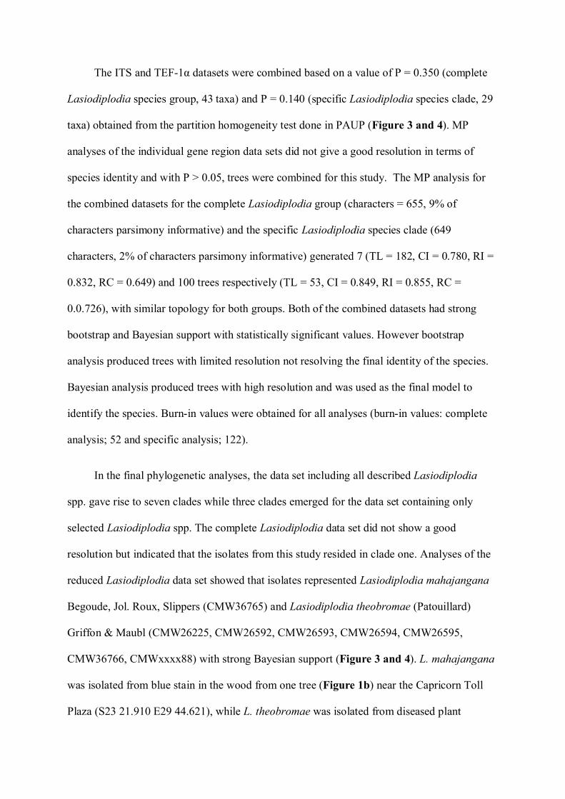
The ITS and TEF-1α datasets were combined based on a value of P = 0.350 (complete
Lasiodiplodia species group, 43 taxa) and P = 0.140 (specific Lasiodiplodia species clade, 29
taxa) obtained from the partition homogeneity test done in PAUP (Figure 3 and 4). MP
analyses of the individual gene region data sets did not give a good resolution in terms of
species identity and with P > 0.05, trees were combined for this study. The MP analysis for
the combined datasets for the complete Lasiodiplodia group (characters = 655, 9% of
characters parsimony informative) and the specific Lasiodiplodia species clade (649
characters, 2% of characters parsimony informative) generated 7 (TL = 182, CI = 0.780, RI =
0.832, RC = 0.649) and 100 trees respectively (TL = 53, CI = 0.849, RI = 0.855, RC =
0.0.726), with similar topology for both groups. Both of the combined datasets had strong
bootstrap and Bayesian support with statistically significant values. However bootstrap
analysis produced trees with limited resolution not resolving the final identity of the species.
Bayesian analysis produced trees with high resolution and was used as the final model to
identify the species. Burn-in values were obtained for all analyses (burn-in values: complete
analysis; 52 and specific analysis; 122).
In the final phylogenetic analyses, the data set including all described Lasiodiplodia
spp. gave rise to seven clades while three clades emerged for the data set containing only
selected Lasiodiplodia spp. The complete Lasiodiplodia data set did not show a good
resolution but indicated that the isolates from this study resided in clade one. Analyses of the
reduced Lasiodiplodia data set showed that isolates represented Lasiodiplodia mahajangana
Begoude, Jol. Roux, Slippers (CMW36765) and Lasiodiplodia theobromae (Patouillard)
Griffon & Maubl (CMW26225, CMW26592, CMW26593, CMW26594, CMW26595,
CMW36766, CMWxxxx88) with strong Bayesian support (Figure 3 and 4). L. mahajangana
was isolated from blue stain in the wood from one tree (Figure 1b) near the Capricorn Toll
Plaza (S23 21.910 E29 44.621), while L. theobromae was isolated from diseased plant

material (CMW26225, CMW26592, CMW26593, CMW26594, CMW26595) form
Mokopane (S24 10.291 E29 01.131) as well as the insects Cyrtogenius africus Wood
(CMW36766) and Cossonus Claireville (CMWxxxx88) from Mokopane and Last Post Private
Nature Reserve (S23 17.738 E29 55.467) (Louis Trichardt site), respectively.
Pathogenicity trials
L. mahajangana (CMW36765) and L. theobromae (CMW36766) produced lesions on
the exterior, cambium and internal core of healthy E. ingens branches (Figure 1c). The most
severe damage caused by the fungi was in the internal core of the succulent branches which
had rotten. Lesions on the exterior were conspicuous at the point of inoculation with necrotic
tissue and a black discharge. L. mahajangana and L. theobromae lesions were brown and
circular at the points of inoculation with necrotic tissue in the internal core. The control also
had small brown circular lesions at the point of inoculation but had no signs of discoloration
in the cambium or internal core (Figure 1d) of the succulent branches. Statistical analysis did
not show significant differences in pathogenicity between species except for the cambium
lesion length with P values of 0.025 (DF = 12.51), 0.1303 (DF = 18.00) and 0.4261 (DF =
14.06) for the cambium lesion, internal lesion depth and internal lesion width data,
respectively (Figure 5). Isolations from the sites of inoculation yielded L. theobromae and L.
mahajangana identified based on characteristic morphological features.
DISCUSSION
Results of this study showed that two species of Lasiodiplodia, L. theobromae and L.
mahajangana are associated with die-back symptoms on E. ingens. These fungi were
identified based on morphological characteristics and DNA sequence comparisons. They are

both well-known from trees in Southern Africa (Crous et al. 2000, Burgess et al. 2003, Pavlic
et al. 2007, Begoude et al. 2010), but have not previously been found on E. ingens.
L. mahajangana is a recently described species from healthy branches of T. catappa in
Madagascar (Begoude et al. 2010). The current study represents only the second report of this
fungus and very limited information is, therefore, available regarding its possible origin or
importance. In this study L. mahajangana was isolated from blue stain in the wood of the
main stem of an E. ingens tree. Blue stain is a common symptom of wood infected by species
in the Botryosphaeriaceae, resulting from the dark colour of the mycelium of these fungi
(Slippers and Wingfield 2007). Blue stain is, however, a secondary symptom on dead or
dying wood, suggesting that L. mahajangana obtained from E. ingens in this study, is not the
primary cause of tree decline but rather causes secondary infections due to the trees being
stressed.
L. theobromae was obtained from diseased plant material and insects collected from the
internal parts of dying E. ingens trees. These insects, Cyrtogenius africus (Curculionidae:
Scolytinae), and a Cossonus sp. (Curculionidae: Cossoninae) were not surface disinfected and
inoculum of L. theobromae could have been on their surfaces or related to tissue that they had
consumed and so occurring in their guts. Based on morphology, variation was found between
previously identified L. theobromae and isolates from this study. Isolates from E. ingens have
smaller conidia than previously described L. theobromae isolates (Table 2).
The Botryosphaeriaceae typically disperse via rain splash and are not adapted to insect
dispersal. But species such as L. theobromae have previously been isolated from insects such
as Hypocryphalus mangiferae Stebbing (Scolytinae) after surface sterilization, implying that
it might be carried in the gut or mycangia (Masood et al. 2010). Both insect families have
previously been associated with succulent Euphorbia (Wollaston) (Jordal 2006, 2009).
Recently, previously described L. theobromae species were re-analysed to determine their

genetic variability and cryptic speciation based on morphology and the combined sequence
analyses of ITS and TEF-1α (Alves et al. 2008). Three distinct morphological groups were
discovered based on conidial size and phylogenetic analysis clearly showing Lasiodiplodia
parva AJL Phillips, A Alves & Crous (small conidia) and Lasiodiplodia pseudotheobromae
AJL Phillips, A Alves & Crous (larger conidia) being separate from L. theobromae as two
new species (Alves et al. 2008). Phylogenetic analyses based on ITS and TEF-1α gene
sequences, in this study, showed poor resolution in the L. theobromae clade (Figure 3 and
4) with limited differences between isolates of L. theobromae and Lasiodiplodia
hormozganensis Abdollahzadeh, Zare & A.J.L. Phillips. Additional analyses will be needed to
resolve this group more clearly, either by including other gene regions or using specific
microsatellite markers to consider the problem at a population level.
Inoculation studies, using L. theobromae and L. mahajangana, showed that both these
fungi have the potential to cause disease on E. ingens. Both produced extensive internal rot of
the succulent branches of these trees, within six weeks. It was not surprising to find that L.
theobromae and L. mahajangana were able to cause disease symptoms on E. ingens.
Previously, L. theobromae was shown to be pathogenic to Eucalyptus clones (GC540) (Pavlic
et al. 2007), grapevines (Úrbez-Torres et al. 2008, Úrbez-Torres and Gubler 2009) and T.
catappa (Begoude et al. 2010). Similar to results in the study by Begoude et al. (2010), in our
study, L. mahajangana produced smaller lesions in artificial inoculation studies than L.
theobromae.
CONCLUSIONS
Since environmental and other stress factors play an important role in the epidemiology
of diseases caused by fungi in the Botryosphaeriaceae, the symptoms observed on dying E.
ingens trees in South Africa could, at least in part, be attributed to L. theobromae and L.

mahajangana. Reports by Van der Linde et al. (2011) of increased temperature and decreased
rain in the study areas in the Limpopo Province over the last 40 years may be a possible stress
factor for E. ingens. It does appear that a link to an environmental and/or an anthropogenic
trigger initiated the the sudden and severe decline of these trees, in combination with various
pathogens and insects. Further and more extensive surveys will be required to fully
understand the diversity and distribution of Botryosphaeriaceae on native Euphorbia trees and
to establish the possible triggers enabling these fungi to attack and thrive on these trees.
ACKNOWLEDGEMENTS
We thank the Department of Science and Technology/National Research Foundation
(DST/NRF) Centre of Excellence in Tree Health Biotechnology (CTHB) and the University
of Pretoria, South Africa, for financial support and Dr Jeff Garnas for assistance with
statistical analysis. We also thank Mr Mark Howitt, Mrs Rentia Malan, Mr Keith Johnson, Mr
Manie Elof, Mr Chris Richards, Ms Hermien Roux and Mr Alf Sephton thanked for
permission to undertake field studies. We also gratefully acknowledge the assistance of Dr.
Norbert Hahn with plant identifications and field work.
REFRENCES
Begoude BAD, Slippers B, Wingfield MJ, Roux J, 2010. Botryosphaeriaceae associated with
Terminalia catappa in Cameroon, South Africa and Madagascar. Mycological Progress
9: 101 ̶ 123.

Burgess T, Wingfield MJ, Wingfield BD, 2003. Development and characterization of
microsatellite loci for the tropical tree pathogen Botryosphaeria rhodina. Molecular
Ecology Notes 3: 91 ̶ 94.
Crous PW, Phillips AJL, Baxter AP, 2000. Phytopathogenic fungi from South Africa.
Stellenbosch, South Africa: University of Stellenbosch, Department of Plant Pathology
Press.
Damm U, Crous PW, Fourie PH, 2007. Botryosphaeriaceae as potential pathogens of Prunus
species in South Africa, with descriptions of Diplodia africana and Lasiodiplodia
plurivora sp. nov. Mycologia 99: 664 ̶ 680.
Farris JS, Kallersjo M, Kluge AG, Bult C, 1995. Testing significance of incongruence.
Cladistics 10: 315 ̶ 319.
Felsenstein J, 1985. Confidence intervals on phylogenetics: an approach using bootstrap.
Evolution 39: 783 ̶ 791.
Gildenhuys S, 2006. The three most abundant tree Euphorbia species of the Transvaal (South
Africa). Euphorbia World 2: 9 ̶ 14.
Huelsenbeck JP, Bull JJ, Cunningham CV, 1996. Combining data in phylogenetic analysis.
Trends in Ecology and Evolution 11: 152 ̶ 158.
Huelsenbeck JP, Ronquist F, 2001. MrBayes: Bayesian inference of phylogenetic trees.
Bioinformatics 17: 754 ̶ 755.
Jacobs K, Bergdahl DR, Wingfield MJ, Halik S, Seifert KA, Bright DE, Wingfield BD, 2004.
Leptographium wingfieldii introduced into North America and found associated with
exotic Tomicus piniperda and native bark beetles. Mycological Research 108: 411 ̶ 418.

Jordal BH, 2006. Community structure and reproductive biology of bark beetles (Coleoptera:
Scolytinae) associated with Macaronesian Euphorbia shrubs. European Journal of
Entomology 103: 71 ̶ 80.
Jordal BH, 2009. Two new species of Aphanarthrum Wollaston (Coleoptera: Curculionidae,
Scolytinae) associated with Euphorbia in South Africa. Zootaxa 1983: 54 ̶ 62.
Katoh K, Misawa K, Kuma KI, Miyata T, 2002. MAFFT: A novel method for rapid sequence
alignment based on fast Fourier transform. Nucleic Acids Research 30: 3059 ̶ 3066.
Laughton EM, 1937. The incidence of fungal disease on timber trees in South Africa. South
African Journal of Science 33: 377 ̶ 382.
Masood A, Saeed S, Erbilgin N, Jung Kwon Y, 2010. Role of stressed mango host conditions
in attraction of colonization by the mango bark beetle Hypocryphalus mangiferae
Stebbing (Coleoptera: Curculionidae: Scolytinae) and in the symptom development of
quick decline of mango trees in Pakistan. Entomological Research 40: 316 ̶ 327.
Mehl JWM, Slippers B, Roux J, Wingfield MJ, 2011. Botryosphaeriaceae associated with
Pterocarpus angolensis (kiaat) in South Africa. Mycologia, doi:10.3852/10 ̶ 003.
Mohali SR, Slippers B, Wingfield MJ, 2007. Identification of Botryosphaeriaceae from
Eucalyptus, Acacia and Pinus in Venezuela. Fungal Diversity 25: 103 ̶ 125.
Möller EM, Bahnweg G, Sanderman H, Geiger HH, 1992. A simple and efficient protocol for
isolation of high molecular weight DNA from filamentous fungi, fruit bodies, and
infected plant tissues. Nucleic Acid Research 22: 6115 ̶ 6116.
Palgrave KC, Drummond RB, Moll EJ, Palgrave MC, 2002. Euphorbia L. In: Moll EJ (ed),
Trees of Southern Africa. Cape Town: Struik Publishers. pp 523 ̶ 535.

Pavlic D, Slippers B, Coutinho TA, Wingfield MJ, 2007. Botryosphaeriaceae occurring on
native Syzygium cordatum in South Africa and their potential threat to Eucalyptus. Plant
Pathology 56: 624 ̶ 636.
Posada D, 2008. jModelTest: phylogenetic model averaging. Molecular Biology and
Evolution 25: 1253 ̶ 1256.
Punithalingam E, 1980. Plant diseases attributed to Botryodiplodia theobromae Pat.
Bibiotheca Mycologica, J. Cramer, Germany.
Rayner RW, 1970. A mycological color chart. Kew, Surrey, U.K: Commonwealth
Mycological Institute and British Mycological Society. 34p.
Roux J, Malan R, Howitt M, Six D, Wingfield MJ, 2008. Discovery of new fungi associated
with the decline and death of Euphorbia ingens in the Limpopo province of South
Africa. South African Journal of Botany 74: 377 ̶ 378.
Roux J, Malan R, Howitt M, Six D, Wingfield MJ, 2009. Fungi associated with diseased
Euphorbia ingens in South Africa.46th Biannual Conference of the Southern African
Society for Plant Pathology, 25th January – 28th January 2009, Gordons Bay, South
Africa.
Roux J, Wingfield MJ, 1997.Survey and virulence of fungi occurring on diseased Acacia
mearnsii in South Africa. Forest Ecology and Management 99: 327 ̶ 336.
Slippers B, Roux J, van der Walt FJJ, Wingfield MJ, Marais GJ, 2011. Rich diversity of
Botryosphaeriaceae associated with native Acacia spp. in South Africa and Namibia.
Submitted to Mycologia.

Slippers B, Smit WA, Crous PW, Coutinho TA, Wingfield BD, Wingfield MJ, 2007.
Taxonomy, phylogeny and identification of Botryosphaeriaceae associated with pome
and stone fruit trees in South Africa and other regions of the world. Plant Pathology 56:
128 ̶ 139.
Slippers B, Wingfield MJ, 2007.Botryosphaeriaceae as endophytes and latent pathogens of
woody plants: diversity, ecology and impact. Fungal Biology Reviews 21: 90 ̶ 106.
Smith H, Wingfield MJ, Petrini O, 1996. Botryosphaeria dothidea endophytic in Eucalyptus
grandis and Eucalyptus nitens in South Africa. Forestry Ecology and Management 89:
189 ̶ 198.
Swart WJ, Knox-Davies PS, Wingfield MJ, 1985.Sphaeropsis sapinea, with special reference
to its occurrence on Pinus spp. in South Africa. South African Forestry Journal
DEC1985: 1 ̶ 8.
Swofford DL, 2002. PAUP: Phylogenetic Analysis Using Parsimony. Version 4.0b10 win 32.
Sunderland, Massachusetts: Sinauer Associates.
Tamura K, Dudley J, Nei M, Kumar S, 2007. MEGA4: molecular evolutionary genetics
analysis (MEGA). Version 4.0. Molecular Biology and Evolution 8: 1596 ̶ 1599.
Úrbez-Torres JR, Gubler WD, 2009. Pathogenicity of Botryosphaeriaceae species isolated
from grapevine cankers in California. Plant Disease 93: 584 ̶ 592.
Úrbez-Torres JR, Leavitt GM, Guerrero JC, Guevara J, Gubler WD, 2008. Identification and
pathogenicity of Lasiodiplodia theobromae and Diplodia seriata, the causal agents of
Bot canker disease of grapevines in Mexico. Plant Disease 92: 519 ̶ 529.

Van der Linde JA, Six DL, Wingfield MJ, Roux J. Die-off of giant tree Euphorbias in South
Africa: symptoms and relationships to climate. MSc thesis, University of Pretoria,
South Africa.
Van Niekerk JM, Crous PW, Groenewald JZ, Fourie PH, Halleen F, 2004. DNA phylogeny,
morphology and pathogenecity of Botryosphaeria species on grapevines. Mycologia 96:
781 ̶ 798.
Van Wyk B, Van Wyk P, 1997. Group 1. In: Joyce P (ed), Field Guide to Trees of Southern
Africa. Cape Town: Struik Nature. pp 38 ̶ 43.
White TJ, Bruns T, Lee S, Taylor J, 1990. Amplification and direct sequencing of fungal
ribosomal RNA genes for phylogenetics. In: Innis MA, Gelfand DH, Sninsky JJ, White
TJ (eds), PCR protocols: a sequencing guide to methods and applications. San Diego:
Academic Press. pp 315 ̶ 322.

Table 1: Isolates of Botryosphaeriaceae used in this study and obtained from E. ingens and GenBank
Species Culture Number Host Origin GenBank Accession Number
ITS TEF-1α
Botryosphaeriasarmentorum CBS120.41 Pyrus communis Norway AY573207 AY573224
Lasiodiplodiacitricola IRAN1521C Citrus sp. Iran GU945353 GU945339
L. citricola IRAN1522C Citrus sp. Iran GU945354 GU945340
L. crassispora CMW13488 Eucalyptus urophylla Venezuela DQ103552 DQ103559
L. crassispora WAC12533 Santalum album Australia DQ103550 DQ103557
L. crassispora UCD27Co Grapevines USA GU799457 GU799488
L. gilanensis IRAN1501C Unknown Iran GU945352 GU945341
L. gilanensis IRAN1523C Unknown Iran GU945351 GU945342
L. gonubiensis CBS115812 Syzygium cordatum South Africa DQ458892 DQ458877
L. gonubiensis CMW14078 S. cordatum South Africa AY639594 DQ103567
L. hormozganensis IRAN1498C Mangifera indica Iran GU945356 GU945344
L. hormozganensis IRAN1500C Oleo sp. Iran GU945355 GU945343
L. iraniensis IRAN921C Mangifera indica Iran GU945346 GU945334

L. iraniensis IRAN1502C Juglans sp. Iran GU945347 GU945335
L. mahajangana CMW36765 Euphorbia ingens South Africa JN098457 JN098464
L. mahajangana CMW27801 Terminalia catappa Madagascar FJ900595 FJ900641
L. mahajangana CMW27818 T. catappa Madagascar FJ900596 FJ900642
L. mahajangana CMW27820 T. catappa Madagascar FJ900597 FJ900643
L. margaritacea CBS122519 Adansonia gibbosa Australia EU144050 EU144065
L. margaritacea CBS122065 Adansonia gibbosa Australia EU144051 EU144066
L. parva CBS494.78 Cassava-field soil Colombia EF622084 EF622064
L. parva CBS456.78 Cassava-field soil Colombia EF622083 EF622063
L. parva CBS356.59 Theobroma cacao Sri Lanka EF622082 EF622062
L. plurivora STEU-5803 Prunuss alicina South Africa EF445362 EF445395
L. plurivora STEU-4583 Vitis vinifera South Africa AY343482 EF445395
L. pseudotheobromae CBS116459 Gmelinea arborea Costa Rica EF622077 EF622057
L. pseudotheobromae CBS374.54 Coffea sp. Zaire EF622080 EF622059
L. pseudotheobromae CBS447.62 Citrus aurantium Suriname EF622081 EF622060
L. rubropurpurea CBS118740 Eucalyptus grandis Australia DQ103553 DQ103571

L. rubropurpurea CMW15207 E. grandis Australia DQ103554 DQ103572
L. theobromae CMW36766 Euphorbia ingens South Africa JN098457 JN098465
L. theobromae CMW88 E. ingens South Africa JN098458 JN098466
L. theobromae CMW26225 E. ingens South Africa JN098459 JN098467
L. theobromae CMW26592 E. ingens South Africa JN098460 JN098468
L. theobromae CMW26593 E. ingens South Africa JN098461 JN098469
L. theobromae CMW26594 E. ingens South Africa JN098462 JN098470
L. theobromae CMW26595 E. ingens South Africa JN098463 JN098471
L. theobromae CBS111530 Unknown Unknown EF622074 EF622054
L. theobromae CMW30105 Syzygium cordatum Zambia FJ747642 FJ871116
L. theobromae CMW30104 S. cordatum Zambia FJ747641 FJ871115
L. theobromae CMW28317 Terminalia catappa Cameroon FJ900602 FJ900648
L. theobromae CMW28319 T. catappa Cameroon FJ900603 FJ900649
L. theobromae IRAN1233C Mangifera indica Iran GU973868 GU973860
L. theobromae IRAN1496C M. indica Iran GU973869 GU973861
L. venezuelensis WAC12539 Acacia mangium Venezuela DQ103547 DQ103568

L. venezuelensis WAC12540 A.mangium Venezuela DQ103548 DQ103569

Table 2: Conidial measurements comparing L. theobromae isolates from E. ingens and previous studies.
Species Previous studies Reference
L. theobromae (Pat.) Griffon & Maubl. 23.6 - 28.8 x 13 - 15.4 Alves et al. 2008
L. theobromae 22.5 - 26 x 12.5 - 15 Begoude et al. 2010
L. theobromae 22.4 - 24.2 x 12.9 - 14.3 Abdollahzadeh et al. 2010
L. theobromae - E. ingens 18.1 - 21.3 x 11.6 - 13.3 This study

List of Figures
Figure 1: Disease symptoms on E. ingens trees. (a) Dying E. ingens trees near Mokopane in
the Limpopo Province. (b) Blue stain in wood from which L. mahajangana was isolated. (c)
Internal lesion produced by L. theobromae (CMW36766) on the succulent branches of E.
ingens during the pathogenicity trial. (d) Healthy control inoculation showing no disease
development.
Figure 2: Lasiodiplodia mahajangana and Lasiodiplodia theobromae culture and conidial
morphology. (a) Culture morphology of L. mahajangana. (b) Pycnidium of L. mahajangana,
with short neck, on sterile pine needle. (c) Immature conidia of L. mahajangana with typical
ellipsoid to ovoid shape. (d) Mature conidia of L. mahajangana being one septate with
characteristic striations. (e) Culture morphology of L. theobromae. (f) Pycnidium of L.
theobromae. (g) Immature conidia of L. theobromae. (h) Mature conidia of L. theobromae.
Bars: b, f = 200 μm; c, g = 20 μm; d, h = 10 μm.
Figure 3: One of the most parsimonious trees obtained from maximum parsimony analyses of
the combined sequences of ITS and TEF-1α (complete) of representative taxa of
Lasiodiplodia. Isolates in bold were collected in this study and stars at the nodes indicate
posterior probabilities higher than 0.90.
Figure 4: One of the most parsimonious trees obtained from maximum parsimony analyses of
the combined sequences of ITS and TEF-1α (clade specific) of the representative taxa of
Lasiodiplodia. Isolates in bold were collected in this study and stars at the nodes indicate
posterior probabilities higher than 0.90.
Figure 5: Histogram of mean lesion lengths (mm) resulting from inoculations with isolates of
L. mahajangana (CMW36765) and L. theobromae (CMW367660) used in the E. ingens
pathogenicity trails. Bars indicate 95% confidence limits for each isolate.

Figure 1

Figure 2

Figure 3
Lasiodiplodia theobromae CMW 26225Lasiodiplodia theobromae CMW26592Lasiodiplodia theobromae CMW 26593Lasiodiplodia theobromae CMW 26594Lasiodiplodia theobromae CMW26595Lasiodiplodia hormozganensis IRAN1498CLasiodiplodia hormozganensis IRAN1500CLasiodiplodia theobromae CMW36766Lasiodiplodia theobromae CMW88Lasiodiplodia theobromae CBS111530Lasiodiplodia theobromae CMW30105Lasiodiplodia theobromae CMW30104Lasiodiplodia theobromae IRAN1233CLasiodiplodia theobromae IRAN1496CLasiodiplodia theobromae CMW28317
Lasiodiplodia theobromae CMW28319Lasiodiplodia mahajangana CMW36765Lasiodiplodia mahajangana CMW 27801Lasiodiplodia mahajangana CMW 27818Lasiodiplodia mahajangana CMW 27820Lasiodiplodia citricola IRAN1521CLasiodiplodia citricola IRAN1522CLasiodiplodia parva CBS494.78Lasiodiplodia parva CBS456.78
Lasiodiplodia gilanensis IRAN1501CLasiodiplodia gilanensis IRAN1523C
Lasiodiplodia plurivora STEU5803Lasiodiplodia plurivora STEU4583Lasiodiplodia iraniensis IRAN921CLasiodiplodia iraniensis IRAN1502CLasiodiplodia pseudotheobromae CBS116459Lasiodiplodia pseudotheobromae CBS374.54
Lasiodiplodia margaritacea CBS122519Lasiodiplodia margaritacea CBS122065Lasiodiplodia crassispora CMW13488Lasiodiplodia crassispora WAC12533
Lasiodiplodia venezuelensis WAC12539Lasiodiplodia venezuelensis WAC12540
Lasiodiplodia rubropurpurea WAC12535Lasiodiplodia rubropurpurea WAC12536Lasiodiplodia gonubiensis CBS115812Lasiodiplodia gonubiensis CMW14078
Botryosphaeria sarmentorum CBS120 41
*
*
*
*
*
*
**
**
**
*
*
*
*

Figure 4
Lasiodiplodia theobromae IRAN1233C
Lasiodiplodia theobromae IRAN1496C
Lasiodiplodia theobromae CMW30105
Lasiodiplodia theobromae CMW30104
Lasiodiplodia theobromae CMW28317
Lasiodiplodia theobromae CMW28319
Lasiodiplodia hormozganensis IRAN1498C
Lasiodiplodia hormozganensis IRAN1500C
Lasiodiplodia theobromae CMW36766
Lasiodiplodia theobromae CMW88
Lasiodiplodia theobromae CBS111530
Lasiodiplodia mahajangana CMW36765
Lasiodiplodia mahajangana CMW 27801
Lasiodiplodia mahajangana CMW 27818
Lasiodiplodia mahajangana CMW 27820
Lasiodiplodia citricola IRAN1521C
Lasiodiplodia citricola IRAN1522C
Lasiodiplodia iraniensis IRAN921C
Lasiodiplodia iraniensis IRAN1502C
Lasiodiplodia pseudotheobromae CBS116459
Lasiodiplodia pseudotheobromae CBS374.54
Lasiodiplodia parva CBS494.78
Lasiodiplodia parva CBS456.78
Lasiodiplodia gonubiensis CMW14078
Lasiodiplodia theobromae CMW 26225
Lasiodiplodia theobromae CMW26592
Lasiodiplodia theobromae CMW 26593
Lasiodiplodia theobromae CMW 26594
Lasiodiplodia theobromae CMW26595
*
*
*
*
*
*
*

Figure 5
0
5
10
15
20
25
30
35
40depthwidthcambium
LASIODIPLODIA ISOLATES USED IN PATHOGENICITY TRIALS
MEA
N L
ESIO
N L
ENG
TH (m
m)