Embed Size (px)
Citation preview
50
Glaucoma
© TOUCH MEDICAL MEDIA 2014
Lasers have been used in ophthalmology since within a decade of its
invention in 1960. The argon laser was found to be most suitable for the eye
as the blue-green wavelengths were better absorbed by hemoglobin and
melanin in ocular tissue. The first reported use of lasers for glaucoma was
in the 1970s, initially for iridotomy and trabeculoplasty. The physician now
has a range of lasers to use for the prevention, modulation, and treatment
of glaucoma and to augment surgical procedures. This article reviews the
types of lasers used in open angle glaucoma, their efficacy, and side effects.
Laser to the Trabecular MeshworkLaser Trabeculoplasty (Argon Laser Trabeculoplasty and Selective Laser Trabeculoplasty) IndicationsOpen angle glaucoma as primary treatment or as an adjunct to
medical therapy.
Efficacy The glaucoma laser trial1 showed that argon laser trabeculoplasty (ALT)
was at least as effective as initial treatment with timolol maleate 0.5 % and
the procedure gained popularity in the 1990s. However, the visible thermal
damage, limited repeatability, late pressure rise, and treatment failure
along with the technical skill required to correctly identify angle structures
likely reduced its popularity among comprehensive ophthalmologists. A
laser that delivers over 100 times less energy than ALT while providing
a similar intraocular pressure (IOP)-lowering effect was developed by
Latina2 in 1995. It is a frequency-doubled, Q-switched, neodymium: yttrium
aluminium garnet (Nd:YAG) laser and delivers a 400 µm diameter treatment
spot in 3 nanoseconds. In addition, the large spot size facilitating less-
precise identification of angle structures during laser application led to
increased interest in this modality.3,4 Table 1 highlights the differences in
the characteristics of ALT and selective laser trabeculoplasty (SLT).
SLT does not cause visible thermal damage to the trabecular meshwork
and its surrounding tissues and the low power and short duration of this
laser is thought to selectively target the pigmented trabecular meshwork
cells. SLT has been shown to be repeatable5 and has theoretical promise
to have greater repeatability than ALT, although this has not been
convincingly proven.6
Two independent meta-analyses in 2013 reported on six randomized
controlled trials (although not the same six) comparing SLT with ALT.
Wang and He reported no significant difference in IOP lowering at all-time
points up to 5 years in patients who were naïve to laser, and no statistical
difference in IOP lowering at 6 months for those who had previous laser
treatments.7 By contrast, Wang and Cheng reported an overall weighted
mean difference of 0.6 although they did not stratify the time to follow
up.8 For repeated treatments, Wang and Cheng reported SLT to be more
effective than ALT with a weighted mean difference of 1.48.
TechniquePretreatment with topical anesthetic and prophylactic hypotensive agent
such as apraclonidine or brimonidine.
Various lenses may be used for ALT including the Goldmann three mirror
lens, the Ritch trabeculoplasty lens, and the LASAG contact glass anterior
(CGA) lens. The Latina lens was designed specifically for the SLT and has
no spot size magnification.
The laser settings are shown in Table 1. The ALT aiming beam is focused
on the junction of the anterior non-pigmented and posterior pigmented
trabecular meshwork with the power titrated to achieve slight blanching.
For SLT, the spot size is fixed at 400 µm and power is titrated to achieve
Abstract
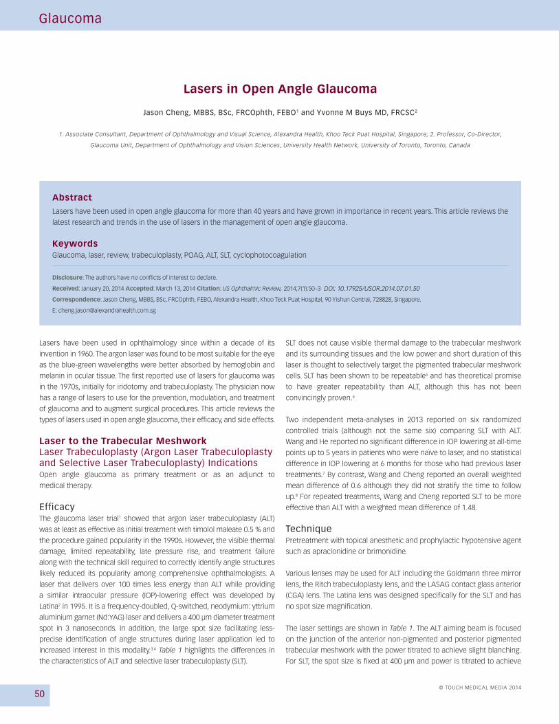
Lasers have been used in open angle glaucoma for more than 40 years and have grown in importance in recent years. This article reviews the
latest research and trends in the use of lasers in the management of open angle glaucoma.
KeywordsGlaucoma, laser, review, trabeculoplasty, POAG, ALT, SLT, cyclophotocoagulation
Disclosure: The authors have no conflicts of interest to declare.
Received: January 20, 2014 Accepted: March 13, 2014 Citation: US Ophthalmic Review, 2014;7(1):50–3
Correspondence: Jason Cheng, MBBS, BSc, FRCOphth, FEBO, Alexandra Health, Khoo Teck Puat Hospital, 90 Yishun Central, 728828, Singapore.
Lasers in Open Angle Glaucoma
Jason Cheng, MBBS, BSc, FRCOphth, FEBO1 and Yvonne M Buys MD, FRCSC2
1. Associate Consultant, Department of Ophthalmology and Visual Science, Alexandra Health, Khoo Teck Puat Hospital, Singapore; 2. Professor, Co-Director,
Glaucoma Unit, Department of Ophthalmology and Vision Sciences, University Health Network, University of Toronto, Toronto, Canada
Cheng_Lasers in Glaucoma_AMc(TL).indd 50 05/04/2014 08:32
DOI: 10.17925/USOR.2014.07.01.50

Lasers in Open Angle Glaucoma
US OPHTHALMIC REVIEW 51
small champagne bubbles. The power setting is usually inversely related
to the amount of pigmentation seen.
Postlaser topical anti-inflammatory medication is typically prescribed for
a few days. Pre-laser hypotensive agents are typically continued and IOP
assessment is recommended 1–4 hours after the laser and again a few
weeks later to assess the response.
ComplicationsComplications following ALT and SLT are similar in frequency and
severity. Early postoperative elevation of IOP of more than 5 mmHg
can occur in up to 34 % of patients in ALT1 and SLT.2 This reduces
significantly to <10 % with the use of alpha-2 adrenergic drops
immediately pre- or postlaser.9
Anterior uveitis occurs up to 83 % of the time but tends to settle without
topical medication.2 Peripheral anterior synaechiae (PAS) although more
common with ALT, can occur with SLT and have been reported in around
1 % of cases.6 Other rare complications include hyphema,10 choroidal
effusion,11 and cystoid macular edema.12 More recently, there have been
reports of corneal stromal haze requiring intensive topical steroids
following SLT. The etiology of this remains unclear.13,14 Higher energy
levels, more segments treated,15 posterior placement of laser, and heavy
angle pigmentation have been associated with postprocedure IOP spikes,
PAS,16 and anterior uveitis.
Newer Lasers for TrabeculoplastyNewer lasers for trabeculoplasty include micropulse diode laser
trabeculoplasty (MLT), titanium sapphire laser trabeculoplasty (TSLT), and
excimer laser trabeculotomy (ELT).
MLT uses a 532/810 nm diode laser. It has a long interval between the
pulses of thermal energy giving sufficient cooling time to avoid injury
to the pigmented trabecular meshwork and its surrounding tissues. In
theory, MLT is less traumatic than ALT and SLT, which potentially may
translate to a better safety profile and repeatability. In a phase II clinical
study, 25 % failed and the remaining 75 % (15 eyes) had a mean reduction
of 22 % at 12 months.17 A retrospective study on MLT showed only three
eyes (7.5 %) had an IOP reduction of more than or equal to 3 mmHg.
However, this was a mixed group of primary and secondary open angle
glaucoma, with 36 % already on three or more glaucoma medications.18
A short-term prospective randomized trial of MLT versus ALT showed
MLT to be less efficacious than ALT. MLT had a mean IOP decrease of
2.5 mmHg (12 %) compared with ALT, which had a mean IOP decrease
of 4.9 mmHg (22 %) at 3 months.19 There are currently two registered trials
that are in the recruitment stage comparing MLT to SLT that may help our
understanding of its efficacy.
TSLT uses a 790 nm laser with a 7 msec exposure time and a spot size
of 200 µm. The near-infrared wavelengths are thought to penetrate
deeper into the pigmented trabecular meshwork and also have an
effect on the juxta-canalucular region and inner wall of Schlemm’s
canal. In a study comparing TSLT to ALT, both lasers had a similar
efficacy (8.3 mmHg versus 6.5 mmHg IOP reduction, respectively
[p>0.05]).20 There are several ongoing trials evaluating the efficacy of
this technology.
ELT is a 308 nm xenon chloride laser with a pulsed delivery of 1.2 mJ over
80 nm duration. It is designed to enhance aqueous outflow by creating
microperforations in the trabecular meshwork and inner wall of Schlemm’s
canal. A recent study looking at phacoemulsification combined with ELT
showed an overall reduction of 4.5 mmHg (23 %) at 12 months. Those
with a higher preoperative IOP (>21 mmHg) had a better IOP response
(9.5 mmHg, 37 %).21 Babighian et al. performed a randomized controlled
trial comparing ELT with SLT. Eight spots were equally distributed at a
distance of 500 µm from one another over the anterior trabeculum in the
ELT group. At 24 months, 53 % of the ELT group and 40 % of the SLT group
had a IOP reduction of 20 % or more.22 Complications included anterior
chamber bleeding (80 %) and transient pressure spikes (20 %).
Laser to the Ciliary BodyTransscleral Cyclodiode Transscleral cyclophotocoagulation has traditionally been reserved for
end-stage glaucoma with little or no visual potential, where all other
treatment options have been tried and failed.
The American Academy of Ophthalmology (AAO) Ophthalmic Technology
Assessment Committee stated that “Cyclophotocoagulation is indicated
for patients with refractory glaucoma who have failed trabeculectomy
or tube shunt procedures, patients with minimal useful vision and
elevated IOP, patients who have no visual potential and need pain relief,
and patients with complicated glaucoma and conjunctival scarring from
previous surgery.”23
This general apprehension stems from the perceived high-risk
complications of phthisis, sympathetic ophthalmia, and vision loss
largely from the historical use of cryotherapy. Newer treatment protocols
with lower energy levels and longer duration have made transscleral
cyclodiode (TSD) safer and more accepted. In recent years, there has
been a shift in the timing at which this treatment is considered in the
glaucoma management paradigm. In a UK survey, 60 % of 180 responders
would perform TSD at any visual acuity24 and there have been reports of
good success in using TSD as a primary treatment in developing countries
where access and compliance is poor.25 TSD has also been advocated
as a safe alternative in acute angle closure that is nonresponsive to
conventional treatment.26
EfficacyThere is a correlation between energy used and treatment success. Many
studies report good IOP reduction with TSD with 54 to 93 % achieving an
IOP of under 21 mmHg.27
Table 1: Selective Laser Trabeculplasty versus Argon Laser Trabeculoplasty Characteristics
Selective Laser Argon Laser Trabeculplasty TrabeculoplastySpot size 400 µm 50 µm
Energy output 0.3–1.5 mJ 500–1,000 mW
Pulse duration 3 ns 0.1 s
Power 600–1,200 mW 0.5–0.8 mJ
Fluence 600 mJ/cm2 60,000 mJ/cm2
Number of burns 45–55 over 180o 45–55 over 180o
Cheng_Lasers in Glaucoma_AMc(TL).indd 51 05/04/2014 08:32
52
Glaucoma
US OPHTHALMIC REVIEW
TechniqueThe G-Probe is designed to direct energy to the ciliary body, some surgeons
prefer to transilluminate through the pupil to identify the location of the
ciliary body as a dark band posterior to the limbus.
The procedure should be performed under retrobulbar anesthesia. The
author recommends the following starting settings: 1,500 mW, 2,000 mS x
20 shots.
ComplicationsThere is a correlation between energy used and complications. In
treatment protocols that use less than 80 J per session, they report
no hypotony or phthisis. Vision loss is also similar to other surgical
procedures. In a summary of 19 studies, many with end-stage secondary
glaucoma, 23 % lost two or more lines (range 0–55 %).27 However, when
TSD is performed on eyes with at least 20/60 vision, 68 % retained 20/60
at 5 years and 31 % lost more than two lines of vision.28 This is comparable
to the Tube vs Trabeculectomy study at 5 years, which reported an overall
44 % loss of 2 or more Snellen lines.29
Endoscopic Cyclophotocoagulation Endoscopic photoablation was first described by Shields in 1986.30 As
the diode laser energy is applied under direct visualization of the ciliary
processes, there is less damage to the surrounding tissue compared with
the TSD. It is reported that only 35 % of TSD energy reaches its intended
target with the remaining causing unwanted trauma.31
IndicationsAs it is an intraocular procedure, it is often combined with phacoemulsification
in patients with good visual prognosis as well as being used independently
for end-stage refractory glaucoma.
EfficacyThe combined phaco-endoscopic cyclophotocoagulation (ECP) has a
reported mean IOP reduction of 7 mmHg at 2 years, with no change in topical
medication use or vision loss.32 However, there are no randomized controlled
trails comparing phacoemulsification alone to combined phaco-ECP. ECP
performs well in refractory glaucoma, with studies reporting a success rate
(<21 mmHg) of around 80 % at 2 years.33,34 ECP also compares favorably with
the Ahmed glaucoma drainage device in refractory glaucoma.35
TechniqueThe ECP laser probe contains a light source, a camera, and helium-neon
aiming beam. The endoscopic view is displayed on a monitor to allow
direct visualization of the ciliary process. Between 180–360° of ciliary
process is treated. Lindfield et al. recommends 250 mW for 3.5 seconds
to be applied to each ciliary process. Intracameral dexamethasone
0.8mg/0.2 ml is also recommended.32
ComplicationsThe ECP Collaborative Study Group reported the following complications
in 5,824 eyes: IOP spike (15 %), hemorrhage (4 %), choroidal effusion
(0.4 %), retinal detachment (0.3 %), vision loss of more than two lines
(1 %), and hypotony or phthisis (0.1 %). The incidence of serious
complications was low and occurred in neovascular glaucoma and
refractory glaucoma groups.36
Lasers to Augment Glaucoma SurgeryLaser Suture LysisTrabeculectomy is the most commonly performed surgery for glaucoma.37
The trabeculectomy scleral flap covers the sclerostomy and is held down
by sutures to reduce postoperative hypotony. These sutures can be later
cut with an argon laser to titrate the postoperative aqueous flow and IOP.
The procedure was originally described by Lieberman and Hoskins
and Migliazzo.38,39 Suture lysis lens (Hoskins or Blumenthal) has a clear
pointed tip that when placed over the scleral suture, blanches the
conjunctival vessels, magnifies the suture, and focuses the laser beam.
A review of 200 consecutive trabeculectomies reported around 50 %
required suture lysis.40
TechniqueRecommended argon laser settings are 50 µm spot size, 0.1 seconds
duration, and power of 400 mW. The beam is focused on the suture. Placing
longer scleral flap sutures and removing thick tenons during surgery
facilitates identifying the flap sutures for postoperative suture lysis.
ComplicationsThis procedure is not without risk. Complications mainly occur due a
sudden decompression of the eye and include: flat anterior chamber
(13 %), malignant glaucoma (2 %), iris incarceration (2 %), hyphema (2 %),
corneal dellen (1 %), hypotony (3 %), hypotonous maculopathy (2 %),
conjunctival burns (<1 %), and wound leaks (<1 %).
Laser Goniopuncture Deep sclerectomy (DS) is a nonpenetrating filtration procedure that
depends on aqueous flow through the trabeculo-Descemet’s membrane
(TDM) into Schlemm’s canal and the subconjunctival space. Laser
goniopuncture (LGP) can be performed in eyes with unsatisfactory IOP post-
DS to puncture the TDM and convert the procedure to a trabeculectomy
to further lower the IOP.41 LGP is considered a minor follow-up procedure
to DS in the same way laser-suture lysis is to trabeculectomy.42
TechniqueA glass CGAL gonioscope contact lens (Haag-Streit AG, Koeniz,
Switzerland) is used to visualize the semi-translucent TDM. Around one
to 20 shots at energy levels of 2–6 mJ are directed to the anterior edge of
the TDM using the Nd:YAG laser.
EfficacyReported LGP rates range from 4.7 % to 63 %.41–44 A critical evaluation of
nonpenetrating surgery by Sarodia et al. suggest a great importance of LGP
in DS, with lower success rates of DS when LGP rates are low. There has
been a steady increase in LGP rates being reported.45 LGP has good efficacy
in reducing IOP after DS. Mermoud reports an 83 % immediate reduction of
IOP and Detry-Morel reported mean IOP of 15 mmHg post-LGP.44
ComplicationsIris incarceration can occur postlaser and prophylactic argon laser
iridoplasty, a smaller size puncture, and targeting a more anterior puncture
site can help prevent this. The incarcerated laser can be released using
argon or Nd:YAG laser. There is also a risk for hypotony and its associated
complications after this procedure.
Cheng_Lasers in Glaucoma_AMc(TL).indd 52 05/04/2014 08:33
Lasers in Open Angle Glaucoma
US OPHTHALMIC REVIEW 53
1. The Glaucoma Laser Trial (GLT). 2. Results of argon laser trabeculoplasty versus topical medicines. The Glaucoma Laser Trial Research Group, Ophthalmology, 1990;97:1403–13.
2. Latina MA, Sibayan SA, Shin DH, et al., Q-switched 532-nm Nd:YAG laser trabeculoplasty (selective laser trabeculoplasty): a multicenter, pilot, clinical study, Ophthalmology, 1998;105:2082–8; discussion 2089–90.
3. Campbell RJ, Trope GE, Rachmiel R, et al., Glaucoma laser and surgical procedure rates in Canada: a long-term profile, Can J Ophthalmol, 2008;43:449–53.
4. Rachmiel R, Trope GE, Chipman ML, et al., Laser trabeculoplasty trends with the introduction of new medical treatments and selective laser trabeculoplasty, J Glaucoma, 2006;15:306–9.
5. Avery N, Ang GS, Nicholas S, et al., Repeatability of primary selective laser trabeculoplasty in patients with primary open-angle glaucoma, Int Ophthalmol, 2013;33:501–6.
6. Damji KF, Bovell AM, Hodge WG, et al., Selective laser trabeculoplasty versus argon laser trabeculoplasty: results from a 1-year randomised clinical trial, Br J Ophthalmol, 2006;90:1490–4.
7. Wang H, Cheng JW, Wei RL et al, Meta-analysis of selective laser trabeculoplasty with argon laser trabeculoplasty in the treatment of open-angle glaucoma, Can J Ophthalmol. 2013;48:186–92.
8. Wang W, He M, Zhou M, Zhang X, Selective laser trabeculoplasty versus argon laser trabeculoplasty in patients with open-angle glaucoma: a systematic review and meta-analysis, PLoS One, 2013;8:e84270.
9. Barkana Y, Belkin M, Selective laser trabeculoplasty, Surv Ophthalmol, 2007;52:634–54.
10. Rhee DJ, Krad O, Pasquale LR, Hyphema following selective laser trabeculoplasty, Ophthalmic Surg Lasers Imaging, 2009;40:493–4.
11. Kim DY, Singh A, Severe iritis and choroidal effusion following selective laser trabeculoplasty, Ophthalmic Surg Lasers Imaging, 2008;39:409–11.
12. Wechsler DZ, Wechsler IB, Cystoid macular oedema after selective laser trabeculoplasty, Eye (Lond), 2010;24:1113.
13. Regina M, Bunya VY, Orlin SE, et al., Corneal edema and haze after selective laser trabeculoplasty, J Glaucoma, 2011;20:327–9.
14. Moubayed SP, Hamid M, Choremis J, et al., An unusual finding of corneal edema complicating selective laser trabeculoplasty, Can J Ophthalmol, 2009;44:337–8.
15. Nagar M, Ogunyomade A, O’Brart DP, et al., A randomised, prospective study comparing selective laser trabeculoplasty
with latanoprost for the control of intraocular pressure in ocular hypertension and open angle glaucoma, Br J Ophthalmol, 2005;89:1413–7.
16. Traverso CE, Greenidge KC, Spaeth GL, Formation of peripheral anterior synechiae following argon laser trabeculoplasty. A prospective study to determine relationship to position of laser burns, Arch Ophthalmol, 1984;102:861–3.
17. Fea AM, Bosone A, Rolle T, et al., Micropulse diode laser trabeculoplasty (MDLT): A phase II clinical study with 12 months follow-up, Clin Ophthalmol, 2008;2:247–52.
18. Rantala E, Valimaki J, Micropulse diode laser trabeculoplasty – 180-degree treatment, Acta Ophthalmol, 2012;90:441–4.
19. Detry-Morel M, Muschart F, Pourjavan S, Micropulse diode laser (810 nm) versus argon laser trabeculoplasty in the treatment of open-angle glaucoma: comparative short-term safety and efficacy profile, Bull Soc Belge Ophtalmol, 2008;308:21–8.
20. Goldenfeld M, Melamed S, Simon G, et al., Titanium:sapphire laser trabeculoplasty versus argon laser trabeculoplasty in patients with open-angle glaucoma, Ophthalmic Surg Lasers Imaging, 2009;40:264–9.
21. Toteberg-Harms M, Hanson JV, Funk J, Cataract surgery combined with excimer laser trabeculotomy to lower intraocular pressure: effectiveness dependent on preoperative IOP, BMC Ophthalmol, 2013;13:24.
22. Babighian S, Caretti L, Tavolato M, et al., Excimer laser trabeculotomy vs 180 degrees selective laser trabeculoplasty in primary open-angle glaucoma. A 2-year randomized, controlled trial, Eye (Lond), 2010;24:632–8.
23. Pastor SA, Singh K, Lee DA, et al., Cyclophotocoagulation: a report by the American Academy of Ophthalmology, Ophthalmology, 2001;108:2130–8.
24. Agrawal P, Dulku S, Nolan W, et al., The UK National Cyclodiode Laser Survey, Eye (Lond), 2011;25:168–73.
25. Schulze Schwering M, Kayange P, Klauss V, et al., Low-dose transscleral diode laser cyclophotocoagulation (TSCPC) as a potential single treatment for primary open-angle glaucoma (POAG) in Malawi?, Graefes Arch Clin Exp Ophthalmol, 2013;251:2389–93.
26. Manna A, Foster P, Papadopoulos M, et al., Cyclodiode laser in the treatment of acute angle closure, Eye (Lond), 2012;26:742–5.
27. Ishida K, Update on results and complications of cyclophotocoagulation, Curr Opin Ophthalmol, 2013;24:102–10.
28. Rotchford AP, Jayasawal R, Madhusudhan S, et al., Transscleral diode laser cycloablation in patients with good vision, Br J Ophthalmol, 2010;94:1180–3.
29. Gedde SJ, Herndon LW, Brandt JD, et al., Postoperative
complications in the Tube Versus Trabeculectomy (TVT) study during five years of follow-up, Am J Ophthalmol, 2012;153:804–14 e1.
30. Shields MB, Intraocular cyclophotocoagulation, Trans Ophthalmol Soc U K, 1986;105:237–41.
31. Bloom PA, Dharmaraj S, Endoscopic and transscleral cyclophotocoagulation, Br J Ophthalmol, 2006;90:666–8.
32. Lindfield D, Ritchie RW, Griffiths MF, ‘Phaco-ECP’: combined endoscopic cyclophotocoagulation and cataract surgery to augment medical control of glaucoma, BMJ Open, 2012;2(3).
33. Chen J, Cohn RA, Lin SC, et al., Endoscopic photocoagulation of the ciliary body for treatment of refractory glaucomas, Am J Ophthalmol, 1997;124:787–96.
34. Murthy GJ, Murthy PR, Murthy KR, et al., A study of the efficacy of endoscopic cyclophotocoagulation for the treatment of refractory glaucomas, Indian J Ophthalmol, 2009;57:127–32.
35. Lima FE, Magacho L, Carvalho DM, et al., A prospective, comparative study between endoscopic cyclophotocoagulation and the Ahmed drainage implant in refractory glaucoma, J Glaucoma, 2004;13:233–7.
36. The ECP Collaborative Study Group. Complications of ECP: a large, long term, multi centre study, Ocul Surg News, 2006.
37. Ramulu PY, Corcoran KJ, Corcoran SL, et al., Utilization of various glaucoma surgeries and procedures in Medicare beneficiaries from 1995 to 2004, Ophthalmology, 2007;114:2265–70.
38. Lieberman MF, Suture lysis by laser and goniolens, Am J Ophthalmol, 1983;95:257–8.
39. Hoskins HD, Migliazzo CV, Argon laser treatment of filtering bleb insufficiency, Klin Monbl Augenheilkd, 1989;195:328–9.
40. Macken P, Buys Y, Trope GE, Glaucoma laser suture lysis, Br J Ophthalmol, 1996;80:398–401.
41. Mermoud A, Karlen ME, Schnyder CC, et al., Nd:Yag goniopuncture after deep sclerectomy with collagen implant, Ophthalmic Surg Lasers, 1999;30:120–5.
42. Anand N, Pilling R, Nd:YAG laser goniopuncture after deep sclerectomy: outcomes, Acta Ophthalmol, 2010;88:110–15.
43. Wishart PK, Wishart MS, Porooshani H, Viscocanalostomy and deep sclerectomy for the surgical treatment of glaucoma: a longterm follow-up, Acta Ophthalmol Scand, 2003;81:343–8.
44. Detry-Morel M, Detry MB, Five-year experience with non penetrating deep sclerectomy, Bull Soc Belge Ophtalmol, 2006;1:83–94.
45. Sarodia U, Shaarawy T, Barton K, Nonpenetrating glaucoma surgery: a critical evaluation, Curr Opin Ophthalmol, 2007;18:152–8.
ConclusionThe management trends of primary open angle glaucoma (POAG) are
constantly evolving. Much recent research has focused on minimizing
the risks for and adverse reactions to current therapy. In glaucoma
medications we have seen the increase number of preservative-
free medications to reduce ocular surface pathology. In surgery, we have
the introduction of minimally invasive glaucoma procedures (MIGS),
which include ECP and ELT. These procedures are often combined with
phacoemulsification and are targeted at ocular hypertensive patients
or early POAG on a low number of medications. They compensate for a
lower efficacy compared with filtration surgery with an improved safety
profile. All these procedures have shown promise in the short term but
will have to prove long-term efficacy in order to replace the current
conventional therapy. n
Cheng_Lasers in Glaucoma_AMc(TL).indd 53 05/04/2014 08:33