Embed Size (px)
Citation preview
Laser-Scanning Microscopy as a Tool to Study the
Spatio-Temporal Organization of InsP3-Mediated Ca 2+ signaling

Xenopus laevis and its oocyte

IP3/Ca2+ signaling pathway in the oocyte

Video-rate confocal microscopy in conjunction with UV photolysis of caged-IP3 and Ca2+
sensitive dyes reveals a high degree of spatio-temporal organization of Ca2+
release in the oocyte
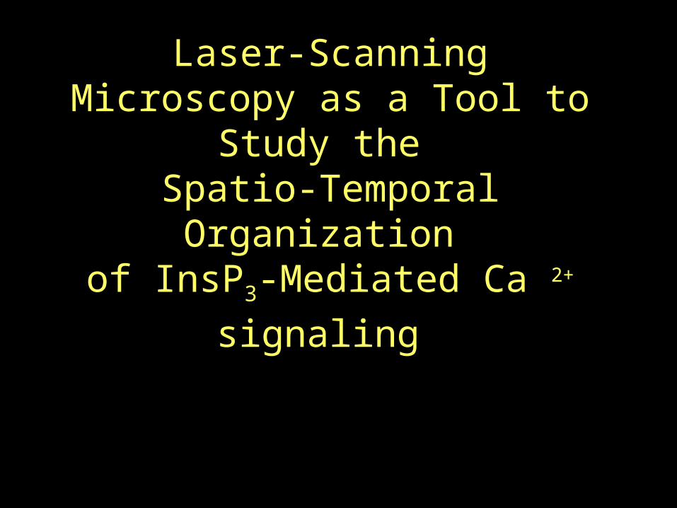
Optical Schematic of Confocal Line-Scan Microscope (CLSM)
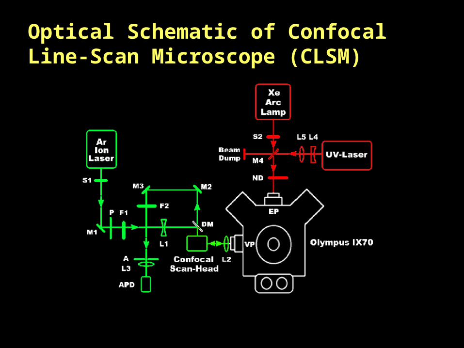
Comparative resolution of the system

Optical Schematic and comparative resolution of the
Video-Rate CLSM

Do elementary events arise from the activity of a
single IP3R?
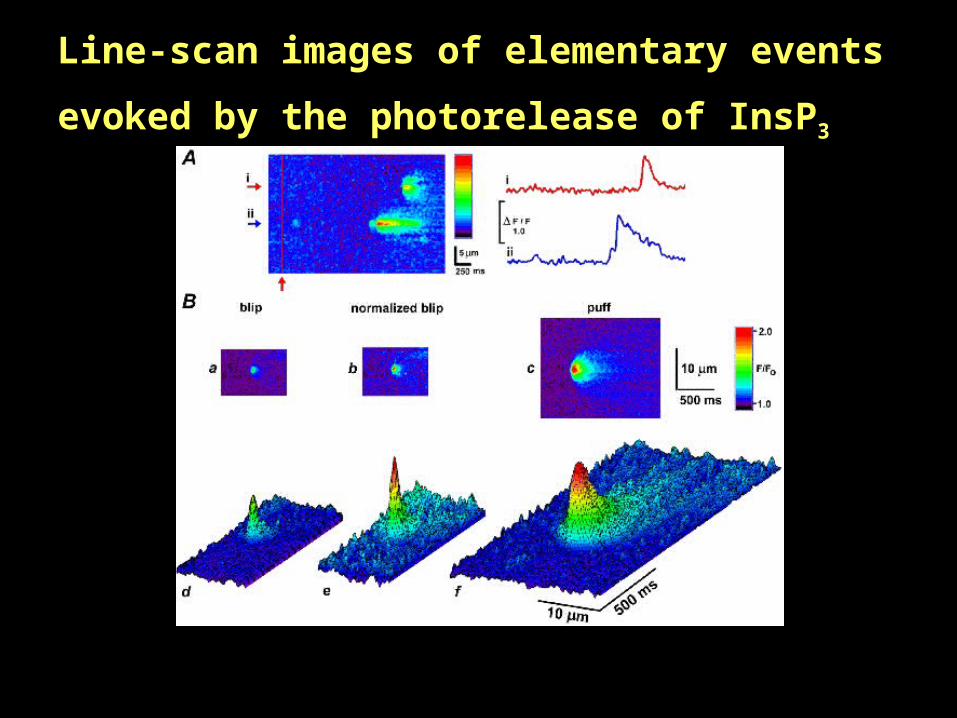
Line-scan images of elementary events evoked by the
photorelease of InsP3

The spread of Ca2+ during elementary events is consistent with passive diffusion from a point source

Does CICR and Ca2+ diffusion between sites give rise to
global waves?
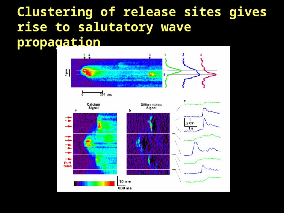
Clustering of release sites gives rise to salutatory wave propagation

EGTA spatially decouples individual release sites to block wave propagation

What is the radial organization of release sites?

Optical schematic of the piezo z-scan unit and representative images of Ca2+ release events in the z-axis

Rapid localized Ca2+ transients as resolved with real-time x-z scanning confocal microscopy

Model of InsP3-mediated Ca2+ release in the oocyte
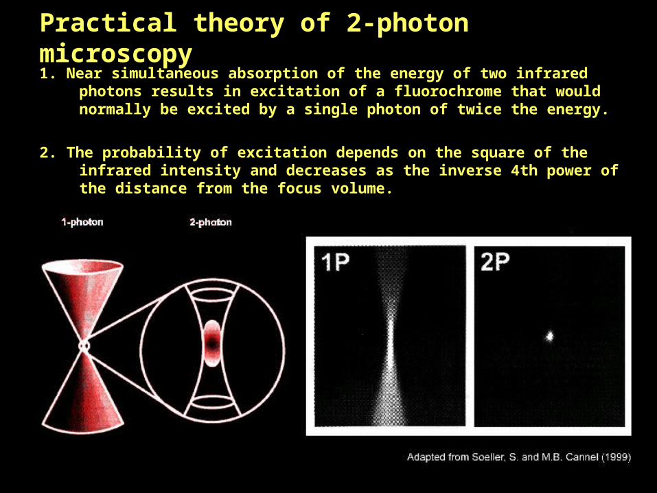
Practical theory of 2-photon microscopy1. Near simultaneous absorption of the energy of two infrared photons results in
excitation of a fluorochrome that would normally be excited by a single photon of twice the energy.
2. The probability of excitation depends on the square of the infrared intensity and decreases as the inverse 4th power of the distance from the focus volume.

Advantages of 2-Photon microscopy
1. Increased penetration of infrared light allows deeper imaging.
2. No out-of-focus fluorescence, thus increased signal to noise.
3. Photo-damage and bleaching are confined to diffraction limited spot.
4. Multiple fluorochrome excitation allows simultaneous, diffraction limited, co-localization.
5. Imaging of UV-excited compounds with conventional optics.
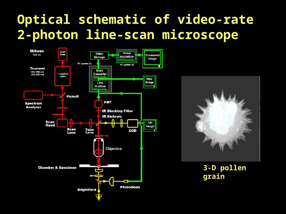
Optical schematic of video-rate 2-photon line-scan microscope
3-D pollen grain

2-photon imaging of pyramidal cells in acute cortical slices

2-Photon Ca 2+ imaging in cortical slices following antidromic stimulation