Embed Size (px)
Citation preview
LASER - OPERCULECTOMY
Oral surgery with laser
Historical overview
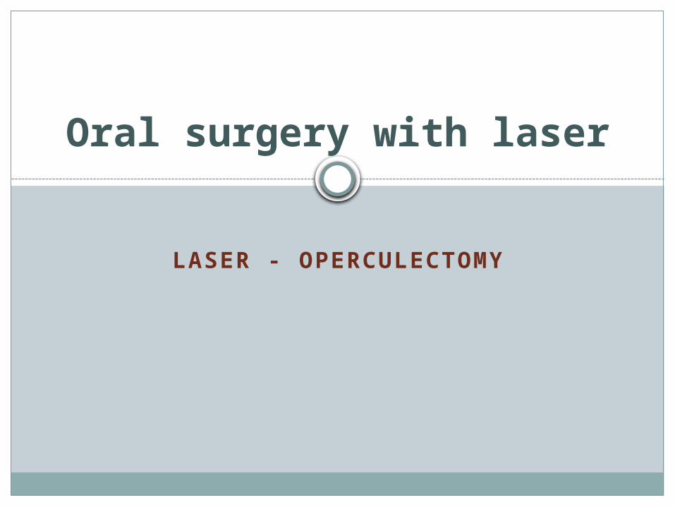
The word laser is an acronym for light amplification by stimulated emission of radiation.
Lasers have been is use in the medical community since the 1970s. In 1989 the first laser specifically designed for use in dentistry
was introduced. Since then, the number of clinical applications is constantly increasing. Laser light is different from ordinary light by being:
- monochromatic (generates a laser beam of a single color) - coherent (identical in phisical size and shape)
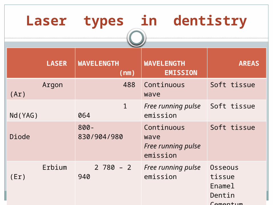
Laser types in dentistry
LASER WAVELENGTH
(nm)WAVELENGTH EMISSION
AREAS
Argon (Ar) 488 Continuous wave Soft tissue
Nd(YAG) 1 064 Free running pulse emission
Soft tissue
Diode 800-830/904/980 Continuous waveFree running pulse emission
Soft tissue
Erbium (Er) 2 780 – 2 940 Free running pulse emission
Osseous tissueEnamelDentinCementum
CO2 10 600 Continuous wave Soft tissue
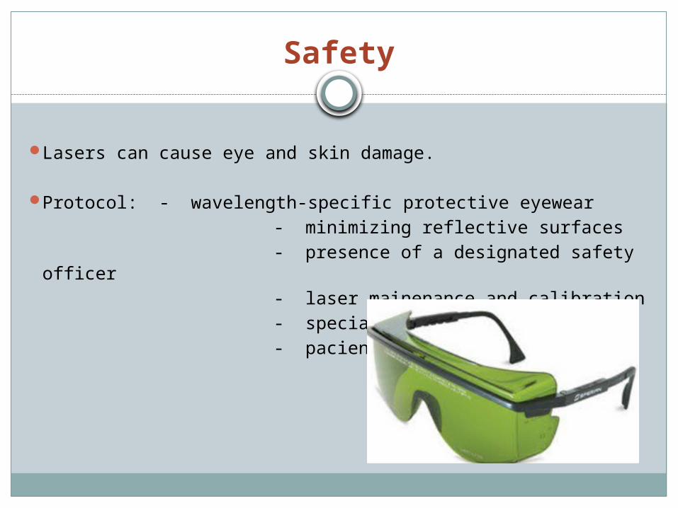
Safety
Lasers can cause eye and skin damage.
Protocol: - wavelength-specific protective eyewear - minimizing reflective surfaces - presence of a designated safety officer - laser mainenance and calibration - specialized staff - pacient education
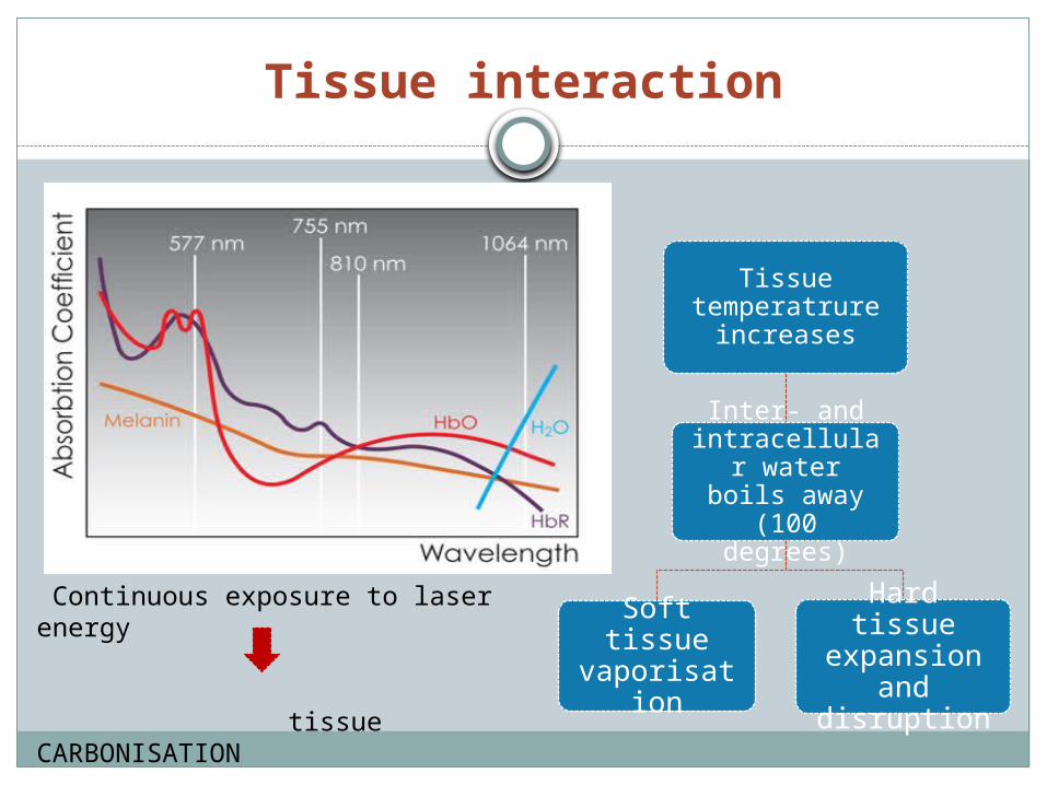
Tissue interaction
Tissue temperatrure
increases
Inter- and intracellular water boils away (100 degrees)
Soft tissue vaporisatio
n
Hard tissue expansion
and disruption
Continuous exposure to laser energy
tissue CARBONISATION
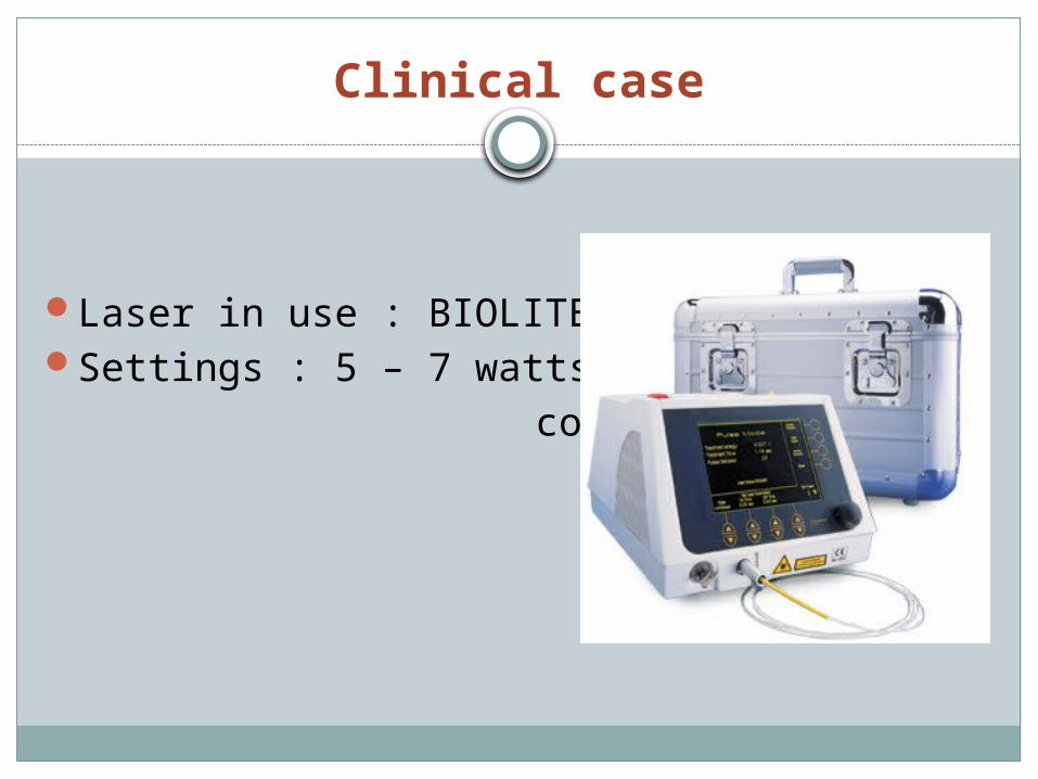
Clinical case
Laser in use : BIOLITECSettings : 5 – 7 watts continuous wave
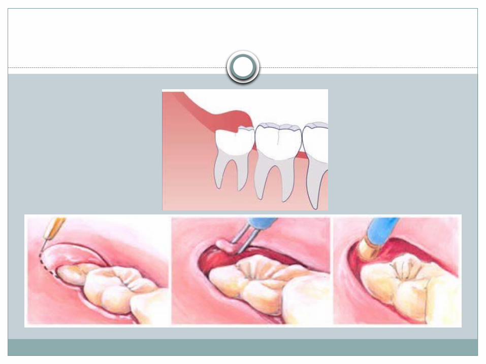
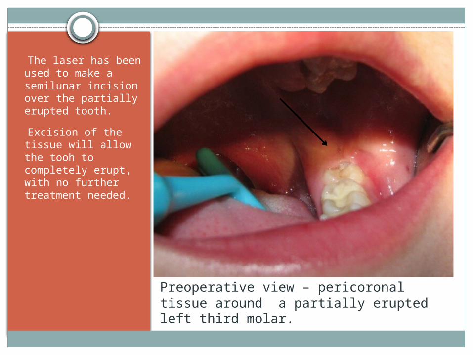
Preoperative view – pericoronal tissue around a partially erupted left third molar.
-The laser has been used to make a semilunar incision over the partially erupted tooth.
-Excision of the tissue will allow the tooh to completely erupt, with no further treatment needed.
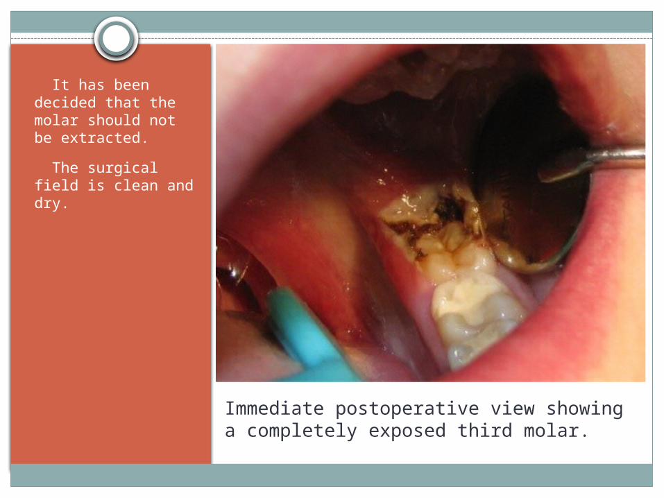
Immediate postoperative view showing a completely exposed third molar.
It has been decided that the molar should not be extracted.
The surgical field is clean and dry.
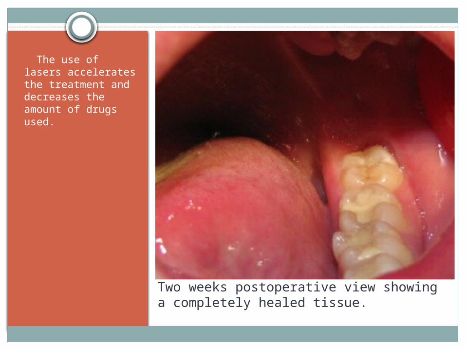
Two weeks postoperative view showing a completely healed tissue.
The use of lasers accelerates the treatment and decreases the amount of drugs used.
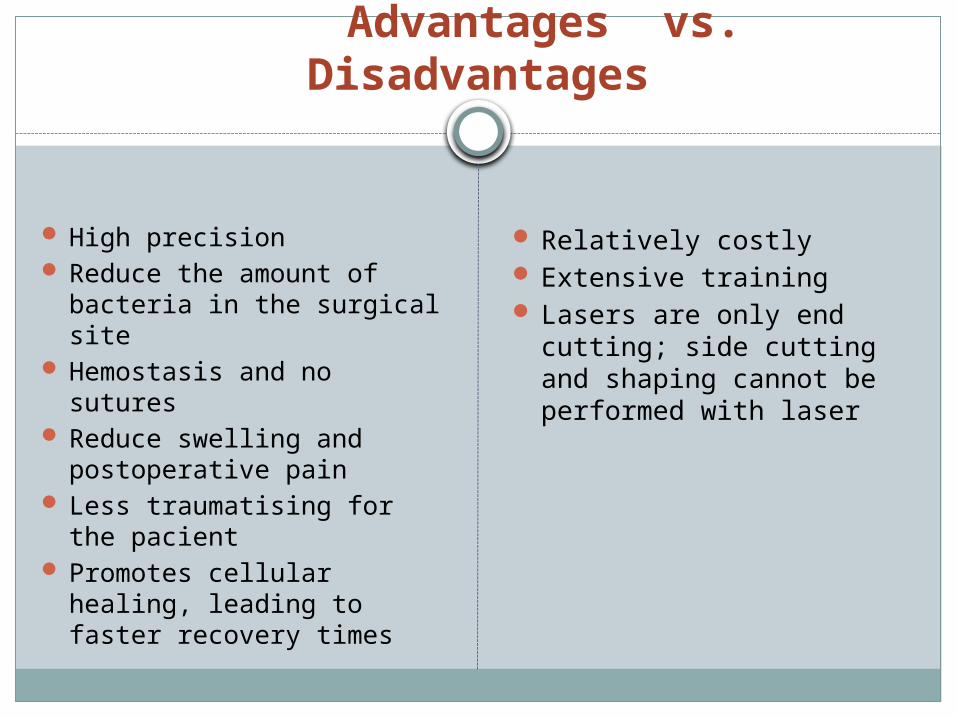
Advantages vs. Disadvantages
High precision Reduce the amount of
bacteria in the surgical site Hemostasis and no sutures Reduce swelling and
postoperative pain Less traumatising for the
pacient Promotes cellular healing,
leading to faster recovery times
Relatively costly Extensive training Lasers are only end
cutting; side cutting and shaping cannot be performed with laser
References
1. Gáspár L.: The use of four different lasers in oral soft tissue surgery In: Loh Hong Sai :Lasers in
Dentistry, Monduzzi Editore, Bologna, Italy, 1995. 2. Waidelich,W., Waidelich,R., Hofstetter,A.: Laser in der Medicin Springer Verlag, Berlin, Heidelberg, 1992. 3. Donald J. Coluzzi,DDS; Robert A. Convissar,DDS:Atlas of laser applications in dentistry,
Quintessence Publishing Co, Inc, 2007.
Thank You for Your attention