Embed Size (px)
Citation preview

Journal of Voice Vol. 7, No. 2, pp. 123-128 © 1993 Raven Press, Lt~l., New York
Laryngeal Biomechanics: An Overview of Mucosal Wave Mechanics
Gerald S. Berke and Bruce R. Gerratt
Head and Neck Surgery, University of California at Los Angeles, and VA Medical Center, West Los Angeles, Los Angeles, California, U.S.A.
Summary: The biomechanics of wave propagation in viscoelastic materials can be useful in understanding the nature of normal and pathologic vocal fold vibration. Mucosal wave movement is the primary means by which the larynx transforms the egressive pulmonary air flow into sound. This short tutorial describes a number of concepts fundamental to the understanding of the vocal fold traveling wave. The displacement velocity of the vocal folds is shown to be proportional to the wave speed, which in turn is proportional to the elastic modulus or stiffness of the vocal folds. Finally, a few cases of unilateral pa- ralysis are used to demonstrate how vocal fold stiffness, entrainment, and degree of vocal fold closure interact to create the complex vibratory patterns that occur in disordered laryngeal states. It is emphasized that surgical voice restoration must consider these properties of the mucosal wave to improve phonatory function. Key Words: Larynx--Mucosal wave movement-- Phonation--Voice.
For many years, it has been known that a funda- mental element of vocal fold vibration is the mu- cosal wave (1). However , its significance in the pro- cess of diagnosing and treating laryngeal disorders is one of the most neglected aspects of laryngology and speech pathology. A greater understanding of the vocal fold traveling wave will lead to an in- creased awareness of the biomechanics of vocal production and its role in procedures used to im- prove voice.
BASICS OF VOCAL FOLD VIBRATION
After the closure of the glottis, the pulmonary- generated air flow directed between the apposed vocal folds causes them to vibrate (2). The forces acting on the folds that sustain self-oscillation are
Accepted April 28, 1992. Address correspondence and reprint requests to Dr. G. S.
Berke, UCLA Head and Neck Surgery, CHS 62-132, 10833 Le Conte Ave., Los Angeles, CA 90024-1624, U.S.A.
This article was presented at the meeting of the Association for Research in Otolaryngology midwinter meeting, St. Petersburg, Florida, Feb. 4, 1991.
not well understood (see Titze, 1980, for example) (3,4). Nevertheless, vibration of the vocal folds is characterized by a tissue wave with compressive and rarefactive phases. The laryngeal tissue wave modulates the air stream flowing through the glottis into a series of puffs (or pulses) of air released into the vocal tract. The physical properties of these re- leased air puffs are the critical elements in the acoustic properties of the sound produced (5).
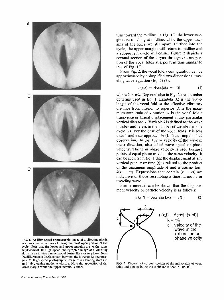
The delay between the compression and rarefac- tion of the tissue wave as it travels the vertical ex- tent of the vocal folds produces a "phase differ- ence" between the opening and closing of the infe- rior and superior portions of the vocal folds (6). Figure IA, B, and C are high-speed photographic images of the vibrating vocal folds in an in vivo canine model. Figure 1A shows the glottis in its most open configuration, in which the upper and lower margins are at the same displacement ampli- tude (i.e., distance from the midline). Figure 1B, slightly further into the cycle, shows the continued compression of the upper margin of the vocal folds, while rarefaction is seen as the lower margins' re-
123
A
B
C
FIG. 1. A: High-speed photographic image of a vibrating glottis in an in vivo canine model during the most open portion of the cycle. Note that the lower and upper margins are at the same displacement. B: High-speed photographic image of a vibrating glottis in an in vivo canine model during the closing phase. Note the difference in displacement between the lower and:upper mar- gins. C: High-speed photographic image of a vibratiflg glottis in an in vivo canine model at closure. Note the apposition of the lower margin while the upper margin is apart.
turn toward the midline. In Fig. 1C, the lower mar- gins are touching at midline, while the upper mar- gins of the folds are still apart. Further into the cycle, the upper margins will return to midline and a subsequent cycle will ensue. Figure 2 depicts a coronal section of the larynx through the midpor- tion of the vocal folds at a point in time similar to that of Fig. 1C.
From Fig. 2, the vocal fold's configuration can be approximated by a simplified two-dimensional trav- eling wave equation (Eq. 1) (7).
u ( x , t ) = a c o s [ k ( x - c t )] (1)
where k ~ -rr//t. Depicted also in Fig. 2 are a number of terms used in Eq. 1. Lambda (X) is the wave- length of the vocal fold or the effective vibratory distance from inferior to superior. A is the maxi- mum amplitude of vibration, u is the vocal fold's transverse or lateral displacement at any particular vertical distance x. Variable k is defined as the wave number and refers to the number of wavelets in one cycle (7). For the case of the vocal folds, k is less than 1 and may approach 1/4 (I. Titze, unpublished observation). In Eq. 1, c = velocity of the wave in the x direction, also called wave speed or phase velocity. The term phase velocity is used because points of equal phase travel at the same velocity. It can be seen from Eq. 1 that the displacement at any vertical point x or time (t) is related to the product of the maximum amplitude A and a cosine term k ( x - c t ) . Expressions that contain ( x - c t ) are indicative of those resembling a time harmonic or traveling wave.
Furthermore, it can be shown that the displace- ment velocity or particle velocity is as follows:
it ( x , t ) = a k c sin [ k ( x - c t ) ] (2)
A
u(x,t) = Acos[k(x-c t ) ] k = ~/~, c = veloc i ty of the
wave in the x d i rect ion or phase veloci ty
FIG. 2. Diagram of coronal section of the midportion of vocal folds and a point in the cycle similar to that in Fig. 1C.
Journal of Voice, Vol. 7, No. 2, 1993

L A R Y N G E A L BIOMECHANICS 125
where A = amplitude, k ~ w/X (vibrates as half a sine wave), and c = phase or wave velocity. Thus, the phase velocity or traveling wave speed may be one of the main controlling factors in determining the displacement velocity. Also, note that if c is directly proportional to the product of the wave- length (X) and frequency, then an increase in c or a decrease in X will produce an increase in vibration frequency. In fact, traveling wave velocity is one of the important factors controlling the fundamental frequency of phonation (8,9).
What controls the wave velocity? It can be shown that the wave velocity (c) in any viscoelastic me- dium such as the vocal folds is related to the fol- lowing equation (10):
C ----
if (resistance to deformation of the medium or E)
(density)
where the elastic modulus (E) is defined as follows:
(force required for displacement)/ (area to which the force is applied)
E = (change in length at a particular length)
In summary, the displacement velocity is propor- tional to the wave speed, which is in turn propor- tional to the elastic modulus of the vocal folds or vocal fold stiffness.
To understand how the stiffness of the vocal folds is controlled, we must examine the intralaryngeal and extralaryngeal innervation of the larynx. Intra- laryngeally, as the recurrent or inferior laryngeal nerve enters the larynx posterior to the inferior cornu of the thyroid cartilage, it immediately di- vides into a posterior division that produces glottal dilatation and an anterior division producing vocal fold adduction (11). The proximal portion of the an- terior division goes on to innervate the lateral crico- arytenoid and interarytenoid muscles, which pro- duce glottal closure. The more distal portion of the anterior division innervates the thyroarytenoid muscle (Fig. 3). The thryoarytenoid muscle runs be- neath the lamina propria from the vocal process to the anterior commissure. The thyroarytenoid mus- cle is one of the major factors in controlling the elastic modulus of the vocal folds (12). Figure 4 is from an in vivo canine experiment (13) in which the transverse elastic modulus (i.e., measured horizon- tally along the medial-lateral plane) of the vocal
FIG. 3. Illustration of intralaryngeal branching pattern of recur- rent laryngeal nerve (RLN). SLN, superior laryngeal nerve; LCA, lateral cricoarytenoid nerve.
fold was measured for increasing recurrent laryn- geal nerve and superior laryngeal nerve stimulation. Because the footplate positioned against the medial edge of the vocal fold contacted the full width of the vocal fold, the elastic modulus represented the stiff- ness of the mucosa, ligament, and muscle of the vocal fold. As depicted, increasing stimulation pro- duced an increase in the fold's elastic modulus.
The extralaryngeal innervation also influences the total elastic modulus or stiffness of the vocal folds. The superior laryngeal nerve external branch runs posterior to the thyroid ala to innervate the cricothyroid muscle, producing a stretching and
o (:3 7 0 0 -
,XX 6 0 0 -
CO 5 0 0 -
- - I 4O0 -
£3 0 3 0 0 -
2 0 0 - O9
loo- z
o O >-
RLN S T I M U L A T I O N ( m A )
Do Do, Dos0 moo
I I I I 0.3 0.4 0 . 4 5 0 .6
SLN STIMULATION (volts) FIG. 4. In vivo canine measurement of the transverse elastic modulus of the vocal fold for increasing recurrent (RLN) and superior laryngeal nerve (SLN) stimulation.
Journal of Voice, Vol. 7, No. 2, 1993

126 G. S. B E R K E A N D B. R . G E R R A T T
thinning of the vocal folds. As shown in Fig. 4, this has the direct effect of increasing the elastic modu- lus of the lamina propria and secondarily acts syn- ergistically with the thyroarytenoid muscle to fur- ther increase its effect on the elastic modulus. Fi- nally, vocal fold thinning produced by cricothyroid muscular contraction changes the geometry of the vocal folds, producing a decrease in the wavelength of vibration and also a decrease in the amplitude of vibration (14). Thus, the strong influence of crico- thyroid contraction on pitch is probably related to its effect on increasing the elastic modulus and de- creasing the vibratory wavelength, i.e., c is propor- tional to h • f.
While thyroarytenoid and cricothyroid muscle activation are synergistic at low to moderate levels of activation, there is some evidence to suggest that high levels of thyroarytenoid muscle activation may be antagonistic to cricothyroid muscle activation and reduce the stiffness of the vocal fold (15). In a system as specialized and complex as the larynx, many other factors also come into play that alter vibration. For example, the amplitude of vibration is quite variable and is influenced by a number of factors such as air flow rate, cricothyroid and vo- calis contraction, and vocal tract configuration. Furthermore, the vocal folds' elastic modulus will be different depending on whether the transverse, longitudinal, or shear modulus is being measured (anisotropy). Also, because of this anisotropy and energy dispersion, the wave speed may not be con- stant but may vary from the lower to upper vertical extent of the folds (6).
One means of gaining further insight into the bio- mechanics of laryngeal vibration is by examining the effect of pathologic states on that vibration. For example, if, as stated earlier, the displacement ve- locity is related to the wave speed, which is propor- tional to the elastic modulus or stiffness, then a uni- lateral decrease in the stiffness would be expected to produce a unilateral decrease in the traveling wave velocity and vocal fold displacement. This type of effect would be observed in patients who exhibit paresis or paralysis of the vocal folds. Un-
fortunately, the two vocal folds interact during vi- bration and so the effect of vocal fold entrainment has to be considered whenever examining the effect of asymmetric pathologic states on bilateral vibra- tion. Entrainment refers to those factors that pro- duce the simultaneous opening and closing of both vocal folds. In general, entrainment occurs through tissue proximity during vocal fold opening and aero-
dynamics during vocal fold closing. For ~example, regardless of which vocal fold's traveling wave is traveling with greater velocity, both folds move lat- erally in glottal opening, and conversely, both the folds move medially in glottal closing. Therefore, differences in velocity of displacement can usually only be observed when the vocal folds are not touching (i.e., during the open portion of a glottal cycle).
Let us first consider a unilateral paralysis when vocal fold closure is adequate, that is to say the vocal folds touch one another during each glottal cycle. A unilateral paralysis should produce a uni- lateral decrease in stiffness, slower traveling wave velocity, and less displacement from midline when compared to the normal fold. However, because of entrainment, differences existing in wave speed be- tween the left and right vocal folds will be mani- fested only during the open interval. The net effect will be that the apparent displacement or vocal fold excursion will be as if the normal fold is greater than
FIG. 5. Digitized most-open and most-closed portions of video- stroboscopic images from a subject following injection of Xy- locaine into the left recurrent laryngeal nerve to produce a left cord paresis.
Journal of Voice, Vol. 7, No. 2, 1993

L A R Y N G E A L BIOMECHANICS 127
the paretic fold. Figure 5 is from a subject who un- derwent Xylocaine injection of his left recurrent la- ryngeal nerve to produce a temporary left vocal fold paresis. The digitized video images are from the most open and most closed portions of a video- stroboscopic cycle. The excursion of each vocal fold was obtained by subtracting the vocal fold width in pixels during complete closure from the vocal fold width during maximal opening, in this case, a - c on the right and b - d on the left. The a - c = 6 pixels and b - d = 3 pixels, indicating that the paretic left-sided vocal fold moved only 50% of the right normal vocal fold.
Using objective analysis of this type, similar find- ings can be observed in nearly all patients with vo- cal fold paresis and paralysis. These results can be interpreted as differences in traveling wave veloci- ties manifesting as differences in displacement ve- locities as discussed earlier (Eq. 2). Furthermore, the asymmetries in wave velocities and vocal fold displacements affect the physical characteristics of the air flow pulses (smaller speed quotient, greater open quotient, and steeper spectral slope) and thus the acoustics and perceived quality of the sound produced (a breathy voice) (16).
A second example is a patient in whom glottal closure is inadequate, in which the vocal folds sel- dom touch during a vocal cycle. In this circum- stance, entrainment may occur through aerodynam- ics only and differences in the wave velocities can be manifested during both opening and closing por- tions of the cycle. These types of asymmetries are observed in patients who exhibit diplophonia (17) (phonation with two simultaneous fundamental fre- quencies). Figure 6 is a fast Fourier transform
ACOUSTIC
PGG
~ A .L 200 480 600 800 I 0J00 1200
HZ FIG. 6. Magnitude spectrum of the acoustic signal from a patient with diplophonia. PGG, photoglottography.
PGG
EGG
ACOUSTIC @ ~ ~
20 40 60 80 NSEC
FIG. 7. Photoglottography (PGG), electroglottography (EGG), and acoustic signals obtained from the patient with diplophonia presented in Fig. 6.
(FFT) from a patient with diplophonia. It can be seen that there are two distinct fundamental fre- quencies. The next, Fig. 7, shows the characteristic photoglot tographie, e lectroglot tographic, and acoustic waveforms obtained from this patient. The electroglottography, displayed as increasing imped- ance toward the top, demonstrates that the vocal folds touch infrequently. Photoglottography shows the characteristic frequency-modulated-type signal often observed in these patients (18), indicating a complex vibratory pattern interpreted as limited en- trainment associated with asymmetric stiffness and two different traveling wave velocities. These are just two examples indicative of the subtle relation- ships that vocal fold stiffness, entrainment, and de- gree of vocal fold closure can form in determining complex vibratory patterns and altered vocal quality.
CONCLUSIONS
Vocal fold stiffness or its manifestation (traveling wave symmetry) is an important consideration in procedures designed to restore voice. Surgical voice restoration procedures must consider the fun- damental biomechanics of the larynx. Their consid- eration will permit modification of the individual elements controlling phonatory function.
Acknowledgment: This research was supported by NIH/NIDCD grant RO1 DC00855-01 and Department of Veterans Affairs Merit Review Funds.
REFERENCES
1. Tirncke R, Von Leden H, Moore P. Laryngeal vibrations: measurement of the glottic wave. Part 1. The normal vibra- tory cycle. Arch Otolaryngol 1958;68:1-9.
Journal of Voice, Vol. 7, No. 2, 1993

128 G. S. BERKE AND B. R. GERRATT
2. Blevins RD. Flow-induced vibration. 2nd ed. New York: Van Nostrand Reinhold, 1990.
3. Titze I. Tutorial: comments on the myoelastic-aerodynamic theory of phonation. J Speech Hear Res 1980;23:495-510.
4. Titze IR. The physics of small-amplitude oscillation of the vocal folds. J Acoust Soc Am 1988;83:1536-52.
5, Fant G. Glottal source and excitation analysis. STL-QPSR 1979;85-107.
6. Berke GS, Moore DM, Hantke DR, Hanson DG, Gerratt BR, Burstein F. Laryngeal modeling: theoretical, in vitro, in vivo. Laryngoscope 1987;97:871-81.
7. Achenbach JD. Wave propagation in elastic solids. New York: Elsevier Science Publishing Co Inc, 1984.
8. Sloan S, Berke GS, Gerratt BR, Ye M. Determination of vocal fold mucosal wave velocity in an in vivo canine model. Laryngoscope (in press).
9. Titze IR, Jiang J J, Tzu-Yu H. Measurements of mucosal wave propagation and vertical phase difference in vocal fold vibration. J Acoust Soc Am 1992;91:2420.
10. Fung YC. Biomechanics, mechanical properties o f living tis- sues. New York: Springer-Verlag, 1981.
11. Green DC, Berke GS, Graves MC. Physiologic motion after
vocal cord reinnervation: a preliminary study. Laryngo- scope 1992;102:14--22.
12. Fujimura O. Body-cover theory of the vocal fold and its phonetic implications. In: Stevens KN, Hirano M, (eds.), Vocal fold physiology. Tokyo: University of Tokyo Press, 1981:271-88.
13. Berke GS, Smith ME. In vivo measurement of the elastic modulus of the vocal fold. Part II: Theoretical consideration. Laryngoscope (in press).
14. Moore DM, Berke GS. The effect of laryngeal nerve stimu- lation on phonation: a glottographic study using an in vivo canine model. J Acoust Soc Am 1988;83:705-15.
15. Harris K. Electromyography as a technique for laryngeal investigation. Proceedings of the conference on the assess- ment of vocal pathology. ASHA 1981 ;11:70-87.
16. Klatt DH, Klatt LC. Analysis synthesis and perception of voice quality variations among female and male talkers. J Acoust Soc Am 1990;87:820-57.
17. Ward PH, Sanders JW, Goldmon R, Moore GP. Diplopho- nia. Ann Otol Rhinol Laryngol 1969;78:771-7.
18. Gerratt BR, Precoda K, Hanson D, Berke GS. Source char- acteristics of diplophonia. J Acoust Soc Am 1988;83:$66.
Journal of Voice, VoL 7, No. 2, 1993