Embed Size (px)
Citation preview
The Metamorphosis of Mosquito Immune and Circulatory Physiology: Comparative Analyses in
Larval and Adult Anopheles gambiae
By
Garrett Philip League
Dissertation
Submitted to the Faculty of the
Graduate School of Vanderbilt University
in partial fulfillment of the requirements
for the degree of
DOCTOR OF PHILOSOPHY
in
Biological Sciences
May, 2017
Nashville, Tennessee
Approved:
Julián F. Hillyer, Ph.D.
Kenneth C. Catania, Ph.D.
Laurence J. Zwiebel, Ph.D.
Antonis Rokas, Ph.D.
Andrea Page-McCaw, Ph.D.

ii
Dedicated to Jesus Christ, the creator and sustainer of all life (Colossians 1:16-18). “For ‘in him
we live and move and have our being’” (The Apostle Paul, quoting from Epimenides of Crete in
Acts 17:28). Soli Deo Gloria!

iii
ACKNOWLEDGEMENTS
I would like to acknowledge my advisor Dr. Julián Hillyer, who has served as a
thoughtful and accessible guide throughout my time in graduate school. I have learned much
from his sound advice and attention to detail and I am deeply thankful for his patient mentorship.
My committee members, including Drs. Ken Catania, Larry Zwiebel, Antonis Rokas, and
Andrea Page-McCaw, provided helpful feedback in shaping my dissertation research, and I am
thankful for their time and input. I thank my all labmates, past and present, especially Leah Sigle,
Tania Estévez-Lao, Dr. Lisa Brown, Dr. Lillian Shapiro, Yan Yan, Oge Onuh, Valeria Garcia,
and former labmate Dr. Jonas King for their support, both personally and professionally. It has
been a joy to work with you. I thank my entire family, especially my parents Scott and Amanda,
for their counsel and encouragement throughout the years, as well as for the countless prayers
and acts of love too numerous to recall, much less list. I reserve my most heartfelt thanks for my
wife Kim for being my closest earthly companion during graduate school. I will always treasure
this most special season of our lives together and I know that our greatest discoveries are still to
come. Finally, in the words of the patriarch Jacob in Genesis 35:3, I thank “the God who answers
me in the day of my distress and has been with me wherever I have gone,” to whom I offer this
work.
iv
TABLE OF CONTENTS
Page
DEDICATION ................................................................................................................................ ii
ACKNOWLEDGEMENTS ........................................................................................................... iii
LIST OF TABLES ......................................................................................................................... vi
LIST OF FIGURES ...................................................................................................................... vii
Chapter
I. Introduction .................................................................................................................................1
Overview .....................................................................................................................................1
Mosquito Life Cycle ....................................................................................................................3
Mosquito Adult Circulatory Physiology .....................................................................................4
Mosquito Adult Immunity in the Hemocoel ...............................................................................6
Larval Immune Competence Relative to Adults .........................................................................8
Summary and Preview of Subsequent Chapters..........................................................................9
II. Comparative structural and functional analysis of the larval and adult dorsal vessel and its role
in hemolymph circulation in the mosquito Anopheles gambiae ...............................................11
Preface .......................................................................................................................................11
Abstract .....................................................................................................................................11
Introduction ...............................................................................................................................12
Materials and Methods ..............................................................................................................15
Results .......................................................................................................................................20
Discussion .................................................................................................................................39
III. Functional integration of the circulatory, immune, and respiratory systems in mosquito larvae:
pathogen killing in the hemocyte-rich tracheal tufts .................................................................46
Preface .......................................................................................................................................46
Abstract .....................................................................................................................................46
Introduction ...............................................................................................................................47
Materials and Methods ..............................................................................................................51
Results .......................................................................................................................................57
Discussion .................................................................................................................................79
IV. Anopheles gambiae larvae mount stronger immune responses against bacterial infection than
adults: evidence of adaptive decoupling in mosquitoes ............................................................87
v
Preface .......................................................................................................................................87
Abstract .....................................................................................................................................87
Introduction ...............................................................................................................................88
Materials and Methods ..............................................................................................................91
Results .......................................................................................................................................99
Discussion ...............................................................................................................................120
V. Conclusions and future directions ...........................................................................................130
Overview .................................................................................................................................130
Larval and Adult Circulatory Physiology ...............................................................................131
Functional Integration of the Larval Circulatory and Immune Systems .................................133
Larval and Adult Cellular and Humoral Immune Responses ..................................................136
Concluding Thoughts ..............................................................................................................138
REFERENCES ............................................................................................................................141
Appendix
A. Gene Names, VectorBase Gene IDs, and Primers Used for Quantitative RT-PCR ...............158
B. Relative mRNA Expression Levels of Individual Immunity Genes .......................................159

vi
LIST OF TABLES
Table Page
1. Mosquito immunity rapidly declines after metamorphosis and with adult age .......................122
vii
LIST OF FIGURES
Figure Page
CHAPTER I
1. The mosquito life cycle ................................................................................................................4
2. Hemolymph circulation through the dorsal vessel of adult mosquitoes ......................................6
CHAPTER II
1. Larval and adult heart contractions in Anopheles gambiae .......................................................22
2. Larval and adult heart contraction rates .....................................................................................24
3. Larval and adult hemolymph flow velocity ...............................................................................26
4. Larval and adult heart structure .................................................................................................28
5. Larval and adult abdominal ostia ...............................................................................................31
6. Larval hemolymph flow patterns and entry through the eighth abdominal segment incurrent
openings of the heart. ................................................................................................................33
7. Larval and adult posterior heart structure ..................................................................................35
8. Larval aorta structure .................................................................................................................37
CHAPTER III
1. Hemolymph flow through the larval and adult dorsal vessels changes during development ....50
2. Fluorescence intensity and optical density at the larval periostial regions and tracheal tufts
were measured using custom-drawn regions of interest ...........................................................55
3. Sessile hemocyte distributions in the abdomen differ between larvae and adults .....................58
4. Larvae contain pericardial cells, but lack periostial hemocytes ................................................60
5. Larval hemocytes associate with the eighth abdominal segment tracheal tufts and are distinct
from the segmental hemocyte bands .........................................................................................63
6. Tracheal tufts are shed in the fourth instar larval exuviae during pupation ...............................64
viii
7. E. coli injected into the larval hemocoel rapidly aggregate in the tracheal tufts. ......................66
8. Tracheal tuft hemocytes show high phagocytic activity ............................................................67
9. Microspheres injected into larvae and adults aggregate in regions of high hemolymph flow and
hemocyte concentration .............................................................................................................68
10. E. coli injected into larvae and adults aggregate in regions of high hemolymph flow and
hemocyte concentration ...........................................................................................................69
11. E. coli aggregate in the tracheal tufts, where they are rapidly destroyed ................................71
12. GFP signal from periostial region ROIs of larvae originate from pericardial cells and
segmental hemocytes ...............................................................................................................72
13. Tracheal tuft hemocytes phagocytose melanized bacteria .......................................................74
14. The melanization immune response is concentrated in the tracheal tufts ................................75
15. Hemocytes are more abundant in the tracheal tufts, but do not increase in response to E. coli
infection ...................................................................................................................................77
16. The circulatory and immune systems of mosquito larvae and adults are functionally
integrated..................................................................................................................................80
CHAPTER IV
1. Fluorescence emitted by sessile hemocytes in larvae and adults was measured using custom-
drawn regions of interest ...........................................................................................................95
2. Larvae kill E. coli in their hemocoels more efficiently than adults .........................................101
3. Larvae contain more circulating hemocytes than adults ..........................................................103
4. Sessile hemocytes differ in spatial arrangement and circulation-dependent aggregation across
life stages and in response to infection ....................................................................................105
5. Sessile hemocytes of segments 2, 3, and 7 in response to infection ........................................106
6. The distribution of sessile hemocytes in the trachea, cuticle, and periostial regions of
abdominal segments 4, 5, and 6 varies with life stage and with infection state. .....................107
7. Hemocytes from adults carry higher phagocytic burdens and spread differently than
hemocytes from larvae ............................................................................................................109
ix
8. Larvae display stronger antibacterial humoral immunity compared to adults .........................111
9. Larval hemolymph has higher phenoloxidase activity than adult hemolymph .......................113
10. Phenoloxidase-based melanization of endogenous substrates is higher in larvae than in adults,
and melanization of exogenous substrates is completely inhibited by DETC .......................114
11. Infection-induced transcriptional regulation of immunity-related genes is higher in larvae
than in adults ..........................................................................................................................118
1
CHAPTER I
INTRODUCTION
Overview
Insects are one of the most abundant, diverse, and successful classes of organisms on
Earth. Within the insects, the Nematocera are an epidemiologically important suborder of
Diptera (flies) that includes, among others, black flies, biting midges, and mosquitoes.
Mosquitoes are responsible for transmitting a number of intractable human diseases which have
historically ranked among the deadliest to ever to plague mankind, including dengue fever,
lymphatic filariasis, and malaria. Numerous methods have been employed with varying degrees
of success to control mosquito populations and the diseases they transmit, including the removal
of mosquito breeding sites, the use of chemical insecticides like DDT, as well as a variety of
integrated pest management strategies (Patterson, 2016; Ramirez et al., 2009). However, due to
the evolution of insecticide resistance and the spread of invasive species, no single control
method has proven to be a panacea. More recently, manipulation of the mosquito host immune
response has been proposed as another viable means of pest control (Clayton et al., 2014;
Ramirez et al., 2009). As host immune responses are a crucial determinant of pathogen viability
and transmission, further investigations into the role of mosquito immunity in determining the
outcomes of various host-pathogen interactions are urgently needed.
Equipped with a potent innate immune system, mosquitoes have developed an effective
series of conserved immune responses that act in concert to limit infections. Mosquitos acquire
pathogens from a number of sources, including the microbe-rich aquatic environments of the
2
immature stages and the infectious blood meals of the adult stage. Upon entering the mosquito
hemocoel (body cavity) either via ingestion and subsequent penetration of the midgut or via
breaches in the cuticle, pathogens are met with a wide range of cellular and humoral host
immune responses (Hillyer, 2010; Hillyer, 2016). In the dynamic environment of the hemocoel,
both pathogens and host-derived immune factors are subject to the forces of the open circulatory
system, where hemolymph (insect blood) is propelled by the dorsal vessel, the main hemolymph
pumping organ in mosquitoes (Glenn et al., 2010). Indeed, the immune and circulatory systems
are so intimately intertwined in the hemocoel that they have become functionally integrated such
that immune responses preferentially occur at areas of highest hemolymph flow, so as to
maximize immune surveillance and pathogen killing efficiency (King and Hillyer, 2012; Sigle
and Hillyer, 2016).
Because mosquitoes are holometabolous insects and undergo dramatic morphological,
physiological, and environmental changes during the larval to adult transition, we predicted that
their circulatory and immune systems, and hence the nature of their interactions, would differ in
important respects between the larval and adult life stages. At the outset of my dissertation work,
however, no studies had examined circulation-dependent immunity in larvae and little data
existed on the relative strength and composition of larval and adult immune responses. In light of
this, the overarching goal of my dissertation research was to compare and contrast circulation-
based immunity and overall immune competence in larval and adult mosquitoes to determine the
extent to which differing larval and adult selection pressures have led to divergent immune
responses between the two life stages.
Regarding circulation-dependent immunity, we hypothesized that key structural and
functional differences between the larval and adult circulatory systems would result in functional
3
integration of the larval circulatory and immune systems that reflected the distinct morphology
and physiology of the larval stage. Furthermore, since larvae encounter an abundance of
microbes in their aquatic environments and survival through the larval stage is essential for
reproduction in the adult life stage, we hypothesized that mosquito larvae have evolved powerful
innate immune responses that wane in the adult life stage due to immune senescence and other
factors. In sum, in this dissertation I document striking differences between the larval and adult
circulatory systems, which have led to stage-specific functional integration of the larval
circulatory and immune systems. Furthermore, I also present compelling evidence that mosquito
larvae display enhanced antibacterial immunity compared to adults.
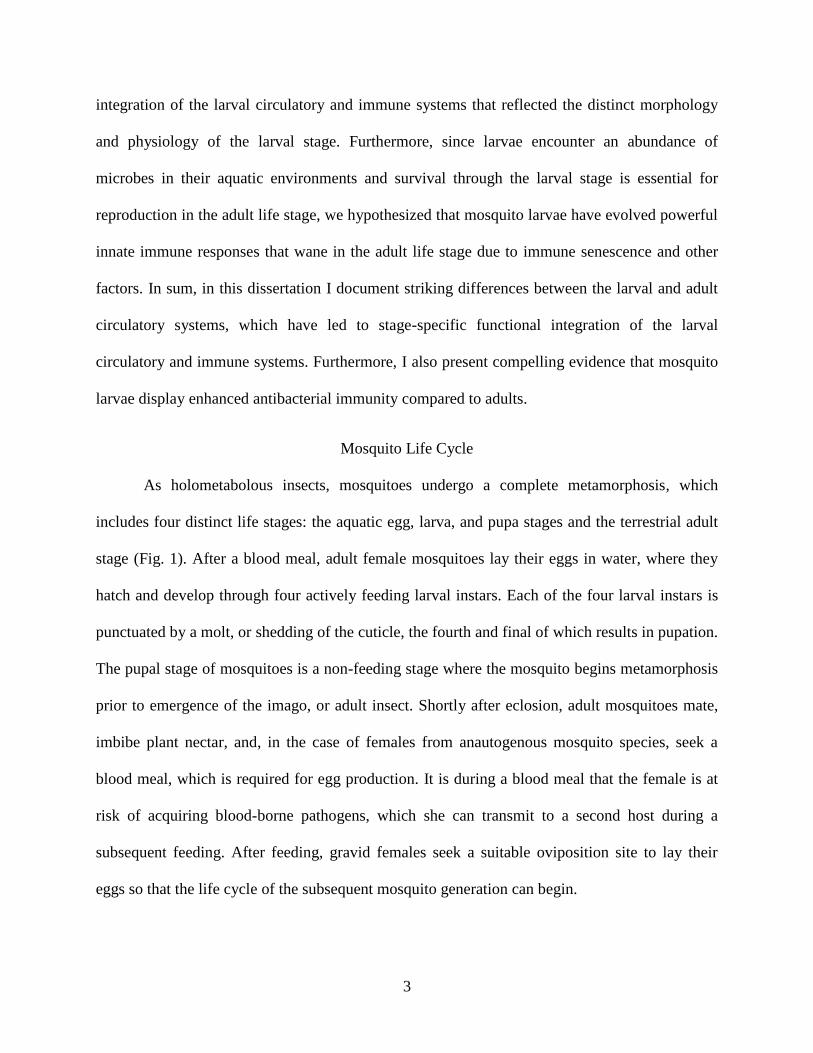
Mosquito Life Cycle
As holometabolous insects, mosquitoes undergo a complete metamorphosis, which
includes four distinct life stages: the aquatic egg, larva, and pupa stages and the terrestrial adult
stage (Fig. 1). After a blood meal, adult female mosquitoes lay their eggs in water, where they
hatch and develop through four actively feeding larval instars. Each of the four larval instars is
punctuated by a molt, or shedding of the cuticle, the fourth and final of which results in pupation.
The pupal stage of mosquitoes is a non-feeding stage where the mosquito begins metamorphosis
prior to emergence of the imago, or adult insect. Shortly after eclosion, adult mosquitoes mate,
imbibe plant nectar, and, in the case of females from anautogenous mosquito species, seek a
blood meal, which is required for egg production. It is during a blood meal that the female is at
risk of acquiring blood-borne pathogens, which she can transmit to a second host during a
subsequent feeding. After feeding, gravid females seek a suitable oviposition site to lay their
eggs so that the life cycle of the subsequent mosquito generation can begin.
4
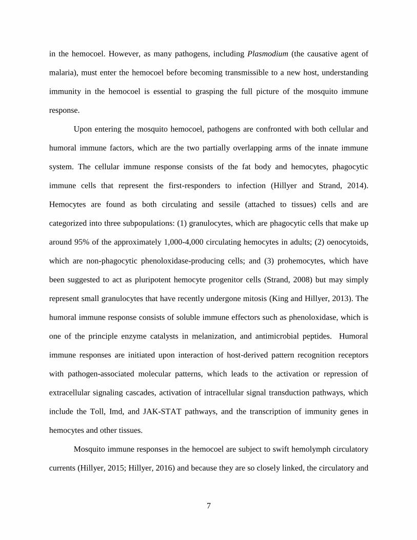
Figure 1. The mosquito life cycle. The mosquito life cycle consists of four general stages that
inhabit both aquatic and terrestrial environments. Adult female mosquitoes lay their eggs in
water where the eggs hatch and develop through four larval instars. After the fourth instar, larvae
pupate and the pupae undergo metamorphosis before emerging from their aquatic habitat as
terrestrial adults.
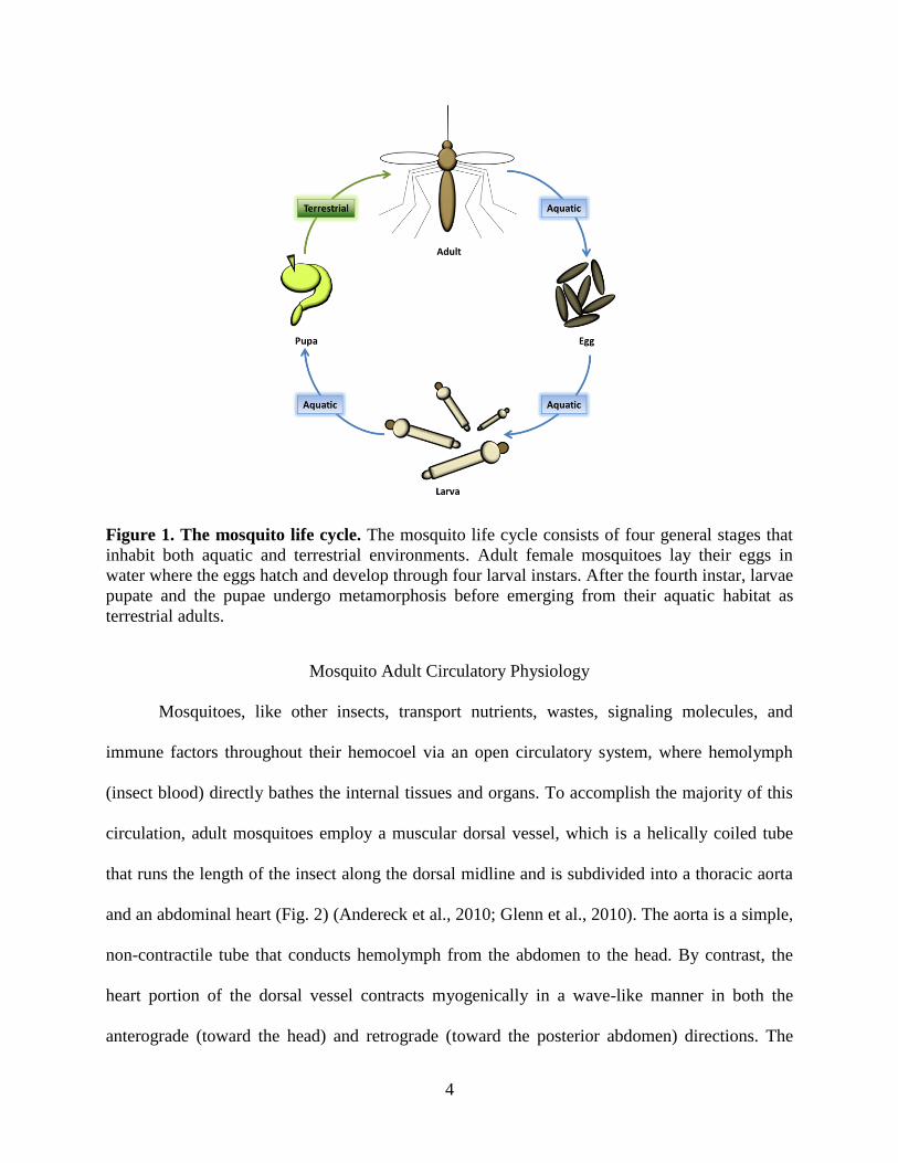
Mosquito Adult Circulatory Physiology
Mosquitoes, like other insects, transport nutrients, wastes, signaling molecules, and
immune factors throughout their hemocoel via an open circulatory system, where hemolymph
(insect blood) directly bathes the internal tissues and organs. To accomplish the majority of this
circulation, adult mosquitoes employ a muscular dorsal vessel, which is a helically coiled tube
that runs the length of the insect along the dorsal midline and is subdivided into a thoracic aorta
and an abdominal heart (Fig. 2) (Andereck et al., 2010; Glenn et al., 2010). The aorta is a simple,
non-contractile tube that conducts hemolymph from the abdomen to the head. By contrast, the
heart portion of the dorsal vessel contracts myogenically in a wave-like manner in both the
anterograde (toward the head) and retrograde (toward the posterior abdomen) directions. The
5
heart is also distinguished from the aorta in that it contains incurrent ostia (valves) at the anterior
portion of abdominal segments 2-7. During anterograde contraction periods, hemolymph enters
the lumen of the dorsal vessel via each of these abdominal ostia and exits the aorta into the head,
where it slowly circulates posteriorly. During retrograde contraction periods, however, the
abdominal ostia remain closed and hemolymph enters the heart via a single pair of ostia at the
thoraco-abdominal junction, exiting the dorsal vessel via a pair of excurrent openings at the
posterior terminus of the heart. Although the structure and function of the dorsal vessel has been
described in detail in adult mosquitoes, prior to the initiation of my dissertation work little was
known regarding the structure and function of the larval circulatory system and how it compared
to that of adults. I have addressed this gap in our understanding of the mosquito circulatory
system in the comparative larval and adult study presented in Chapter II.
6
Figure 2. Hemolymph circulation through the dorsal vessel of adult mosquitoes. (A) During
periods of anterograde heart contraction, hemolymph enters the heart through 6 paired ostia
located at the anterior portions of abdominal segments 2-7 and exits into the head via an anterior
excurrent opening of the aorta. (B) During periods of retrograde heart contraction, hemolymph
enters a single pair of ostia at the thoraco-abdominal junction and exits into the abdomen via
excurrent openings of the posterior terminus of the heart. Figure adapted from (League and
Hillyer, 2016).
Mosquito Adult Immunity in the Hemocoel
Mosquitoes, unlike vertebrates, do not have an adaptive immune system, but instead rely
on a highly conserved innate immune system to kill pathogens by phagocytosis, lysis,
melanization, and other mechanisms (Hillyer, 2016). The mosquito immune system is spatially
organized into three major immune compartments, namely, the midgut, the hemocoel, and the
salivary glands, as well as conceptually organized into both cellular and humoral immune
responses (Hillyer, 2010). Although much research effort has been expended on describing
immunity in the midgut (Abraham and Jacobs-Lorena, 2004; Cirimotich et al., 2010; Whitten et
al., 2006; Yassine and Osta, 2010), comparatively few studies have been conducted on immunity
7
in the hemocoel. However, as many pathogens, including Plasmodium (the causative agent of
malaria), must enter the hemocoel before becoming transmissible to a new host, understanding
immunity in the hemocoel is essential to grasping the full picture of the mosquito immune
response.
Upon entering the mosquito hemocoel, pathogens are confronted with both cellular and
humoral immune factors, which are the two partially overlapping arms of the innate immune
system. The cellular immune response consists of the fat body and hemocytes, phagocytic
immune cells that represent the first-responders to infection (Hillyer and Strand, 2014).
Hemocytes are found as both circulating and sessile (attached to tissues) cells and are
categorized into three subpopulations: (1) granulocytes, which are phagocytic cells that make up
around 95% of the approximately 1,000-4,000 circulating hemocytes in adults; (2) oenocytoids,
which are non-phagocytic phenoloxidase-producing cells; and (3) prohemocytes, which have
been suggested to act as pluripotent hemocyte progenitor cells (Strand, 2008) but may simply
represent small granulocytes that have recently undergone mitosis (King and Hillyer, 2013). The
humoral immune response consists of soluble immune effectors such as phenoloxidase, which is
one of the principle enzyme catalysts in melanization, and antimicrobial peptides. Humoral
immune responses are initiated upon interaction of host-derived pattern recognition receptors
with pathogen-associated molecular patterns, which leads to the activation or repression of
extracellular signaling cascades, activation of intracellular signal transduction pathways, which
include the Toll, Imd, and JAK-STAT pathways, and the transcription of immunity genes in
hemocytes and other tissues.
Mosquito immune responses in the hemocoel are subject to swift hemolymph circulatory
currents (Hillyer, 2015; Hillyer, 2016) and because they are so closely linked, the circulatory and
8
immune systems of adults are functionally integrated and work cooperatively to limit systemic
infections (King and Hillyer, 2012; Sigle and Hillyer, 2016). Although other studies have
observed immune responses on the heart (Hernández-Martínez et al., 2013; Hillyer et al., 2007;
Michel et al., 2005; Schnitger et al., 2007; Yassine et al., 2014; Yassine et al., 2012), neither
these studies. nor the studies documenting functional integration in adults, have examined
whether these phenomena occur in the larval stage. To address this gap in our knowledge, I
examined Anopheles gambiae larvae after infection to determine whether immune responses in
this life stage are functionally coordinated with their circulatory system, and this study is
presented in Chapter III.
Larval Immune Competence Relative to Adults
Anopheles mosquitoes oviposit in small, warm bodies of water that are also natural
breeding grounds for bacteria (Laird, 1988), and therefore the risk of bacterial infections must be
high during the larval life stage. Because of this, and the fact that mosquitoes must survive the
larval stage in order to reproduce, larvae are likely to have evolved more proficient means of
neutralizing infections than adults. Although the mosquito adult immune response has been
studied in depth, little is known about larval immunity and how larval immune responses
compare to those of adults. Differences in the expression patterns of some immunity genes occur
across developmental stages (Christophides et al., 2002; Dimopoulos et al., 1997), however no
study has functionally described the cellular and humoral antibacterial immune responses of
mosquito larvae, let alone placed these responses in the context of adult immunity. If larval and
adult immune responses indeed differ in either strength or composition, this would imply that
future studies could no longer assume either complete continuity or discontinuity in immune
responses across life stages and that metamorphosis has, to some extent, decoupled the larval and
9
adult immune systems, thus enabling their independent evolution (Moran, 1994). Furthermore, if
immune responses differ across life stages, this would have important implications for the
creation of stage-specific control measures that are better tailored to the specific immune
responses of each life stage.
Because hemocyte numbers decline with adult age (Castillo et al., 2006; Hillyer et al.,
2005; King and Hillyer, 2013; Pigeault et al., 2015), increase in response to immune stimuli
(Baton et al., 2009; Castillo et al., 2011; Christensen et al., 1989; Coggins et al., 2012; King and
Hillyer, 2013), and may also increase in preparation for tissue restructuring during
metamorphosis (Castillo et al., 2006; King and Hillyer, 2013), it is likely that mosquito larvae
display enhanced antibacterial cellular immune responses compared to adults. Furthermore, as
bacteria are also subject to melanization in the hemocoel (Hillyer et al., 2004), and the strength
of the melanization response has also been shown to decline with adult age (Christensen et al.,
1986; Chun et al., 1995; Cornet et al., 2013; Li et al., 1992), we also expect humoral immunity to
be stronger in larvae compared to adults. To test whether A. gambiae larvae have a more potent
antibacterial immune response compared to differently aged adults, I compared the strength of a
range of immune responses between larvae, newly emerged adults and older adults, and present
the findings in Chapter IV.
Summary and Preview of Subsequent Chapters
Mosquitoes vector numerous deadly diseases that cause tremendous human suffering
worldwide. Understanding how the mosquito circulatory and immune systems work in
conjunction to fight pathogens is critical to potentially limiting their transmission to human
hosts. Furthermore, because mosquitoes are holometabolous insects, studies of the circulatory
and immune systems in adults should be complemented by studies in larvae, as these life stages
10
differ, yet are inextricably linked to one another. Understanding how larval and adult immunity
relate to each other could yield insight into how larval infections prime adults against subsequent
infections (Bargielowski and Koella, 2009; Moreno-García et al., 2015), and why larvae tend to
evolve resistance to pesticides faster than adults (Koella et al., 2009), as the mechanisms behind
both of these phenomena are not fully understood. To being to more fully understand the
mosquito larval immune system and how it compares to that of adults, the dissertation work
presented in the subsequent chapters describes how the larval and adult circulatory systems are
structurally and functionally adapted to meet the unique physiological needs of each life stage,
and how these adaptations have led to spatially distinct yet functionally analogous integration of
the circulatory and immune systems. Finally, by comparing both cellular and humoral immunity
across life stages and adult ages, we show that larvae have more robust immune responses than
adults, and that immunity declines with adult age. Taken together, these findings suggest that a
holistic approach to the study of mosquito immunity that incorporates both larval and adult life
stages yields important insights that could aid in the creation and novel pest and disease control
measures.
11
CHAPTER II
COMPARATIVE STRUCTURAL AND FUNCTIONAL ANALYSIS OF THE LARVAL AND
ADULT DORSAL VESSEL AND ITS ROLE IN HEMOLYMPH CIRCULATION IN THE
MOSQUITO ANOPHELES GAMBIAE
Preface
This chapter provides a detailed comparison of the Anopheles gambiae larval and adult
circulatory systems and shows that though the heart is structurally similar in both life stages, it
differs with respect to both contraction and hemolymph flow dynamics. This work laid the
foundation for the study on the coordinated function of the larval circulatory and immune
systems presented in Chapter III. I led the experimental effort and conducted the majority of the
experiments, and did so by adapting for the larval stage a series of techniques previously applied
only to the adult life stage. During the course of this project I mentored Ogechukwu “Oge”
Onuh, a School for Science and Math at Vanderbilt high school student, and her contributions
during experiments on larval heart contraction and dorsal vessel hemolymph flow rates were
incorporated into this chapter. All experiments were designed, analyzed, and written with my
advisor, Dr. Julián Hillyer. This chapter is adapted from the final transcript of this work, which
was published in the February 1, 2015 issue of The Journal of Experimental Biology (volume
218, pages 370-380).
Abstract
Hemolymph circulation in insects is driven primarily by the contractile action of a dorsal
vessel, which is divided into an abdominal heart and a thoracic aorta. As holometabolous insects,
12
mosquitoes undergo striking morphological and physiological changes during metamorphosis.
This study presents a comprehensive structural and functional analysis of the larval and adult
dorsal vessel in the malaria mosquito Anopheles gambiae. Using intravital video imaging we
show that, unlike the adult heart, the larval heart contracts exclusively in the anterograde
direction and does not undergo heartbeat directional reversals. The larval heart contracts 24%
slower than the adult heart, and hemolymph travels across the larval dorsal vessel at a velocity
that is 68% slower than what is seen in adults. By fluorescently labeling muscle tissue we show
that although the general structure of the heart and its ostia are similar across life stages, the
heart-associated alary muscles are significantly less robust in larvae. Furthermore, unlike the
adult ostia, which are the entry points for hemolymph into the heart, the larval ostia are almost
entirely lacking in incurrent function. Instead, hemolymph enters the larval heart through
incurrent openings located at the posterior terminus of the heart. These posterior openings are
structurally similar across life stages, but in adults have an opposite, excurrent function. Finally,
the larval aorta and heart differ significantly in the arrangement of their cardiomyocytes. In
summary, this study provides an in-depth developmental comparison of the circulatory system of
larval and adult mosquitoes.
Introduction
Circulation of hemolymph (blood) in the insect open circulatory system is essential for
the transport of nutrients, waste, signaling molecules and immune factors throughout the
hemocoel (body cavity) (Chapman et al., 2013; Klowden, 2013). Hemolymph circulation is
accomplished primarily via the pumping action of a muscular dorsal vessel that lies beneath the
dorsal cuticle and runs the length of the body along the dorsal midline. The dorsal vessel consists
of two distinct regions: the abdominal heart and the thoracic aorta. The heart is a pulsatile organ

13
that contains ostia (valves) that allow hemolymph to enter the lumen of the vessel, whereas the
aorta serves as a passive conduit for hemolymph propelled into the thorax by the contractile
action of the heart.
In adult mosquitoes, intravital imaging has revealed that the heart contracts
bidirectionally, propelling hemolymph towards the head (anterograde) and towards the posterior
of the abdomen (retrograde) (Andereck et al., 2010; Glenn et al., 2010). During anterograde heart
contractions, hemolymph enters the lumen of the dorsal vessel via incurrent ostia located at the
anterior portion of abdominal segments 2-7 and exits into the head via an excurrent opening
located at the anterior end of the aorta. During retrograde heart contractions, however,
hemolymph enters the dorsal vessel via a single pair of ostia located at the thoraco-abdominal
junction and exits into the abdominal hemocoel via a pair of excurrent openings located at the
posterior terminus of the heart (Glenn et al., 2010).
Studies on heart function and hemolymph flow dynamics in anopheline mosquitoes have
yielded important insights into both immunity (King and Hillyer, 2012) and circulation
(Andereck et al., 2010; Boppana and Hillyer, 2014; Estévez-Lao et al., 2013; Glenn et al., 2010).
However, these studies in A. gambiae have focused on the adult life stage without addressing
how hemolymph is propelled in the immature stages. As holometabolous insects, mosquitoes
undergo dramatic changes en route to adulthood. Mosquitoes lay their eggs in water, where they
hatch into larvae, develop through four larval instars, pupate and, finally, emerge into terrestrial
environments as airborne adults. Because larvae are adapted to swimming and feeding in aquatic
habitats and have yet to undergo the metamorphic changes necessary for flight and reproduction,
the larval body plan differs significantly from that of adults. A recent study in the distantly
related culicine mosquito, Aedes aegypti, compared the ultrastructure of the larval, pupal and
14
adult heart, and found that the structure and arrangement of the cardiomyocytes is similar across
all life stages, with major differences occurring primarily in heart-associated tissues such as the
alary muscles (Leódido et al., 2013). However, no mosquito study on the larval stage has
coupled structural data on the dorsal vessel with functional data on hemolymph flow. The only
study that has ventured into a related area used bright-field light microscopy to compare, at a
gross level, larval and adult dorsal vessel structure and heart contractions, but this study did not
visualize hemolymph flow or flow mechanics (Jones, 1954).
Although little attention has been given to larval circulation and heart structure in
mosquitoes, larval studies in the fellow dipteran Drosophila melanogaster have increased our
understanding of cardiac function in immature insects while also serving as a model for human
cardiac physiology (Babcock et al., 2008; Curtis et al., 1999; Lehmacher et al., 2012; Molina and
Cripps, 2001; Piazza and Wessells, 2011; Sláma and Farkas, 2005). Furthermore, as the insect
heart and associated tissues are restructured or even destroyed during the pupa to adult transition
(King and Hillyer, 2013; Lehmacher et al., 2012; Leódido et al., 2013; Molina and Cripps, 2001;
Smits et al., 2000), larval heart structure and circulatory dynamics cannot merely be inferred
from observations in adults. Here, we used live imaging techniques to visualize and quantify
heart contraction dynamics and hemolymph flow velocity in A. gambiae fourth instar larvae and
adults. We show that the larval heart contracts exclusively in the anterograde direction, and that
heart contraction rates and hemolymph flow velocity are slower in larvae when compared with
adults. Furthermore, we present a comprehensive structural comparison of the dorsal vessel in
both life stages and highlight differences that may account for the markedly different
hemolymph flow patterns observed between larval and adult mosquitoes.
15
Materials and Methods
Mosquito rearing and maintenance
Anopheles gambiae Giles sensu stricto (G3 strain; Diptera: Culicidae) were reared as
described (Estévez-Lao et al., 2013). Briefly, eggs were hatched in distilled water and larvae
were fed a combination of Koi food and yeast. Upon pupation, mosquitoes were transferred to
plastic containers with a marquisette top where adults emerged and were fed a 10% sucrose
solution ad libitum. All mosquito stages were maintained in an environmental chamber set to
27°C and 75% relative humidity under a 12 h:12 h light:dark photoperiod. All experiments were
performed on fourth instar larvae and adult female mosquitoes at 5 days post-eclosion.
Mosquito injection
Larvae were immobilized by removing excess water and were then injected at the
mesothorax with either ~0.1 μl PBS (pH 7.0) or 0.008% solids 2 μm diameter red fluorescent
(580/605) carboxylate-modified microspheres (Invitrogen, Carlsbad, CA, USA) in PBS. Injured
and naïve larvae received a needle wound or no treatment at all, respectively. Larvae were
restrained for video recording by placing them in water that was pooled between two stacks of
coverslips that were resting on a glass slide (Fig. 1A). This mode of restraint maintains
anopheline larvae in their natural horizontal orientation with respect to the water surface. In these
experiments, larvae had access to oxygen but in some cases they remained completely
submerged during video recording. Thus it is possible that in these cases the restraint method
limited the access of larvae to oxygen. However, it is highly unlikely that the restraint method
had any effect on the larval heart for two reasons. Firstly, larvae were restrained for only a brief
period (~90 s) and their heart rates did not change during the 60 s heart recording. Secondly,
anopheline larvae often subject themselves to extensive anoxic conditions due to foraging as well
16
as their ‘flight’ response, where they swim to the bottom of the larval pool and remain immobile
until the perceived threat has disappeared.
Adult mosquitoes were cold-anesthetized prior to injection and restrained dorsal-side up
on Sylgard 184 silicone elastomer (Dow Corning Corporation, Midland, MI, USA) plates using a
non-invasive method previously described (Fig. 1D) (Andereck et al., 2010; Glenn et al., 2010).
After acclimating to room temperature, adults were subjected to the same treatments as the
larvae, with injections taking place at the thoracic anepisternal cleft.
Measurement of heart contractions
Immediately after treatment (naïve, injury or injection), 60 s intravital videos of the
dorsal abdomen of larvae and adult mosquitoes were recorded under bright-field trans-
illumination using a Nikon SMZ1500 stereo microscope (Nikon, Tokyo, Japan) connected to a
Hamamatsu ORCA-Flash 2.8 digital CMOS camera (Hamamatsu Photonics, Hamamatsu, Japan)
and Nikon Advanced Research NIS-Elements software. Videos were captured at ~20 frames
s−1
and manually analyzed using NIS-Elements software. Larval heart rates were quantified by
visualizing the movement of the dorsal tracheal trunks, a technique previously employed to
measure heart rates in Drosophila larvae (Dasari and Cooper, 2006). Adult heart rates were
quantified by visualizing the direction and frequency of wave-like contractions of cardiac muscle
throughout the length of the abdomen (Glenn et al., 2010). Because the heart rate in adult
mosquitoes does not differ between anterograde and retrograde contraction periods (Chen and
Hillyer, 2013; Estévez-Lao et al., 2013; Glenn et al., 2010; Hillyer et al., 2012), only adult total
contraction rates are presented. Four independent trials of 10 individuals per treatment group
were conducted. Data were analyzed by two-way ANOVA, using stage and treatment as the
variables, followed by Šidák's post hoc test.
17
Graphical representations of individual larval heart contractions were rendered using
NIS-Elements software by selecting an area of the heart immediately medial to the longitudinal
tracheal trunks and quantifying how the sum light intensity changed as each heart contraction
shifted the trachea in and out of the selected area. Graphical representations of individual adult
heart contractions were rendered by selecting an area immediately medial to the dorsal
diaphragm at the anterior-most region of the abdomen and quantifying changes in sum light
intensity as the heart wall moved in and out of the selected area. For visualization ease, all values
were multiplied by −1 such that each peak (not valley) signified a contraction.
Measurement of hemolymph flow velocity
Adults and larvae were restrained and injected as described above. For dorsal,
intracardiac particle tracking, mosquitoes were injected with ~0.1 μl of 0.008% solids 2 μm
diameter red fluorescent microspheres in PBS whereas for ventral, extracardiac particle tracking
they were injected with 0.004% solids microspheres. Immediately following injection, adults and
larvae were video recorded for 60 s using low-level fluorescence illumination on the SMZ1500
microscope ensemble described above.
Larval videos were acquired through the dorsal and ventral abdomen using either the
Hamamatsu ORCA-Flash 2.8 digital CMOS camera or a Photometrics CoolSNAP HQ2 high
sensitivity monochrome CCD camera (Roper Scientific, Ottobrunn, Germany) at 20-25 frames
s−1
. The manual feature of the Object Tracker module of NIS-Elements was used to
quantitatively track the trajectory of the neutral density microspheres as they flowed through the
dorsal vessel or the ventral hemocoel. Hemolymph flow velocity was calculated from these
measurements by dividing the path length of an individually tracked microsphere by the amount
of time it was tracked. For each larva assayed, five microspheres were tracked as they travelled
18
through the heart or through the ventral abdomen. A total of 40 larvae were assayed: 20 were
assayed dorsally and 20 were assayed ventrally.
Videos of adult mosquitoes were acquired using the Photometrics CoolSNAP HQ2
camera at 24 frames s−1
. A total of 20 adult mosquitoes were assayed, and for each individual 10
microspheres were tracked: five as they travelled in the anterograde direction and five as they
travelled in the retrograde direction. All microspheres were tracked for a minimum distance of
500 μm. Particle tracking data were analyzed using the Kruskal-Wallis test, followed Dunn's
multiple comparisons post hoc test.
Quantification of hemolymph flow into the larval ostia and eighth abdominal segment incurrent
opening
Larvae were injected with 0.008% solids 2 μm diameter red fluorescent microspheres in
PBS and video recorded for 60 s using low-level fluorescence illumination on the SMZ1500
microscope equipped with the Photometrics CoolSNAP HQ2 camera. The number of fluorescent
microspheres entering the dorsal vessel through the incurrent openings of the eighth abdominal
segment and the abdominal ostia of segments 2-7 were manually counted. Twenty mosquitoes
were assayed and the raw data were analyzed using the Wilcoxon matched pairs test.
Light and fluorescence microscopy of aldehyde-fixed mosquito whole mounts
For fluorescence labeling of larval muscle, whole larvae were first fixed by immersion in
8% formaldehyde (Electron Microscopy Sciences, Hatfield, PA, USA) in PBS for 1 h. The larval
thorax and abdomen were then bisected along a coronal plane, cleared of all internal organs,
rinsed in PBS, and incubated for 1 h in a solution consisting of 0.3 μmol l−1
phalloidin-Alexa
Fluor 488 (Invitrogen) and 1% Triton X-100 (Thermo Fisher Scientific, Waltham, MA, USA) in
19
PBS. After rinsing in PBS, specimens were mounted on glass slides using Aqua-Poly/Mount
(Polysciences Inc., Warrington, PA, USA).
For fluorescence labeling of adult muscle, adults were intrathoracically injected with 4%
formaldehyde in PBS and allowed to fix for 15 min. Abdomens were bisected along a coronal
plane and, after removal of internal organs, placed in 0.5% Tween in PBS for 15 min, rinsed
three times briefly in PBS, fixed in 4% formaldehyde for 5 min, and incubated for 1 h in 0.3
μmol l−1
phalloidin-Alexa Fluor 488 and 1% Triton in PBS. After rinsing in PBS, adult dorsal
abdomens were mounted on a glass slide using Aqua-Poly/Mount. For some adult and larval
preparations, cell nuclei were fluorescently labelled by adding Hoechst 33342 (Invitrogen) to the
phalloidin-containing solution.
Larval and adult samples were imaged under bright-field and fluorescence illumination
using a Nikon 90i compound microscope connected to a Nikon Digital Sight DS-Qi1Mc
monochrome digital camera. For the rendering of detailed fluorescence images with extended
focal depth, Z-stacks of whole mounts were acquired using a linear encoded Z-motor and all
images in a stack were combined to form a single focused image using the Extended Depth of
Focus (EDF) module of NIS-Elements. For three-dimensional rendering, Z-stacks were created
by acquiring images at 0.5 μm Z-intervals for a total Z-range of 30 μm. Z-stacks were then
quantitatively deconvolved using the AQ 3D Blind Deconvolution module of NIS-Elements and
rendered using the volume view feature.
20
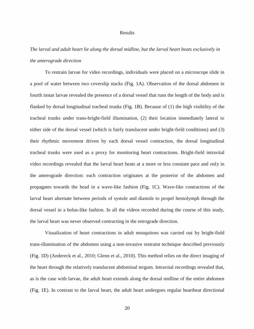
Results
The larval and adult heart lie along the dorsal midline, but the larval heart beats exclusively in
the anterograde direction
To restrain larvae for video recordings, individuals were placed on a microscope slide in
a pool of water between two coverslip stacks (Fig. 1A). Observation of the dorsal abdomen in
fourth instar larvae revealed the presence of a dorsal vessel that runs the length of the body and is
flanked by dorsal longitudinal tracheal trunks (Fig. 1B). Because of (1) the high visibility of the
tracheal trunks under trans-bright-field illumination, (2) their location immediately lateral to
either side of the dorsal vessel (which is fairly translucent under bright-field conditions) and (3)
their rhythmic movement driven by each dorsal vessel contraction, the dorsal longitudinal
tracheal trunks were used as a proxy for monitoring heart contractions. Bright-field intravital
video recordings revealed that the larval heart beats at a more or less constant pace and only in
the anterograde direction: each contraction originates at the posterior of the abdomen and
propagates towards the head in a wave-like fashion (Fig. 1C). Wave-like contractions of the
larval heart alternate between periods of systole and diastole to propel hemolymph through the
dorsal vessel in a bolus-like fashion. In all the videos recorded during the course of this study,
the larval heart was never observed contracting in the retrograde direction.
Visualization of heart contractions in adult mosquitoes was carried out by bright-field
trans-illumination of the abdomen using a non-invasive restraint technique described previously
(Fig. 1D) (Andereck et al., 2010; Glenn et al., 2010). This method relies on the direct imaging of
the heart through the relatively translucent abdominal tergum. Intravital recordings revealed that,
as is the case with larvae, the adult heart extends along the dorsal midline of the entire abdomen
(Fig. 1E). In contrast to the larval heart, the adult heart undergoes regular heartbeat directional

21
reversals, beating for longer periods of time in the anterograde direction but switching to the
retrograde direction at regular intervals (Fig. 1F). When compared with larvae, the adult heart
appears to constrict more narrowly during systole, particularly when contracting in the retrograde
direction.
22
Figure 1. Larval and adult heart contractions in Anopheles gambiae. (A) Larval restraint
technique for bright-field intravital video recording of heart contractions. Larvae were restrained
in water that was pooled between two stacks of coverslips. (B) Bright-field image of the larval
abdomen showing that the heart (arrowheads) is located between the dorsal longitudinal tracheal
trunks (arrows). (C) Graphical representation of larval heart contractions, where each peak
represents a contraction. Contractions from the middle third of the 60 s recording are magnified
in the lower graph. All larval contractions propagate in the anterograde direction. (D) Adult
restraint technique for bright-field intravital video recording of heart contractions. Adults were
cold-anesthetized and pins were placed through non-vascular portions of the wings and over the
neck. (E) Bright-field image of the adult abdomen showing the heart (arrowheads). (F) Graphical
representation of adult heart contractions, where each peak represents a heart contraction.
Contractions from the middle third of the 60 s recording are magnified in the lower graph.
Unshaded and shaded areas represent periods of anterograde and retrograde heart contraction,
respectively.
23
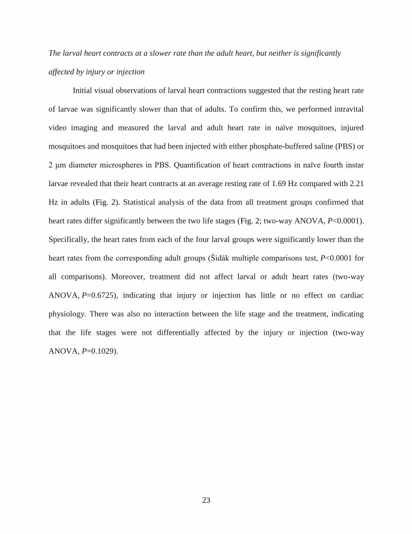
The larval heart contracts at a slower rate than the adult heart, but neither is significantly
affected by injury or injection
Initial visual observations of larval heart contractions suggested that the resting heart rate
of larvae was significantly slower than that of adults. To confirm this, we performed intravital
video imaging and measured the larval and adult heart rate in naïve mosquitoes, injured
mosquitoes and mosquitoes that had been injected with either phosphate-buffered saline (PBS) or
2 μm diameter microspheres in PBS. Quantification of heart contractions in naïve fourth instar
larvae revealed that their heart contracts at an average resting rate of 1.69 Hz compared with 2.21
Hz in adults (Fig. 2). Statistical analysis of the data from all treatment groups confirmed that
heart rates differ significantly between the two life stages (Fig. 2; two-way ANOVA, P<0.0001).
Specifically, the heart rates from each of the four larval groups were significantly lower than the
heart rates from the corresponding adult groups (Šidák multiple comparisons test, P<0.0001 for
all comparisons). Moreover, treatment did not affect larval or adult heart rates (two-way
ANOVA, P=0.6725), indicating that injury or injection has little or no effect on cardiac
physiology. There was also no interaction between the life stage and the treatment, indicating
that the life stages were not differentially affected by the injury or injection (two-way
ANOVA, P=0.1029).
24
Figure 2. Larval and adult heart contraction rates. Heart contraction rates were quantified in
larvae and adults after receiving no treatment (naïve), a needle wound (injury), an injection with
PBS, or an injection with 2 μm fluorescent microspheres in PBS. Across all comparisons, larvae
displayed lower heart rates than adults (two-way ANOVA, P<0.0001), but treatment did not
affect heart rates (P=0.6725). Whiskers denote the SEM.
25
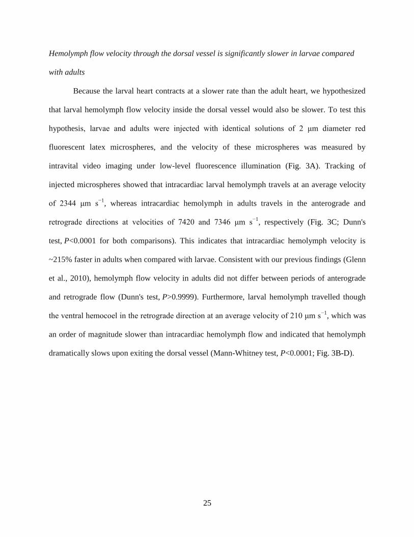
Hemolymph flow velocity through the dorsal vessel is significantly slower in larvae compared
with adults
Because the larval heart contracts at a slower rate than the adult heart, we hypothesized
that larval hemolymph flow velocity inside the dorsal vessel would also be slower. To test this
hypothesis, larvae and adults were injected with identical solutions of 2 μm diameter red
fluorescent latex microspheres, and the velocity of these microspheres was measured by
intravital video imaging under low-level fluorescence illumination (Fig. 3A). Tracking of
injected microspheres showed that intracardiac larval hemolymph travels at an average velocity
of 2344 μm s−1
, whereas intracardiac hemolymph in adults travels in the anterograde and
retrograde directions at velocities of 7420 and 7346 μm s−1
, respectively (Fig. 3C; Dunn's
test, P<0.0001 for both comparisons). This indicates that intracardiac hemolymph velocity is
~215% faster in adults when compared with larvae. Consistent with our previous findings (Glenn
et al., 2010), hemolymph flow velocity in adults did not differ between periods of anterograde
and retrograde flow (Dunn's test, P>0.9999). Furthermore, larval hemolymph travelled though
the ventral hemocoel in the retrograde direction at an average velocity of 210 μm s−1
, which was
an order of magnitude slower than intracardiac hemolymph flow and indicated that hemolymph
dramatically slows upon exiting the dorsal vessel (Mann-Whitney test, P<0.0001; Fig. 3B-D).
26
Figure 3. Larval and adult hemolymph flow velocity. (A) Diagrammatic representation
(dorsal view; anterior on top) of intracardiac hemolymph flow in larvae (left) and the adult
abdomen (middle and right). Hemolymph inside the larval heart is only propelled in the
anterograde direction (AG; red arrow) whereas hemolymph is propelled across the adult heart in
both anterograde (red arrow) and retrograde (RG; blue arrow) directions. (B) Diagrammatic
representation (ventral view) of extracardiac hemolymph flow in the larval ventral abdomen,
showing that hemolymph only moves in the retrograde direction (blue arrows). (C) Hemolymph
velocity in the heart of larvae and adults, as determined by the tracking of neutral density
fluorescent microspheres. Intracardiac (anterograde) hemolymph in larvae travels significantly
more slowly than intracardiac (anterograde and retrograde) hemolymph in adults. (D)
Extracardiac retrograde hemolymph flow in the ventral abdomen of larvae. In larvae,
extracardiac flow is significantly slower than intracardiac flow. For box plots, the centerline
marks the median, the box marks 50% of the data, and the whiskers mark 90% of the data.
27
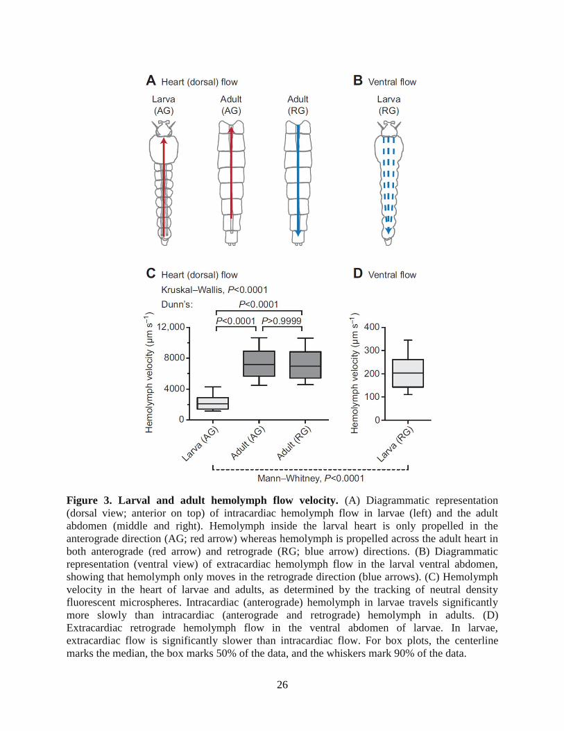
The larval heart is structurally similar to the adult heart, but its associated abdominal wall
musculature and alary muscles differ drastically
The differences observed in larval and adult heart physiology probably result from
underlying structural changes that occur during metamorphosis. To uncover these changes, we
performed a structural analysis of the musculature associated with the dorsal abdomen of both
larvae and adults. Specifically, we treated the dorsal abdomens with Alexa Fluor-conjugated
phalloidin, which binds F-actin, and visualized the specimens by fluorescence microscopy.
Muscle staining of the larval abdomen revealed a dorsal vessel consisting of an
abdominal heart and a thoracic aorta that spans the length of the body along the dorsal midline
(Fig. 4A). The larval heart is nestled within a thin dorsal cavity that is reminiscent of the adult
pericardial sinus and is created by massive, segmentally arranged swim muscles that flank the
heart and intersect it at nearly 45 deg angles (Fig. 4A-C). Under low magnification, the larval
heart was often difficult to distinguish from the surrounding tissues because the fluorescence
emitted by the heart, though similar to that of adults, was nearly overwhelmed by that of the
surrounding swim muscles (Fig. 4A, B). The larval heart muscle is composed of a single layer of
spirally arranged cardiomyocytes which, when viewed from the ventral side, form a clockwise,
left-handed helical twist with respect to the lumen of the vessel (Fig. 4C). Three-dimensional
rendering of deconvolved Z-stacks showed that the larval heart is dorsal and medial to the
longitudinal tracheal trunks.
28
Figure 4. Larval and adult heart structure. All images are fluorescence micrographs of
specimens treated with Alexa Fluor conjugated phalloidin, which labels muscles green. (A)
Image of the larval dorsal thorax and abdomen showing that the dorsal vessel extends the length
of the body along the dorsal midline and is subdivided into an abdominal heart (arrows) and a
thoracic aorta (arrowheads). The dashed line delineates the thoraco-abdominal junction. (B) A
portion of the larval abdomen illustrated in panel A, magnified to show the heart (arrows) and
associated alary muscles (e.g. arrowheads). The larval heart is flanked on either side by large
swim muscles that approach the heart at ~45 deg angles. (C) High magnification view of two
segments of the larval heart (arrows) showing the spiral arrangement of cardiomyocytes, the
dorsal longitudinal tracheal trunks (T) and the alary muscles (arrowheads). Also visible along the
surface of the heart are numerous pericardial cells. (D) Adult dorsal abdomen showing the heart
(arrows), which runs the length of the abdomen along the dorsal midline. (E) A portion of the
adult abdomen magnified to show the heart (arrows) and the extensive alary muscles
(arrowheads). All images are oriented with anterior on top. Scale bars: A and D, 500 μm; B and
E, 200 μm; C, 100 μm.
Extending towards the dorsal midline from areas near the tergum-pleuron junction at a
location that is immediately posterior to each abdominal suture are six complete and three
incomplete pairs of bilaterally symmetrical alary muscles (Fig. 4B, C). These muscles attach to
29
the heart on either side of the abdominal suture and tether it to the dorsal abdominal cuticle. The
complete alary muscle pairs are located in abdominal segments 2-7. One incomplete alary
muscle pair is located at the thoraco-abdominal junction and the other two are present at the
posterior suture where the seventh and eighth abdominal segments are joined. Each alary muscle
branches once, and each branch splits again to form two connections at the ventrolateral surface
of the heart at a location near the pericardial cells. The points where the alary muscles connect to
the heart were usually too tenuous to remain intact in our mounted specimens, but using a
different imaging technique other researchers have observed the connection between the alary
muscles and the heart of Aedes aegypti larvae (Leódido et al., 2013).
Consistent with our video recordings, muscle staining revealed that the larval and adult
heart lie in the same location and span the same length along the dorsal midline of the abdomen
(Fig. 4). However, comparative analyses revealed a significant disparity in overall abdominal
musculature between the larva and adult stages. Specifically, compared with the larval swim
muscles, the adult abdomen displays a significantly smaller array of intrasegmental lateral
muscle fibers, which are oriented at 90 deg angles with respect to the heart (Fig. 4D, E). In both
larvae and adults the structure of the heart varied depending on the contraction state at the time
of fixation. However, the spiral arrangement of cardiomyocytes was similar in both life stages
(Fig. 4).
Although the alary muscles of larvae and adults share the same point of origin at the body
wall, alary muscle connections to the heart are far more extensive in adults when compared with
larvae. In adults, each alary muscle branches once and then divides again to form anywhere from
10 to >30 myofiber connections to the heart. So extensive are these connections in adults that the
anterior-most connection of one alary muscle extends to the posterior-most connection of the
30
alary muscle located in the adjacent abdominal segment (Fig. 4D, E). Together, these structures
form the basket-like muscular network that comprises the incomplete dorsal diaphragm in adults,
a structure that is essentially absent in larvae due to their immature alary muscles.
Larval and adult abdominal ostia are in the same location and display a similar structure
The larval heart contains paired ostia, or valves, at the anterior portion of abdominal
segments 2-7. An additional pair of ostia is located at the thoraco-abdominal junction, where the
heart joins the aorta (discussed below). The ostia are located on the lateral sides of the heart near
the anterior of each abdominal segment (Fig. 5A-C). Near each ostium is the junction of the
posterior branch of each alary muscle, and each ostium is flanked ventrally by a pair of large
pericardial cells (Fig. 4C; Fig. 5A). The heart muscle near the ostia typically bulges out slightly,
giving the periostial regions a slightly wider diameter than other regions of the heart, perhaps due
to increased tension created by the attachment of the alary muscles. Each ostium consists of two
specialized cardiomyocytes that contain prominent nuclei and form funnel-shaped lips that
extend into the lumen of the vessel (Fig. 5B, C).
31
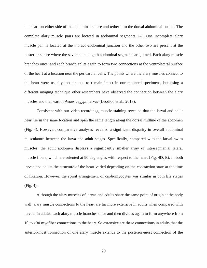
Figure 5. Larval and adult abdominal ostia. Phalloidin stained larval (A-C) and adult (D-F)
hearts showing the location and structure of the ostia. (A) Larval ostia (arrows) are located on the
lateral sides of the heart in between and dorsal to a pair of enlarged pericardial cells
(arrowheads). (B) Ostia (arrows) are oriented towards the anterior of the organism as they extend
into the lumen of the vessel. (C) Each ostium (arrows) is composed of a pair of cardiomyocytes
(nuclei are labelled blue with Hoechst 33342; circles). (D) Adult ostia (arrows) are located on the
lateral sides of the heart near the posterior of each alary muscle pair. Pericardial cells are present
but are not labelled. (E) Ostia (arrows) are oriented towards the anterior of the organism as they
extend into the lumen of the vessel. (F) Each ostium is composed of two cardiomyocytes (nuclei
are labelled blue; circles). Note how the ostia extend into the lumen of the vessel (arrows). All
images are oriented with anterior at the top, and the abdominal sutures are marked with a dashed
line. Scale bars: A and D, 50 μm; B, C, E and F, 50 μm.
The ostia of adults are also positioned at the anterior portions of abdominal segments 2-7
(Fig. 5D-F), suggesting that all of the larval abdominal ostia are maintained into adulthood.
Likewise, adult abdominal ostia form funnel-shaped lips that protrude into the heart lumen in an
anterior direction and contain paired nuclei (Fig. 5E, F). Although the muscle that comprises
these lips was occasionally observed in larval ostia, it was significantly more prominent in
32
adults. Furthermore, larval ostial nuclei were typically found closer to the heart wall rather than
projecting into the lumen.
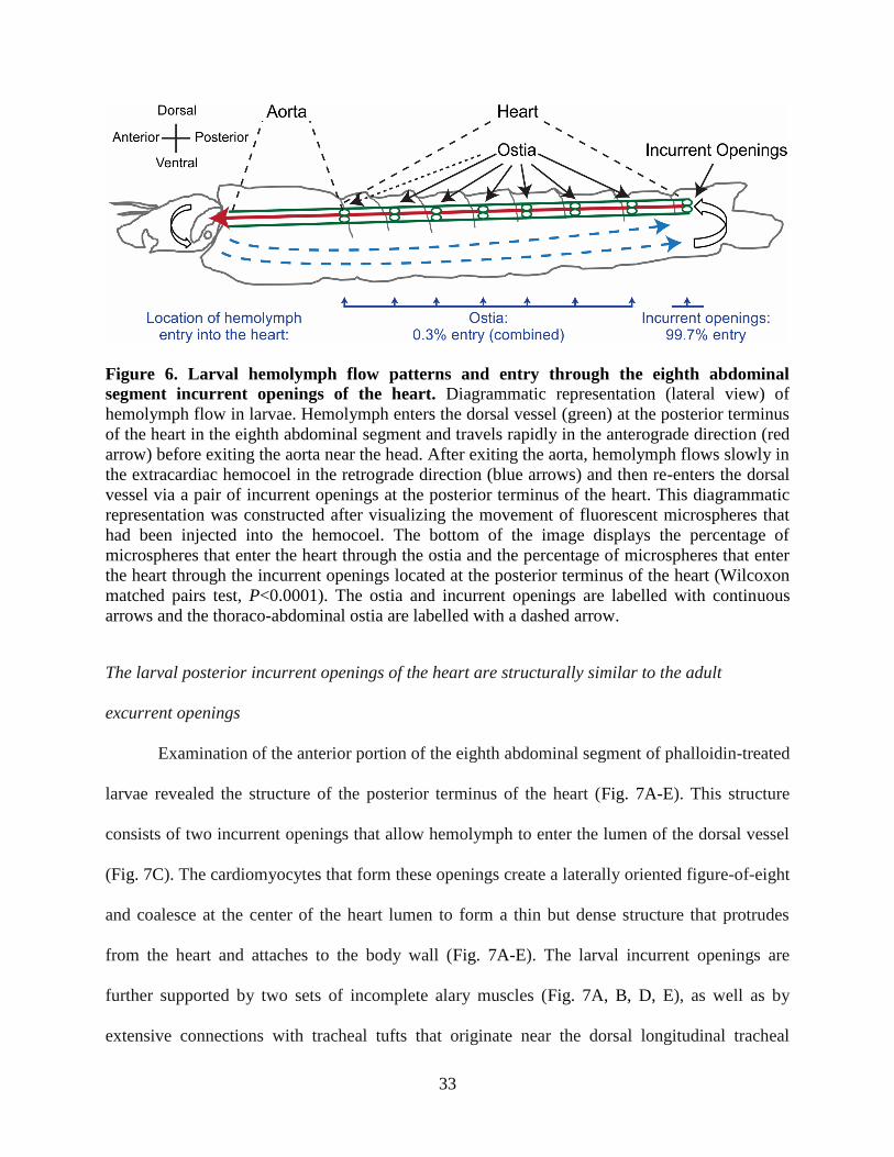
Larval hemolymph enters the dorsal vessel primarily through incurrent openings in the eighth
abdominal segment and not the abdominal ostia
Intravital video imaging revealed that 2 μm diameter red fluorescent microspheres
injected into the larval ventral hemocoel migrate slowly in the retrograde direction, bypass the
abdominal ostia and enter the dorsal vessel at the posterior terminus of the heart (Fig. 3B). To
gauge the amount of hemolymph that enters the larval heart through the terminal opening in the
eighth abdominal segment relative to the ostia located in abdominal segments 2-7, microspheres
were injected into the larval thorax and the number of microspheres that entered the dorsal vessel
within each abdominal segment over a 60 s time period was quantified. Although microspheres
were occasionally observed entering the heart through the abdominal ostia, the vast majority of
microspheres (>99%) entered the dorsal vessel through the terminal openings of the heart in the
eighth abdominal segment (Wilcoxon matched pairs test, P<0.0001; Fig. 6). Even when injecting
larvae with higher concentrations of fluorescent microspheres, regardless of the site of injection,
the microspheres consistently migrated posteriorly and only entered the dorsal vessel after
reaching the eighth abdominal segment.
33
Figure 6. Larval hemolymph flow patterns and entry through the eighth abdominal
segment incurrent openings of the heart. Diagrammatic representation (lateral view) of
hemolymph flow in larvae. Hemolymph enters the dorsal vessel (green) at the posterior terminus
of the heart in the eighth abdominal segment and travels rapidly in the anterograde direction (red
arrow) before exiting the aorta near the head. After exiting the aorta, hemolymph flows slowly in
the extracardiac hemocoel in the retrograde direction (blue arrows) and then re-enters the dorsal
vessel via a pair of incurrent openings at the posterior terminus of the heart. This diagrammatic
representation was constructed after visualizing the movement of fluorescent microspheres that
had been injected into the hemocoel. The bottom of the image displays the percentage of
microspheres that enter the heart through the ostia and the percentage of microspheres that enter
the heart through the incurrent openings located at the posterior terminus of the heart (Wilcoxon
matched pairs test, P<0.0001). The ostia and incurrent openings are labelled with continuous
arrows and the thoraco-abdominal ostia are labelled with a dashed arrow.
The larval posterior incurrent openings of the heart are structurally similar to the adult
excurrent openings
Examination of the anterior portion of the eighth abdominal segment of phalloidin-treated
larvae revealed the structure of the posterior terminus of the heart (Fig. 7A-E). This structure
consists of two incurrent openings that allow hemolymph to enter the lumen of the dorsal vessel
(Fig. 7C). The cardiomyocytes that form these openings create a laterally oriented figure-of-eight
and coalesce at the center of the heart lumen to form a thin but dense structure that protrudes
from the heart and attaches to the body wall (Fig. 7A-E). The larval incurrent openings are
further supported by two sets of incomplete alary muscles (Fig. 7A, B, D, E), as well as by
extensive connections with tracheal tufts that originate near the dorsal longitudinal tracheal

34
trunks at the posterior of the eighth abdominal segment (Fig. 7D-F). The tracheal tufts are a
dense mass of thin trachea that is suspended in the hemolymph and shifts with each heart
contraction.
35
Figure 7. Larval and adult posterior heart structure. (A-C) Image series showing, in
increasing magnification, phalloidin staining of the larval posterior incurrent openings of the
heart. The posterior terminus of the larval heart ends in the anterior portion of the eighth
abdominal segment, lies between the dorsal longitudinal tracheal trunks (T), and is supported by
two incomplete pairs of alary muscles (white arrowheads). The posterior terminus of the larval
heart contains two incurrent openings (arrows) that contain a muscular tether (yellow arrowhead)
that attaches the heart to the abdominal wall. (D-F) Series of fluorescence (D, E) and bright-field
(F; same specimen as E) images showing that the posterior terminus of the larval heart is also
attached to an extensive network of thin tracheoles called the tracheal tufts (TT), which extend
from a location near the posterior base of the dorsal longitudinal tracheal trunks (T). (G-I) Image
series showing, in increasing magnification, phalloidin staining of the adult posterior excurrent
openings of the heart. The posterior terminus of the adult heart ends in the anterior portion of the
eighth abdominal segment and is supported by two incomplete pairs of alary muscles (white
arrowheads). The posterior terminus of the adult heart contains two excurrent openings (arrows)
that contain a muscular tether (yellow arrowhead) that attaches the heart to the abdominal wall.
All images are oriented with anterior at the top. Scale bars: A, D and G, 100 μm; B, E, F and H,
50 μm; C and I, 20 μm.
36
The posterior terminus of the adult heart is exclusively excurrent (Glenn et al., 2010), and
thus serves the opposite function to the one found in larvae. The adult posterior heart shares the
same paired openings as the larval posterior heart, as well as a similar point of attachment to the
body wall (Fig. 7G-I). Also similar to larvae, the adult posterior terminus of the heart is
supported by two sets of incomplete alary muscles, although, as is the case with the other alary
muscles, these form more extensive connections to the heart in adults when compared with
larvae.
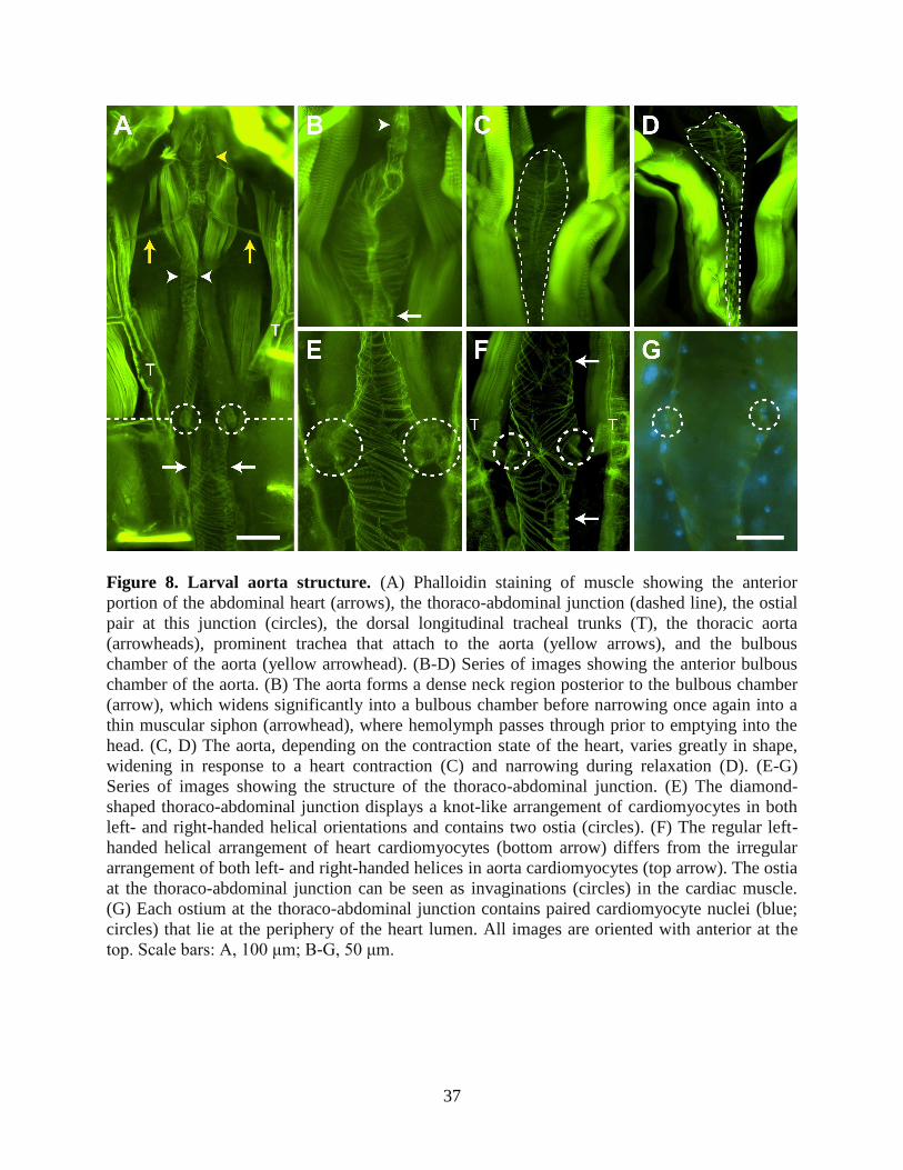
The larval aorta extends from the thoraco-abdominal junction to the base of the head and differs
in structure from the heart
Imaging of the larval thoracic musculature after phalloidin staining revealed the presence
of a thoracic aorta that extends from the thoraco-abdominal junction to the anterior portion of the
thorax (Fig. 8A). The shape of the aorta varies substantially depending on the contraction state of
the heart; during contraction, the aorta widens (Fig. 8B, C), while during relaxation the aorta
narrows (Fig. 8D). Although no alary muscle attachments were observed supporting the aorta,
two prominent trachea that originate from the longitudinal tracheal trunks were seen attached to
the anterior portion of the vessel, perhaps providing structural support in addition to meeting
oxygen demands (Fig. 8A).
37
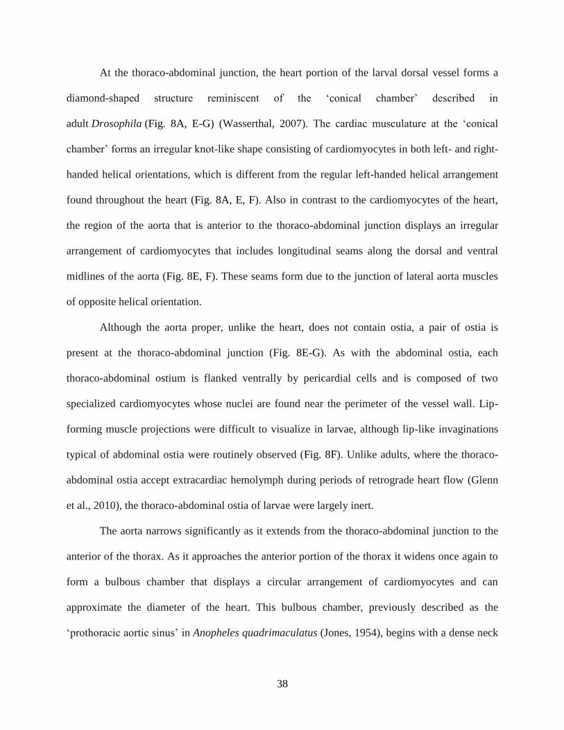
Figure 8. Larval aorta structure. (A) Phalloidin staining of muscle showing the anterior
portion of the abdominal heart (arrows), the thoraco-abdominal junction (dashed line), the ostial
pair at this junction (circles), the dorsal longitudinal tracheal trunks (T), the thoracic aorta
(arrowheads), prominent trachea that attach to the aorta (yellow arrows), and the bulbous
chamber of the aorta (yellow arrowhead). (B-D) Series of images showing the anterior bulbous
chamber of the aorta. (B) The aorta forms a dense neck region posterior to the bulbous chamber
(arrow), which widens significantly into a bulbous chamber before narrowing once again into a
thin muscular siphon (arrowhead), where hemolymph passes through prior to emptying into the
head. (C, D) The aorta, depending on the contraction state of the heart, varies greatly in shape,
widening in response to a heart contraction (C) and narrowing during relaxation (D). (E-G)
Series of images showing the structure of the thoraco-abdominal junction. (E) The diamond-
shaped thoraco-abdominal junction displays a knot-like arrangement of cardiomyocytes in both
left- and right-handed helical orientations and contains two ostia (circles). (F) The regular left-
handed helical arrangement of heart cardiomyocytes (bottom arrow) differs from the irregular
arrangement of both left- and right-handed helices in aorta cardiomyocytes (top arrow). The ostia
at the thoraco-abdominal junction can be seen as invaginations (circles) in the cardiac muscle.
(G) Each ostium at the thoraco-abdominal junction contains paired cardiomyocyte nuclei (blue;
circles) that lie at the periphery of the heart lumen. All images are oriented with anterior at the
top. Scale bars: A, 100 μm; B-G, 50 μm.
38
At the thoraco-abdominal junction, the heart portion of the larval dorsal vessel forms a
diamond-shaped structure reminiscent of the ‘conical chamber’ described in
adult Drosophila (Fig. 8A, E-G) (Wasserthal, 2007). The cardiac musculature at the ‘conical
chamber’ forms an irregular knot-like shape consisting of cardiomyocytes in both left- and right-
handed helical orientations, which is different from the regular left-handed helical arrangement
found throughout the heart (Fig. 8A, E, F). Also in contrast to the cardiomyocytes of the heart,
the region of the aorta that is anterior to the thoraco-abdominal junction displays an irregular
arrangement of cardiomyocytes that includes longitudinal seams along the dorsal and ventral
midlines of the aorta (Fig. 8E, F). These seams form due to the junction of lateral aorta muscles
of opposite helical orientation.
Although the aorta proper, unlike the heart, does not contain ostia, a pair of ostia is
present at the thoraco-abdominal junction (Fig. 8E-G). As with the abdominal ostia, each
thoraco-abdominal ostium is flanked ventrally by pericardial cells and is composed of two
specialized cardiomyocytes whose nuclei are found near the perimeter of the vessel wall. Lip-
forming muscle projections were difficult to visualize in larvae, although lip-like invaginations
typical of abdominal ostia were routinely observed (Fig. 8F). Unlike adults, where the thoraco-
abdominal ostia accept extracardiac hemolymph during periods of retrograde heart flow (Glenn
et al., 2010), the thoraco-abdominal ostia of larvae were largely inert.
The aorta narrows significantly as it extends from the thoraco-abdominal junction to the
anterior of the thorax. As it approaches the anterior portion of the thorax it widens once again to
form a bulbous chamber that displays a circular arrangement of cardiomyocytes and can
approximate the diameter of the heart. This bulbous chamber, previously described as the
‘prothoracic aortic sinus’ in Anopheles quadrimaculatus (Jones, 1954), begins with a dense neck
39
region before widening and then tapering off ventrally into a narrow, muscular siphon, which
hemolymph passes through just prior to exiting into the head (Fig. 8A, B). Intravital imaging
near the base of the head confirmed the presence of the excurrent opening of the aorta, which
releases hemolymph into the head where it remains briefly before gradually flowing in the
posterior direction. Although hemolymph flow patterns in adults suggest a similar location and
function of the adult aorta and anterior excurrent opening, no reliable images of these structures
have been obtained (Glenn et al., 2010).
Discussion
The comparative structural and functional data presented herein advance our
understanding of the insect circulatory system by providing novel insights into its development
across the life stages of the African malaria mosquito, A. gambiae. Overall, the larval pattern of
unidirectional heart contraction and hemolymph flow reflects a simpler version of the adult
circulatory system, a difference in complexity that is probably a function of the more streamlined
body plan and overall morphology of the larval life stages. Likewise, the increased complexity of
the adult circulatory system may reflect adaptations to the morphology and physiological
demands specific to the adult stage, such as those involved in flight, blood feeding, and
reproduction.
In this study we found that while the adult heart contracts in both the anterograde and
retrograde directions, the larval heart only contracts in an anterograde direction. Unidirectional
anterograde heart contraction is a common feature of insect larvae (Dulcis et al., 2001; Gerould,
1924; Jones, 1954; Jones, 1977; Miller et al., 1985; Sláma and Farkas, 2005; Smits et al., 2000).
Heartbeat directional reversals, however, have been observed primarily in adult insects from a
variety of holometabolous orders, with contraction reversals generally beginning either at the

40
adult stage or around the time of pupation (Gerould, 1933; Miller, 1997). Although the late-stage
larvae of some insects undergo heartbeat reversals prior to pupation (Gerould, 1933; Matsushita
et al., 2002; Uchimura et al., 2006), we never observed mosquito larvae, even when nearing
pupation, undergoing heartbeat directional reversals. The reason mosquito adults, but not larvae,
undergo periodic heartbeat directional reversals remains unknown, but in other insects several
environmental and neural stimuli are known to control contraction directionality. For example,
the larval heart of Manduca sexta contracts only in the anterograde direction whereas the pupal
and adult heart periodically reverses contraction direction (Davis et al., 2001). The rhythmicity
of adult heartbeat reversals is altered by tactile stimuli, eliminated by the treatment with
tetrodotoxin (a neurotoxin), and controlled by specific motor neurons (Dulcis et al., 2001).
Neural and hormonal stimuli also modulate the proportional directionality of heart contractions
in other insects (Dulcis and Levine, 2005; Dulcis et al., 2005; Kuwasawa et al., 1999), indicating
that although the insect heart is myogenic (Chapman et al., 2013; Jones, 1977; Klowden, 2013),
contraction dynamics are under partial neural control. In D. melanogaster, anterograde heart
contractions are thought to be modulated by a putative pacemaker located at the posterior end of
the heart (Dulcis and Levine, 2003), whereas retrograde heart contractions are thought to be
modulated by a pacemaker located in the conical chamber, which represents the anterior-most
region of the heart (Dulcis and Levine, 2005). This conical chamber develops during
metamorphosis, and thus is not present in larvae (Curtis et al., 1999; Dulcis and Levine, 2003),
suggesting that the larval heart may lack the inherent ability to contract in the retrograde
direction. Indeed, the larval heart of most insects appears to lack direct innervation and the
formation of an anterior retrograde pacemaker in adults probably coincides with the formation of
new neuronal inputs during metamorphosis to coordinate the action of the anterograde and
41
retrograde pacemakers (Davis et al., 2001; Dulcis et al., 2001; Dulcis and Levine, 2003; Dulcis
and Levine, 2004; Dulcis and Levine, 2005). This notion is consistent with the findings of the
present study as well as our recent studies that have shown that the adult heart of A. gambiae is
both innervated and under partial hormonal control, with the identified cardiomyotropic
neuropeptides having a stronger effect during periods of anterograde heart contraction (Chen and
Hillyer, 2013; Estévez-Lao et al., 2013; Hillyer et al., 2014).
The finding that the larval mosquito heart contracts more slowly than the adult heart is
consistent with earlier findings in A. quadrimaculatus (Jones, 1954) as well as the increased ‘fast
forward’ phase beating that occurs in M. sexta following eclosion (Dulcis et al., 2001; Smits et
al., 2000). Similarly, the heart rate of Drosophila adults is approximately double that of the third
instar larvae (Sláma and Farkas, 2005). We also observed that hemolymph flow velocity was
slower in larvae compared with adults, with intracardiac larval hemolymph travelling at less than
one-third of the speed of intracardiac hemolymph flow in adults. Because this slower flow
velocity correlates with a slower contraction rate in larva when compared with adults, it appears
that the more rapid heart rate in adults allows this life stage to propel hemolymph at vastly
greater velocities than larvae. Furthermore, as systole appears to constrict the heart of adults to a
greater degree than occurs in larvae, the increased reduction in heart luminal volume that
presumably accompanies each adult contraction may explain why the developmentally
associated increase in intracardiac hemolymph velocity is much greater than the increase in the
heart rate (~215% versus ~30%, respectively).
The relative paucity of abdominal body wall musculature in adults compared with larvae
indicates a drastic reduction in muscle mass after metamorphosis. However, despite lying in the
center of and in close proximity to these abdominal wall muscles, both the larval and adult
42
hearts: (1) are composed of a thoracic aorta and an abdominal heart; (2) span the same length
along the dorsal midline of the abdomen; (3) display the same spiral arrangement of
cardiomyocytes; (4) contain the same number of ostia; and (5) retain ostia in the same position
and at regular intervals that closely align with the anterior portion of each abdominal segment.
Together with the fact that larvae and adults have the same number of abdominal segments, these
findings suggest that all portions of the larval heart are maintained following eclosion. In
contrast to this high degree of continuity, the adult dorsal vessel in Drosophila differs greatly
from its larval predecessor in each of the aforementioned areas: (1) unlike adults, the larval aorta
spans both thoracic and abdominal segments (T3 to A5) (Curtis et al., 1999; Molina and Cripps,
2001; Wasserthal, 2007); (2) the posterior two segments of the larval heart are lost during
metamorphosis (Curtis et al., 1999; Lehmacher et al., 2012; Molina and Cripps, 2001); (3) unlike
the regular circular arrangement of adult cardiomyocytes, the larval heart contains a mixture of
both circular and helically oriented muscles (Lehmacher et al., 2012); (4) only the first of the
three larval ostial pairs are retained into adulthood, with four additional pairs arising during
metamorphosis from progenitor cells in the larval aorta (Curtis et al., 1999; Molina and Cripps,
2001; Wasserthal, 2007); and (5) the ostia and the alary muscles of both larvae and adults are not
evenly spaced across the abdominal segments. This extensive restructuring of
the Drosophila dorsal vessel, which is absent in mosquitoes, may be due to the greater disparity
between the larval and adult body plans of Drosophila when compared with A. gambiae, whose
larvae and adults, unlike Drosophila, both have a distinct head, thorax and abdomen.
In contrast to the relative stability of the heart across mosquito life stages, the supporting alary
muscles are grossly under-developed in larvae when compared with adults, an observation
recently made across life stages in A. aegypti (Leódido et al., 2013) that is also consistent with
43
structural data from adults of other mosquito species (Martins et al., 2011). This disparity in
alary muscle support may partly account for the differences in contraction and hemolymph flow
dynamics observed between the two life stages, as alary muscles are believed to not only aid in
the relaxation of the heart muscle during diastole (Lehmacher et al., 2012) but also to facilitate
the opening and closing of the ostia (Glenn et al., 2010). During our particle-tracking
experiments we observed that nearly all of the injected microspheres entered the lumen of the
larval dorsal vessel through the posterior incurrent openings of the heart instead of the ostia, a
finding that is in agreement with indirect observations in A. quadrimaculatus larvae (Jones,
1954). Although larval ostia typically contained lips that were less prominent than those of adults
and lay close to the perimeter of the heart, thus resembling ostia in the ‘closed’ position, the
orientation of larval ostia towards the anterior of the insect is consistent with their eventual
function in the adult stage as incurrent valves for hemolymph entry into the heart during periods
of anterograde heart contractions (Glenn et al., 2010; Jones, 1977). Furthermore, as microspheres
occasionally (though rarely) entered the larval heart through the abdominal ostia, we hypothesize
that the developmental differences in the mechanics of hemolymph entry into the dorsal vessel
are not due to major stage-specific structural differences in the ostia themselves but instead due
to differences in the degree of nearby alary muscle support. Indeed, the ostia described here
appear to be both developmentally and evolutionarily conserved, as structurally similar ostia
have been described in both the larval and adult stages of A. aegypti (Leódido et al., 2013)
and Drosophila (Curtis et al., 1999; Lehmacher et al., 2012; Molina and Cripps, 2001;
Wasserthal, 2007).
We have also shown that the function of the posterior terminus of the heart changes with
developmental stage: larvae have incurrent openings whereas adults have excurrent openings.
44
Incurrent function of the posterior larval heart has been inferred from observations in A.
quadrimaculatus (Jones, 1954), and described in Drosophila (Babcock et al., 2008), as well as in
some lepidopteran larvae (Locke, 1997; Rao et al., 2009). Each of these insect orders contains
several known examples of adults with excurrent posterior heart function, suggesting that the
functional change of the posterior terminus of the heart is evolutionarily conserved (Glenn et al.,
2010; Wasserthal, 1980; Wasserthal, 1981; Wasserthal, 2007; Wasserthal, 2012). In A. gambiae,
the posterior end of the heart is structurally similar in both larvae and adults, and the excurrent
function that develops following eclosion is a direct result of the onset of retrograde heart
contraction periods. The reason the posterior opening of the adult heart does not retain incurrent
function even during periods of anterograde heart contractions remains unknown, but it is
possible that the high efficiency of the adult abdominal ostia, which are inert in the larvae, has
rendered posterior incurrent function unnecessary.
This study also provides the first structural description of the aorta of A. gambiae, as this
portion of the dorsal vessel has proved difficult to visualize in adults due to its location in the
highly sclerotized thorax (Glenn et al., 2010). The aorta differs significantly from the heart in the
arrangement of its cardiomyocytes; whereas the heart contains regular, spirally arranged
cardiomyocytes that form a left-handed helical twist, the aorta displays muscle cells arranged in
both right- and left-handed helices. This distinct arrangement of cardiomyocytes may contribute
to the capacity of the aorta for dramatic alterations in its diameter in response to heart
contractions. Although similar irregularities in cardiomyocyte orientation have been noted in the
heart and aorta of Drosophila larvae (Lehmacher et al., 2012), in mosquito larvae we found these
irregularities exclusively in the aorta. However, the narrow diameter of the mosquito aorta and
its lack of ostia are common features among insect aortas, including that
45
of Drosophila (Chapman et al., 2013; Jones, 1954; Lehmacher et al., 2012; Molina and Cripps,
2001; Wasserthal, 2007).
Hemolymph circulation is an essential yet understudied aspect of insect biology and has
been shown to influence thermoregulation, tracheal ventilation and the transport of nutrients,
waste, hormones and immune factors (Babcock et al., 2008; Harrison et al., 2012; King and
Hillyer, 2012; Klowden, 2013; Nation, 2008; Wasserthal, 1999; Wasserthal, 2003; Wasserthal,
2012). The present study on the circulatory system of A. gambiae, a primary disease vector of
malaria in sub-Saharan Africa, increases our understanding of key physiological processes in this
epidemiologically important mosquito. Specifically, the data presented herein provide novel
insights into larval circulatory physiology and describe numerous differences between the larval
and adult dorsal vessel, providing a developmental perspective on this organ system. Larvae are
a common target in mosquito control strategies (Fillinger and Lindsay, 2011) and many chemical
and biological pesticides exert their effect at the larval stage after penetrating the cuticle or
midgut and circulating throughout the hemocoel (Favia et al., 2007; Otieno-Ayayo et al., 2008;
Paily et al., 2012). Furthermore, numerous mosquito-borne pathogens must complete an obligate
migration from the midgut to the salivary glands that involves traversing the hemocoel, where
they are subject to hemolymph flow currents (Hillyer et al., 2007). Thus understanding both
larval and adult circulatory physiology can yield important insights into current control methods
as well as inform the creation of novel ones.
46
CHAPTER III
FUNCTIONAL INTEGRATION OF THE CIRCULATORY, IMMUNE, AND RESPIRATORY
SYSTEMS IN MOSQUITO LARVAE: PATHOGEN KILLING IN THE HEMOCYTE-RICH
TRACHEAL TUFTS
Preface
This chapter builds on the findings pertaining to the structure and physiology of the larval
circulatory system that are described in Chapter II by identifying the mosquito larval tracheal
tufts as the main site of immune activity in larvae. This finding is significant because immune
responses at the tracheal tufts occur at the sole region of hemolymph flow into the heart, and
thus, it shows the functional integration of the circulatory and immune systems of mosquito
larvae. I carried out the experiments presented in this chapter, which involved adapting injection,
staining, imaging, and analytic techniques to larvae. All experiments were designed, analyzed,
and written with my advisor, Dr. Julián Hillyer. I thank Dr. Patrick Abbot, Dr. Lisa Brown, Leah
Sigle, and Tania Estévez-Lao for providing critical feedback during the writing of this
manuscript. This chapter is adapted from the final transcript of this work, which was published in
September of 2016 in the journal BMC Biology (volume 14, article 78).
Abstract
As both larvae and adults, mosquitoes encounter a barrage of immune insults, ranging
from microbe-rich communities in larval habitats to ingested blood-borne pathogens in adult
blood meals. Given that mosquito adults have evolved an efficient means of eliminating
infections in their hemocoel (body cavity) via the coordinated action of their immune and
47
circulatory systems, the goal of the present study was to determine whether such functional
integration is also present in larvae. By fluorescently labeling hemocytes (immune cells),
pericardial cells, and the heart, we discovered that fourth instar larvae, unlike adults, contain
segmental hemocytes but lack the periostial hemocytes that surround the ostia (heart valves) in
abdominal segments 2-7. Instead, larvae contain an abundance of sessile hemocytes at the
tracheal tufts, which are respiratory structures that are unique to larvae, are located in the
posterior-most abdominal segment, and surround what in larvae are the sole incurrent openings
for hemolymph entry into the heart. Injection of fluorescent immune elicitors and bacteria into
the larval hemocoel then showed that tracheal tuft hemocytes mount rapid and robust immune
responses against foreign insults. Indeed, green fluorescent protein-labeled Escherichia
coli flowing with the hemolymph rapidly aggregate exclusively at the tracheal tufts, where they
are killed within 24 h post-infection via both phagocytosis and melanization. Together, these
findings show that the functional integration of the circulatory, respiratory, and immune systems
of mosquitoes varies drastically across life stages.
Introduction
Insects have evolved powerful innate immune responses to neutralize infectious agents
(Chapman et al., 2013; Hillyer, 2016). Pathogens, including bacteria, viruses, malaria parasites,
and fungi, invade the insect hemocoel (body cavity) after ingestion and penetration of the
midgut, through breaches in the cuticle, or through the tracheal system. Upon entering the
hemocoel, cellular and humoral immune factors kill pathogens by phagocytosis, melanization,
lysis, and other mechanisms (Hillyer, 2010; Hillyer, 2016). The primary immune cells that drive
these processes are the hemocytes, which are found both circulating with the hemolymph and
attached to tissues (sessile) (Hillyer and Strand, 2014; Lavine and Strand, 2002; Strand, 2008).
48
Immune responses in the hemocoel occur in a dynamic space, where hemocytes, humoral
immune factors, and pathogens are propelled through the body by the action of an open
circulatory system (Hillyer, 2015; Hillyer, 2016). This circulation is primarily mediated by the
contractile action of a muscular dorsal vessel that comprises two distinct regions: the abdominal
heart and the thoracic aorta (Chapman et al., 2013; Klowden, 2013). Because circulatory currents
move both pathogens and immune components, functional integration is expected to exist
between the immune and circulatory systems. Such integration was recently documented in the
adult stage of the malaria mosquito A. gambiae, where cellular immune responses are rapid and
inducible on the surface of the heart (King and Hillyer, 2012). Specifically, in adult mosquitoes,
hemolymph enters the lumen of the dorsal vessel via valves, called ostia, which are located in the
anterior portion of abdominal segments 2-7 (Glenn et al., 2010; League et al., 2015). A
population of sessile hemocytes, called periostial hemocytes, is always present on the surface of
the heart in the regions that flank the ostia, where they phagocytose pathogens as they are swept
with the hemolymph toward the heart (King and Hillyer, 2012; King and Hillyer, 2013; Sigle and
Hillyer, 2016). This initial capture of circulating pathogens induces the migration of additional
hemocytes to each periostial region, where they aggregate with the initial population of first
responders and amplify the phagocytosis response. Although heart-associated immune
aggregations have been observed in multiple mosquito studies (Hernández-Martínez et al., 2013;
Hillyer et al., 2007; King and Hillyer, 2012; King and Hillyer, 2013; Michel et al., 2005;
Schnitger et al., 2007; Sigle and Hillyer, 2016; Yassine et al., 2014; Yassine et al., 2012), they
are not unique to this group of insects. Such aggregations have also been directly or indirectly
observed in Drosophila melanogaster adults (Akbar et al., 2011; Chambers et al., 2012; Cuttell
et al., 2008; Elrod-Erickson et al., 2000; Ghosh et al., 2015; Kocks et al., 2005; Shiratsuchi et al.,
49
2012; Stone et al., 2012), though these studies did not seek to link immune responses with the
functional mechanics of the heart or with hemolymph flow.
To date, no study has examined whether similar interactions between the immune and
circulatory systems occur in insect larvae. However, prior to the initiation of this work, several
lines of evidence suggested that any such interactions, if they were to exist in mosquito larvae,
would differ from what is seen in adults (Glenn et al., 2010; League et al., 2015). First,
intracardiac hemolymph in larvae flows solely in the anterograde direction (toward the head), as
the larval heart, unlike the adult heart, does not undergo heartbeat directional reversals (Fig. 1).
Second, although the ostia of larvae and adults are similar in number, structure, and position,
larval ostia are inert and do not serve as incurrent (inflow) valves for hemolymph entry into the
heart (Fig. 1A, B). Third, despite sharing a similar structure, the posterior terminus of the larval
heart serves as the sole incurrent opening for hemolymph entry into the dorsal vessel, whereas
this same posterior terminus in adults only has excurrent (outflow) function (Fig. 1A, C).
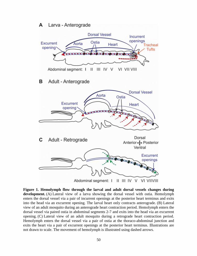
50
Figure 1. Hemolymph flow through the larval and adult dorsal vessels changes during
development. (A) Lateral view of a larva showing the dorsal vessel with ostia. Hemolymph
enters the dorsal vessel via a pair of incurrent openings at the posterior heart terminus and exits
into the head via an excurrent opening. The larval heart only contracts anterograde. (B) Lateral
view of an adult mosquito during an anterograde heart contraction period. Hemolymph enters the
dorsal vessel via paired ostia in abdominal segments 2-7 and exits into the head via an excurrent
opening. (C) Lateral view of an adult mosquito during a retrograde heart contraction period.
Hemolymph enters the dorsal vessel via a pair of ostia at the thoraco-abdominal junction and
exits the heart via a pair of excurrent openings at the posterior heart terminus. Illustrations are
not drawn to scale. The movement of hemolymph is illustrated using dashed arrows.
51
In the present study we assessed whether the circulatory and immune systems of
mosquito larvae interact during the course of an infection, and describe a pattern that is
remarkably different from what is seen in adults. Specifically, we show that, unlike in adults,
infection of a mosquito larva does not induce the aggregation of hemocytes and pathogens in the
periostial regions of the heart. Instead, infection induces the rapid aggregation and killing of
pathogens on the surface of the tracheal tufts, which are respiratory structures that are unique to
larvae and associate with the posterior terminus of the heart in the eighth abdominal segment. In
a process that is functionally analogous to what is seen in the periostial regions of adults, the
tracheal tuft hemocytes of larvae phagocytose pathogens as they near entry into the dorsal vessel,
and thus sequester and kill pathogens in the area of the body with the highest hemolymph flow.
Taken together, these findings show that the functional integration of the circulatory and immune
systems of mosquitoes displays drastic stage-specific differences that reflect the broad changes in
body plan that occur during metamorphosis.
Materials and Methods
Mosquito rearing and maintenance
Anopheles gambiae Giles sensu stricto (G3 strain; Diptera: Culicidae) were reared as
previously described (Estévez-Lao et al., 2013). Briefly, larvae were reared in deionized water
and fed a mixture of koi food and baker’s yeast. Upon pupation, mosquitoes were transferred to
plastic containers with a marquisette net top, and adults were fed a solution of 10 % sucrose ad
libitum. All life stages were maintained in an environmental chamber set to 27 °C and 75 %
relative humidity under a 12 h:12 h, light:dark cycle. Experiments were conducted on fourth
instar larvae and adult female mosquitoes at 5 days post-eclosion.
52
Mosquito injection, inoculation of immune elicitors, and bacterial infection
Mosquito larvae were immobilized by removing excess water and then injected at the
lateral center of the mesothorax. Mosquito adults were cold anesthetized and then injected at the
thoracic anepisternal cleft. Finely pulled glass capillary tubes were used as needles, and 0.2 μl of
the following substances, alone or in combination, were injected: 0.08 % solids 1-μm diameter
green fluorescent (505/515) carboxylate-modified microspheres (Invitrogen, Carlsbad, CA,
USA) in phosphate-buffered saline (PBS) (pH 7.0), GFP-expressing E. coli (modified DH5α) in
Luria-Bertani’s rich nutrient medium (LB), LB medium, 1 mg/ml pHrodo Red E. coli Bio
Particles (560/585 nm; Invitrogen) in PBS, or cellular staining solutions (see the next section).
For bacterial infections, E. coli were grown in LB overnight at 37 °C in a shaking incubator until
cultures reached approximately OD600 = 5, as measured using a BioPhotometer Plus
spectrophotometer (Eppendorf AG, Hamburg, Germany).
Staining of hemocytes, pericardial cells, and heart muscle
To fluorescently label hemocytes, live larvae or adults were injected with 67 μM Vybrant
CM-DiI or DiO Cell-Labeling Solutions (Invitrogen) in PBS and were incubated at 27 °C and
75 % relative humidity for 20 min or 1 h, respectively. Larvae were then fixed in 16 %
paraformaldehyde (Electron Microscopy Sciences, Hatfield, PA, USA) for 1 h and either rinsed
briefly in PBS and the whole body mounted directly on a glass slide using Aqua-Poly/Mount
(Polysciences Inc., Warrington, PA, USA), or dissected along a coronal plane, rinsed briefly in
PBS, and the dorsal abdomen sans the internal organs mounted. Adults were fixed by injecting
16 % paraformaldehyde, and 10 min later the specimens were dissected along a coronal plane,
placed in 0.5 % Tween in PBS to reduce surface tension, rinsed briefly in PBS, and the dorsal
abdomen sans the internal organs mounted on a glass slide using Aqua-Poly/Mount.
53
To fluorescently co-stain phagocytic events and hemocytes, larvae were injected with
green fluorescent microspheres in PBS, GFP-E. coli in LB, or pHrodo Red E. coli Bio Particles
in PBS and allowed to incubate for 4 h at 27 °C and 75 % relative humidity prior to staining with
either CM-DiI or DiO. To co-stain hemocytes and heart muscle, following staining with CM-DiI
the larvae were incubated in 16 % formaldehyde for 1 h, dissected, and incubated in 0.7 μM
phalloidin-Alexa Fluor 488 (Invitrogen) in PBS on a Pelco R1 Rotary Mixer (Pelco Instruments,
Redding, CA, USA) for 1 h. To fluorescently stain pericardial cells, larvae and adults were
injected with 0.2 mg/ml Alexa Fluor conjugated IgG 568 nm (Invitrogen) and 0.8 mM Hoechst
33342 nuclear stain (350/461; Invitrogen) in PBS and allowed to incubate for 2 h. To co-stain
pericardial cells and hemocytes, larvae were injected with 0.2 mg/ml IgG 488 nm and allowed to
incubate for 2 h prior to staining hemocytes with CM-DiI. After staining, specimens were
dissected and mounted as described above.
Light and fluorescence microscopy of aldehyde-fixed mosquito specimens
Larval and adult specimens were imaged under bright-field and fluorescence illumination
using a Nikon 90i compound microscope connected to a Nikon Digital Sight DS-Qi1Mc
monochrome digital camera (Nikon, Tokyo, Japan). For the rendering of detailed fluorescence
images with extended focal depth, Z-stacks of whole mounts were acquired using a linear
encoded Z-motor, and all images in a stack were combined to form a single focused image using
the Extended Depth of Focus (EDF; for image viewing) or Maximum Intensity Projection (for
image quantification) modules of Nikon’s NIS Elements software. For three-dimensional
rendering, Z-stacks were quantitatively deconvolved using the AQ 3D Blind Deconvolution
module of NIS Elements and rendered using the volume view feature.
54
Quantification of fluorescently labeled E. coli and hemocyte fluorescence intensity
GFP-E. coli and CM-DiI-labeled hemocyte fluorescence intensity were quantified in
abdominal segments 1-8 of larvae using maximum intensity projections of Z-stacks created in
NIS Elements software. For abdominal segments 1-7, custom polygonal regions of interest
(ROIs) were drawn over the anterior portion of each abdominal segment, encompassing the
periostial regions of the heart (Fig. 2). Specifically, these ROIs contained the area in each
abdominal segment that lay between the dorsal longitudinal tracheal trunks, and stretched from
the abdominal suture to the dorsal abdominal tracheal commissure. For segment 8, two custom
ROIs circumscribing each tracheal tuft were constructed. The mean pixel intensity, defined as
the average intensity of pixels within a given ROI, was then calculated to compare fluorescence
emission across ROIs quantitatively.
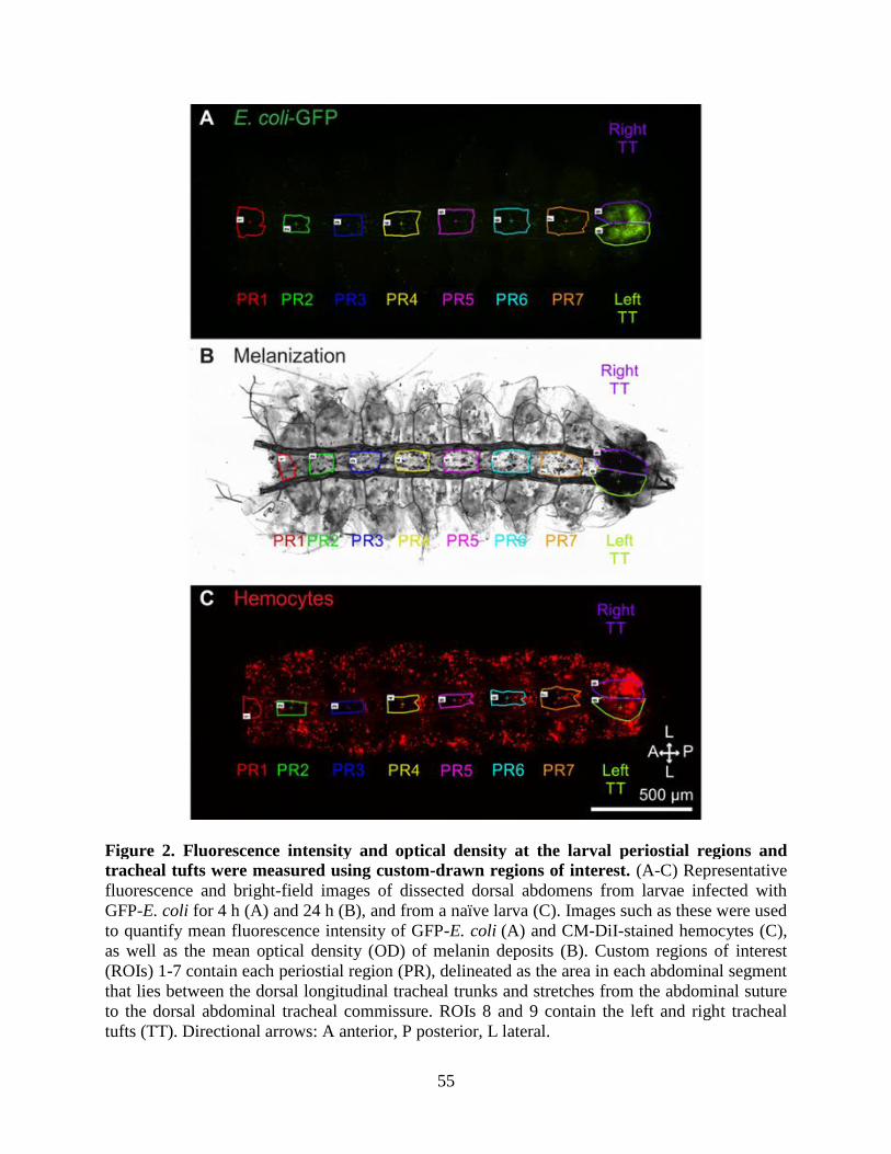
55
Figure 2. Fluorescence intensity and optical density at the larval periostial regions and
tracheal tufts were measured using custom-drawn regions of interest. (A-C) Representative
fluorescence and bright-field images of dissected dorsal abdomens from larvae infected with
GFP-E. coli for 4 h (A) and 24 h (B), and from a naïve larva (C). Images such as these were used
to quantify mean fluorescence intensity of GFP-E. coli (A) and CM-DiI-stained hemocytes (C),
as well as the mean optical density (OD) of melanin deposits (B). Custom regions of interest
(ROIs) 1-7 contain each periostial region (PR), delineated as the area in each abdominal segment
that lies between the dorsal longitudinal tracheal trunks and stretches from the abdominal suture
to the dorsal abdominal tracheal commissure. ROIs 8 and 9 contain the left and right tracheal
tufts (TT). Directional arrows: A anterior, P posterior, L lateral.
56
For GFP-E. coli quantification, intensity measurements for any given ROI in the GFP-E.
coli and injury (injected with LB alone) groups were normalized by subtracting the intensity
values from the corresponding ROI from the naïve group, as naïve mosquitoes represent
background fluorescence. Three independent trials were conducted at both 4 and 24 h post-
treatment, and each trial consisted of at least 10 mosquitoes per group. For CM-DiI-labeled
hemocyte quantification, intensity measurements for any given ROI in the CM-DiI-stained naïve,
injured, and E. coli groups were normalized by subtracting the intensity values from the
corresponding ROI of the unstained naïve group. Four and five independent trials consisting of
on average 8 and 6 mosquitoes per group were conducted at 4 and 24 h post-treatment,
respectively. Because for each mosquito measurements were obtained for all periostial regions
and the tracheal tufts (left and right), data were analyzed by repeated measures two-way
ANOVA (RM 2 W ANOVA; repeated measures being the nine regions of the body of an
individual mosquito), using treatment and ROI as the variables, followed by a Šidák post
hoc test.
Quantification of melanization in response to E. coli infection
Melanization was quantified in abdominal segments 1-8 of larvae using maximum
intensity projections created from the bright-field images of the Z-stacks of the same specimens
used to measure GFP-E. coli fluorescence intensity. These data were analyzed using the same
ROI scheme and statistical tests described above. For the melanization analysis, the mean optical
pixel density (OD), defined as the average OD of pixels within a given ROI, was calculated to
compare melanization across ROIs quantitatively. The OD measurements for any given ROI in
the E. coli and injured groups were normalized by subtracting the OD values from the
corresponding ROI from the naïve group.
57
Results
Larval sessile hemocytes are segmentally arranged and enriched in the eighth abdominal
segment
In adult mosquitoes, sessile hemocytes that are associated with the periostial regions of
the heart work in concert with hemolymph flow to clear infections (King and Hillyer, 2012; Sigle
and Hillyer, 2016). To determine whether a similar phenomenon occurs in larvae, we initially
injected fourth instar larvae and adult mosquitoes with CM-DiI cell labeling solution, which
stains hemocytes (King and Hillyer, 2012; King and Hillyer, 2013), and examined the
distribution of these cells in the dorsal abdomen by fluorescence microscopy. Analyses of whole
and dissected mosquitoes revealed that larvae and adults have a markedly different pattern of
sessile hemocyte distribution (Fig. 3). Five-day-old adult females showed a dispersed
distribution of hemocytes with no clear segmentation pattern other than the aggregation of
hemocytes at the periostial regions of the heart (Fig. 3A, B). In contrast, larvae displayed dense
bands of segmentally arranged hemocytes (Fig. 3C, D). These segmental hemocyte bands form
continuous rings that encircle each abdominal segment, as this banding is identical in the dorsal
and ventral halves of the body (Fig. 3C-H). Furthermore, larvae do not contain the distinct
periostial hemocyte aggregates that are typically observed along the heart of adults. Instead, they
contain a high concentration of hemocytes in the eighth abdominal segment, a region of the body
that contains far fewer hemocytes in adults (Fig. 3B, D).
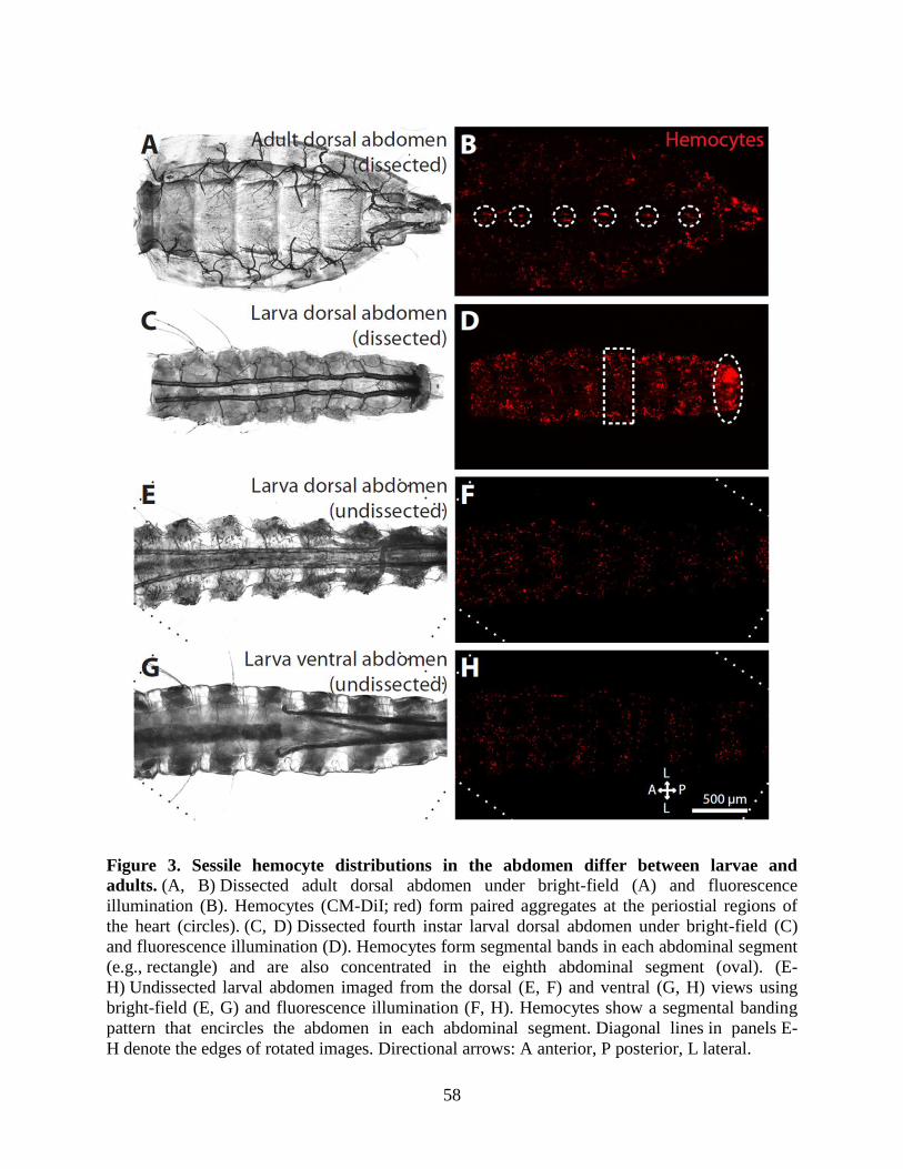
58
Figure 3. Sessile hemocyte distributions in the abdomen differ between larvae and
adults. (A, B) Dissected adult dorsal abdomen under bright-field (A) and fluorescence
illumination (B). Hemocytes (CM-DiI; red) form paired aggregates at the periostial regions of
the heart (circles). (C, D) Dissected fourth instar larval dorsal abdomen under bright-field (C)
and fluorescence illumination (D). Hemocytes form segmental bands in each abdominal segment
(e.g., rectangle) and are also concentrated in the eighth abdominal segment (oval). (E-
H) Undissected larval abdomen imaged from the dorsal (E, F) and ventral (G, H) views using
bright-field (E, G) and fluorescence illumination (F, H). Hemocytes show a segmental banding
pattern that encircles the abdomen in each abdominal segment. Diagonal lines in panels E-
H denote the edges of rotated images. Directional arrows: A anterior, P posterior, L lateral.
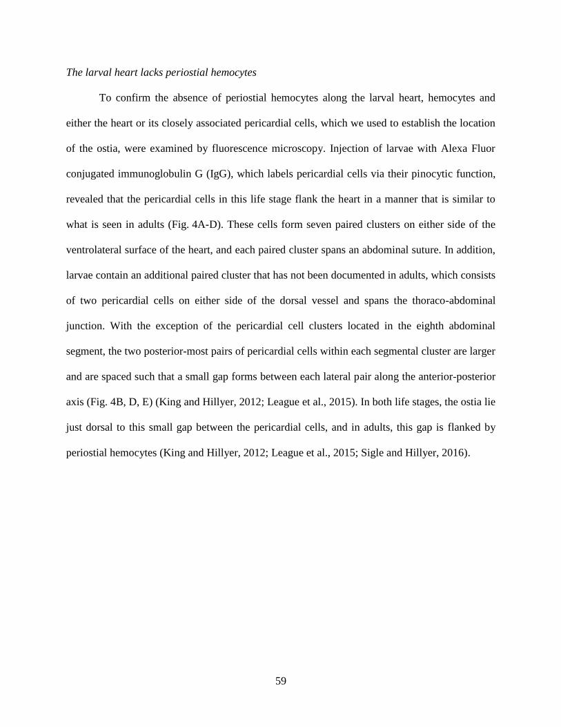
59
The larval heart lacks periostial hemocytes
To confirm the absence of periostial hemocytes along the larval heart, hemocytes and
either the heart or its closely associated pericardial cells, which we used to establish the location
of the ostia, were examined by fluorescence microscopy. Injection of larvae with Alexa Fluor
conjugated immunoglobulin G (IgG), which labels pericardial cells via their pinocytic function,
revealed that the pericardial cells in this life stage flank the heart in a manner that is similar to
what is seen in adults (Fig. 4A-D). These cells form seven paired clusters on either side of the
ventrolateral surface of the heart, and each paired cluster spans an abdominal suture. In addition,
larvae contain an additional paired cluster that has not been documented in adults, which consists
of two pericardial cells on either side of the dorsal vessel and spans the thoraco-abdominal
junction. With the exception of the pericardial cell clusters located in the eighth abdominal
segment, the two posterior-most pairs of pericardial cells within each segmental cluster are larger
and are spaced such that a small gap forms between each lateral pair along the anterior-posterior
axis (Fig. 4B, D, E) (King and Hillyer, 2012; League et al., 2015). In both life stages, the ostia lie
just dorsal to this small gap between the pericardial cells, and in adults, this gap is flanked by
periostial hemocytes (King and Hillyer, 2012; League et al., 2015; Sigle and Hillyer, 2016).
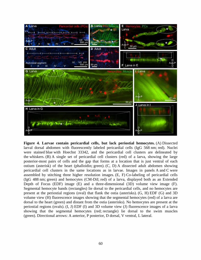
60
Figure 4. Larvae contain pericardial cells, but lack periostial hemocytes. (A) Dissected
larval dorsal abdomen with fluorescently labeled pericardial cells (IgG 568 nm; red). Nuclei
were stained blue with Hoechst 33342, and the pericardial cell clusters are delineated by
the whiskers. (B) A single set of pericardial cell clusters (red) of a larva, showing the large
posterior-most pairs of cells and the gap that forms at a location that is just ventral of each
ostium (asterisk) of the heart (phalloidin; green). (C, D) A dissected adult abdomen showing
pericardial cell clusters in the same locations as in larvae. Images in panels A and C were
assembled by stitching three higher resolution images. (E, F) Co-labeling of pericardial cells
(IgG 488 nm; green) and hemocytes (CM-DiI; red) of a larva, displayed both as an Extended
Depth of Focus (EDF) image (E) and a three-dimensional (3D) volume view image (F).
Segmental hemocyte bands (rectangles) lie dorsal to the pericardial cells, and no hemocytes are
present at the periostial regions (oval) that flank the ostia (asterisks). (G, H) EDF (G) and 3D
volume view (H) fluorescence images showing that the segmental hemocytes (red) of a larva are
dorsal to the heart (green) and distant from the ostia (asterisks). No hemocytes are present at the
periostial regions (ovals). (I, J) EDF (I) and 3D volume view (J) fluorescence images of a larva
showing that the segmental hemocytes (red; rectangle) lie dorsal to the swim muscles
(green). Directional arrows: A anterior, P posterior, D dorsal, V ventral, L lateral.
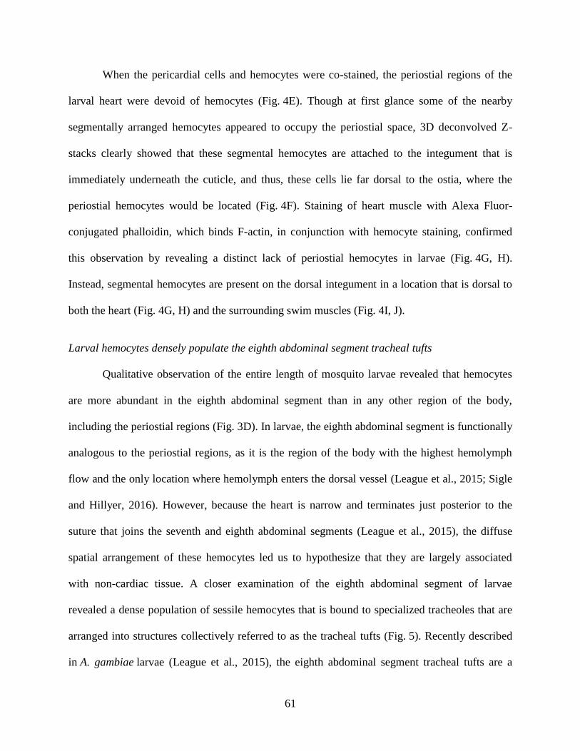
61
When the pericardial cells and hemocytes were co-stained, the periostial regions of the
larval heart were devoid of hemocytes (Fig. 4E). Though at first glance some of the nearby
segmentally arranged hemocytes appeared to occupy the periostial space, 3D deconvolved Z-
stacks clearly showed that these segmental hemocytes are attached to the integument that is
immediately underneath the cuticle, and thus, these cells lie far dorsal to the ostia, where the
periostial hemocytes would be located (Fig. 4F). Staining of heart muscle with Alexa Fluor-
conjugated phalloidin, which binds F-actin, in conjunction with hemocyte staining, confirmed
this observation by revealing a distinct lack of periostial hemocytes in larvae (Fig. 4G, H).
Instead, segmental hemocytes are present on the dorsal integument in a location that is dorsal to
both the heart (Fig. 4G, H) and the surrounding swim muscles (Fig. 4I, J).
Larval hemocytes densely populate the eighth abdominal segment tracheal tufts
Qualitative observation of the entire length of mosquito larvae revealed that hemocytes
are more abundant in the eighth abdominal segment than in any other region of the body,
including the periostial regions (Fig. 3D). In larvae, the eighth abdominal segment is functionally
analogous to the periostial regions, as it is the region of the body with the highest hemolymph
flow and the only location where hemolymph enters the dorsal vessel (League et al., 2015; Sigle
and Hillyer, 2016). However, because the heart is narrow and terminates just posterior to the
suture that joins the seventh and eighth abdominal segments (League et al., 2015), the diffuse
spatial arrangement of these hemocytes led us to hypothesize that they are largely associated
with non-cardiac tissue. A closer examination of the eighth abdominal segment of larvae
revealed a dense population of sessile hemocytes that is bound to specialized tracheoles that are
arranged into structures collectively referred to as the tracheal tufts (Fig. 5). Recently described
in A. gambiae larvae (League et al., 2015), the eighth abdominal segment tracheal tufts are a

62
dense network of thin tracheoles that emanate from the ventral base of the dorsal longitudinal
tracheal trunks (Fig. 5A, B; Fig. 6A). Although these tracheoles are predominantly suspended in
the hemolymph, some are attached to the posterior terminus of the heart, thus causing them to
move with each heartbeat. Furthermore, examination of the exuviae of fourth instar larvae and
the dissected dorsal abdomens of pupae and adults showed that the tracheal tufts are unique to
larvae and are shed during pupation (Fig. 6).
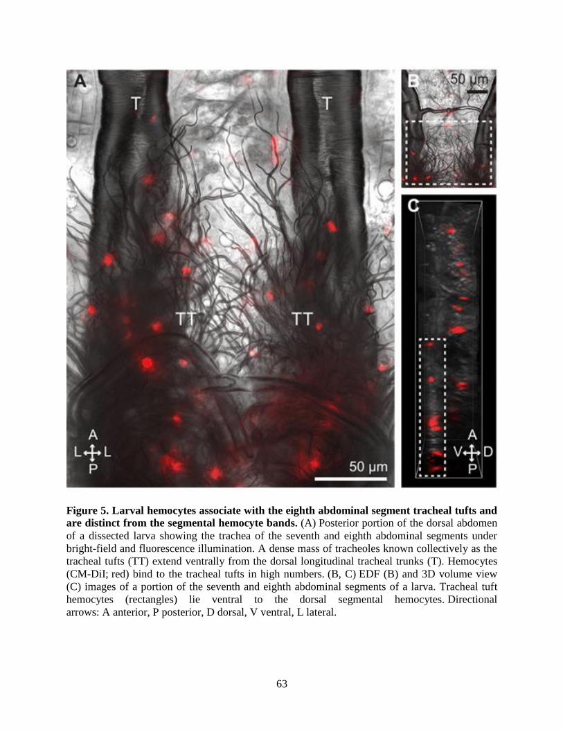
63
Figure 5. Larval hemocytes associate with the eighth abdominal segment tracheal tufts and
are distinct from the segmental hemocyte bands. (A) Posterior portion of the dorsal abdomen
of a dissected larva showing the trachea of the seventh and eighth abdominal segments under
bright-field and fluorescence illumination. A dense mass of tracheoles known collectively as the
tracheal tufts (TT) extend ventrally from the dorsal longitudinal tracheal trunks (T). Hemocytes
(CM-DiI; red) bind to the tracheal tufts in high numbers. (B, C) EDF (B) and 3D volume view
(C) images of a portion of the seventh and eighth abdominal segments of a larva. Tracheal tuft
hemocytes (rectangles) lie ventral to the dorsal segmental hemocytes. Directional
arrows: A anterior, P posterior, D dorsal, V ventral, L lateral.
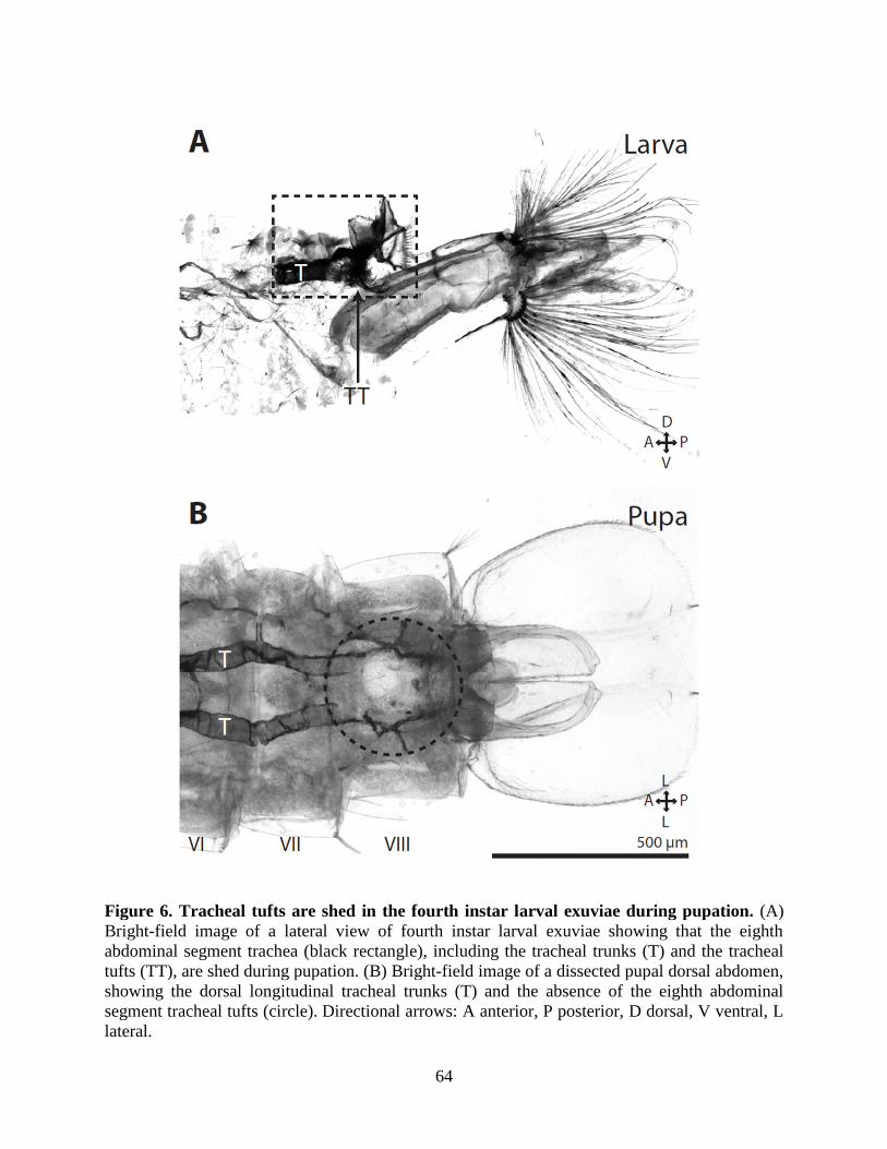
64
Figure 6. Tracheal tufts are shed in the fourth instar larval exuviae during pupation. (A)
Bright-field image of a lateral view of fourth instar larval exuviae showing that the eighth
abdominal segment trachea (black rectangle), including the tracheal trunks (T) and the tracheal
tufts (TT), are shed during pupation. (B) Bright-field image of a dissected pupal dorsal abdomen,
showing the dorsal longitudinal tracheal trunks (T) and the absence of the eighth abdominal
segment tracheal tufts (circle). Directional arrows: A anterior, P posterior, D dorsal, V ventral, L
lateral.
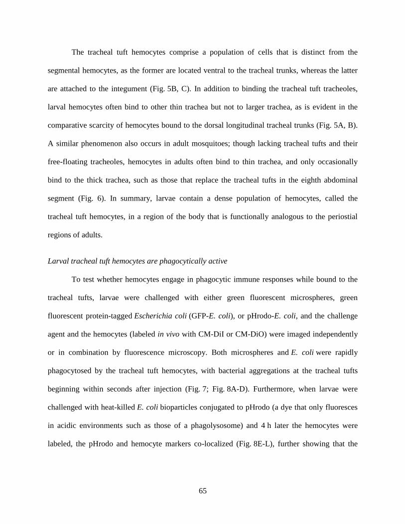
65
The tracheal tuft hemocytes comprise a population of cells that is distinct from the
segmental hemocytes, as the former are located ventral to the tracheal trunks, whereas the latter
are attached to the integument (Fig. 5B, C). In addition to binding the tracheal tuft tracheoles,
larval hemocytes often bind to other thin trachea but not to larger trachea, as is evident in the
comparative scarcity of hemocytes bound to the dorsal longitudinal tracheal trunks (Fig. 5A, B).
A similar phenomenon also occurs in adult mosquitoes; though lacking tracheal tufts and their
free-floating tracheoles, hemocytes in adults often bind to thin trachea, and only occasionally
bind to the thick trachea, such as those that replace the tracheal tufts in the eighth abdominal
segment (Fig. 6). In summary, larvae contain a dense population of hemocytes, called the
tracheal tuft hemocytes, in a region of the body that is functionally analogous to the periostial
regions of adults.
Larval tracheal tuft hemocytes are phagocytically active
To test whether hemocytes engage in phagocytic immune responses while bound to the
tracheal tufts, larvae were challenged with either green fluorescent microspheres, green
fluorescent protein-tagged Escherichia coli (GFP-E. coli), or pHrodo-E. coli, and the challenge
agent and the hemocytes (labeled in vivo with CM-DiI or CM-DiO) were imaged independently
or in combination by fluorescence microscopy. Both microspheres and E. coli were rapidly
phagocytosed by the tracheal tuft hemocytes, with bacterial aggregations at the tracheal tufts
beginning within seconds after injection (Fig. 7; Fig. 8A-D). Furthermore, when larvae were
challenged with heat-killed E. coli bioparticles conjugated to pHrodo (a dye that only fluoresces
in acidic environments such as those of a phagolysosome) and 4 h later the hemocytes were
labeled, the pHrodo and hemocyte markers co-localized (Fig. 8E-L), further showing that the
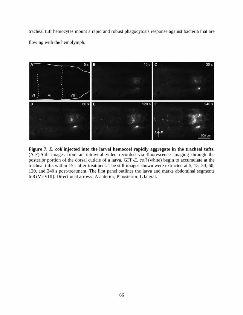
66
tracheal tuft hemocytes mount a rapid and robust phagocytosis response against bacteria that are
flowing with the hemolymph.
Figure 7. E. coli injected into the larval hemocoel rapidly aggregate in the tracheal tufts. (A-F) Still images from an intravital video recorded via fluorescence imaging through the
posterior portion of the dorsal cuticle of a larva. GFP-E. coli (white) begin to accumulate at the
tracheal tufts within 15 s after treatment. The still images shown were extracted at 5, 15, 30, 60,
120, and 240 s post-treatment. The first panel outlines the larva and marks abdominal segments
6-8 (VI-VIII). Directional arrows: A anterior, P posterior, L lateral.
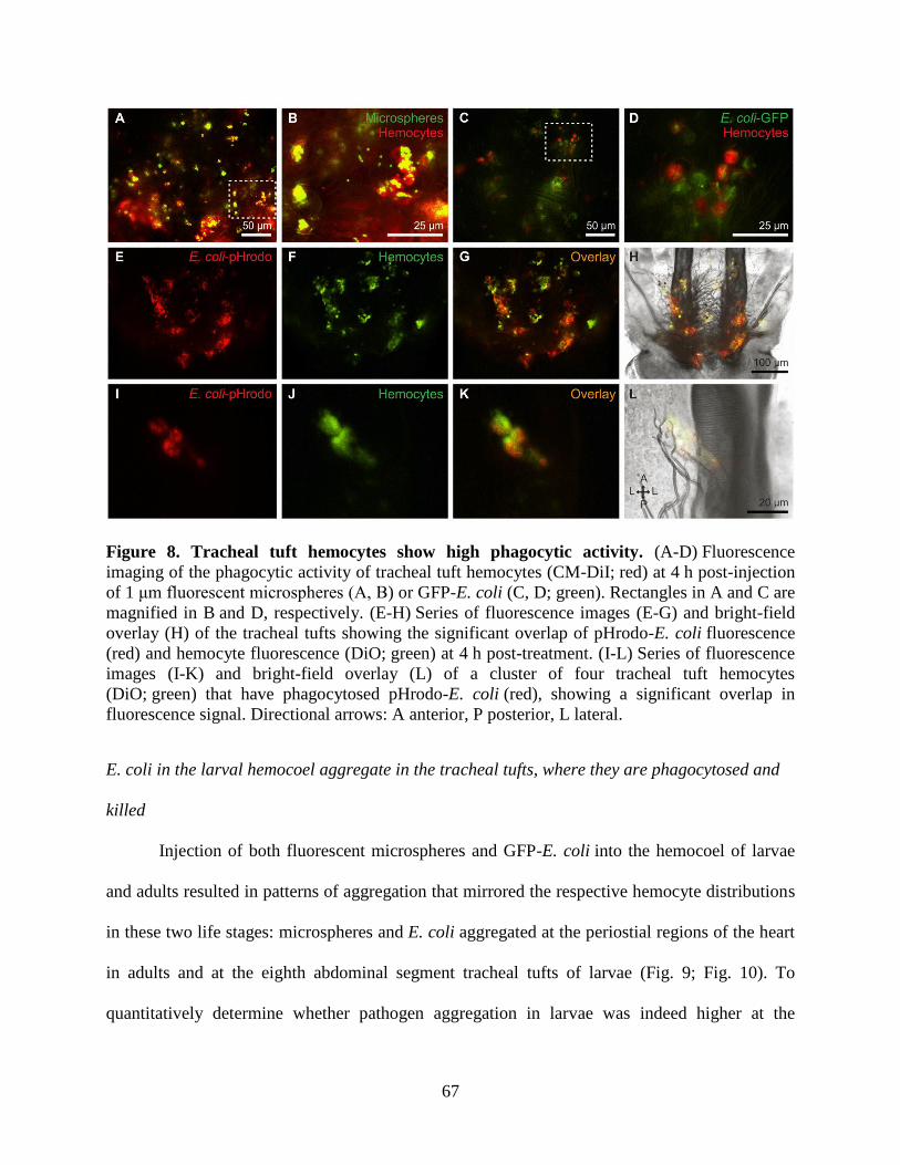
67
Figure 8. Tracheal tuft hemocytes show high phagocytic activity. (A-D) Fluorescence
imaging of the phagocytic activity of tracheal tuft hemocytes (CM-DiI; red) at 4 h post-injection
of 1 μm fluorescent microspheres (A, B) or GFP-E. coli (C, D; green). Rectangles in A and C are
magnified in B and D, respectively. (E-H) Series of fluorescence images (E-G) and bright-field
overlay (H) of the tracheal tufts showing the significant overlap of pHrodo-E. coli fluorescence
(red) and hemocyte fluorescence (DiO; green) at 4 h post-treatment. (I-L) Series of fluorescence
images (I-K) and bright-field overlay (L) of a cluster of four tracheal tuft hemocytes
(DiO; green) that have phagocytosed pHrodo-E. coli (red), showing a significant overlap in
fluorescence signal. Directional arrows: A anterior, P posterior, L lateral.
E. coli in the larval hemocoel aggregate in the tracheal tufts, where they are phagocytosed and
killed
Injection of both fluorescent microspheres and GFP-E. coli into the hemocoel of larvae
and adults resulted in patterns of aggregation that mirrored the respective hemocyte distributions
in these two life stages: microspheres and E. coli aggregated at the periostial regions of the heart
in adults and at the eighth abdominal segment tracheal tufts of larvae (Fig. 9; Fig. 10). To
quantitatively determine whether pathogen aggregation in larvae was indeed higher at the
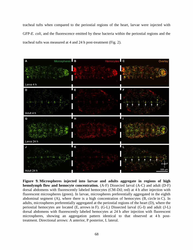
68
tracheal tufts when compared to the periostial regions of the heart, larvae were injected with
GFP-E. coli, and the fluorescence emitted by these bacteria within the periostial regions and the
tracheal tufts was measured at 4 and 24 h post-treatment (Fig. 2).
Figure 9. Microspheres injected into larvae and adults aggregate in regions of high
hemolymph flow and hemocyte concentration. (A-F) Dissected larval (A-C) and adult (D-F)
dorsal abdomens with fluorescently labeled hemocytes (CM-DiI; red) at 4 h after injection with
fluorescent microspheres (green). In larvae, microspheres preferentially aggregated in the eighth
abdominal segment (A), where there is a high concentration of hemocytes (B, circle in C). In
adults, microspheres preferentially aggregated at the periostial regions of the heart (D), where the
periostial hemocytes are located (E, arrows in F). (G-L) Dissected larval (G-I) and adult (J-L)
dorsal abdomens with fluorescently labeled hemocytes at 24 h after injection with fluorescent
microspheres, showing an aggregation pattern identical to that observed at 4 h post-
treatment. Directional arrows: A anterior, P posterior, L lateral.
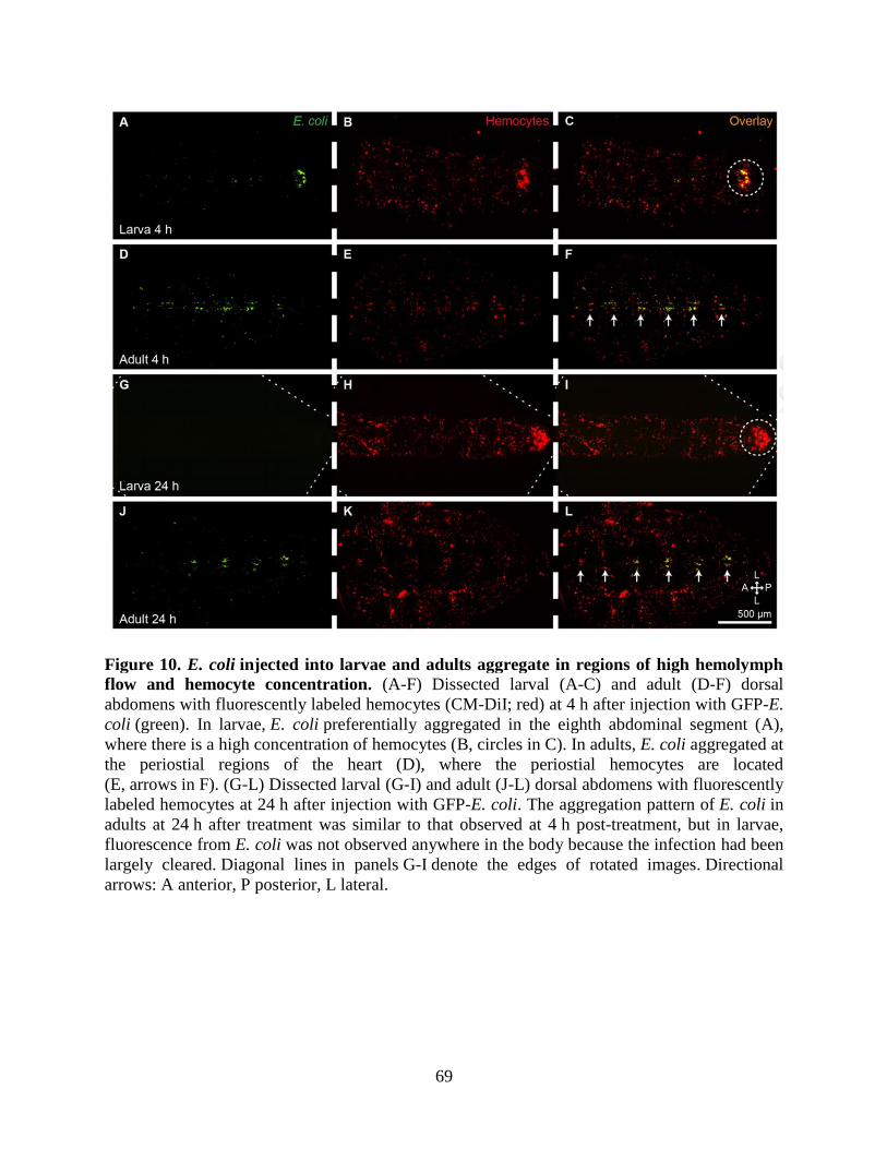
69
Figure 10. E. coli injected into larvae and adults aggregate in regions of high hemolymph
flow and hemocyte concentration. (A-F) Dissected larval (A-C) and adult (D-F) dorsal
abdomens with fluorescently labeled hemocytes (CM-DiI; red) at 4 h after injection with GFP-E.
coli (green). In larvae, E. coli preferentially aggregated in the eighth abdominal segment (A),
where there is a high concentration of hemocytes (B, circles in C). In adults, E. coli aggregated at
the periostial regions of the heart (D), where the periostial hemocytes are located
(E, arrows in F). (G-L) Dissected larval (G-I) and adult (J-L) dorsal abdomens with fluorescently
labeled hemocytes at 24 h after injection with GFP-E. coli. The aggregation pattern of E. coli in
adults at 24 h after treatment was similar to that observed at 4 h post-treatment, but in larvae,
fluorescence from E. coli was not observed anywhere in the body because the infection had been
largely cleared. Diagonal lines in panels G-I denote the edges of rotated images. Directional
arrows: A anterior, P posterior, L lateral.
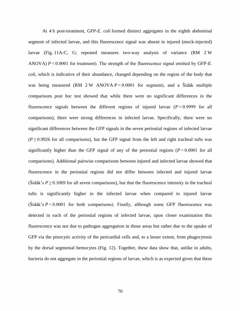
70
At 4 h post-treatment, GFP-E. coli formed distinct aggregates in the eighth abdominal
segment of infected larvae, and this fluorescence signal was absent in injured (mock-injected)
larvae (Fig. 11A-C, G; repeated measures two-way analysis of variance (RM 2 W
ANOVA) P < 0.0001 for treatment). The strength of the fluorescence signal emitted by GFP-E.
coli, which is indicative of their abundance, changed depending on the region of the body that
was being measured (RM 2 W ANOVA P < 0.0001 for segment), and a Šidák multiple
comparisons post hoc test showed that while there were no significant differences in the
fluorescence signals between the different regions of injured larvae (P > 0.9999 for all
comparisons), there were strong differences in infected larvae. Specifically, there were no
significant differences between the GFP signals in the seven periostial regions of infected larvae
(P ≥ 0.9926 for all comparisons), but the GFP signal from the left and right tracheal tufts was
significantly higher than the GFP signal of any of the periostial regions (P < 0.0001 for all
comparisons). Additional pairwise comparisons between injured and infected larvae showed that
fluorescence in the periostial regions did not differ between infected and injured larvae
(Šidák’s P ≥ 0.1069 for all seven comparisons), but that the fluorescence intensity in the tracheal
tufts is significantly higher in the infected larvae when compared to injured larvae
(Šidák’s P < 0.0001 for both comparisons). Finally, although some GFP fluorescence was
detected in each of the periostial regions of infected larvae, upon closer examination this
fluorescence was not due to pathogen aggregation in those areas but rather due to the uptake of
GFP via the pinocytic activity of the pericardial cells and, to a lesser extent, from phagocytosis
by the dorsal segmental hemocytes (Fig. 12). Together, these data show that, unlike in adults,
bacteria do not aggregate in the periostial regions of larvae, which is as expected given that these
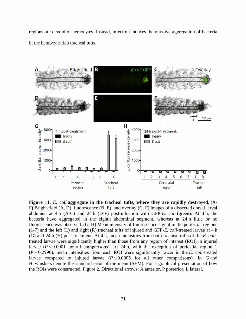
71
regions are devoid of hemocytes. Instead, infection induces the massive aggregation of bacteria
in the hemocyte-rich tracheal tufts.
Figure 11. E. coli aggregate in the tracheal tufts, where they are rapidly destroyed. (A-
F) Bright-field (A, D), fluorescence (B, E), and overlay (C, F) images of a dissected dorsal larval
abdomen at 4 h (A-C) and 24 h (D-F) post-infection with GFP-E. coli (green). At 4 h, the
bacteria have aggregated in the eighth abdominal segment, whereas at 24 h little or no
fluorescence was observed. (G, H) Mean intensity of fluorescence signal in the periostial regions
(1-7) and the left (L) and right (R) tracheal tufts of injured and GFP-E. coli-treated larvae at 4 h
(G) and 24 h (H) post-treatment. At 4 h, mean intensities from both tracheal tufts of the E. coli-
treated larvae were significantly higher than those from any region of interest (ROI) in injured
larvae (P < 0.0001 for all comparisons). At 24 h, with the exception of periostial region 1
(P = 0.2990), mean intensities from each ROI were significantly lower in the E. coli-treated
larvae compared to injured larvae (P ≤ 0.0005 for all other comparisons). In G and
H, whiskers denote the standard error of the mean (SEM). For a graphical presentation of how
the ROIs were constructed, Figure 2. Directional arrows: A anterior, P posterior, L lateral.
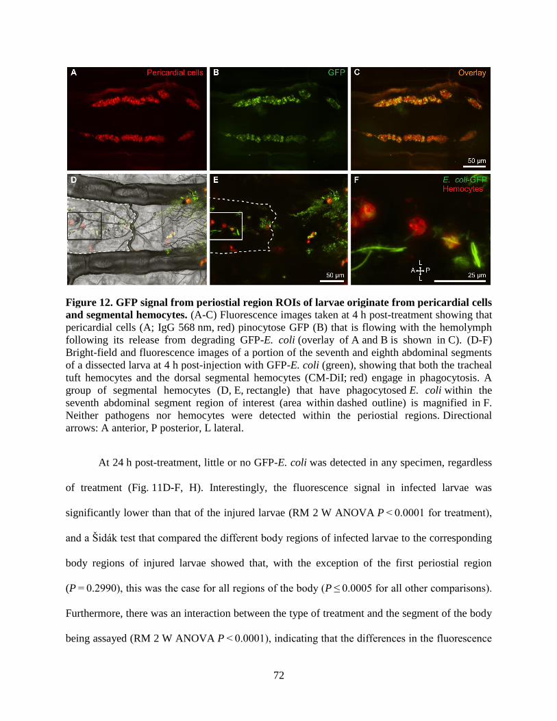
72
Figure 12. GFP signal from periostial region ROIs of larvae originate from pericardial cells
and segmental hemocytes. (A-C) Fluorescence images taken at 4 h post-treatment showing that
pericardial cells (A; IgG 568 nm, red) pinocytose GFP (B) that is flowing with the hemolymph
following its release from degrading GFP-E. coli (overlay of A and B is shown in C). (D-F)
Bright-field and fluorescence images of a portion of the seventh and eighth abdominal segments
of a dissected larva at 4 h post-injection with GFP-E. coli (green), showing that both the tracheal
tuft hemocytes and the dorsal segmental hemocytes (CM-DiI; red) engage in phagocytosis. A
group of segmental hemocytes (D, E, rectangle) that have phagocytosed E. coli within the
seventh abdominal segment region of interest (area within dashed outline) is magnified in F.
Neither pathogens nor hemocytes were detected within the periostial regions. Directional
arrows: A anterior, P posterior, L lateral.
At 24 h post-treatment, little or no GFP-E. coli was detected in any specimen, regardless
of treatment (Fig. 11D-F, H). Interestingly, the fluorescence signal in infected larvae was
significantly lower than that of the injured larvae (RM 2 W ANOVA P < 0.0001 for treatment),
and a Šidák test that compared the different body regions of infected larvae to the corresponding
body regions of injured larvae showed that, with the exception of the first periostial region
(P = 0.2990), this was the case for all regions of the body (P ≤ 0.0005 for all other comparisons).
Furthermore, there was an interaction between the type of treatment and the segment of the body
being assayed (RM 2 W ANOVA P < 0.0001), indicating that the differences in the fluorescence
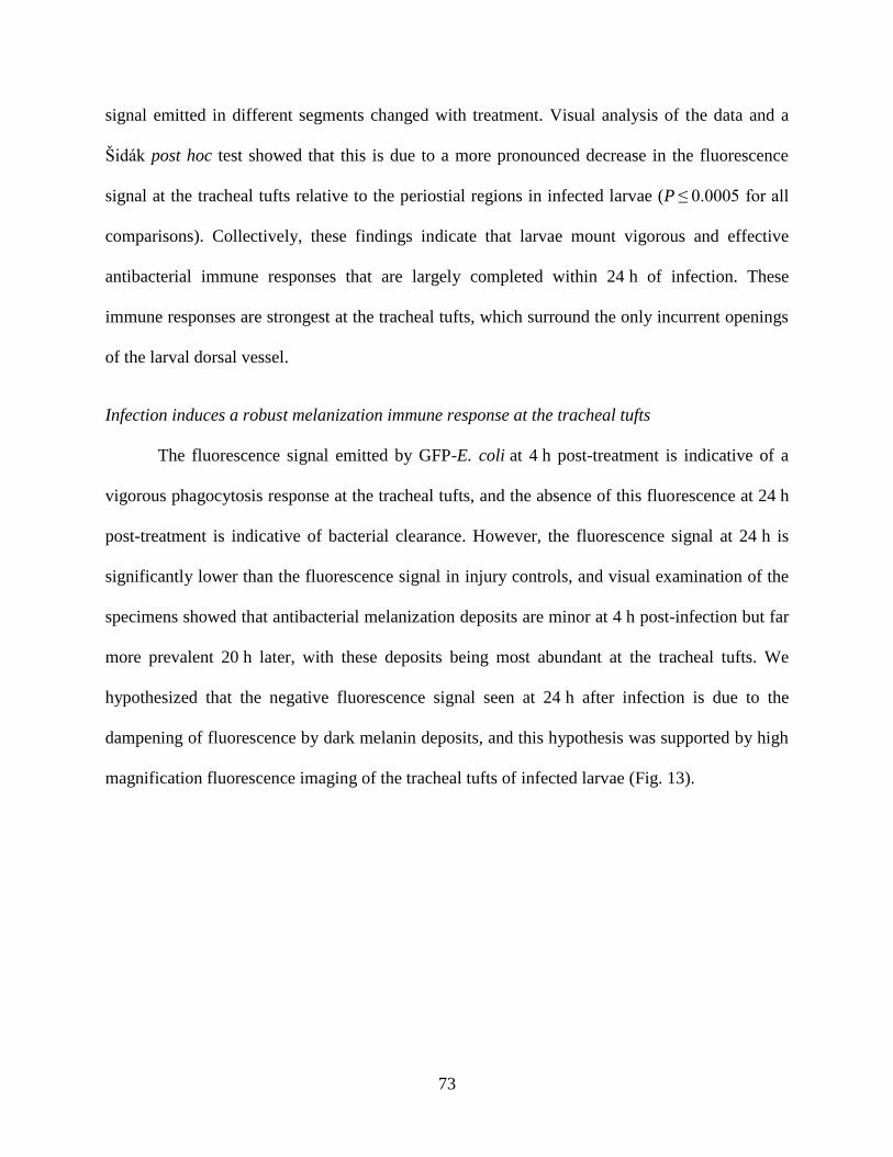
73
signal emitted in different segments changed with treatment. Visual analysis of the data and a
Šidák post hoc test showed that this is due to a more pronounced decrease in the fluorescence
signal at the tracheal tufts relative to the periostial regions in infected larvae (P ≤ 0.0005 for all
comparisons). Collectively, these findings indicate that larvae mount vigorous and effective
antibacterial immune responses that are largely completed within 24 h of infection. These
immune responses are strongest at the tracheal tufts, which surround the only incurrent openings
of the larval dorsal vessel.
Infection induces a robust melanization immune response at the tracheal tufts
The fluorescence signal emitted by GFP-E. coli at 4 h post-treatment is indicative of a
vigorous phagocytosis response at the tracheal tufts, and the absence of this fluorescence at 24 h
post-treatment is indicative of bacterial clearance. However, the fluorescence signal at 24 h is
significantly lower than the fluorescence signal in injury controls, and visual examination of the
specimens showed that antibacterial melanization deposits are minor at 4 h post-infection but far
more prevalent 20 h later, with these deposits being most abundant at the tracheal tufts. We
hypothesized that the negative fluorescence signal seen at 24 h after infection is due to the
dampening of fluorescence by dark melanin deposits, and this hypothesis was supported by high
magnification fluorescence imaging of the tracheal tufts of infected larvae (Fig. 13).
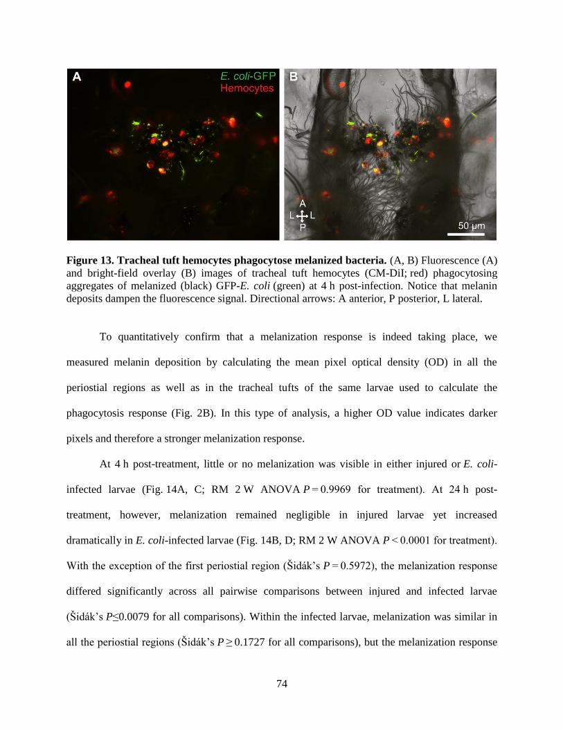
74
Figure 13. Tracheal tuft hemocytes phagocytose melanized bacteria. (A, B) Fluorescence (A)
and bright-field overlay (B) images of tracheal tuft hemocytes (CM-DiI; red) phagocytosing
aggregates of melanized (black) GFP-E. coli (green) at 4 h post-infection. Notice that melanin
deposits dampen the fluorescence signal. Directional arrows: A anterior, P posterior, L lateral.
To quantitatively confirm that a melanization response is indeed taking place, we
measured melanin deposition by calculating the mean pixel optical density (OD) in all the
periostial regions as well as in the tracheal tufts of the same larvae used to calculate the
phagocytosis response (Fig. 2B). In this type of analysis, a higher OD value indicates darker
pixels and therefore a stronger melanization response.
At 4 h post-treatment, little or no melanization was visible in either injured or E. coli-
infected larvae (Fig. 14A, C; RM 2 W ANOVA P = 0.9969 for treatment). At 24 h post-
treatment, however, melanization remained negligible in injured larvae yet increased
dramatically in E. coli-infected larvae (Fig. 14B, D; RM 2 W ANOVA P < 0.0001 for treatment).
With the exception of the first periostial region (Šidák’s P = 0.5972), the melanization response
differed significantly across all pairwise comparisons between injured and infected larvae
(Šidák’s P≤0.0079 for all comparisons). Within the infected larvae, melanization was similar in
all the periostial regions (Šidák’s P ≥ 0.1727 for all comparisons), but the melanization response
75
in the tracheal tufts was significantly stronger than in any of the periostial regions
(Šidák’s P < 0.0001 for all comparisons).
Figure 14. The melanization immune response is concentrated in the tracheal tufts. (A,
B) Bright-field image of a dissected larval dorsal abdomen at 4 h (A) and 24 h (B) post-treatment
with GFP-E. coli. At 4 h there is little or no visible melanization, whereas at 24 h widespread
melanization is observed, including the uptake of melanin by the pericardial cells and an
enrichment of melanin deposits in the tracheal tufts. (C, D) Melanization, as measured by mean
pixel optical density (OD), in the periostial regions (1-7) and the left (L) and right (R) tracheal
tufts of injured and GFP-E. coli-treated larvae at 4 h (C) and 24 h (D) post-treatment. At 4 h, OD
values are similar in injured and E. coli-treated larvae (P = 0.9969), whereas at 24 h mean OD for
all pairwise comparisons was significantly higher in infected larvae when compared to injured
larvae (P ≤ 0.0079 for all other comparisons). In C and D, whiskers denote the SEM. For a
graphical presentation of how the ROIs were constructed, see Figure 2B. Directional
arrows: A anterior, P posterior, L lateral.
Because the periostial regions of larvae are devoid of hemocytes, we scrutinized the
location of melanin deposits in these regions using a combination of fluorescence and bright-
field microscopy, and found that melanin accumulates at the periostial regions of infected larvae
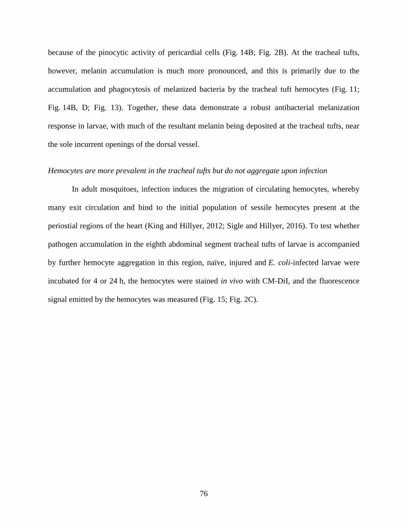
76
because of the pinocytic activity of pericardial cells (Fig. 14B; Fig. 2B). At the tracheal tufts,
however, melanin accumulation is much more pronounced, and this is primarily due to the
accumulation and phagocytosis of melanized bacteria by the tracheal tuft hemocytes (Fig. 11;
Fig. 14B, D; Fig. 13). Together, these data demonstrate a robust antibacterial melanization
response in larvae, with much of the resultant melanin being deposited at the tracheal tufts, near
the sole incurrent openings of the dorsal vessel.
Hemocytes are more prevalent in the tracheal tufts but do not aggregate upon infection
In adult mosquitoes, infection induces the migration of circulating hemocytes, whereby
many exit circulation and bind to the initial population of sessile hemocytes present at the
periostial regions of the heart (King and Hillyer, 2012; Sigle and Hillyer, 2016). To test whether
pathogen accumulation in the eighth abdominal segment tracheal tufts of larvae is accompanied
by further hemocyte aggregation in this region, naïve, injured and E. coli-infected larvae were
incubated for 4 or 24 h, the hemocytes were stained in vivo with CM-DiI, and the fluorescence
signal emitted by the hemocytes was measured (Fig. 15; Fig. 2C).
77
Figure 15. Hemocytes are more abundant in the tracheal tufts, but do not increase in
response to E. coli infection. (A) Mean intensity of CM-DiI-stained hemocytes in the periostial
regions (1-7) and the left (L) and right (R) tracheal tufts of naïve, injured, and GFP-E. coli-
treated larvae at 4 h post-treatment. Hemocyte intensity was not significantly affected by
treatment (RM 2 W ANOVA P = 0.1123) but was significantly higher in the tracheal tufts
compared to the periostial regions in all three groups (Šidák’s P ≤ 0.0312 for 39 out of 42
comparisons). (B) Mean intensity of CM-DiI stained hemocytes in the periostial regions and
tracheal tufts of naïve, injured, and GFP-E. coli-treated larvae at 24 h post-treatment. Hemocyte
intensity was significantly affected by treatment (RM 2 W ANOVA P=0.0012), with hemocyte
intensities from GFP-E. coli-treated larvae being significantly lower than those of both naïve and
injured larvae (Šidák’s P < 0.0001 for all comparisons). Hemocyte intensity was significantly
higher in the tracheal tufts compared to the periostial regions in all three groups
(Šidák’s P ≤ 0.0003 for all comparisons). Whiskers denote the SEM. For a graphical presentation
of how the ROIs were constructed, see Figure 2C.
At 4 h post-treatment, there was no difference in the fluorescence signal emitted by
hemocytes in naïve, injured, and infected mosquitoes (Fig. 15A; RM 2 W ANOVA P = 0.1123
for treatment). However, there was a significant difference between the regions of the body being
measured (RM 2 W ANOVA P < 0.0001). Specifically, for naïve, injured, and infected
mosquitoes, the fluorescence intensities of the two tracheal tufts were significantly higher than
the fluorescence intensities in the corresponding periostial regions (Šidák’s P ≤ 0.0312 for 39 out
of 42 comparisons). This indicates that hemocytes are absent from the periostial regions of
infected larvae (any signal emanates from segmental hemocytes) but are abundant at the tracheal
78
tufts. This also indicates that at 4 h post-infection the relative number of tracheal tuft hemocytes
in infected larvae is similar to that found in naïve and injured larvae.
At 24 h post-treatment, hemocyte intensity was significantly different between naïve,
injured, and infected larvae (Fig. 15B; RM 2 W ANOVA P=0.0012). There was also a
significant difference between the regions of the body being measured (RM 2 W
ANOVA P < 0.0001), as the fluorescence signal in the tracheal tufts remained elevated relative
to the periostial regions (Šidák’s P ≤ 0.0003 for all comparisons). Furthermore, there was a
significant interaction between the region of the body and the treatment (RM 2 W
ANOVA P < 0.0001 for interaction), indicating that the distribution of fluorescence signal in the
different body regions changes depending on the infection status of the larva. Specifically, the
fluorescence signal emitted by segmental hemocytes was similar between the periostial ROIs of
naïve, injured, and infected larvae (Šidák’s P ≥ 0.1528 for all comparisons), but the signal in the
tracheal tufts was lower in infected larvae when compared to naïve and injured larvae
(Šidák’s P < 0.0001 for all comparisons).
Taken together, these results suggest that although hemocytes are more abundant at the
tracheal tufts, infection does not recruit circulating hemocytes to the periostial regions or to the
tracheal tufts. At 24 h post-treatment, hemocyte fluorescence in the tracheal tufts of infected
larvae was lower than that of naïve and injured larvae, but visual analysis of these specimens
strongly suggests that this is not due to a reduction in the hemocyte population but rather due to
melanization-induced dampening of the fluorescence signal (Fig. 11H; Fig. 14D; Fig. 2B and
Fig. 13).
79
Discussion
Mosquitoes undergo a dramatic metamorphosis as they transition from an aquatic
environment during the larval and pupal stages to terrestrial and aerial environments during the
adult stage. In the present study, we show that the changes in circulatory and respiratory
physiology that occur during the larval-to-adult transition are accompanied by stage-specific
interactions between the circulatory, immune, and respiratory systems (Fig. 16). These systems
are functionally integrated such that structures once thought to act solely in oxygen
dissemination have been co-opted by the immune system to act as both a sieve to sequester
circulating pathogens and as an ideal staging ground for mounting cellular and humoral immune
responses at the sole entry points for hemolymph into the heart.
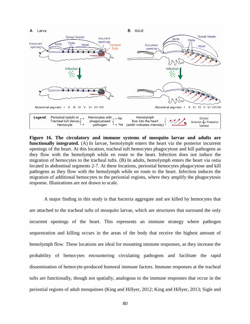
80
Figure 16. The circulatory and immune systems of mosquito larvae and adults are
functionally integrated. (A) In larvae, hemolymph enters the heart via the posterior incurrent
openings of the heart. At this location, tracheal tuft hemocytes phagocytose and kill pathogens as
they flow with the hemolymph while en route to the heart. Infection does not induce the
migration of hemocytes to the tracheal tufts. (B) In adults, hemolymph enters the heart via ostia
located in abdominal segments 2-7. At these locations, periostial hemocytes phagocytose and kill
pathogens as they flow with the hemolymph while en route to the heart. Infection induces the
migration of additional hemocytes to the periostial regions, where they amplify the phagocytosis
response. Illustrations are not drawn to scale.
A major finding in this study is that bacteria aggregate and are killed by hemocytes that
are attached to the tracheal tufts of mosquito larvae, which are structures that surround the only
incurrent openings of the heart. This represents an immune strategy where pathogen
sequestration and killing occurs in the areas of the body that receive the highest amount of
hemolymph flow. These locations are ideal for mounting immune responses, as they increase the
probability of hemocytes encountering circulating pathogens and facilitate the rapid
dissemination of hemocyte-produced humoral immune factors. Immune responses at the tracheal
tufts are functionally, though not spatially, analogous to the immune responses that occur in the
periostial regions of adult mosquitoes (King and Hillyer, 2012; King and Hillyer, 2013; Sigle and
81
Hillyer, 2016), as in both life stages pathogens and hemocytes associate with the entry points for
hemolymph into the heart. Similar aggregations of pathogens at the periostial regions have also
been observed in adult Drosophila, though these aggregations have not been linked to
hemolymph flow or the structural mechanics of the heart, and have not been scrutinized at the
cellular level (Elrod-Erickson et al., 2000; Horn et al., 2014). Furthermore, although melanotic
nodules and melanized bacteria accumulate on the surface of the heart of lepidopteran larvae
(Bao et al., 2011; Pereira et al., 2015), these melanin deposits are diffused along the length of the
heart and do not associate specifically with the periostial regions (the tracheal tufts were not
examined). Thus, it remains unclear whether these melanization responses in larval Lepidoptera
are modulated by hemolymph flow.
A difference between the immune response at the periostial regions of adult mosquitoes
and the immune response at the tracheal tufts of larvae is that infection induces the migration of
additional hemocytes to the former but not the latter. The reason for this is unclear, but it may be
due to the kinetics of the antibacterial immune response in these two life stages. In adults, we
have observed bacteria at the periostial regions as far as 12 days post-infection (the last time
point that was studied) (King and Hillyer, 2012), and other studies have documented the
presence of bacteria in the hemocoel days or weeks after infection (Gorman and Paskewitz,
2000; Hillyer et al., 2005). In larvae, however, our method measuring fluorescence pixel
intensity could not detect any bacteria at 24 h post-infection, and higher magnification
observation of larval tissues detected few bacteria. Another possibility for the lack of an
infection-induced increase in the number of hemocytes in the tracheal tufts is that, unlike in
adults, larval hemocytes do not increase in number following immune stimulation or alter their
adherence properties upon infection, as occurs in adults after blood feeding or immune
82
stimulation (Bryant and Michel, 2014; Bryant and Michel, 2016; Castillo et al., 2011;
Christensen et al., 1989; King and Hillyer, 2012; King and Hillyer, 2013; Sigle and Hillyer,
2016). However, as these properties have yet to be tested in larval hemocytes, we presently
hypothesize that the basal number of hemocytes at the tracheal tufts, in combination with the
activity of other sessile and circulating hemocytes, is sufficient to rapidly clear systemic
infections. This immune proficiency may in part be facilitated by the streamlined circulatory
system of larvae, which contains a single pair of incurrent heart openings (League et al., 2015),
as opposed to that of adults, which contains seven pairs of incurrent ostia (Glenn et al., 2010).
In adult mosquitoes, the primary cause of pathogen death at the periostial regions is
phagocytosis (King and Hillyer, 2012; Sigle and Hillyer, 2016), and our data show that this
immune response is also prominent at the tracheal tufts of mosquito larvae. This response begins
within seconds of infection, which is similar to the speed of the phagocytosis response by the
periostial, non-cardiac sessile, and circulating hemocytes of adult mosquitoes (Hillyer and
Christensen, 2002; Hillyer et al., 2003a; Hillyer et al., 2003b; Hillyer et al., 2004; Hillyer and
Strand, 2014; King and Hillyer, 2012; Sigle and Hillyer, 2016). Furthermore, much like in adult
mosquitoes (Hillyer, 2016; Hillyer et al., 2003a; Hillyer et al., 2003b; King and Hillyer, 2012),
melanization of bacteria is prevalent in larvae. Our observation that melanin deposits are
predominant at the tracheal tufts suggests that hemolymph flow drives the accumulation of
melanized bacteria at these respiratory structures, and some of the melanized bacteria are
phagocytosed by hemocytes. These observations also provide a functional explanation for the
spatial distribution of the melanization response against vertically transmitted Elizabethkingia
meningoseptica that has been observed in A. gambiae larvae (Akhouayri et al., 2013), which also
occurs in the eighth abdominal segment. Furthermore, our findings provide a new host-immunity
83
perspective on a previous report linking parasitoid wasp development to the tracheal tufts
of Heliothis virescens caterpillars, where the authors suggest that the trapping of Toxoneuron
nigriceps eggs in the tracheal tufts is advantageous for the development of the parasite because it
places them in an environment that is rich in oxygen (Rao et al., 2009). Although the authors
note that the propensity of the eggs to aggregate near the tracheal tufts is due to the natural flow
of hemolymph, it is possible that the host immune response at the tracheal tufts outweighs any
respiration-related benefits gained by the wasp eggs.
Our discovery that a high concentration of immunologically active hemocytes is
associated with the eighth abdominal segment tracheal tufts of A. gambiae larvae represents the
first description of this unique population of sessile hemocytes in Diptera. Populations of
hemocytes in the posterior abdominal segments have been observed in other dipteran larvae,
such as Drosophila (Babcock et al., 2008; Honti et al., 2014; Kurucz et al., 2007; Lanot et al.,
2001; Makhijani et al., 2011; Márkus et al., 2009; Shrestha and Gateff, 1982b; Stofanko et al.,
2008; Welman et al., 2010; Zettervall et al., 2004), Calliphora sp. (Arvy, 1953; Crossley, 1964;
Zachary and Hoffmann, 1973), and Musca sp. (Arvy, 1953; Arvy, 1954). However, unlike the
tracheal tuft-bound sessile hemocytes we describe in mosquito larvae, the posterior hemocytes
described in other Diptera have not been documented as a hot spot of phagocytic activity during
systemic infections or described as being associated with tracheal tufts.
Mosquito larvae are metapneustic, meaning they breathe through a single pair of
posterior spiracles (Clements, 1992). Air enters these spiracles and is transported the length of
the body by a pair of large dorsal longitudinal tracheal trunks. As they traverse the body, these
tracheal trunks branch into a series of smaller segmental branches, which branch into
increasingly smaller trachea, finally terminating as thin-walled tracheoles that directly oxygenate
84
the tissues to which they are permanently attached. However, the tracheoles that comprise the
eighth abdominal segment tracheal tufts differ from other tracheoles in that, aside from some
attachments to the posterior terminus of the heart, they float freely in the hemolymph instead of
being attached to tissues (League et al., 2015).
Tracheal tufts, which have also been referred to as the “tracheole plexus” or “tracheal
lungs,” have been observed in the larvae of the mosquitoes A. aegypti (Christophers,
1960), Culex sp. (Imms, 1907; Raschke, 1887), Anopheles maculipennis (Imms, 1907;
Wigglesworth, 1949), and Anopheles quadrimaculatus (Jones, 1954), as well as in the larvae of
other dipterans (Dell, 1905; Imms, 1907; Vaney, 1902), but not in Drosophila, as our own
experiments have confirmed. However, the tracheal tufts have been most comprehensively
described in the lepidopteran Calpodes ethlius (Locke, 1997). Because of their obvious role in
the oxygenation of hemolymph and their close association with hemocytes, the tracheal tufts
were originally hypothesized to act as a “lung” for hemocytes. However, given that circulating
and sessile hemocytes persist following eclosion and the tracheal tufts do not, we hypothesize
that the tracheal tufts are not necessary for the direct oxygenation of hemocytes and instead
function in the dissolution of oxygen into the hemolymph. This is supported by our observation
that hemolymph flow is highest at the tracheal tufts (League et al., 2015), and thus, the
contractile action of the heart disseminates newly dissolved oxygen to other parts of the body.
More relevant to immune defense, we hypothesize that hemocytes associate with the tracheal
tufts because, in a process analogous to the function of periostial hemocytes (King and Hillyer,
2012; King and Hillyer, 2013; Sigle and Hillyer, 2016), the attachment of immune cells to
structures that surround the only incurrent opening of the heart places them in an ideal location to
survey the hemolymph for foreign invaders. Additionally, we observed that hemocytes, both in
85
larvae and adults, have a propensity for binding to thin trachea, and we hypothesize that this also
confers an immunological advantage, because the sites for gas exchange are also locations where
pathogens often invade the hemocoel (Bukhari et al., 2010; Clark et al., 1968; Engelhard et al.,
1994; Franz et al., 2015; King and Hillyer, 2013; Lacey et al., 1988; Miranpuri and
Khachatourians, 1991; Romoser et al., 2004; Scholte et al., 2004).
To our knowledge, this study contains the first description of segmentally arranged
sessile hemocytes in larval mosquitoes. These segmentally arranged cells are phagocytic, but
infection does not induce the aggregation of hemocytes or the dense aggregation of pathogens
within these locations. Because of their immune competence and their sheer abundance in the
larval stage, this newly discovered population of sessile hemocytes represents a formidable
immune reservoir. A similar arrangement of hemocytes, referred to as the segmentally arranged
sessile hemocyte islets or patches, has been documented in Drosophila (Honti et al., 2014; Lanot
et al., 2001; Makhijani et al., 2011; Moreira et al., 2013; Narita et al., 2004; Stofanko et al.,
2008; Zettervall et al., 2004), as well as in other Diptera, such as the larger Calliphora flies
(Crossley, 1964). In Drosophila, larval segmental hemocytes detach at the onset of pupation to
engage in tissue remodeling (Lanot et al., 2001; Stofanko et al., 2008; Tepass et al., 1994) and in
order to encapsulate parasitoid wasp eggs (Márkus et al., 2009; Zettervall et al., 2004). In adult
mosquitoes, segmentally arranged hemocytes participate in the consumption of autolysing larval
swim muscles during the first couple of days post-eclosion, and then dissipate (King and Hillyer,
2013). Our finding that these cells also contribute to bacterial clearance points to an even broader
role for these sessile hemocytes in larval insect immune responses.
A. gambiae is a major vector of malaria in sub-Saharan Africa, and as such,
understanding the innate immune defenses of this mosquito against invading pathogens is crucial
86
to the development of novel pest and disease control programs. In spite of this, the host immune
response in larvae has received scant attention compared to the adult stage, a significant
oversight considering that larvae are often a major target in pest control efforts (Fillinger and
Lindsay, 2011; Tusting et al., 2013), and many common bacterial and fungal pesticides are
seeded into mosquito breeding sites where they exert their effects in the hemocoel of larvae
(Bukhari et al., 2011; Capone et al., 2013; Federici et al., 2003; Otieno-Ayayo et al., 2008). The
present study uncovers an immune strategy that mosquito larvae use in combating microbial
infections, which relies on the coordinated action of their circulatory, immune, and respiratory
systems to sequester and kill pathogens present in the hemocoel.
87
CHAPTER IV
ANOPHELES GAMBIAE LARVAE MOUNT STRONGER IMMUNE RESPONSES AGAINST
BACTERIAL INFECTION THAN ADULTS: EVIDENCE OF ADAPTIVE DECOUPLING IN
MOSQUITOES
Preface
This chapter explores the relative strength and composition of larval and adult immune
responses, as our previous comparative data on larval and adult circulation and immunity
(Chapters II and III) show that differences exist in immune parameters between these life stages.
I oversaw all of the work, and conducted, in whole or in part, all of the experiments. I carried out
this work with Tania Estévez-Lao, who synthetized cDNA and ran the qPCR analyses, Yan Yan,
who contributed to experiments on sessile hemocyte quantification, and Valeria Garcia-Lopez, a
School for Science and Math at Vanderbilt high school student, who participated in the in vivo E.
coli killing experiments. All experiments were designed, analyzed, and written with my advisor,
Dr. Julián Hillyer, and, as of this writing, the manuscript from which this chapter is adapted has
been submitted for publication.
Abstract
The immune system of adult mosquitoes has received significant attention because of the
ability of females to ingest blood meals and vector disease-causing pathogens. However, few
studies have focused on the immune system of larvae, which we hypothesize is highly robust due
to the high density and diversity of microorganisms that larvae encounter in their aquatic
environments, and the strong selection pressures at work in the larval stage to ensure survival to
88
reproductive maturity. Here, we surveyed a broad range of cellular and humoral immune
parameters in larvae of the malaria mosquito, Anopheles gambiae, and compared their potency to
that of newly-emerged adults and older adults. We found that larvae kill bacteria in their
hemocoel with equal or greater efficiency compared to newly-emerged adults, and that
antibacterial ability declines further with adult age, indicative of senescence. This phenotype
correlates with more circulating hemocytes and a differing spatial arrangement of sessile
hemocytes in larvae relative to adults, as well as with the individual hemocytes of adults carrying
a greater phagocytic burden. The hemolymph of larvae also possesses markedly stronger
antibacterial lytic and melanization activity than the hemolymph of adults. Finally, infection
induces a stronger transcriptional upregulation of immunity genes in larvae than in adults,
including differences in the immunity genes that are regulated. These results demonstrate that
immunity is strongest in larvae and declines after metamorphosis and with adult age, and suggest
that adaptive decoupling, or the independent evolution of larval and adult traits made possible by
metamorphosis, has occurred in the mosquito lineage.
Introduction
Mosquitoes face the threat of infection during all stages of their holometabolous life
cycle. These threats arise from numerous sources, including the microbe-rich aquatic
environments where larvae reside and the infected blood meals that adults ingest. To combat
pathogens that invade their hemocoel (body cavity), mosquitoes have evolved a diverse array of
cellular and humoral immune responses (Hillyer, 2010). These responses begin when pathogen-
associated molecular patterns are recognized by host-derived pattern recognition receptors. This
recognition triggers the amplification or repression of extracellular signaling cascades, the
initiation of intracellular signal transduction pathways, and, ultimately, the activation or
89
expression of immune effectors. These immune effectors, together with the circulating and
sessile hemocytes (insect blood cells) that produce many of them, kill pathogens via
phagocytosis, lysis, melanization, and other mechanisms (Hillyer, 2016; Hillyer and Strand,
2014).
Despite the critical importance of immune responses for survival, the vast majority of
what we know about the mosquito immune system comes exclusively from experiments
conducted on adult females, as this stage and sex is responsible for the transmission of blood-
borne pathogens such as those that cause dengue (Blair and Olson, 2014; Cheng et al., 2016; Sim
et al., 2014), lymphatic filariasis (Bartholomay, 2014; Erickson et al., 2009), and malaria
(Clayton et al., 2014; Severo and Levashina, 2014). Recently, we described the functional
integration of the circulatory and immune systems of mosquito larvae, whereby pathogens are
phagocytosed and melanized by tracheal tuft hemocytes that surround the sole entry point of
hemolymph (insect blood) into the heart (League and Hillyer, 2016). However, few other studies
have described immune responses in larvae. Most of these studies have focused on uninfected
larvae by assaying the developmental expression of immunity genes (Christophides et al., 2002;
Costa-da-Silva et al., 2014; Estevez-Lao and Hillyer, 2014; Gupta et al., 2009; Koutsos et al.,
2007; Li et al., 2005; Müller et al., 1999; Neira Oviedo et al., 2009; Suwanchaichinda and
Kanost, 2009; Ursic Bedoya et al., 2005; Venancio et al., 2009; Vizioli et al., 2000; Vizioli et al.,
2001a; Volz et al., 2005), or by describing hemocyte subpopulations (Castillo et al., 2006; Wang
et al., 2011b). A minority of studies describing immune response in larvae assessed immune
gene expression or protein levels in infected larvae (Biron et al., 2005; Dimopoulos et al., 1997;
Meredith et al., 2008; Richman et al., 1996; Shin et al., 2005), and prior to our recently published
work (League and Hillyer, 2016), to our knowledge only one study had described a larval
90
immune response: the encapsulation of the aquatic fungus, Lagenidium giganteum, by Aedes
aegypti (Brey et al., 1988).
Although the immune system of mosquito larvae remains largely unexplored, two general
lines of reasoning led us to hypothesize that larvae have evolved more proficient means of
neutralizing infections than adults. First, by virtue of inhabiting aquatic environments that are
rife with microorganisms, mosquito larvae are under constant threat of infection, whereas adults
are less likely to encounter pathogens in their terrestrial and aerial habitats. Second, larvae have
yet to fulfill any of their reproductive potential, and hence, strong selection pressures must be at
work on the larval immune system to increase the probability of their survival to reproductive
maturity (Hamilton, 1966; Koella et al., 2009; Monaghan et al., 2008). As a corollary to this, as
mosquito adults age and fulfill their reproductive potential, investment in the immune system is
expected to wane in order to reallocate resources to other adult processes, such as reproduction
(Hurd et al., 2005; Pompon and Levashina, 2015; Rono et al., 2010). If larval and adult immune
responses indeed differ in either strength or composition, this would imply that future studies
could no longer assume complete continuity in immune responses across life stages, and that
metamorphosis has, to some extent, decoupled the larval and adult immune systems, thus
enabling their independent evolution (Fellous and Lazzaro, 2011; Moran, 1994). Furthermore, if
immune responses differ across life stages, this would have important implications for the
creation of stage-specific control measures that are better tailored to the specific immune
responses of each life stage.
To test the hypothesis that larvae display stronger immune responses compared to adults,
and to gain a better mechanistic understanding of the larval response to infection, we conducted a
series of comparative analyses of immune responses in larval and adult Anopheles gambiae. We
91
show that mosquito larvae are more proficient than adults in killing bacteria, and that this
correlates with stronger cellular and humoral immune responses in larvae compared to adults.
Furthermore, we uncovered evidence of immune senescence within the adult stage, as the
antimicrobial immune response of adults declines over the first five days of life.
Materials and Methods
Mosquito rearing and maintenance
Anopheles gambiae Giles sensu stricto (G3 strain) were reared and maintained at 27°C
and 75% relative humidity in an environmental chamber set to a 12 h: 12 h light:dark cycle.
Larvae were reared in plastic tubs containing deionized water and fed a combination of koi food
and baker’s yeast. The resulting pupae were separated from the larvae and placed into plastic
containers with marquisette tops, and adults that emerged were fed a 10% sucrose solution. All
experiments were performed on fourth instar larvae and adult female mosquitoes at 1 and 5 days
post-emergence.
Mosquito injection and bacterial infection
Mosquito larvae were immobilized by removal of excess water and injected at the
mesothorax (League et al., 2015). Mosquito adults were cold anesthetized and injected at the
thoracic anepisternal cleft. For all experiments involving bacterial injection, a Nanoject II Auto-
Nanoliter Injector (Drummond Scientific Company, Broomall, PA, USA) was used to inject 69
nl of the following: live, tetracycline resistant, GFP-expressing E. coli (modified DH5α; GFP-E.
coli) in Luria-Bertani’s rich nutrient medium (LB), heat-killed GFP-E. coli, heat-killed
Micrococcus luteus, heat-killed Enterobacter sp. isolate Ag1 (Jiang et al., 2012; Wang et al.,
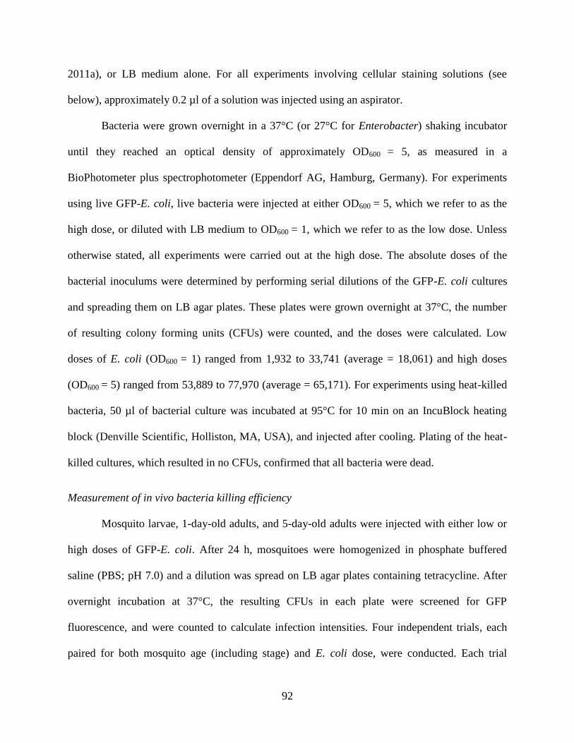
92
2011a), or LB medium alone. For all experiments involving cellular staining solutions (see
below), approximately 0.2 µl of a solution was injected using an aspirator.
Bacteria were grown overnight in a 37°C (or 27°C for Enterobacter) shaking incubator
until they reached an optical density of approximately OD600 = 5, as measured in a
BioPhotometer plus spectrophotometer (Eppendorf AG, Hamburg, Germany). For experiments
using live GFP-E. coli, live bacteria were injected at either OD600 = 5, which we refer to as the
high dose, or diluted with LB medium to OD600 = 1, which we refer to as the low dose. Unless
otherwise stated, all experiments were carried out at the high dose. The absolute doses of the
bacterial inoculums were determined by performing serial dilutions of the GFP-E. coli cultures
and spreading them on LB agar plates. These plates were grown overnight at 37°C, the number
of resulting colony forming units (CFUs) were counted, and the doses were calculated. Low
doses of E. coli (OD600 = 1) ranged from 1,932 to 33,741 (average = 18,061) and high doses
(OD600 = 5) ranged from 53,889 to 77,970 (average = 65,171). For experiments using heat-killed
bacteria, 50 µl of bacterial culture was incubated at 95°C for 10 min on an IncuBlock heating
block (Denville Scientific, Holliston, MA, USA), and injected after cooling. Plating of the heat-
killed cultures, which resulted in no CFUs, confirmed that all bacteria were dead.
Measurement of in vivo bacteria killing efficiency
Mosquito larvae, 1-day-old adults, and 5-day-old adults were injected with either low or
high doses of GFP-E. coli. After 24 h, mosquitoes were homogenized in phosphate buffered
saline (PBS; pH 7.0) and a dilution was spread on LB agar plates containing tetracycline. After
overnight incubation at 37°C, the resulting CFUs in each plate were screened for GFP
fluorescence, and were counted to calculate infection intensities. Four independent trials, each
paired for both mosquito age (including stage) and E. coli dose, were conducted. Each trial
93
consisted of approximately 10 mosquitoes per group, and the data were combined and analyzed
using the Kruskal-Wallis test, followed by Dunn’s multiple comparisons post hoc test.
Quantification of circulating hemocytes
For each mosquito, an incision was made across the lateral and ventral portions of the
suture that joins the 7th
and 8th
abdominal segment of naïve, injured (LB-injected), and E. coli-
infected larvae, 1-day-old adults, and 5-day-old adults at 24 h post-treatment. A finely pulled
glass capillary needle was then inserted into the thorax, approximately 200 µl of Schneider’s
Drosophila medium was perfused through the mosquito, and the diluted hemolymph was
collected within two 1 cm diameter etched rings on a Rite-On glass slide (Gold Seal; Portsmouth,
NH, USA) (King and Hillyer, 2013). After allowing the hemocytes to adhere to the slide for 20
min, cells were fixed and stained using Hema 3 (Fisher Scientific, Pittsburgh, PA, USA), and,
after drying, mounted with coverslips using Poly-Mount (Polysciences, Warrington, PA, USA)
(Coggins et al., 2012; Hillyer et al., 2005). The total number of hemocytes was then counted
under bright-field conditions at 40x magnification using either a Nikon 90i compound
microscope (Nikon, Tokyo, Japan) or an Olympus BH-2 microscope (Olympus, Tokyo, Japan).
Three independent trials consisting of approximately 5 individuals per treatment group were
conducted (N=14-16 per group). Data were analyzed by two-way ANOVA, using age (including
stage) and treatment as variables, followed by Šidák’s post hoc test.
Quantification of sessile hemocytes associated with the dorsal abdomen
The hemocytes from naïve, injured, and E. coli-challenged larvae, 1-day-old adults, and
5-day-old adults were fluorescently labelled in vivo at 24 h post-treatment by injecting live
animals with 67 µM of Vybrant CM-DiI Cell-Labeling Solution (Invitrogen, Carlsbad, CA,
USA) and 1.08 mM Hoechst 33342 nuclear stain (350/461; Invitrogen) in PBS, and incubating
94
them at 27°C and 75% relative humidity for 20 min (King and Hillyer, 2012; League and
Hillyer, 2016). Larvae were fixed for 5-10 min by immersion in 4% paraformaldehyde (Electron
Microscopy Sciences, Hatfield, PA, USA) and then dissected along a coronal plane. Specimens
were rinsed in PBS, and the dorsal abdomens, sans the internal organs, were mounted on a glass
slide using Aqua-Poly/Mount (Polysciences Inc., Warrington, PA, USA). Adults were fixed for
5-10 min by injection of 4% paraformaldehyde and then dissected along a coronal plane.
Specimens were placed in 0.5% Tween in PBS, rinsed briefly in PBS, and the dorsal abdomens,
sans the internal organs, were mounted on a glass slide using Aqua-Poly/Mount.
Dissected larval and adult dorsal abdomens were imaged under bright-field and
fluorescence illumination using a Nikon 90i microscope connected to a Nikon Digital Sight DS-
Qi1 Mc monochrome digital camera and Nikon’s Advanced Research NIS Elements software.
To render images with extended focal depth, Z-stacks of specimens were acquired using a linear
encoded Z-motor, and all images within a stack were combined to form a single focused image
using either NIS Elements’ Maximum Intensity Projection (for quantification) or Extended
Depth of Focus (EDF; for viewing) modules.
Sessile hemocytes attached to the dorsal abdominal wall were quantified by two methods.
In the first method, custom polygonal regions of interest (ROI) were drawn on the maximum
intensity projections to delineate the dorsal and lateral portions of abdominal segments 2-8, as
was done previously for the periostial regions and tracheal tufts of larvae (League and Hillyer,
2016), and the mean pixel intensities of CM-DiI fluorescence within the ROIs were measured
and recorded for each abdominal segment as well as for the entire dorsal and lateral abdomen
(Fig. 1). For each specimen, background fluorescence was removed by subtracting the intensity
values from the abdominal segments of each group’s corresponding unstained naïve specimens.
95
In the second method, EDF images were used to manually count the sessile hemocytes within
abdominal segments 4, 5 and 6, with these hemocytes categorized as being bound to one of three
tissues: (1) the cuticular integument, (2) the heart (periostial hemocytes), or (3) the trachea. Data
were analyzed by two-way ANOVA, using age (including stage) and treatment as variables,
followed by Šidák’s post hoc test.
Figure 1. Fluorescence emitted by sessile hemocytes in larvae and adults was measured
using custom-drawn regions of interest. Representative images of dissected larva (A), 1-day-
old adult (B) and 5-day-old adult (C) dorsal and lateral abdomens imaged under bright-field and
fluorescence illumination at 24 h post-infection with E. coli. Hemocytes were stained with CM-
DiI (red). Seven custom regions of interest (ROIs), encompassing abdominal segments 2-8, were
used to quantify mean fluorescence intensity of CM-DiI stained hemocytes. ROIs delineated the
dorsal (tergum) and lateral (pleuron) abdominal cuticle that lies between the abdominal sutures
of adjoining abdominal segments. Directional arrows: A, anterior; P, posterior; L, lateral.
Quantification of phagocytosis by circulating hemocytes
Larvae, 1-day-old adults, and 5-day-old adults were injected with E. coli, and 1 h later,
hemocytes were perfused as described above, except that each mosquito was perfused with
approximately 50 µl of Schneider’s medium and all the perfusate was collected onto a single
etched ring. After allowing the hemocytes to adhere to the slide for 20 min, cells were fixed for 5
min by adding 50 µl of PBS containing 4% formaldehyde and 0.03 mM Hoechst 33342 nuclear
96
stain. After removal of the fixation and staining solution, slides were mounted with coverslips
using Aqua-Poly/Mount. Specimens were then examined under 100x magnification using
simultaneous DIC and fluorescence illumination on a Nikon 90i microscope. For each mosquito,
the number of bacteria that had been phagocytosed by each of the first 100 hemocytes viewed
was counted. These values were used to calculate two parameters: (1) the phagocytic index,
which is defined as the percentage of cells that engage in phagocytosis; and (2) the phagocytic
capacity, which is defined as the average number of phagocytosed bacteria per hemocyte
(Coggins et al., 2012). Three independent trials consisting of 10 individuals per age were
conducted. Data were analyzed by the Kruskal-Wallis test, followed by Dunn’s post hoc test.
Quantification of antimicrobial hemolymph activity
Using a hemolymph extraction method we adapted from a protocol used for Drosophila
larvae and adults (Musselman et al., 2011), hemolymph was extracted at 24 h after treatment
from larvae, 1-day-old adults, and 5-day-old adults that were naïve, injured, or treated with heat-
killed E. coli, M. luteus, or Enterobacter sp. Briefly, an incision approximately 5 mm in length
was made into the bottom of a 0.6 ml microfuge tube using a feather blade, and freshly wounded
larvae or adults were placed inside. This tube was then inserted into a 1.5 ml microfuge tube,
which was centrifuged to collect the hemolymph. For each larval collection, up to ten individuals
were placed on a Kimwipe to remove all external water before placing the larvae on the inner
rim of the 0.6 ml tube. An insect pin was used to puncture the thorax of each larva and the tubes
were centrifuged for 10 sec in a mini centrifuge. This resulted in the collection of 1-2 µl of
hemolymph. For each adult collection, 20-30 individuals were cold-anesthetized and an insect
pin was used to puncture the lateral thorax. The mosquitoes were then placed in the 0.6 ml tube
97
and centrifuged at 2,152 rcf for 5 min at 4°C. This resulted in the collection of 1-2 µl of
hemolymph. Immediately after collection, hemolymph was stored in a -20°C freezer until use.
Antimicrobial activity of hemolymph was measured via a zone of inhibition assay that we
adapted from protocols developed for bumblebee workers (Moret and Schmid-Hempel, 2000)
and mealworm beetle larvae (Haine et al., 2008). Briefly, a 2 ml M. luteus culture was grown
overnight in LB broth at 37°C in a shaking incubator until it reached approximately OD600 = 10.
The entire M. luteus culture was then added to 20 ml of sterile, liquid 1% LB agar that was
maintained at 55°C by immersion in a water bath. The solution was mixed, and 5 ml of the M.
luteus-LB agar solution was poured into 9 cm diameter Petri dish plates. After solidifying,
equidistant holes were created in the agar 1.5 cm from the outer edge of the Petri dish by
piercing and suctioning using a 1 mm tip sterile glass Pasteur pipette. Each hole was then loaded
with 1 µl of hemolymph and, once the hemolymph had been absorbed by the agar, the plates
were placed upside down in a 37°C incubator for 16 h. Plates were then scanned at 1200 pixels
per inch using an Epson Perfection V600 scanner (Epson America, Inc., Long Beach, CA), and
images were analyzed using ImageJ software (Schneider et al., 2012). For each sample, the
diameters of the individual zones of bacterial growth inhibition were measured three times at
different locations, and the average of these diameters was used to calculate the areas of the
zones of inhibition. Five independent trials for each of the three bacterial treatments were
conducted, each consisting of 2-3 larval and 1-2 adult pooled hemolymph samples per treatment
group (naïve, injured, and treated with heat-killed bacteria; N=12-15 and N=5-6 for larval and
adult groups, respectively). The data were combined and analyzed by two-way ANOVA, using
age (including stage) and treatment as variables, followed by Šidák’s post hoc test.
98
Quantification of the phenoloxidase activity of hemolymph
Using the method described for the zone of inhibition assay, hemolymph was extracted
from naïve, injured, and E. coli-infected larvae, 1-day-old adults, and 5-day-old adults at 24 h
post-treatment. Because we noted that using PBS as a solvent causes auto-oxidation of DOPA,
and hence darkening that is not caused by phenoloxidase activity, all experiments utilized sterile
water as the diluent, and not PBS. After adding 1 µl of extracted hemolymph to 50 µl of water,
10 µl of the diluted hemolymph solution (or water alone in the case of the negative control) was
added to a cuvette containing 90 µl of one of the following three solutions: (1) 4 mg/mL 3,4-
Dihydroxy-L-phenylalanine (L-Dopa; Sigma); (2) 20 mg/mL Sodium Diethyldithiocarbamate
Trihydrate (DETC; Fisher Scientific) and 4 mg/mL L-DOPA; or (3) water alone. Absorbance
readings were measured at 490 nm every 5 min for 30 min using a BioPhotometer plus
spectrophotometer. Five independent trials were conducted, each consisting of 1 larval or adult
sample per treatment group. The data were combined and analyzed by repeated measures two-
way ANOVA, using age (including stage) and treatment as variables, followed by Šidák’s post
hoc test.
Quantification of immunity gene expression by qPCR
Total RNA was extracted from the whole body of approximately forty larvae, twenty 1-
day-old adults and twenty 5-day-old adults that were naïve, or had been injected 24 h earlier with
LB (injury), or GFP-E. coli. RNA was purified after homogenization in TRIzol reagent
(Invitrogen), and repurified using the PureLink Micro-to-Midi Total RNA Purification System
(Invitrogen). For each sample, 5 μg of RNA was treated with RQ1 RNase-Free DNAse I
(Promega, Madison, WI, USA) to remove any contaminating genomic DNA, and then used as
template for cDNA synthesis using an Oligo(dT)20 primer and the SuperScript III First-Strand
99
Synthesis System for RT-PCR (Invitrogen). Real-time quantitative PCR was performed using
Power SYBR Green PCR Master Mix (Applied Biosystems, Foster City, CA) on a Bio Rad CFX
Connect Real-Time PCR Detection System (Hercules, CA, USA). Relative quantification of
mRNA levels was performed using the 2−ΔΔC
T method (Livak and Schmittgen, 2001), and mRNA
levels were calculated relative to the naïve groups of each mosquito age. In all experiments,
ribosomal protein gene RPS7 was used as the reference gene, and the ribosomal protein
gene RPS17 was used as a control. Primer sequences are listed in Appendix A. Melting curve
analyses after RT-PCR confirmed that the cDNAs used in the experiments were free of genomic
DNA contamination and that only the gene of interest was amplified. Three independent trials
were conducted, and each trial was analyzed in duplicate. Data is presented as the average fold-
change relative to the naïve group of a given life stage or adult age. Data for each gene were
analyzed separately by age (stage) using the Kruskal-Wallis test, followed by Dunn’s post hoc
test.
Results
Larvae and newly-emerged adult mosquitoes kill bacteria in their hemocoels more efficiently
than older adult mosquitoes
To begin to test whether larval and adult mosquitoes have different immune
proficiencies, we infected larvae and differently aged adults with either low (OD600 = 1) or high
(OD600 = 5) doses of E. coli, and measured their bacterial load 24 h later. We found that, for
both doses, bacterial killing proficiency differed significantly between the larval and adult stages
(Fig. 2; Kruskal-Wallis P<0.0001 for both doses). Specifically, at 24 h following a low dose
infection, the bacterial load in fourth instar larvae, 1-day-old (newly-emerged) adults, and 5-day-
old adults had decreased by 94%, 86% and 22%, respectively. At 24 h following a high dose
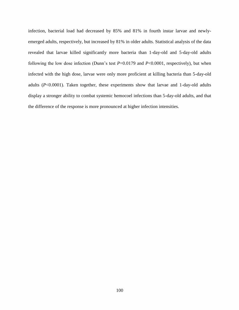
100
infection, bacterial load had decreased by 85% and 81% in fourth instar larvae and newly-
emerged adults, respectively, but increased by 81% in older adults. Statistical analysis of the data
revealed that larvae killed significantly more bacteria than 1-day-old and 5-day-old adults
following the low dose infection (Dunn’s test P=0.0179 and P<0.0001, respectively), but when
infected with the high dose, larvae were only more proficient at killing bacteria than 5-day-old
adults (P<0.0001). Taken together, these experiments show that larvae and 1-day-old adults
display a stronger ability to combat systemic hemocoel infections than 5-day-old adults, and that
the difference of the response is more pronounced at higher infection intensities.
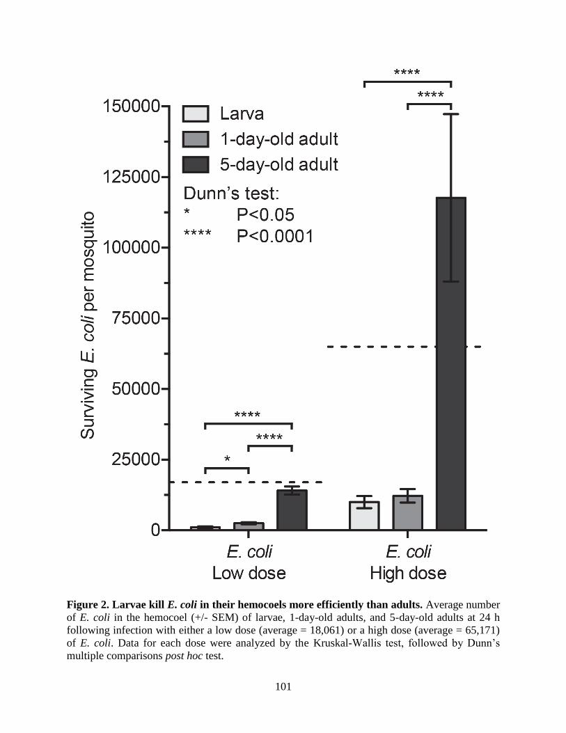
101
Figure 2. Larvae kill E. coli in their hemocoels more efficiently than adults. Average number
of E. coli in the hemocoel (+/- SEM) of larvae, 1-day-old adults, and 5-day-old adults at 24 h
following infection with either a low dose (average = 18,061) or a high dose (average = 65,171)
of E. coli. Data for each dose were analyzed by the Kruskal-Wallis test, followed by Dunn’s
multiple comparisons post hoc test.
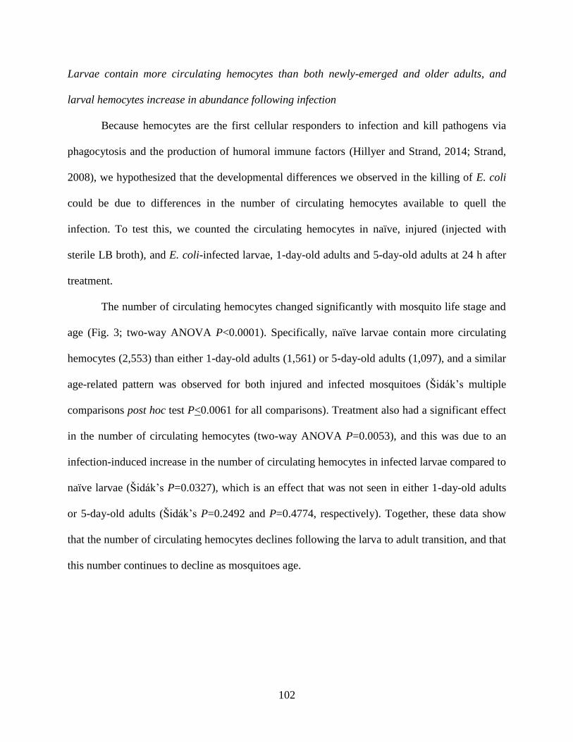
102
Larvae contain more circulating hemocytes than both newly-emerged and older adults, and
larval hemocytes increase in abundance following infection
Because hemocytes are the first cellular responders to infection and kill pathogens via
phagocytosis and the production of humoral immune factors (Hillyer and Strand, 2014; Strand,
2008), we hypothesized that the developmental differences we observed in the killing of E. coli
could be due to differences in the number of circulating hemocytes available to quell the
infection. To test this, we counted the circulating hemocytes in naïve, injured (injected with
sterile LB broth), and E. coli-infected larvae, 1-day-old adults and 5-day-old adults at 24 h after
treatment.
The number of circulating hemocytes changed significantly with mosquito life stage and
age (Fig. 3; two-way ANOVA P<0.0001). Specifically, naïve larvae contain more circulating
hemocytes (2,553) than either 1-day-old adults (1,561) or 5-day-old adults (1,097), and a similar
age-related pattern was observed for both injured and infected mosquitoes (Šidák’s multiple
comparisons post hoc test P<0.0061 for all comparisons). Treatment also had a significant effect
in the number of circulating hemocytes (two-way ANOVA P=0.0053), and this was due to an
infection-induced increase in the number of circulating hemocytes in infected larvae compared to
naïve larvae (Šidák’s P=0.0327), which is an effect that was not seen in either 1-day-old adults
or 5-day-old adults (Šidák’s P=0.2492 and P=0.4774, respectively). Together, these data show
that the number of circulating hemocytes declines following the larva to adult transition, and that
this number continues to decline as mosquitoes age.
103
Figure 3. Larvae contain more circulating hemocytes than adults. Average number of
circulating hemocytes (+/- SEM) in naïve, injured, or E. coli-infected (high dose) larvae, 1-day-
old adults, and 5-day-old adults at 24 h post-treatment. Data were analyzed by two-way
ANOVA, followed by Šidák’s multiple comparisons post hoc test.
The spatial arrangement of sessile hemocytes differs between larvae and differently aged adults,
and sessile hemocytes increase after infection in newly-emerged adults
We have previously shown that, regardless of the age of an adult mosquito, 75% of
hemocytes circulate with the hemolymph whereas 25% are sessile, or attached to tissues (King
and Hillyer, 2013). Furthermore, the majority of sessile hemocytes are present on the dorsal
abdominal wall or the tissues associated with it (King and Hillyer, 2013). Thus, to compare the
sessile hemocyte populations of larvae and adults, we labeled hemocytes in vivo using CM-DiI
and quantified fluorescence intensity in the dorsal abdomen (Fig. 1).
Spatially, sessile hemocytes in larvae and one-day-old adults are arranged in segmental
bands that encircle each abdominal segment, but hemocytes do not retain this clear segmental
pattern in 5-day-old adults due to fewer hemocytes on the dorsal tergum (Fig. 4A-F).
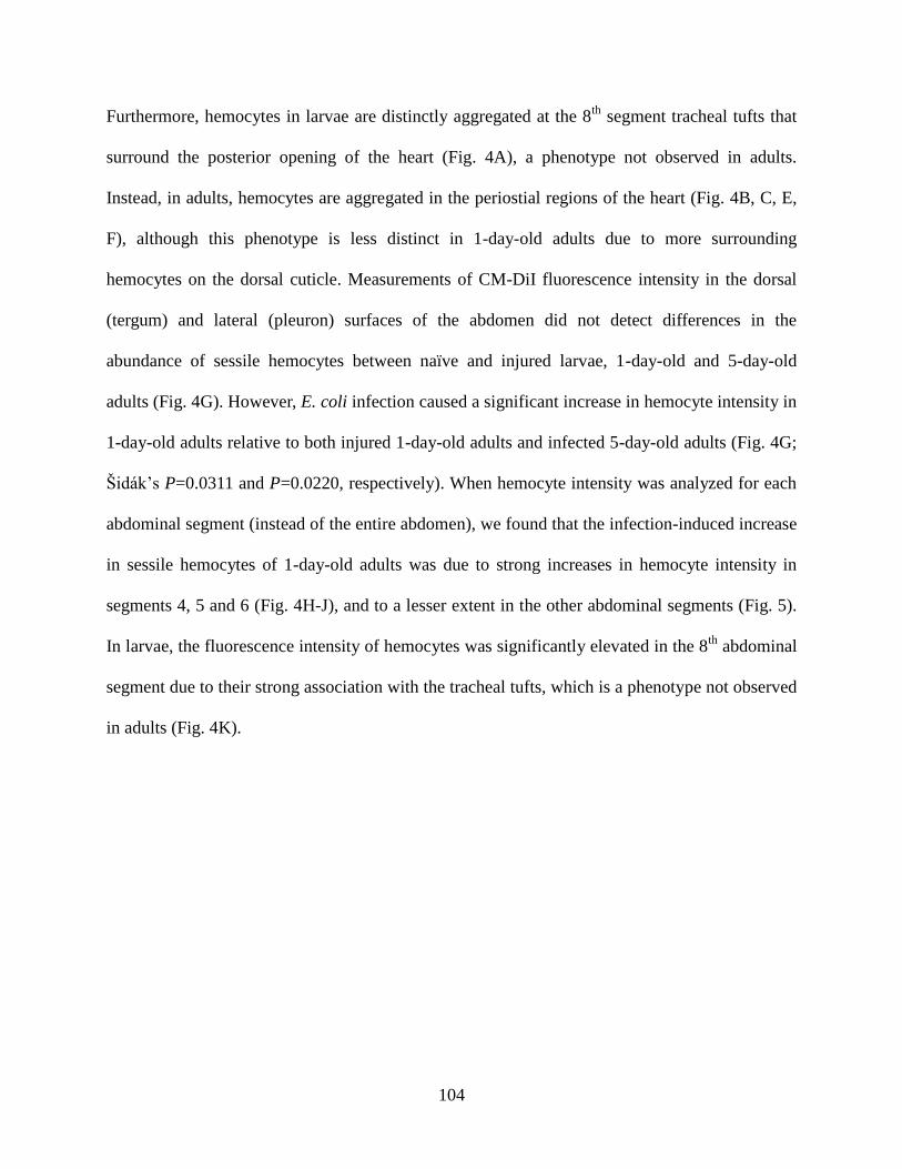
104
Furthermore, hemocytes in larvae are distinctly aggregated at the 8th
segment tracheal tufts that
surround the posterior opening of the heart (Fig. 4A), a phenotype not observed in adults.
Instead, in adults, hemocytes are aggregated in the periostial regions of the heart (Fig. 4B, C, E,
F), although this phenotype is less distinct in 1-day-old adults due to more surrounding
hemocytes on the dorsal cuticle. Measurements of CM-DiI fluorescence intensity in the dorsal
(tergum) and lateral (pleuron) surfaces of the abdomen did not detect differences in the
abundance of sessile hemocytes between naïve and injured larvae, 1-day-old and 5-day-old
adults (Fig. 4G). However, E. coli infection caused a significant increase in hemocyte intensity in
1-day-old adults relative to both injured 1-day-old adults and infected 5-day-old adults (Fig. 4G;
Šidák’s P=0.0311 and P=0.0220, respectively). When hemocyte intensity was analyzed for each
abdominal segment (instead of the entire abdomen), we found that the infection-induced increase
in sessile hemocytes of 1-day-old adults was due to strong increases in hemocyte intensity in
segments 4, 5 and 6 (Fig. 4H-J), and to a lesser extent in the other abdominal segments (Fig. 5).
In larvae, the fluorescence intensity of hemocytes was significantly elevated in the 8th
abdominal
segment due to their strong association with the tracheal tufts, which is a phenotype not observed
in adults (Fig. 4K).
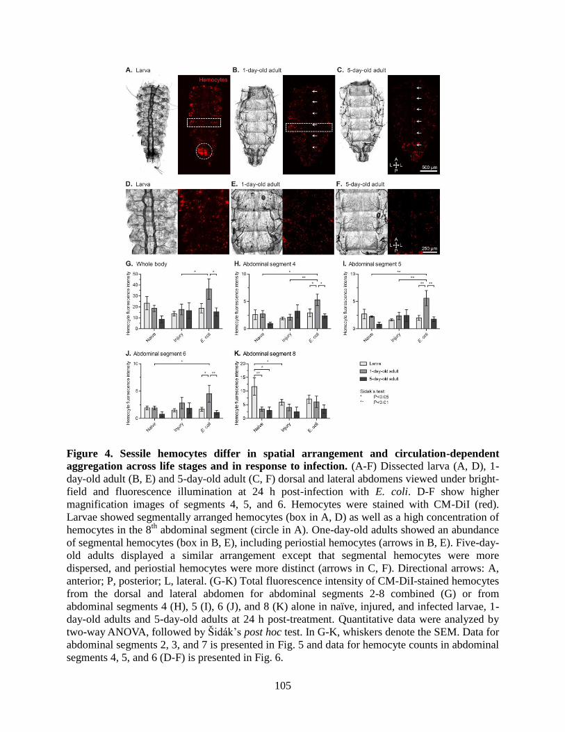
105
Figure 4. Sessile hemocytes differ in spatial arrangement and circulation-dependent
aggregation across life stages and in response to infection. (A-F) Dissected larva (A, D), 1-
day-old adult (B, E) and 5-day-old adult (C, F) dorsal and lateral abdomens viewed under bright-
field and fluorescence illumination at 24 h post-infection with E. coli. D-F show higher
magnification images of segments 4, 5, and 6. Hemocytes were stained with CM-DiI (red).
Larvae showed segmentally arranged hemocytes (box in A, D) as well as a high concentration of
hemocytes in the 8th
abdominal segment (circle in A). One-day-old adults showed an abundance
of segmental hemocytes (box in B, E), including periostial hemocytes (arrows in B, E). Five-day-
old adults displayed a similar arrangement except that segmental hemocytes were more
dispersed, and periostial hemocytes were more distinct (arrows in C, F). Directional arrows: A,
anterior; P, posterior; L, lateral. (G-K) Total fluorescence intensity of CM-DiI-stained hemocytes
from the dorsal and lateral abdomen for abdominal segments 2-8 combined (G) or from
abdominal segments 4 (H), 5 (I), 6 (J), and 8 (K) alone in naïve, injured, and infected larvae, 1-
day-old adults and 5-day-old adults at 24 h post-treatment. Quantitative data were analyzed by
two-way ANOVA, followed by Šidák’s post hoc test. In G-K, whiskers denote the SEM. Data for
abdominal segments 2, 3, and 7 is presented in Fig. 5 and data for hemocyte counts in abdominal
segments 4, 5, and 6 (D-F) is presented in Fig. 6.
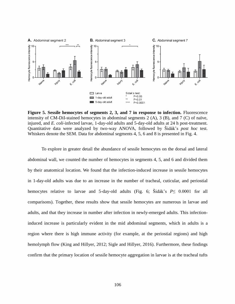
106
Figure 5. Sessile hemocytes of segments 2, 3, and 7 in response to infection. Fluorescence
intensity of CM-DiI-stained hemocytes in abdominal segments 2 (A), 3 (B), and 7 (C) of naïve,
injured, and E. coli-infected larvae, 1-day-old adults and 5-day-old adults at 24 h post-treatment.
Quantitative data were analyzed by two-way ANOVA, followed by Šidák’s post hoc test.
Whiskers denote the SEM. Data for abdominal segments 4, 5, 6 and 8 is presented in Fig. 4.
To explore in greater detail the abundance of sessile hemocytes on the dorsal and lateral
abdominal wall, we counted the number of hemocytes in segments 4, 5, and 6 and divided them
by their anatomical location. We found that the infection-induced increase in sessile hemocytes
in 1-day-old adults was due to an increase in the number of tracheal, cuticular, and periostial
hemocytes relative to larvae and 5-day-old adults (Fig. 6; Šidák’s P≤ 0.0001 for all
comparisons). Together, these results show that sessile hemocytes are numerous in larvae and
adults, and that they increase in number after infection in newly-emerged adults. This infection-
induced increase is particularly evident in the mid abdominal segments, which in adults is a
region where there is high immune activity (for example, at the periostial regions) and high
hemolymph flow (King and Hillyer, 2012; Sigle and Hillyer, 2016). Furthermore, these findings
confirm that the primary location of sessile hemocyte aggregation in larvae is at the tracheal tufts
107
of the eighth abdominal segment, which are structures that surround the posterior openings of the
larval heart (League and Hillyer, 2016).
Figure 6. The distribution of sessile hemocytes in the trachea, cuticle, and periostial regions
of abdominal segments 4, 5, and 6 varies with life stage and with infection state. Total
number of CM-DiI-stained hemocytes attached to the trachea (A), cuticle (B, integument) and
periostial regions of the heart (C) in abdominal segments 4, 5, and 6 of naïve, injured, and E.
coli-infected larvae, 1-day-old adults and 5-day-old adults at 24 h post-treatment. Data were
analyzed by two-way ANOVA, followed by Šidák’s post hoc test. Whiskers denote the SEM.
The phagocytic activity of individual hemocytes is higher in adult mosquitoes compared to
larvae
Our initial hypothesis postulated that changes in the abundance of hemocytes
underscored the increased resistance of larvae and newly-emerged adults to a bacterial infection.
However, differences in immune proficiency may also arise from differences in the frequency or
degree to which hemocytes engage in phagocytosis. To test phagocytic activity across life stages,
larvae and adults were infected with GFP-E. coli and the phagocytic activity of circulating
hemocytes was quantified 1 h later. The phagocytic index, defined as the percentage of
hemocytes that engage in phagocytosis following an immune challenge (Coggins et al., 2012;
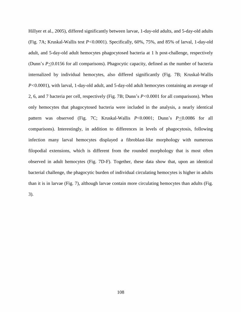
108
Hillyer et al., 2005), differed significantly between larvae, 1-day-old adults, and 5-day-old adults
(Fig. 7A; Kruskal-Wallis test P<0.0001). Specifically, 60%, 75%, and 85% of larval, 1-day-old
adult, and 5-day-old adult hemocytes phagocytosed bacteria at 1 h post-challenge, respectively
(Dunn’s P<0.0156 for all comparisons). Phagocytic capacity, defined as the number of bacteria
internalized by individual hemocytes, also differed significantly (Fig. 7B; Kruskal-Wallis
P<0.0001), with larval, 1-day-old adult, and 5-day-old adult hemocytes containing an average of
2, 6, and 7 bacteria per cell, respectively (Fig. 7B; Dunn’s P<0.0001 for all comparisons). When
only hemocytes that phagocytosed bacteria were included in the analysis, a nearly identical
pattern was observed (Fig. 7C; Kruskal-Wallis P<0.0001; Dunn’s P<0.0086 for all
comparisons). Interestingly, in addition to differences in levels of phagocytosis, following
infection many larval hemocytes displayed a fibroblast-like morphology with numerous
filopodial extensions, which is different from the rounded morphology that is most often
observed in adult hemocytes (Fig. 7D-F). Together, these data show that, upon an identical
bacterial challenge, the phagocytic burden of individual circulating hemocytes is higher in adults
than it is in larvae (Fig. 7), although larvae contain more circulating hemocytes than adults (Fig.
3).
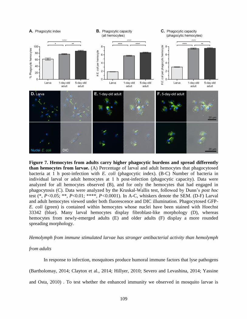
109
Figure 7. Hemocytes from adults carry higher phagocytic burdens and spread differently
than hemocytes from larvae. (A) Percentage of larval and adult hemocytes that phagocytosed
bacteria at 1 h post-infection with E. coli (phagocytic index). (B-C) Number of bacteria in
individual larval or adult hemocytes at 1 h post-infection (phagocytic capacity). Data were
analyzed for all hemocytes observed (B), and for only the hemocytes that had engaged in
phagocytosis (C). Data were analyzed by the Kruskal-Wallis test, followed by Dunn’s post hoc
test (*, P<0.05; **, P<0.01; ****, P<0.0001). In A-C, whiskers denote the SEM. (D-F) Larval
and adult hemocytes viewed under both fluorescence and DIC illumination. Phagocytosed GFP-
E. coli (green) is contained within hemocytes whose nuclei have been stained with Hoechst
33342 (blue). Many larval hemocytes display fibroblast-like morphology (D), whereas
hemocytes from newly-emerged adults (E) and older adults (F) display a more rounded
spreading morphology.
Hemolymph from immune stimulated larvae has stronger antibacterial activity than hemolymph
from adults
In response to infection, mosquitoes produce humoral immune factors that lyse pathogens
(Bartholomay, 2014; Clayton et al., 2014; Hillyer, 2010; Severo and Levashina, 2014; Yassine
and Osta, 2010) . To test whether the enhanced immunity we observed in mosquito larvae is
110
correlated with enhanced humoral antimicrobial activity in their hemolymph, we employed a
zone of inhibition assay to measure the general antibacterial activity of hemolymph.
At 24 h following immune stimulation with heat-killed E. coli, the antibacterial activity
of hemolymph differed significantly between larvae, 1-day-old, and 5-day-old adults (Fig. 8A-B;
two-way ANOVA P<0.0001), and this was due to the significantly higher antibacterial activity
of larval hemolymph compared to both adult groups (Šidák’s P<0.0001 for both comparisons).
Furthermore, this difference in antibacterial activity was due to an induced response, rather than
a constitutive response, as the antibacterial activity in the hemolymph of naïve and injured
individuals did not differ between larvae and adults, with the exception of when injured larvae
were compared to 1-day-old adults (Šidák’s P=0.0387). To test whether this finding was unique
to immune stimulation with Gram (-) E. coli, we repeated the experiment by stimulating
mosquitoes with heat-killed Gram (+) M. luteus (Fig. 8C-D) and heat-killed Gram (-)
Enterobacter sp. isolate Ag1 (Fig. 8E-F), a bacterial strain that naturally colonizes the midgut of
A. gambiae and is a core adult bacterial symbiont taxa (Jiang et al., 2012; Wang et al., 2011a). In
both sets of experiments, antibacterial activity of hemolymph was significantly higher in larvae
compared to adults (Fig. 8D, F; two-way ANOVA P<0.0001 for both bacteria; Šidák’s
P<0.0252 for all comparisons). This resulted from an induced response, as the antibacterial
activity of hemolymph from naïve and injured mosquitoes did not differ between larvae and
adults (Šidák’s P>0.2856 for all comparisons). In sum, these experiments show that the strength
of humoral antibacterial activity of hemolymph is stronger in larvae compared to adults.
111
Figure 8. Larvae display stronger antibacterial humoral immunity compared to adults. (A-
B) Zone of inhibition plating assay (A), including quantitative measurement of the zones of
inhibition (B), showing the extent of hemolymph lytic activity in mosquitoes that were naïve,
injured, or challenged with heat-killed E. coli 24 h earlier. (C-F) Similar experiment except using
heat-killed M. luteus (C, D) or heat-killed Enterobacter sp. (E, F) as the challenge agent.
Abbreviations in panels A, C, and E: L4, 4th
instar larvae; 1D, 1-day-old adult; 5D, 5-day-old
adult; N, naïve; LB, injured; E.c., E. coli; M.l., M. luteus; Eb, Enterobacter. In B, D, and F, data
were analyzed by two-way ANOVA, followed by Šidák’s post hoc test. Whiskers denote the
S.E.M.
Phenoloxidase activity is higher in larvae compared to adults
While conducting experiments measuring the antibacterial activity of hemolymph, we
noticed that hemolymph, especially from larvae, had a tendency to darken rapidly after
extraction, a phenomenon suggestive of the activation of phenoloxidase-mediated melanization.
Because melanization is a major component of the humoral immune response of mosquitoes
(Christensen et al., 2005), we tested the relative strength of melanization in larval and adult
hemolymph using a phenoloxidase activity assay (Sidjanski et al., 1997; Thomas et al., 2011)
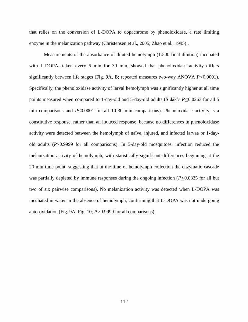
112
that relies on the conversion of L-DOPA to dopachrome by phenoloxidase, a rate limiting
enzyme in the melanization pathway (Christensen et al., 2005; Zhao et al., 1995) .
Measurements of the absorbance of diluted hemolymph (1:500 final dilution) incubated
with L-DOPA, taken every 5 min for 30 min, showed that phenoloxidase activity differs
significantly between life stages (Fig. 9A, B; repeated measures two-way ANOVA P<0.0001).
Specifically, the phenoloxidase activity of larval hemolymph was significantly higher at all time
points measured when compared to 1-day-old and 5-day-old adults (Šidák’s P<0.0263 for all 5
min comparisons and P<0.0001 for all 10-30 min comparisons). Phenoloxidase activity is a
constitutive response, rather than an induced response, because no differences in phenoloxidase
activity were detected between the hemolymph of naïve, injured, and infected larvae or 1-day-
old adults (P>0.9999 for all comparisons). In 5-day-old mosquitoes, infection reduced the
melanization activity of hemolymph, with statistically significant differences beginning at the
20-min time point, suggesting that at the time of hemolymph collection the enzymatic cascade
was partially depleted by immune responses during the ongoing infection (P<0.0335 for all but
two of six pairwise comparisons). No melanization activity was detected when L-DOPA was
incubated in water in the absence of hemolymph, confirming that L-DOPA was not undergoing
auto-oxidation (Fig. 9A; Fig. 10; P>0.9999 for all comparisons).
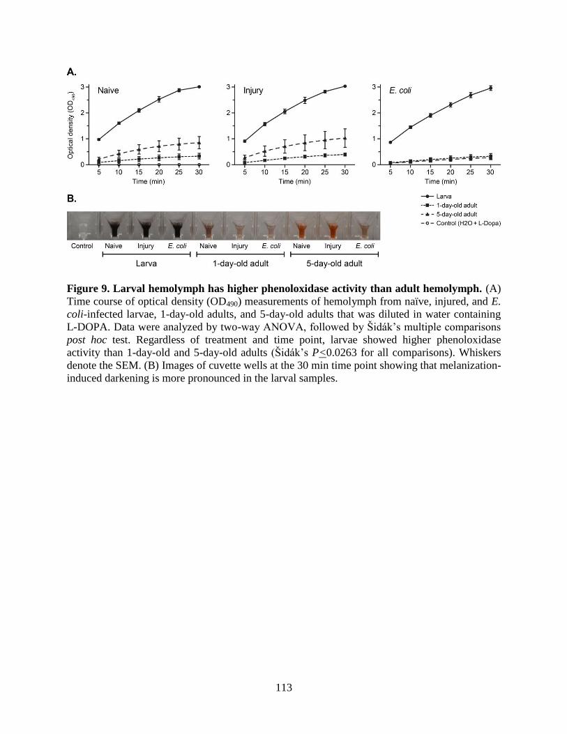
113
Figure 9. Larval hemolymph has higher phenoloxidase activity than adult hemolymph. (A)
Time course of optical density (OD490) measurements of hemolymph from naïve, injured, and E.
coli-infected larvae, 1-day-old adults, and 5-day-old adults that was diluted in water containing
L-DOPA. Data were analyzed by two-way ANOVA, followed by Šidák’s multiple comparisons
post hoc test. Regardless of treatment and time point, larvae showed higher phenoloxidase
activity than 1-day-old and 5-day-old adults (Šidák’s P<0.0263 for all comparisons). Whiskers
denote the SEM. (B) Images of cuvette wells at the 30 min time point showing that melanization-
induced darkening is more pronounced in the larval samples.
114
Figure 10. Phenoloxidase-based melanization of endogenous substrates is higher in larvae
than in adults, and melanization of exogenous substrates is completely inhibited by DETC.
(A-B) Time course of optical density (OD490) measurements of hemolymph from naïve, injured,
and E. coli-infected larvae, 1-day-old adults and 5-day-old adults diluted in water. The scale in
panel A is amplified and separated by treatment in panel B. Larval hemolymph was significantly
darker than adult hemolymph (Šidák’s P<0.0086 for all comparisons) and melanization levels
did not change when comparing the initial and final readings of any age or treatment group
(Šidák’s P>0.1690 for all comparisons). (C-D) Time course of optical density (OD490)
measurements of hemolymph from naïve, injured, and E. coli-infected larvae, 1-day-old adults
and 5-day-old adults diluted in saturated L-DOPA with DETC, which is a phenoloxidase
inhibitor. The scale in panel C is amplified and separated by treatment in panel D. Larval
hemolymph was significantly darker than adult hemolymph (Šidák’s P<0.0013 for all
comparisons) and melanization levels did not change from the initial to final readings of any age
or treatment group (P>0.5840 for all comparisons). Data were analyzed by two-way ANOVA,
followed by Šidák’s post hoc test. Whiskers denote the SEM.
115
To quantify the melanization of endogenous substrates in diluted hemolymph, as opposed
to the conversion of exogenous L-DOPA as in the above experiment, we repeated the experiment
by diluting hemolymph samples in water alone, without L-DOPA. As expected, because of the
absence of exogenous substrate, the absorbance values in all samples were very low (Fig. 10A).
Nevertheless, larval hemolymph was significantly darker than adult hemolymph (Fig. 10B;
Šidák’s P<0.0086 for all comparisons), and no change in melanization occurred when comparing
the initial and final readings, indicating that by the initial reading all endogenous substrates had
been consumed in melanization reactions (P>0.1690 for all comparisons).
Finally, to ensure that the darkening of hemolymph did not occur because of the activity
of enzymes other than phenoloxidase, including iron-based peroxidase and catalase (Christensen
et al., 2005), we mixed diluted hemolymph with a solution containing both L-DOPA and
diethyldithiocarbamate (DETC), a copper-specific chelator that inhibits copper-based
phenoloxidase (Ajamhassani et al., 2012; Christensen et al., 2005; Feng et al., 2008; Ryazanova
et al., 2012). Although larval hemolymph was again significantly darker than adult hemolymph
because of the rapid melanization of endogenous substrates soon after hemolymph collection
(Fig. 10C, D; Šidák’s P<0.0013 for all comparisons), melanization levels did not change
following the addition of the solution of L-DOPA and DETC (P>0.5840 for all comparisons),
confirming that the melanization activity of hemolymph is driven by the activity of
phenoloxidase. Together, these findings demonstrate that larval hemolymph contains greater
melanization potential than adult hemolymph, and that this is due to the activity of
phenoloxidase.
116
Infection-induced transcriptional upregulation of immunity-related genes is higher in larvae
than in adults
To determine whether larvae and differently aged adults differ with respect to the
expression of immunity genes, relative mRNA fold change was assayed by qPCR at 24 h post-
treatment in naïve, injured, and E. coli-infected larvae, 1-day-old adults, and 5-day-old adults
(Fig. 11; Appendix A and B). A broad range of immune responses was surveyed by measuring
the transcription of 24 immunity genes representing four major categories of immune function:
(1) pathogen recognition, specifically CTL4 (Osta et al., 2004; Pinto et al., 2009; Schnitger et al.,
2009), FREP13 (Christophides et al., 2002; Dong and Dimopoulos, 2009), GNBPB4 (Warr et al.,
2008), TEP1 (Levashina et al., 2001; Moita et al., 2005), Eater, Nimrod, and Draper (Estévez-
Lao and Hillyer, 2014), and SCRBQ2 (Dimopoulos et al., 2002; González-Lázaro et al., 2009);
(2) signal modulation, specifically CLIPB15 (Christophides et al., 2002; Pinto et al., 2009; Volz
et al., 2005) and SRPN6 (Abraham et al., 2005; Pinto et al., 2008); (3) signal transduction,
specifically Toll pathway members MYD88 and CACTUS (Christophides et al., 2002; Ramirez
and Dimopoulos, 2010; Xi et al., 2008), Imd pathway members CASPL1 and CASPAR (Garver et
al., 2012), and Jak/Stat pathway members AGSTAT-A and PIAS (Dong et al., 2012; Gupta et al.,
2009; Souza-Neto et al., 2009); and (4) effector responses, specifically CECA (Christophides et
al., 2002; Kim et al., 2004; Vizioli et al., 2000), GAM1 (Christophides et al., 2002; Vizioli et al.,
2001a), DEF1 (Blandin et al., 2002; Christophides et al., 2002; Dimopoulos et al., 1997;
Richman et al., 1996; Vizioli et al., 2001b), PPO1 (Ahmed et al., 1999; Baton et al., 2009;
Christophides et al., 2002; Müller et al., 1999), PPO6 (Baton et al., 2009; Müller et al., 1999),
LYSC1 (Kajla et al., 2010; Kajla et al., 2011; Li et al., 2005; Li and Paskewitz, 2006), NOS
(Hillyer and Estévez-Lao, 2010), and DUOX (Kumar et al., 2003; Kumar et al., 2010; Molina-
117
Cruz et al., 2008; Oliveira et al., 2011). In addition, the expression of two genes involved in the
ecdysteroid biosynthetic pathway, CYP302A1 and CYP315A1 (Pondeville et al., 2008; Sieglaff et
al., 2005), was measured, as ecdysone peaks in the late larval and pupal stages, and ecdysone
induces the transcriptional upregulation of phenoloxidase genes in an A. gambiae hemocyte-like
cell line (Ahmed et al., 1999; Müller et al., 1999). RPS7 was used as the reference gene, and
RPS17 was used as a control (Coggins et al., 2012).
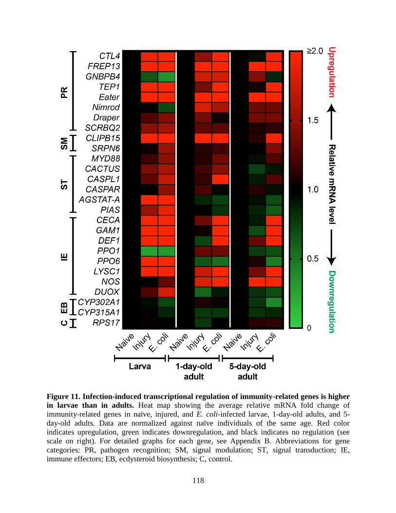
118
Figure 11. Infection-induced transcriptional regulation of immunity-related genes is higher
in larvae than in adults. Heat map showing the average relative mRNA fold change of
immunity-related genes in naïve, injured, and E. coli-infected larvae, 1-day-old adults, and 5-
day-old adults. Data are normalized against naïve individuals of the same age. Red color
indicates upregulation, green indicates downregulation, and black indicates no regulation (see
scale on right). For detailed graphs for each gene, see Appendix B. Abbreviations for gene
categories: PR, pathogen recognition; SM, signal modulation; ST, signal transduction; IE,
immune effectors; EB, ecdysteroid biosynthesis; C, control.
119
In larvae, 17 immunity genes were either strongly (greater than 2-fold; 11) or weakly
(between 1.5 and 2-fold; 6) upregulated by infection relative to naïve larvae, whereas in 1-day-
old and 5-day-old adults, only 13 (11 strongly and 2 weakly) and 10 (9 strongly and 1 weakly)
immunity genes were upregulated by infection, respectively, relative to naïve adults of their
respective age (Fig 11; Appendix B). Transcription of ecdysteroid biosynthesis genes and, as
expected, RPS17 was not regulated by infection in any age group. In general, larvae and adults
showed a similar level of induction of some pathogen recognition and signal modulation genes,
as well as some immune effector genes, such as cecropin (CECA), gambicin (GAM1), defensin
(DEF1) and lysozyme (LYSC1).
When comparing the levels of induction, larvae showed greater than 2-fold higher
induction (that is, the fold change of the larval group divided by the fold change of the adult
group was >2) of AGSTAT-A, PIAS, PPO6, and dual oxidase (DUOX) relative to both adult
groups. Both adult groups showed greater than 2-fold higher induction of nitric oxide synthase
(NOS) and a fibrinogen-related protein gene (FREP13, also known as FBN9) relative to larvae,
and 1-day-old adults showed greater than 2-fold higher induction of a CLIP domain serine
protease (CLIPB15) relative to larvae, despite FREP13 and CLIPB15 being greater than 2-fold
upregulated in both life stages (Fig. 11; Appendix B).
In addition to differences in the level of induction of specific immunity genes across life
stages, some genes were only upregulated in a single life stage. For example, infection in larvae,
but not adults, induced the upregulation of Jak/Stat essential pathway genes AGSTAT-A and
PIAS, as well as the immune effectors PPO6 and DUOX. Conversely, infection in adults, but not
larvae, strongly induced the upregulation of NOS. Interestingly, although the Gram (-) binding
protein gene, GNBPB4, was transcriptionally downregulated in larvae following infection, when
120
all the data was instead normalized against naïve 5-day-old adults, basal expression of GNBPB4
in naïve larvae was 470- and 710-fold higher than in naïve 1-day-old and 5-day-old adults,
respectively. A similar finding was made with respect to one of the phenoloxidase genes assayed,
PPO1, which was expressed 170- and 110-fold higher in naïve larvae relative to naïve 1-day-old
and 5-day-old adults, respectively. Collectively, these data show that induction of immunity
genes differs with life stage and adult age, with infection-induced expression being highest in
larvae and declining shortly after eclosion and as adults age.
Discussion
Mosquitoes are susceptible to infections during all stages of their life cycle, and in
response have evolved a robust innate immune system (Hillyer, 2010). Because the adult stage is
responsible for the transmission of disease-causing pathogens such as malaria (Clayton et al.,
2014; Severo and Levashina, 2014), the vast majority of research on mosquito immunology has
focused on that life stage, and in particular, on the adult females that feed on blood. In contrast to
this, research on the immune system of the larvae of other insects, such as Manduca sexta,
Bombyx mori, and Drosophila melanogaster has proven invaluable for uncovering fundamental
aspects of the cellular and humoral immune response of insects, and animals in general (Honti et
al., 2014; Jiang et al., 2010; Lemaitre and Hoffmann, 2007). Nevertheless, lacking in Insecta are
comparative studies that examine differences in immunity across the different life stages of a
given species. As noted by Fellous and Lazzaro in 2011, “Almost all studies of the immune
system of animals with metamorphosis have focused on either larval or on adult immunity,
implicitly assuming that these traits are either perfectly correlated or evolutionarily independent”
(Fellous and Lazzaro, 2011). Because mosquito larvae live in aquatic environments rife with
microorganisms and survival through the larval stages is required to reach sexual maturity, at the
121
onset of this study we hypothesized that larvae have evolved more proficient means of
neutralizing infections than adults. To compare larval and adult immunity in A. gambiae, we
measured a broad range of immune parameters that were previously unexamined in larvae, and
show that immune activity is strongest in larvae and wanes in the adult life stage, which is
indicative of immune senescence (Table 1). Furthermore, because immunity phenotypes differ
between life stages that are separated by metamorphosis, these findings suggest that adaptive
decoupling, or the independent evolution of larval and adult traits made possible by
metamorphosis (Moran, 1994), has occurred in A. gambiae.
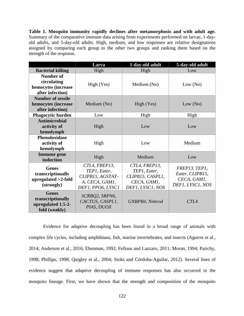
122
Table 1. Mosquito immunity rapidly declines after metamorphosis and with adult age.
Summary of the comparative immune data arising from experiments performed on larvae, 1-day-
old adults, and 5-day-old adults. High, medium, and low responses are relative designations
assigned by comparing each group to the other two groups and ranking them based on the
strength of the response.
Larva 1-day-old adult 5-day-old adult
Bacterial killing High High Low
Number of
circulating
hemocytes (increase
after infection)
High (Yes) Medium (No) Low (No)
Number of sessile
hemocytes (increase
after infection)
Medium (No) High (Yes) Low (No)
Phagocytic burden Low High High
Antimicrobial
activity of
hemolymph
High Low Low
Phenoloxidase
activity of
hemolymph
High Low Medium
Immune gene
induction High Medium Low
Genes
transcriptionally
upregulated >2-fold
(strongly)
CTL4, FREP13,
TEP1, Eater,
CLIPB15, AGSTAT-
A, CECA, GAM1,
DEF1, PPO6, LYSC1
CTL4, FREP13,
TEP1, Eater,
CLIPB15, CASPL1,
CECA, GAM1,
DEF1, LYSC1, NOS
FREP13, TEP1,
Eater, CLIPB15,
CECA, GAM1,
DEF1, LYSC1, NOS
Genes
transcriptionally
upregulated 1.5-2-
fold (weakly)
SCRBQ2, SRPN6,
CACTUS, CASPL1,
PIAS, DUOX
GNBPB4, Nimrod CTL4
Evidence for adaptive decoupling has been found in a broad range of animals with
complex life cycles, including amphibians, fish, marine invertebrates, and insects (Aguirre et al.,
2014; Anderson et al., 2016; Ebenman, 1992; Fellous and Lazzaro, 2011; Moran, 1994; Parichy,
1998; Phillips, 1998; Quigley et al., 2004; Stoks and Córdoba-Aguilar, 2012). Several lines of
evidence suggest that adaptive decoupling of immune responses has also occurred in the
mosquito lineage. First, we have shown that the strength and composition of the mosquito
123
immune response differs between immature larvae, newly-emerged adults, and reproductively
active older adults. Second, we have previously shown (King and Hillyer, 2012; League and
Hillyer, 2016; Sigle and Hillyer, 2016), and recapitulate here, that the mosquito circulatory and
immune systems display stage-specific functional integration, enabling functionally analogous,
yet anatomically disparate immune strategies in larvae and adults. Third, mosquitoes have
complex life cycles with ecologically distinct larval and adult stages that are subject to different
selection pressures due to differences in pathogen exposure and reproductive status. Thus, we
expect that a decoupling of key traits between these life stages, which are separated by
metamorphosis, is advantageous to both stages in responding independently to differing selection
pressures and in the performance of different fitness tasks (for example, food consumption and
growth in larvae and reproduction in adults). Taken together, these arguments suggest that
adaptive decoupling has occurred in mosquitoes, enabling the independent evolution of key
larval and adult immune traits.
Our data show that larvae kill bacteria at a higher rate, possess more circulating
hemocytes, have higher lytic and melanization activity in their hemolymph, and have higher
infection-induced expression of immunity genes than adults, thus strongly supporting our
hypothesis that mosquito larvae invest more in immunity than adults. In our view this is not
surprising; larvae inhabit bacteria-rich pools and display higher gut microbial diversity than
adults (Rani et al., 2009; Wang et al., 2011a), suggesting that mosquitoes are exposed to a higher
density and diversity of bacteria as larvae than as adults, and require more robust immune
responses as larvae, as has been hypothesized previously (Cornet et al., 2013). Thus, given the
vastly different ecologies between larvae and adults, coupled with the fact that evolutionary
pressures are strongest on younger individuals that have yet to reach their reproductive potential
124
(Hamilton, 1966; Koella et al., 2009), we hypothesize that the stronger selection pressures at
work in the larval stage compared to the adult stage have over time caused larvae to invest more
greatly in immunity than adults.
Our findings on immune proficiency in larvae and adults is also indicative of immune
senescence, as immunity in larvae is stronger than in adults, and immunity continues to weaken
after metamorphosis, with 5-day-old adults mounting weaker immune responses than adults that
were 4 days younger. This finding is consistent with well-attested evolutionary theories of
senescence, which state that greater selection pressures are at work in the larval stage to ensure
survival to the reproductively mature adult life stage (Hamilton, 1966; Monaghan et al., 2008).
According to the “Disposable Soma” theory of aging, this occurs in part due to immune trade-
offs that would not be in play at the larval stage, particularly those related to reproduction
(Kirkwood, 1977). For example, resistance to pathogens in adult mosquitoes has been associated
with reproductive costs (Ahmed et al., 2002; Ahmed and Hurd, 2006; Ferdig et al., 1993; Yan et
al., 1997). Furthermore, numerous studies have document age-associated declines in immune
functions in mosquitoes (Castillo et al., 2006; Christensen et al., 1986; Chun et al., 1995; Cornet
et al., 2013; Estevez-Lao and Hillyer, 2014; Hillyer et al., 2005; King and Hillyer, 2013; Li et al.,
1992; Pigeault et al., 2015; Schwartz and Koella, 2002), fruit flies (Felix et al., 2012; Horn et al.,
2014; Mackenzie et al., 2011), butterflies (Prasai and Karlsson, 2012; Stoehr, 2007), bees
(Doums et al., 2002; Laughton et al., 2011; Moret and Schmid-Hempel, 2009; Schmid et al.,
2008; Wilson-Rich et al., 2008), and other insects (Adamo et al., 2001; Eleftherianos et al., 2008;
Kaaya and Darji, 1988; Kurtz, 2002; Rolff, 2001). However, unlike these earlier studies, which
focused almost exclusively on adults, our findings show that this trend extends into the earlier
larval life stage. This suggests that mosquito adults employ a “live fast, die young” life history
125
strategy (Travers et al., 2015), whereby immunity declines and mortality increases after adults
reach reproductive maturity and selection for their survival wanes (Hamilton, 1966).
The age-dependent decrease in immune proficiency correlates with age-related changes
in the number of circulating hemocytes present in the hemocoel. We found that mosquito larvae
contain more circulating hemocytes than adult mosquitoes, and that the number of larval
hemocytes increases in response to infection. This is consistent with earlier studies conducted in
Anopheles gambiae and Aedes aegypti adults that showed that immune stimulation or blood
feeding increases the number of circulating hemocytes (Baton et al., 2009; Bryant and Michel,
2014; Bryant and Michel, 2016; Castillo et al., 2011; Castillo et al., 2006; Christensen et al.,
1989; Coggins et al., 2012; Foley, 1978), and that the number of circulating hemocytes declines
with adult age (Castillo et al., 2006; Hillyer et al., 2005; King and Hillyer, 2013; Pigeault et al.,
2015). Hemocytes are the first cellular responders to infection, phagocytosing pathogens within
minutes of infection (Hillyer et al., 2003a; Hillyer et al., 2003b; King and Hillyer, 2012; League
and Hillyer, 2016; Nazario-Toole and Wu, 2017; Sigle and Hillyer, 2016), and producing factors
that limit both bacteria and Plasmodium infection (Bartholomay et al., 2007; Baton et al., 2009;
Pinto et al., 2009; Smith et al., 2016). Thus, developmental changes in hemocyte profiles in part
explain the higher bacteria killing efficiency of larvae relative to adults.
Although sessile hemocytes were abundant in both larvae and adults, the spatial
arrangement of sessile hemocytes differed as adults aged, with newly-emerged adults displaying
a pronounced segmental arrangement of hemocytes, which is more akin to what is observed in
larvae than what is observed in 5-day-old adults (King and Hillyer, 2013; League and Hillyer,
2016). Furthermore, whereas hemocytes in adults aggregate at the periostial regions of the heart,
hemocytes in larvae aggregate in the tracheal tufts of the 8th
abdominal segment, where the sole
126
incurrent openings into the heart is located. These differences are due to the stage-specific
functional integration of the immune and circulatory systems, as hemocytes aggregate in the
areas of highest hemolymph flow (King and Hillyer, 2012; King and Hillyer, 2013; League and
Hillyer, 2016; Sigle and Hillyer, 2016). Overall, these data show that sessile hemocyte
populations undergo developmentally-related changes in spatial configuration and infection-
induced abundance.
The finding that larvae have more circulating hemocytes than adults may account for the
seemingly counterintuitive finding that larvae show lower phagocytic indices and capacities than
adults. This is because larvae have more hemocytes available to phagocytose pathogens than
adults, and based on our finding on the lytic and melanization activity of hemolymph, they also
have a greater recourse to other means of pathogen clearance. Thus, an accumulation of
phagocytic events in adults may signify a greater need for this immune mechanism, or lower
phagocytic turnover rates, which has also been observed in aging fruit fly adults (Horn et al.,
2014). Interestingly, these differences in phagocytosis are accompanied by changes in hemocyte
morphology. Larval hemocytes tended to have a fibroblast-like spread morphology, which has
been observed in other insect hemocytes (Ribeiro and Brehélin, 2006; Wang et al., 2011b), as
opposed to the more rounded spread morphology typical of mosquito adult hemocytes (Castillo
et al., 2006; Hillyer and Christensen, 2002; Hillyer et al., 2003a; Hillyer et al., 2003b; King and
Hillyer, 2012; King and Hillyer, 2013).
In addition to stage-specific differences in hemocyte biology, we detected profound
differences in the melanization activity of hemolymph. Melanization is commonly used in the
immune response against bacteria, malaria, and filarial worms (Bartholomay, 2014; Christensen
et al., 2005; Hillyer, 2016), and we found that mosquito larvae display a far greater capacity for
127
phenoloxidase-mediated humoral melanization than adults. This finding is consistent with our
recent observations of rapid and extensive melanin deposits in the abdomen of larvae following a
bacterial infection (League and Hillyer, 2016), as well as with the decline in phenoloxidase
activity that occurs with adult age in Aedes aegypti (Li et al., 1992), and across life stage and
with adult age in Culex pipiens (Cornet et al., 2013). In addition to enhancing humoral immunity,
larvae may increase the melanization activity of hemolymph in preparation for cuticle tanning
during and immediately after ecdysis, and for the rapid melanization of wounds in the more
perilous aquatic environment of larvae.
Finally, we show that immunity gene induction is generally stronger in larvae compared
to adults, with the difference being exacerbated as adults age, and that larvae and adults
differentially regulate the expression of some immunity genes. The higher rate of E. coli killing
in larvae is consistent with the expression of immune effector genes such as the Gram (-) binding
protein gene, GNBPB4, which was expressed 470- and 710-fold higher in naïve larvae compared
to naïve 1-day-old and 5-day-old adults, respectively, as well as the higher expression of the
phenoloxidase gene PPO1, which was expressed 170- and 110-fold higher in larvae relative to
naïve 1-day-old and 5-day-old adults, respectively. The higher rate of melanization we observed
in larval hemolymph is also consistent with the higher PPO6 gene induction we observed in
larvae, and supports our previous finding of higher infection-induced melanization in the
abdomen of larvae when compared adults (League and Hillyer, 2016). Furthermore, the higher
melanization activity in the hemolymph of larvae is also consistent with the high level of dual
oxidase (DUOX) gene induction detected in this life stage, as increased reactive oxygen species
levels have been correlated with increased melanization (Kumar et al., 2003). In addition to the
immune effector genes PPO6 and DUOX, we also found that the Jak/Stat essential pathway
128
member AGSTAT-A and, to a lesser extent, the negative regulator of this pathway, PIAS, were
induced in response to E. coli infection in larvae and not in adults, showing that the same
infection results in stage-specific induction of signaling pathway genes. The upregulation of Imd
pathway member in larvae and 1-day-old adults is consistent with this pathway’s role in
combating Gram (-) bacterial infections, however the cause for Jak/Stat pathways involvement in
the larval response alone is unknown, though it may have to do with its function in mosquito
development (Souza-Neto et al., 2009) as this pathway is involved in Drosophila development
(Arbouzova and Zeidler, 2006). Adults, and not larvae, showed distinct upregulation of nitric
oxide synthase, an important component of the adult immune response (Hillyer and Estévez-Lao,
2010; Luckhart et al., 1998), as well as higher upregulation of the pathogen recognition gene
FREP13 and the signal modulation gene CLIPB15. These differences in immunity gene
induction across life stages could result from changes in the tissues expressing these genes
during metamorphosis, as has been hypothesized previously (Dimopoulos et al., 1997; Fellous
and Lazzaro, 2011). More broadly, however, these differences in immune gene regulation in
larvae and adults are suggestive of adaptive decoupling, which would permit the independent
regulation of larval and adult immune gene expression (Fellous and Lazzaro, 2011; Moran,
1994).
Studying larval immunity is crucial to our general understanding of mosquito biology,
disease transmission, and vector control for several reasons. First, because mosquitoes are
holometabolous insects, studying the larval immune system could yield important insights into
how immune responses change over the course of development (League and Hillyer, 2016).
Second, larval environmental factors, including food availability, temperature, population
density, competition, and chemical insecticide exposure (Alto, 2011; Kim and Muturi, 2013;

129
Moller-Jacobs et al., 2014; Mourya et al., 2004; Murdock et al., 2012; Muturi, 2013; Muturi et
al., 2011; Telang et al., 2012; Yadav et al., 2005), as well as exposure to bacterial and fungal
pesticides (Bukhari et al., 2011; Capone et al., 2013; Federici et al., 2003; Federici et al., 2007;
Kala and Gunasekaran, 1999; Mahapatra et al., 1999; Otieno-Ayayo et al., 2008; Paily et al.,
2012), have all been shown to impact adult vector competence. Third, many of the most
widespread and effective mosquito control methods directly target the larval stages (Fillinger and
Lindsay, 2011; Floore, 2006; Tusting et al., 2013), and resistance to insecticides evolves more
rapidly in larvae compared to adults, likely due to the stronger selection pressures at work in the
larval stage (Koella et al., 2009). The present study highlights both continuities and
discontinuities between the larval and adult immune systems, and shows that mosquito larvae
possess an enhanced immune system compared to adults. Hence, understanding the larval
immune system of Anopheles gambiae, a major vector of malaria in Sub Saharan Africa, could
prove critical to the development and implementation of novel pest and disease control methods
that are tailored to each life stage.
130
CHAPTER V
CONCLUSION AND FUTURE DIRECTIONS
Overview
As holometabolous insects, mosquitoes undergo dramatic changes in their morphology,
ecology, and behavior as they develop from aquatic larvae to terrestrial and aerial adults.
However, perhaps due to the imminent threat posed to human health by the adult life stage, most
studies of the mosquito immune system have focused exclusively on adult females and have not
addressed immunity in larvae. As noted by Fellous and Lazzaro “Almost all studies of the
immune system of animals with metamorphosis have focused on either larval or on adult
immunity, implicitly assuming that these traits are either perfectly correlated or evolutionarily
independent” (Fellous and Lazzaro, 2011). In contrast to this “either-or” assumption, we suggest
adopting a holistic, “both-and” approach to the study of mosquito immunity by examining this
system throughout the complex lifecycle of the organism. The work presented in this dissertation
adopts just such an approach by using a comparative approach to analyze circulatory and
immune processes in both the larva and adult life stages to show that: (1) the larval and adult
circulatory systems are functionally distinct; (2) the larval circulatory and immune systems are
functionally integrated in a manner that is functionally analogous but spatially distinct from what
is seen in adults; and (3) larvae mount more robust cellular and humoral immune responses
compared to adults, especially as they age.
131
Larval and Adult Circulatory Physiology
Our investigation into the larval circulatory system, done side-by-side with adults, shows
how this system changes, and remains stable, across metamorphosis. Overall, this study showed
that the larval heart, though in most respects anatomically similar to the adult heart in location
and structure, differs functionally from the adult heart in two important ways: (1) the larval heart
does not undergo heartbeat directional reversals, but instead beats in the anterograde direction
only; and (2) the larval heart does not accept hemolymph through the abdominal ostia, but
instead through incurrent openings at the posterior terminus of the heart, which in adults serve an
exclusively excurrent function during periods of retrograde flow (Andereck et al., 2010; Glenn et
al., 2010).
The first finding from this study suggests that heartbeat directional reversals begin at the
onset of, during, or shortly after pupal metamorphosis, though the exact timing of this transition
has not been determined. Furthermore, the precise cause of heartbeat directional reversals is still
unknown, though evidence from studies in other insects suggests that it may be due to new
innervation of the heart that occurs during metamorphosis or differences in the activities of
myotropic hormones (Davis et al., 2001; Dulcis et al., 2001; Dulcis and Levine, 2003; Dulcis and
Levine, 2004; Dulcis and Levine, 2005; Kuwasawa et al., 1999). Larvae provide a helpful system
for studying the potential causes that trigger the onset of retrograde heartbeats in adults, as their
constant anterograde heart contractions provides a natural negative control environment into
which cardiomyotropic neuropeptides or other stimuli, such as those which have been tested on
the mosquito adult heart, could be added to test whether they have an impact on heartbeat
directionality (Chen and Hillyer, 2013; Estévez-Lao et al., 2013; Hillyer et al., 2014; Hillyer et
al., 2012). The presence of heart innervations in the larval and pupal stages could also be
132
examined, as previous work from our lab has detected such innervations in the mosquito adult
heart using antibody-based immunohistochemical staining methods (Estévez-Lao et al., 2013).
Discovering the causes behind the developmental onset of heartbeat reversals would not only
shed light as to why they are absent in larvae, but could also lead to a means of manipulating
their occurrence in adults to better understand their biological significance.
The second finding regarding hemolymph flow patterns in larvae show that the larval
heart accepts hemolymph via posterior incurrent openings, but does not accept hemolymph
through the ostia, though the precise reason for this is still unknown. We note that ostia are
present in larvae yet remain in the closed position, and hence are unable to accept hemolymph.
On a mechanistic level, one potential explanation for this is that the larval ostia are unable to
open because they lack sufficient support from the nearby alary muscles, which are
underdeveloped in larvae compared to adults (Glenn et al., 2010; League et al., 2015). Another
possibility is that the ostia themselves are not fully developed, and hence would be non-
functional regardless of the degree of alary muscle support, though we were unable to detect
structural differences between larval and adult ostia using our imaging methods. However, on a
broader evolutionary level, our data on hemolymph flow and ostia function in larvae suggests
that their heart structure, function, and resultant hemolymph flow patterns are adapted to
maintain proper hemolymph pressure and volume throughout their more uniformly-shaped
hemocoel and to meet demands associated with their unique feeding and swimming behaviors. In
adults, however, the heart has adapted to contract bidirectionally and accept hemolymph through
abdominal and thoraco-abdominal ostia during anterograde and retrograde contraction periods,
respectively, to accommodate the hemolymph distribution needs of their more
133
compartmentalized body plan as well as to meet the demands of processes unique to their life
stage, such as flight, blood feeding, and reproduction.
The method for measuring ostia usage I developed for use in larvae has already been used
in adults to show which ostia experience the highest levels of hemolymph flow (Sigle and
Hillyer, 2016). The findings from that study suggest that immune activity along the heart,
including pathogen sequestration and recruitment of hemocytes to the periostial regions, is at
least partially dependent upon hemolymph circulation patterns. This is further confirmed by our
finding that neither hemocytes nor pathogens aggregate near the larval ostia, which do not
experience incurrent hemolymph flow (League and Hillyer, 2016; League et al., 2015). The work
described here could also serve as a foundation for the construction of more exhaustive
hemolymph flow maps throughout the hemocoel, as this has yet to be done in either larvae or
adults. Finally, the descriptions of the larval aorta detailed in this study are currently aiding
ongoing attempts in our lab to characterize the aorta of adults, which to date has proven an
elusive structure to describe due to its fragility and position within the thorax (Glenn et al.,
2010). Taken together, these comparative analyses lend credence to the notion that neither life
stage can be fully understood in isolation from the other.
Functional Integration of the Larval Circulatory and Immune Systems
A principal location where immune responses occur in adult mosquitoes is the surface of
the heart, which is the region of the body with the highest hemolymph flow (King and Hillyer,
2012; Sigle and Hillyer, 2016). The work on the coordinated function of the larval circulatory
and immune systems presented in Chapter III represents the first detailed description of
circulation-dependent immunity at the larval tracheal tufts, which are respiratory structures that
are located at the sole entry point of hemolymph into the larval heart. These findings also
134
provide the first detailed description of the tracheal tufts in any mosquito species as well as the
first time these structures have been assigned a central role in hemocyte-mediated immunity in
any insect.
The tracheal tufts had previously been described in detail in the larvae of only one
lepidopteran species, Calpodes ethlius, where they were said to function as a sort of “lung” for
hemocyte oxygenation (Locke, 1997). Although tracheal tufts had been previously linked to
parasitoid wasp egg oxygenation in another lepidopteran (Rao et al., 2009), and had been noted
in passing in some dipteran larvae (Dell, 1905; Imms, 1907; Vaney, 1902), including mosquitoes
(Christophers, 1960; Imms, 1907; Jones, 1954; Wigglesworth, 1949), until now their role in
immunity had gone unstudied. We found that the tracheal tufts are the principal locations where
larvae sequester bacterial pathogens that have entered the hemocoel. Furthermore, we found that,
unlike the periostial hemocyte aggregation response that occurs on the heart of infected adults
(King and Hillyer, 2012; Sigle and Hillyer, 2016), additional hemocytes do not aggregate at the
tracheal tufts in response to infection. It remains unclear why pathogens, but not additional
hemocytes, aggregate at the tracheal tufts during infection, particularly given the high rates of
hemolymph flow in this region. We propose that the need to recruit additional hemocytes to the
site of pathogen aggregation is unnecessary due to high number of hemocytes already bound to
the tracheal tufts. However, the absence of infection-induced hemocyte recruitment at the tufts
may also have to do with the signaling mechanisms underlying the periostial hemocyte
aggregation response in adults, which may involve an immune factor that is not expressed in
hemocytes at the larval stage. To test this, hemocyte aggregation candidates arising from
ongoing work in our lab could potentially be validated by their absence or reduced expression in
larval tissues.
135
The effects of varying oxygen levels on the functioning of the tracheal tufts was not a
focus of this study, but such experiments could be performed to test whether these structures are
indeed necessary for the oxygenation of hemocytes, as has been previously suggested in other
insects (Hilken et al., 2003; Locke, 1997). However, this is not expected to be the case for
mosquito larvae because hemocytes persist in adults in the absence of comparable tracheal
structures, arguing against their function as hemocyte lungs in larvae. Rather, because hemocytes
cover these thin trachea, and such trachea serve not only as the sole site of pathogen
sequestration at the region of highest hemolymph flow into the heart but also as sites of potential
pathogen invasion into the hemocoel from the external environment (Bukhari et al., 2010; Clark
et al., 1968; Engelhard et al., 1994; Franz et al., 2015; King and Hillyer, 2013; Lacey et al.,
1988; Miranpuri and Khachatourians, 1991; Romoser et al., 2004; Scholte et al., 2004), it is more
likely that hemocytes associate with the tracheal tufts solely for the purpose of immune
surveillance.
Although not associated with tracheal tufts in Drosophila (dissections in our lab
confirmed that these structures are absent in fruit fly larvae), eighth abdominal segment
hemocytes comprise a major blood cell compartment in Drosophila and serve an important role
in larval immunity and hematopoiesis (Honti et al., 2014; Kurucz et al., 2007; Márkus et al.,
2009; Shrestha and Gateff, 1982a; Stofanko et al., 2008). However, despite the observation in
some insects that eighth segment hemocytes, and sessile hemocytes in general, serve a
hematopoietic function (Arvy, 1953; Arvy, 1954; Ghosh et al., 2015; Lanot et al., 2001; Márkus
et al., 2009), this function has thus far not been supported in mosquito larvae. In fact, a
hematopoietic organ has not been documented in mosquito larvae (Strand, 2008) and their eighth
segment sessile hemocytes, unlike those of Drosophila, have not been implicated as a
136
hematopoietic compartment for specialized larval immune responses (Márkus et al., 2009).
Future studies in mosquitoes investigating developmental hematopoiesis may either help explain
the apparent absence of a hematopoietic organ in mosquito larvae, or actually discover one, as
well as determine whether the eighth segment hemocytes of mosquito larvae have a
hematopoietic function. One possible explanation for the apparent lack of a larval hematopoietic
organ is that it is unnecessary because larval circulating hemocytes are capable of replicating, as
is true in the larvae of other insects (Arnold and Hinks, 1976; Feir and McClain, 1968; Gardiner
and Strand, 2000; Kiuchi et al., 2008) as well as in mosquito adults (King and Hillyer, 2013).
This could be tested by treating larvae with chemicals that interfere with mitosis such as taxol or
colchicine, thus enriching the number of potentially dividing cells, and directly visualizing
mitosis, if present, by labelling hemocytes with anti-tubulin antibodies (King and Hillyer, 2013).
Larval and Adult Cellular and Humoral Immune Responses
Larval and adult immune responses have seldom been compared in tandem and many of
the immune measurements detailed in Chapter IV represent the first of their kind in the larval life
stage. This may in part be due to the technical difficulties of working with small, soft-bodied
larvae, but it is mainly due to the fact that it is the female adult, and not the larva, that vectors
human pathogens. However, our findings suggest that studies of the adult immune system should
be accompanied by complementary studies in larvae. Such an approach assumes neither that
larval and adult immunity are exactly the same (complete evolutionary coupling), therefore
rendering the study of both redundant, nor that they are completely different (complete
evolutionary decoupling), therefore rendering the study of one irrelevant to the other (Fellous
and Lazzaro, 2011). Our comparative investigation of the larval and adult immune systems
137
suggests that the truth lies somewhere in between these extremes, with both continuities and
discontinuities existing between larval and adult immunity.
Similar to what we observed in the functional integration of the circulatory and immune
systems in larvae and adults, all of the cellular and humoral immune parameters we measured are
functional at both stages, though to varying degrees and with some developmental differences.
We note an overall increased ability of larvae to kill E. coli in vivo, however, no other pathogens
were investigated in this assay. Future studies could use this assay with other bacteria to see how
typical this pattern is, although our other immune assays would suggest that this would be true
for a broad range of pathogens. For example, the zone of inhibition assay included immune
challenges with several types of bacteria and found that in each instance, the larval humoral
antibacterial immune response was stronger compared to adults.
We also noted decreases of hemocyte numbers with age, and increases with infection, at
least in the larvae (adults showed a similar, albeit non-significant trend). Although the precise
source of these hemocytes remains unknown, they most likely arise from hemocyte replication in
response to infection, and not from the release of sessile hemocytes into circulation upon
infection (King and Hillyer, 2013). We have not observed sessile hemocytes in larvae entering
circulation en masse upon infection, as occurs in response to parasitoid wasp infections in
Drosophila larvae (Márkus et al., 2009), although modest levels of interchange between sessile
and circulating hemocyte populations have been observed in adult mosquitoes (Sigle and Hillyer,
2016).
Furthermore, our comparison of immunity gene expression between life stages and adult
ages shows that larvae and adults differentially express some immunity genes and that larvae
generally display stronger levels of immune gene induction than adults, especially as they age.
138
On an evolutionary level, the differences in the strength of adult immunity gene expression are
no doubt related to processes involved in senesce (Kirkwood, 1977), and the stronger immune
gene induction observed in larvae likely result from the stronger evolutionary pressures at work
on younger individuals to ensure their survival to reproductive maturity (Hamilton, 1966).
However, the proximate causes behind both the cis- and trans-stadial (that is, within and between
stage) differences we observed in immunity gene expression remain unknown. We examined the
expression of ecdysone biosynthetic pathway genes in larvae and adults, as ecdysone is high in
the larval stages and is known to regulate some immune functions (Ahmed et al., 1999; Müller et
al., 1999). However, we found that these genes were not regulated by infection in any of our
larval or adult groups, nor was their expression elevated in one stage compared to the other.
Future studies could further probe into the reasons underlying these differences in immunity
gene expression between life stages by examining whether larvae and adults differ in tissue-
specific immunity gene expression due to metamorphosis (Dimopoulos et al., 1997) as well as
how the differences we observed with E. coli infections vary with different bacterial insults.
Furthermore, by employing RNA sequencing technology, as is currently being done in our lab to
identify factors involved in periostial hemocyte aggregation on the heart of adults, we could
obtain a broader look at the whole transcriptome in larvae and adults after infection and identify
novel factors that may explain the differences we observed in their gene expression.
Concluding Thoughts
The work presented in this dissertation shows that the larval and adult circulatory and
immune systems differ in important ways, thus necessitating their continued study in both life
stages. Although all of the basic immune functions examined thus far are present in both life
stages, there are distinct differences in the potency, composition, and regulation of these
139
responses, suggesting a decoupling of immunity in larvae and adults made possible by
metamorphosis, the process that separates the two life stages and enables their independent
evolution (Fellous and Lazzaro, 2011; Moran, 1994). However, this decoupling is clearly only
partial, as some of the immune parameters we measured were similar between larvae and adults.
Furthermore, it is also known that the fitness of adult mosquitoes is partially dependent upon
rearing conditions and a number of stressors experienced during the larval stages (Alto, 2011;
Bukhari et al., 2011; Capone et al., 2013; Favia et al., 2007; Federici et al., 2003; Federici et al.,
2007; Kala and Gunasekaran, 1999; Kim and Muturi, 2013; Mahapatra et al., 1999; Moller-
Jacobs et al., 2014; Mourya et al., 2004; Muturi, 2013; Muturi et al., 2011; Otieno-Ayayo et al.,
2008; Paily et al., 2012; Telang et al., 2012; Yadav et al., 2005), thus limiting the degree to
which these two stages could evolve independently. Thus, studying the larval immune system is
useful not only in uncovering how it differs, but also how it is similar to that of adults, and
therefore the extent to which it has diverged between the life stages. Such findings could have
broad implications for epidemiological studies in mosquito adults as well as the implementation
of control measures in the field.
Lastly, there are good reasons to believe that infections in the larval life stage could affect
subsequent adult-acquired infections. Although insects do not possess an adaptive immune
response, the innate immune systems of some insects display memory-like qualities, referred to
as “immune priming” (Schmid-Hempel, 2005). Current studies in our lab are underway to
determine whether transstadial immune priming can occur with bacterial insults in A. gambiae,
as there is evidence that this occurs in mosquitoes (Bargielowski and Koella, 2009; Moreno-
García et al., 2015) as well as other insects (Cisarovsky et al., 2012; Jacot et al., 2005; Thomas
and Rudolf, 2010). In further support of this notion, we already know that immune priming
140
occurs within the adult life stage, as mosquitoes that have been pre-challenged with bacteria or
Plasmodium prior to infection, or reinfection, with Plasmodium show increased parasite
resistance (Lowenberger et al., 1999; Ramirez et al., 2015; Rodrigues et al., 2010). However,
such resistance often comes at a price, as resistance to pathogens in mosquitoes has been
associated with reproductive costs (Ahmed et al., 2002; Ahmed and Hurd, 2006; Ferdig et al.,
1993; Yan et al., 1997) and altered developmental timelines (Koella and Boëte, 2002). Indeed,
some authors have proposed taking advantage of certain insecticides that induce rapid resistance
in larvae due to the trade-offs they cause in adult mosquitoes, which have the effect of lowering
their ability to transmit malaria (Koella et al., 2009).
Given these ties between larval and adult immunity, future studies must take these
observations further and delve into the cellular and molecular mechanisms that lie beneath them.
This can only be accomplished by viewing the mosquito as a complex series of life stages that
are both distinct, yet inseparable from each another. As a major vector of Plasmodium in sub-
Saharan Africa, A. gambiae must be studied from new and compelling angles in order to
discover novel pest control strategies for this seemingly uncontrollable pest. Investigating
immunity at the larval stage provides just such an angle, and could very well lead to the veritable
metamorphosis of our understanding of mosquito immunity as a whole.
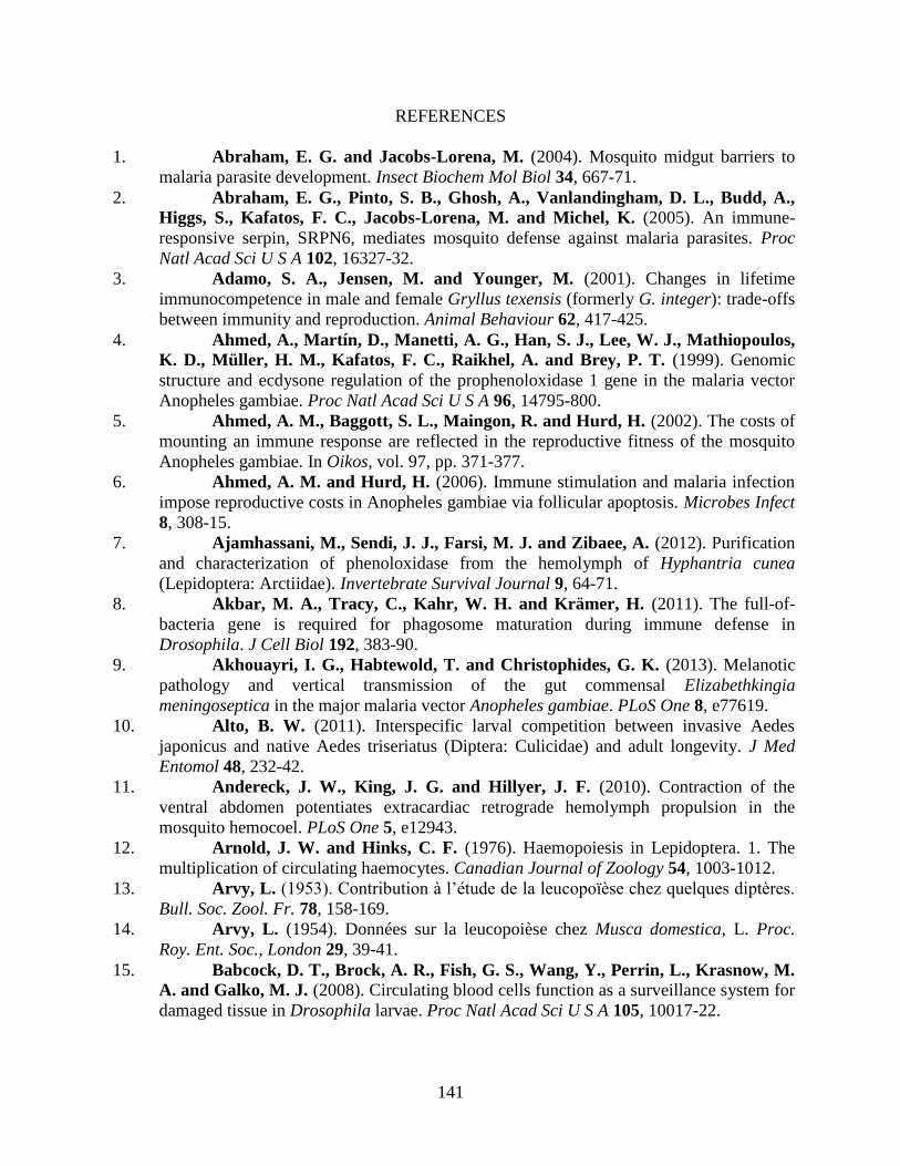
141
REFERENCES
1. Abraham, E. G. and Jacobs-Lorena, M. (2004). Mosquito midgut barriers to
malaria parasite development. Insect Biochem Mol Biol 34, 667-71.
2. Abraham, E. G., Pinto, S. B., Ghosh, A., Vanlandingham, D. L., Budd, A.,
Higgs, S., Kafatos, F. C., Jacobs-Lorena, M. and Michel, K. (2005). An immune-
responsive serpin, SRPN6, mediates mosquito defense against malaria parasites. Proc
Natl Acad Sci U S A 102, 16327-32.
3. Adamo, S. A., Jensen, M. and Younger, M. (2001). Changes in lifetime
immunocompetence in male and female Gryllus texensis (formerly G. integer): trade-offs
between immunity and reproduction. Animal Behaviour 62, 417-425.
4. Ahmed, A., Martín, D., Manetti, A. G., Han, S. J., Lee, W. J., Mathiopoulos,
K. D., Müller, H. M., Kafatos, F. C., Raikhel, A. and Brey, P. T. (1999). Genomic
structure and ecdysone regulation of the prophenoloxidase 1 gene in the malaria vector
Anopheles gambiae. Proc Natl Acad Sci U S A 96, 14795-800.
5. Ahmed, A. M., Baggott, S. L., Maingon, R. and Hurd, H. (2002). The costs of
mounting an immune response are reflected in the reproductive fitness of the mosquito
Anopheles gambiae. In Oikos, vol. 97, pp. 371-377.
6. Ahmed, A. M. and Hurd, H. (2006). Immune stimulation and malaria infection
impose reproductive costs in Anopheles gambiae via follicular apoptosis. Microbes Infect
8, 308-15.
7. Ajamhassani, M., Sendi, J. J., Farsi, M. J. and Zibaee, A. (2012). Purification
and characterization of phenoloxidase from the hemolymph of Hyphantria cunea
(Lepidoptera: Arctiidae). Invertebrate Survival Journal 9, 64-71.
8. Akbar, M. A., Tracy, C., Kahr, W. H. and Krämer, H. (2011). The full-of-
bacteria gene is required for phagosome maturation during immune defense in
Drosophila. J Cell Biol 192, 383-90.
9. Akhouayri, I. G., Habtewold, T. and Christophides, G. K. (2013). Melanotic
pathology and vertical transmission of the gut commensal Elizabethkingia
meningoseptica in the major malaria vector Anopheles gambiae. PLoS One 8, e77619.
10. Alto, B. W. (2011). Interspecific larval competition between invasive Aedes
japonicus and native Aedes triseriatus (Diptera: Culicidae) and adult longevity. J Med
Entomol 48, 232-42.
11. Andereck, J. W., King, J. G. and Hillyer, J. F. (2010). Contraction of the
ventral abdomen potentiates extracardiac retrograde hemolymph propulsion in the
mosquito hemocoel. PLoS One 5, e12943.
12. Arnold, J. W. and Hinks, C. F. (1976). Haemopoiesis in Lepidoptera. 1. The
multiplication of circulating haemocytes. Canadian Journal of Zoology 54, 1003-1012.
13. Arvy, L. (1953). Contribution à l’étude de la leucopoïèse chez quelques diptères.
Bull. Soc. Zool. Fr. 78, 158-169.
14. Arvy, L. (1954). Données sur la leucopoièse chez Musca domestica, L. Proc.
Roy. Ent. Soc., London 29, 39-41.
15. Babcock, D. T., Brock, A. R., Fish, G. S., Wang, Y., Perrin, L., Krasnow, M.
A. and Galko, M. J. (2008). Circulating blood cells function as a surveillance system for
damaged tissue in Drosophila larvae. Proc Natl Acad Sci U S A 105, 10017-22.
142
16. Bao, Y. Y., Xue, J., Wu, W. J., Wang, Y., Lv, Z. Y. and Zhang, C. X. (2011).
An immune-induced reeler protein is involved in the Bombyx mori melanization cascade.
Insect Biochem Mol Biol 41, 696-706.
17. Bargielowski, I. and Koella, J. C. (2009). A possible mechanism for the
suppression of Plasmodium berghei development in the mosquito Anopheles gambiae by
the microsporidian Vavraia culicis. PLoS One 4, e4676.
18. Bartholomay, L. C. (2014). Infection barriers and responses in mosquito-filarial
worm interactions. Current Opinion in Insect Science 3, 37-42.
19. Baton, L. A., Robertson, A., Warr, E., Strand, M. R. and Dimopoulos, G. (2009). Genome-wide transcriptomic profiling of Anopheles gambiae hemocytes reveals
pathogen-specific signatures upon bacterial challenge and Plasmodium berghei infection.
BMC Genomics 10, 257.
20. Biron, D. G., Agnew, P., Marché, L., Renault, L., Sidobre, C. and Michalakis,
Y. (2005). Proteome of Aedes aegypti larvae in response to infection by the intracellular
parasite Vavraia culicis. Int J Parasitol 35, 1385-97.
21. Blair, C. D. and Olson, K. E. (2014). Mosquito immune responses to arbovirus
infections. Curr Opin Insect Sci 3, 22-29.
22. Blandin, S., Moita, L. F., Köcher, T., Wilm, M., Kafatos, F. C. and
Levashina, E. A. (2002). Reverse genetics in the mosquito Anopheles gambiae: targeted
disruption of the Defensin gene. EMBO Rep 3, 852-6.
23. Boppana, S. and Hillyer, J. F. (2014). Hemolymph circulation in insect sensory
appendages: functional mechanics of antennal accessory pulsatile organs (auxiliary
hearts) in the mosquito Anopheles gambiae. J Exp Biol.
24. Brey, P. T., Lebrun, R. A., Papierok, B., Ohayon, H., Vennavalli, S. and
Hafez, J. (1988). Defense reactions by larvae of Aedes aegypti during infection by the
aquatic fungus Lagenidium giganteum (Oomycete). Cell Tissue Res 253, 245-50.
25. Bryant, W. B. and Michel, K. (2014). Blood feeding induces hemocyte
proliferation and activation in the African malaria mosquito, Anopheles gambiae Giles. J
Exp Biol 217, 1238-45.
26. Bryant, W. B. and Michel, K. (2016). Anopheles gambiae hemocytes exhibit
transient states of activation. Dev Comp Immunol 55, 119-29.
27. Bukhari, T., Middelman, A., Koenraadt, C. J., Takken, W. and Knols, B. G. (2010). Factors affecting fungus-induced larval mortality in Anopheles gambiae and
Anopheles stephensi. Malar J 9, 22.
28. Bukhari, T., Takken, W. and Koenraadt, C. J. (2011). Development of
Metarhizium anisopliae and Beauveria bassiana formulations for control of malaria
mosquito larvae. Parasit Vectors 4, 23.
29. Capone, A., Ricci, I., Damiani, C., Mosca, M., Rossi, P., Scuppa, P., Crotti,
E., Epis, S., Angeletti, M., Valzano, M. et al. (2013). Interactions between Asaia,
Plasmodium and Anopheles: new insights into mosquito symbiosis and implications in
Malaria Symbiotic Control. Parasit Vectors 6, 182.
30. Castillo, J., Brown, M. R. and Strand, M. R. (2011). Blood feeding and insulin-
like peptide 3 stimulate proliferation of hemocytes in the mosquito Aedes aegypti. PLoS
Pathog 7, e1002274.
143
31. Castillo, J. C., Robertson, A. E. and Strand, M. R. (2006). Characterization of
hemocytes from the mosquitoes Anopheles gambiae and Aedes aegypti. Insect Biochem
Mol Biol 36, 891-903.
32. Chambers, M. C., Lightfield, K. L. and Schneider, D. S. (2012). How the fly
balances its ability to combat different pathogens. PLoS Pathog 8, e1002970.
33. Chapman, R. F., Douglas, A. E. and Siva-Jothy, M. T. (2013). Circulatory
system, blood, and the immune system. In The insects: structure and function, eds. S. J.
Simpson and A. E. Douglas), pp. 107-131. Cambridge: Cambridge University Press.
34. Chen, W. and Hillyer, J. F. (2013). FlyNap (triethylamine) increases the heart
rate of mosquitoes and eliminates the cardioacceleratory effect of the neuropeptide
CCAP. PLoS One 8, e70414.
35. Cheng, G., Liu, Y., Wang, P. and Xiao, X. (2016). Mosquito Defense Strategies
against Viral Infection. Trends Parasitol 32, 177-86.
36. Christensen, B. M., Huff, B. M., Miranpuri, G. S., Harris, K. L. and
Christensen, L. A. (1989). Hemocyte population changes during the immune response of
Aedes aegypti to inoculated microfilariae of Dirofilaria immitis. J Parasitol 75, 119-23.
37. Christensen, B. M., LaFond, M. M. and Christensen, L. A. (1986). Defense
reactions of mosquitoes to filarial worms: effect of host age on the immune response to
Dirofilaria immitis microfilariae. J Parasitol 72, 212-5.
38. Christensen, B. M., Li, J., Chen, C. C. and Nappi, A. J. (2005). Melanization
immune responses in mosquito vectors. Trends Parasitol 21, 192-9.
39. Christophers, S. R. (1960). Aedes Aegypti (L.) The Yellow Fever Mosquito: Its
Life History, Bionomics and Structure. New York: Cambridge University Press.
40. Christophides, G. K., Zdobnov, E., Barillas-Mury, C., Birney, E., Blandin, S.,
Blass, C., Brey, P. T., Collins, F. H., Danielli, A., Dimopoulos, G. et al. (2002).
Immunity-related genes and gene families in Anopheles gambiae. Science 298, 159-65.
41. Chun, J., Riehle, M. and Paskewitz, S. M. (1995). Effect of mosquito age and
reproductive status on melanization of sephadex beads in Plasmodium-refractory and -
susceptible strains of Anopheles gambiae. J Invertebr Pathol 66, 11-7.
42. Cirimotich, C. M., Dong, Y., Garver, L. S., Sim, S. and Dimopoulos, G. (2010). Mosquito immune defenses against Plasmodium infection. Dev Comp Immunol
34, 387-95.
43. Cisarovsky, G., Schmid-Hempel, P. and Sadd, B. M. (2012). Robustness of the
outcome of adult bumblebee infection with a trypanosome parasite after varied parasite
exposures during larval development. J Evol Biol 25, 1053-9.
44. Clark, T. B., Kellen, W. R., Fukuda, T. and Lindegren, J. E. (1968). Field and
laboratory studies on the pathogenicity of the fungus Beauveria bassiana to three genera
of mosquitoes. J Invertebr Pathol 11, 1-7.
45. Clayton, A. M., Dong, Y. and Dimopoulos, G. (2014). The Anopheles innate
immune system in the defense against malaria infection. J Innate Immun 6, 169-81.
46. Clements, A. N. (1992). The Biology of Mosquitoes: Development, Nutrition and
Reproduction, vol. 1, pp. 100-123. London: Chapman and Hall.
47. Coggins, S. A., Estévez-Lao, T. Y. and Hillyer, J. F. (2012). Increased
survivorship following bacterial infection by the mosquito Aedes aegypti as compared to
Anopheles gambiae correlates with increased transcriptional induction of antimicrobial
peptides. Dev Comp Immunol 37, 390-401.
144
48. Cornet, S., Gandon, S. and Rivero, A. (2013). Patterns of phenoloxidase
activity in insecticide resistant and susceptible mosquitoes differ between laboratory-
selected and wild-caught individuals. Parasit Vectors 6, 315.
49. Crossley, A. C. S. (1964). An experimental analysis of the origins and physiology
of haemocytes in the blue blow-fly Calliphora erythrocephala, (Meig). In Journal of
Experimental Zoology, vol. 157, pp. 375-397.
50. Curtis, N. J., Ringo, J. M. and Dowse, H. B. (1999). Morphology of the pupal
heart, adult heart, and associated tissues in the fruit fly, Drosophila melanogaster. J
Morphol 240, 225-35.
51. Cuttell, L., Vaughan, A., Silva, E., Escaron, C. J., Lavine, M., Van Goethem,
E., Eid, J. P., Quirin, M. and Franc, N. C. (2008). Undertaker, a Drosophila
Junctophilin, links Draper-mediated phagocytosis and calcium homeostasis. Cell 135,
524-34.
52. Dasari, S. and Cooper, R. L. (2006). Direct influence of serotonin on the larval
heart of Drosophila melanogaster. J Comp Physiol B 176, 349-57.
53. Davis, N. T., Dulcis, D. and Hildebrand, J. G. (2001). Innervation of the heart
and aorta of Manduca sexta. J Comp Neurol 440, 245-60.
54. Dell, J. A. (1905). On the structure and life-history of Psychoda sexpunctata.
Trans. Ent. Soc. Lond., 293.
55. Dimopoulos, G., Christophides, G. K., Meister, S., Schultz, J., White, K. P.,
Barillas-Mury, C. and Kafatos, F. C. (2002). Genome expression analysis of
Anopheles gambiae: responses to injury, bacterial challenge, and malaria infection. Proc
Natl Acad Sci U S A 99, 8814-9.
56. Dimopoulos, G., Richman, A., Müller, H. M. and Kafatos, F. C. (1997).
Molecular immune responses of the mosquito Anopheles gambiae to bacteria and malaria
parasites. Proc Natl Acad Sci U S A 94, 11508-13.
57. Dong, Y. and Dimopoulos, G. (2009). Anopheles fibrinogen-related proteins
provide expanded pattern recognition capacity against bacteria and malaria parasites. J
Biol Chem 284, 9835-44.
58. Dong, Y., Morton, J. C., Ramirez, J. L., Souza-Neto, J. A. and Dimopoulos,
G. (2012). The entomopathogenic fungus Beauveria bassiana activate toll and JAK-
STAT pathway-controlled effector genes and anti-dengue activity in Aedes aegypti.
Insect Biochem Mol Biol 42, 126-32.
59. Doums, C., Moret, Y., Benelli, E. and Schmid-Hempel, P. (2002). Senescence
of immune defence in Bombus workers. Ecological Entomology 27, 138-144.
60. Dulcis, D., Davis, N. T. and Hildebrand, J. G. (2001). Neuronal control of heart
reversal in the hawkmoth Manduca sexta. J Comp Physiol A 187, 837-49.
61. Dulcis, D. and Levine, R. B. (2003). Innervation of the heart of the adult fruit fly,
Drosophila melanogaster. J Comp Neurol 465, 560-78.
62. Dulcis, D. and Levine, R. B. (2004). Remodeling of a larval skeletal muscle
motoneuron to drive the posterior cardiac pacemaker in the adult moth, Manduca sexta. J
Comp Neurol 478, 126-42.
63. Dulcis, D. and Levine, R. B. (2005). Glutamatergic innervation of the heart
initiates retrograde contractions in adult Drosophila melanogaster. J Neurosci 25, 271-80.
64. Dulcis, D., Levine, R. B. and Ewer, J. (2005). Role of the neuropeptide CCAP
in Drosophila cardiac function. J Neurobiol 64, 259-74.
145
65. Eleftherianos, I., Baldwin, H., ffrench-Constant, R. H. and Reynolds, S. E. (2008). Developmental modulation of immunity: changes within the feeding period of the
fifth larval stage in the defence reactions of Manduca sexta to infection by Photorhabdus.
J Insect Physiol 54, 309-18.
66. Elrod-Erickson, M., Mishra, S. and Schneider, D. (2000). Interactions between
the cellular and humoral immune responses in Drosophila. Curr Biol 10, 781-4.
67. Engelhard, E. K., Kam-Morgan, L. N., Washburn, J. O. and Volkman, L. E. (1994). The insect tracheal system: a conduit for the systemic spread of Autographa
californica M nuclear polyhedrosis virus. Proc Natl Acad Sci U S A 91, 3224-7.
68. Erickson, S. M., Xi, Z., Mayhew, G. F., Ramirez, J. L., Aliota, M. T.,
Christensen, B. M. and Dimopoulos, G. (2009). Mosquito infection responses to
developing filarial worms. PLoS Negl Trop Dis 3, e529.
69. Estevez-Lao, T. Y. and Hillyer, J. F. (2014). Involvement of the Anopheles
gambiae Nimrod gene family in mosquito immune responses. Insect Biochem Mol Biol
44, 12-22.
70. Estévez-Lao, T. Y., Boyce, D. S., Honegger, H. W. and Hillyer, J. F. (2013).
Cardioacceleratory function of the neurohormone CCAP in the mosquito Anopheles
gambiae. J Exp Biol 216, 601-13.
71. Estévez-Lao, T. Y. and Hillyer, J. F. (2014). Involvement of the Anopheles
gambiae Nimrod gene family in mosquito immune responses. Insect Biochem Mol Biol
44, 12-22.
72. Favia, G., Ricci, I., Damiani, C., Raddadi, N., Crotti, E., Marzorati, M.,
Rizzi, A., Urso, R., Brusetti, L., Borin, S. et al. (2007). Bacteria of the genus Asaia
stably associate with Anopheles stephensi, an Asian malarial mosquito vector. Proc Natl
Acad Sci U S A 104, 9047-51.
73. Federici, B. A., Park, H. W., Bideshi, D. K., Wirth, M. C. and Johnson, J. J. (2003). Recombinant bacteria for mosquito control. J Exp Biol 206, 3877-85.
74. Federici, B. A., Park, H. W., Bideshi, D. K., Wirth, M. C., Johnson, J. J.,
Sakano, Y. and Tang, M. (2007). Developing recombinant bacteria for control of
mosquito larvae. J Am Mosq Control Assoc 23, 164-75.
75. Feir, D. and McClain, E. (1968). Induced changes in the mitotic activity of
hemocytes of the large milkweed bug, Oncopeltus fasciatus. Annals of the Entomological
Society of America 61, 416-421.
76. Felix, T. M., Hughes, K. A., Stone, E. A., Drnevich, J. M. and Leips, J. (2012).
Age-specific variation in immune response in Drosophila melanogaster has a genetic
basis. Genetics 191, 989-1002.
77. Fellous, S. and Lazzaro, B. P. (2011). Potential for evolutionary coupling and
decoupling of larval and adult immune gene expression. Mol Ecol 20, 1558-67.
78. Feng, C., Song, Q., Lü, W. and Lu, J. (2008). Purification and characterization
of hemolymph prophenoloxidase from Ostrinia furnacalis (Lepidoptera: Pyralidae)
larvae. Comp Biochem Physiol B Biochem Mol Biol 151, 139-46.
79. Ferdig, M. T., Beerntsen, B. T., Spray, F. J., Li, J. and Christensen, B. M. (1993). Reproductive costs associated with resistance in a mosquito-filarial worm system.
Am J Trop Med Hyg 49, 756-62.
80. Fillinger, U. and Lindsay, S. W. (2011). Larval source management for malaria
control in Africa: myths and reality. Malar J 10, 353.
146
81. Foley, D. A. (1978). Innate Cellular Defense by Mosquito Hemocytes. In
Invertebrate Models for Biomedical Research, eds. L. A. Bulla Jr. and T. C. Cheng), pp.
113-144. New York: Springer US.
82. Franz, A. W., Kantor, A. M., Passarelli, A. L. and Clem, R. J. (2015). Tissue
Barriers to Arbovirus Infection in Mosquitoes. Viruses 7, 3741-67.
83. Gardiner, E. M. and Strand, M. R. (2000). Hematopoiesis in larval
Pseudoplusia includens and Spodoptera frugiperda. Arch Insect Biochem Physiol 43, 147-
64.
84. Garver, L. S., Bahia, A. C., Das, S., Souza-Neto, J. A., Shiao, J., Dong, Y. and
Dimopoulos, G. (2012). Anopheles Imd pathway factors and effectors in infection
intensity-dependent anti-Plasmodium action. PLoS Pathog 8, e1002737.
85. Gerould, J. H. (1924). Periodic reversal of heart-beat in a chrysalis. Science 60,
570-2.
86. Gerould, J. H. (1933). Orders of insects with heart-beat reversal. Biological
Bulletin 64, 424-431.
87. Ghosh, S., Singh, A., Mandal, S. and Mandal, L. (2015). Active hematopoietic
hubs in Drosophila adults generate hemocytes and contribute to immune response. Dev
Cell 33, 478-88.
88. Glenn, J. D., King, J. G. and Hillyer, J. F. (2010). Structural mechanics of the
mosquito heart and its function in bidirectional hemolymph transport. J Exp Biol 213,
541-50.
89. González-Lázaro, M., Dinglasan, R. R., Hernández-Hernández, F. e. L.,
Rodríguez, M. H., Laclaustra, M., Jacobs-Lorena, M. and Flores-Romo, L. (2009).
Anopheles gambiae Croquemort SCRBQ2, expression profile in the mosquito and its
potential interaction with the malaria parasite Plasmodium berghei. Insect Biochem Mol
Biol 39, 395-402.
90. Gorman, M. J. and Paskewitz, S. M. (2000). Persistence of infection in
mosquitoes injected with bacteria. J Invertebr Pathol 75, 296-7.
91. Gupta, L., Molina-Cruz, A., Kumar, S., Rodrigues, J., Dixit, R., Zamora, R.
E. and Barillas-Mury, C. (2009). The STAT pathway mediates late-phase immunity
against Plasmodium in the mosquito Anopheles gambiae. Cell Host Microbe 5, 498-507.
92. Haine, E. R., Pollitt, L. C., Moret, Y., Siva-Jothy, M. T. and Rolff, J. (2008).
Temporal patterns in immune responses to a range of microbial insults (Tenebrio
molitor). J Insect Physiol 54, 1090-7.
93. Hamilton, W. D. (1966). The moulding of senescence by natural selection. J
Theor Biol 12, 12-45.
94. Harrison, J. F., Woods, H. A. and Roberts, S. P. (2012). Ecological and
environmental physiology of insects. New York: Oxford University Press.
95. Hernández-Martínez, S., Lanz-Mendoza, H., Martínez-Barnetche, J. and
Rodríguez, M. H. (2013). Antimicrobial properties of Anopheles albimanus pericardial
cells. Cell Tissue Res 351, 127-37.
96. Hilken, G., Brockmann, C. and Nevermann, L. (2003). Hemocytes of the
centipede Scutigera coleoptrata (Chilopoda, Notostigmophora) with notes on their
interactions with the tracheae. J Morphol 257, 181-9.
97. Hillyer, J. F. (2009). Transcription in mosquito hemocytes in response to
pathogen exposure. J Biol 8, 51.
147
98. Hillyer, J. F. (2010). Mosquito immunity. Adv Exp Med Biol 708, 218-38.
99. Hillyer, J. F. (2015). Integrated Immune and Cardiovascular Function in
Pancrustacea: Lessons from the Insects. Integr Comp Biol 55, 843-55.
100. Hillyer, J. F. (2016). Insect immunology and hematopoiesis. Dev Comp Immunol
58, 102-18.
101. Hillyer, J. F., Barreau, C. and Vernick, K. D. (2007). Efficiency of salivary
gland invasion by malaria sporozoites is controlled by rapid sporozoite destruction in the
mosquito haemocoel. Int J Parasitol 37, 673-81.
102. Hillyer, J. F. and Christensen, B. M. (2002). Characterization of hemocytes
from the yellow fever mosquito, Aedes aegypti. Histochem Cell Biol 117, 431-40.
103. Hillyer, J. F. and Estévez-Lao, T. Y. (2010). Nitric oxide is an essential
component of the hemocyte-mediated mosquito immune response against bacteria. Dev
Comp Immunol 34, 141-9.
104. Hillyer, J. F., Estévez-Lao, T. Y. and de la Parte, L. E. (2014). Myotropic
effects of FMRFamide containing peptides on the heart of the mosquito Anopheles
gambiae. Gen Comp Endocrinol 202, 15-25.
105. Hillyer, J. F., Estévez-Lao, T. Y., Funkhouser, L. J. and Aluoch, V. A. (2012).
Anopheles gambiae corazonin: gene structure, expression and effect on mosquito heart
physiology. Insect Mol Biol 21, 343-55.
106. Hillyer, J. F., Schmidt, S. L. and Christensen, B. M. (2003a). Hemocyte-
mediated phagocytosis and melanization in the mosquito Armigeres subalbatus following
immune challenge by bacteria. Cell Tissue Res 313, 117-27.
107. Hillyer, J. F., Schmidt, S. L. and Christensen, B. M. (2003b). Rapid
phagocytosis and melanization of bacteria and Plasmodium sporozoites by hemocytes of
the mosquito Aedes aegypti. J Parasitol 89, 62-9.
108. Hillyer, J. F., Schmidt, S. L. and Christensen, B. M. (2004). The antibacterial
innate immune response by the mosquito Aedes aegypti is mediated by hemocytes and
independent of Gram type and pathogenicity. Microbes Infect 6, 448-59.
109. Hillyer, J. F., Schmidt, S. L., Fuchs, J. F., Boyle, J. P. and Christensen, B. M. (2005). Age-associated mortality in immune challenged mosquitoes (Aedes aegypti)
correlates with a decrease in haemocyte numbers. Cell Microbiol 7, 39-51.
110. Hillyer, J. F. and Strand, M. R. (2014). Mosquito hemocyte-mediated immune
responses. Curr Opin Insect Sci 3, 14-21.
111. Honti, V., Csordás, G., Kurucz, É., Márkus, R. and Andó, I. (2014). The cell-
mediated immunity of Drosophila melanogaster: hemocyte lineages, immune
compartments, microanatomy and regulation. Dev Comp Immunol 42, 47-56.
112. Horn, L., Leips, J. and Starz-Gaiano, M. (2014). Phagocytic ability declines
with age in adult Drosophila hemocytes. Aging Cell.
113. Hurd, H., Taylor, P. J., Adams, D., Underhill, A. and Eggleston, P. (2005).
Evaluating the costs of mosquito resistance to malaria parasites. Evolution 59, 2560-72.
114. Imms, A. D. (1907). On the Larval and Pupal Stages of Anopheles maculipennis,
Meigen. J Hyg (Lond) 7, 291-318.3.
115. Jacot, A., Scheuber, H., Kurtz, J. and Brinkhof, M. W. (2005). Juvenile
immune system activation induces a costly upregulation of adult immunity in field
crickets Gryllus campestris. Proc Biol Sci 272, 63-9.
148
116. Jiang, H., Vilcinskas, A. and Kanost, M. R. (2010). Immunity in lepidopteran
insects. Adv Exp Med Biol 708, 181-204.
117. Jiang, J., Alvarez, C., Kukutla, P., Yu, W. and Xu, J. (2012). Draft genome
sequences of Enterobacter sp. isolate Ag1 from the midgut of the malaria mosquito
Anopheles gambiae. J Bacteriol 194, 5481.
118. Jones, J. C. (1954). The heart and associated tissues of Anopheles
quadrimaculatus say (Diptera: Culicidae). In Journal of Morphology, vol. 94, pp. 71-123.
119. Jones, J. C. (1977). The circulatory system of insects. Springfield IL: Charles C.
Thomas.
120. Kaaya, G. P. and Darji, N. (1988). The humoral defense system in tsetse:
differences in response due to age, sex and antigen types. Dev Comp Immunol 12, 255-
68.
121. Kajla, M. K., Andreeva, O., Gilbreath, T. M. and Paskewitz, S. M. (2010).
Characterization of expression, activity and role in antibacterial immunity of Anopheles
gambiae lysozyme c-1. Comp Biochem Physiol B Biochem Mol Biol 155, 201-9.
122. Kajla, M. K., Shi, L., Li, B., Luckhart, S., Li, J. and Paskewitz, S. M. (2011).
A new role for an old antimicrobial: lysozyme c-1 can function to protect malaria
parasites in Anopheles mosquitoes. PLoS One 6, e19649.
123. Kala, M. K. and Gunasekaran, K. (1999). Effect of bacillus thuringiensis ssp.
israelensis on the development of Plasmodium gallinaceum in Aedes aegypti (Diptera:
Culicidae). Ann Trop Med Parasitol 93, 89-95.
124. Kim, C. H. and Muturi, E. J. (2013). Effect of larval density and Sindbis virus
infection on immune responses in Aedes aegypti. J Insect Physiol 59, 604-10.
125. Kim, W., Koo, H., Richman, A. M., Seeley, D., Vizioli, J., Klocko, A. D. and
O'Brochta, D. A. (2004). Ectopic expression of a cecropin transgene in the human
malaria vector mosquito Anopheles gambiae (Diptera: Culicidae): effects on
susceptibility to Plasmodium. J Med Entomol 41, 447-55.
126. King, J. G. and Hillyer, J. F. (2012). Infection-induced interaction between the
mosquito circulatory and immune systems. PLoS Pathog 8, e1003058.
127. King, J. G. and Hillyer, J. F. (2013). Spatial and temporal in vivo analysis of
circulating and sessile immune cells in mosquitoes: hemocyte mitosis following
infection. BMC Biol 11, 55.
128. Kirkwood, T. B. (1977). Evolution of ageing. Nature 270, 301-4.
129. Kiuchi, T., Aoki, F. and Nagata, M. (2008). Effects of high temperature on the
hemocyte cell cycle in silkworm larvae. J Insect Physiol 54, 454-61.
130. Klowden, A. J. (2013). Circulatory systems. In Physiological systems in insects,
pp. 365-413. Boston: Academic Press.
131. Kocks, C., Cho, J. H., Nehme, N., Ulvila, J., Pearson, A. M., Meister, M.,
Strom, C., Conto, S. L., Hetru, C., Stuart, L. M. et al. (2005). Eater, a transmembrane
protein mediating phagocytosis of bacterial pathogens in Drosophila. Cell 123, 335-46.
132. Koella, J. C. and Boëte, C. (2002). A genetic correlation between age at
pupation and melanization immune response of the yellow fever mosquito Aedes aegypti.
Evolution 56, 1074-9.
133. Koella, J. C., Lynch, P. A., Thomas, M. B. and Read, A. F. (2009). Towards
evolution-proof malaria control with insecticides. Evolutionary Applications Volume 2,
Issue 4. In Evolutionary Applications, vol. 2, pp. 469-480.
149
134. Kumar, S., Christophides, G. K., Cantera, R., Charles, B., Han, Y. S.,
Meister, S., Dimopoulos, G., Kafatos, F. C. and Barillas-Mury, C. (2003). The role of
reactive oxygen species on Plasmodium melanotic encapsulation in Anopheles gambiae.
Proc Natl Acad Sci U S A 100, 14139-44.
135. Kumar, S., Molina-Cruz, A., Gupta, L., Rodrigues, J. and Barillas-Mury, C. (2010). A peroxidase/dual oxidase system modulates midgut epithelial immunity in
Anopheles gambiae. Science 327, 1644-8.
136. Kurtz, J. (2002). Phagocytosis by invertebrate hemocytes: causes of individual
variation in Panorpa vulgaris scorpionflies. Microsc Res Tech 57, 456-68.
137. Kurucz, E., Váczi, B., Márkus, R., Laurinyecz, B., Vilmos, P., Zsámboki, J.,
Csorba, K., Gateff, E., Hultmark, D. and Andó, I. (2007). Definition of Drosophila
hemocyte subsets by cell-type specific antigens. Acta Biol Hung 58 Suppl, 95-111.
138. Kuwasawa, K., Ai, H. and Matsushita, T. (1999). Cardiac reflexes and their
neural pathways in lepidopterous insects. Comparative Biochemistry and Physiology -
Part A Molecular & Integrative Physiology 124, 581-586.
139. Lacey, C. M., Lacey, L. A. and Roberts, D. R. (1988). Route of invasion and
histopathology of Metarhizium anisopliae in Culex quinquefasciatus. J Invertebr Pathol
52, 108-18.
140. Laird, M. (1988). The natural history of larval mosquito habitats. London ; San
Diego: Academic.
141. Lanot, R., Zachary, D., Holder, F. and Meister, M. (2001). Postembryonic
hematopoiesis in Drosophila. Dev Biol 230, 243-57.
142. Laughton, A. M., Boots, M. and Siva-Jothy, M. T. (2011). The ontogeny of
immunity in the honey bee, Apis mellifera L. following an immune challenge. J Insect
Physiol 57, 1023-32.
143. Lavine, M. D. and Strand, M. R. (2002). Insect hemocytes and their role in
immunity. Insect Biochem Mol Biol 32, 1295-309.
144. League, G. P. and Hillyer, J. F. (2016). Functional integration of the circulatory,
immune, and respiratory systems in mosquito larvae: pathogen killing in the hemocyte-
rich tracheal tufts. BMC Biol 14, 78.
145. League, G. P., Onuh, O. C. and Hillyer, J. F. (2015). Comparative structural
and functional analysis of the larval and adult dorsal vessel and its role in hemolymph
circulation in the mosquito Anopheles gambiae. J Exp Biol 218, 370-80.
146. Lehmacher, C., Abeln, B. and Paululat, A. (2012). The ultrastructure of
Drosophila heart cells. Arthropod Struct Dev 41, 459-74.
147. Lemaitre, B. and Hoffmann, J. (2007). The host defense of Drosophila
melanogaster. Annu Rev Immunol 25, 697-743.
148. Levashina, E. A. (2004). Immune responses in Anopheles gambiae. Insect
Biochem Mol Biol 34, 673-8.
149. Levashina, E. A., Moita, L. F., Blandin, S., Vriend, G., Lagueux, M. and
Kafatos, F. C. (2001). Conserved role of a complement-like protein in phagocytosis
revealed by dsRNA knockout in cultured cells of the mosquito, Anopheles gambiae. Cell
104, 709-18.
150. Leódido, A. C., Ramalho-Ortigão, M. and Martins, G. F. (2013). The
ultrastructure of the Aedes aegypti heart. Arthropod Struct Dev 42, 539-50.
150
151. Li, B., Calvo, E., Marinotti, O., James, A. A. and Paskewitz, S. M. (2005).
Characterization of the c-type lysozyme gene family in Anopheles gambiae. Gene 360,
131-9.
152. Li, B. and Paskewitz, S. M. (2006). A role for lysozyme in melanization of
Sephadex beads in Anopheles gambiae. J Insect Physiol 52, 936-42.
153. Li, J., Tracy, J. W. and Christensen, B. M. (1992). Relationship of hemolymph
phenol oxidase and mosquito age in Aedes aegypti. J Invertebr Pathol 60, 188-91.
154. Livak, K. J. and Schmittgen, T. D. (2001). Analysis of relative gene expression
data using real-time quantitative PCR and the 2(-Delta Delta C(T)) Method. Methods 25,
402-8.
155. Locke, M. (1997). Caterpillars have evolved lungs for hemocyte gas exchange. J
Insect Physiol 44, 1-20.
156. Lowenberger, C. A., Kamal, S., Chiles, J., Paskewitz, S., Bulet, P., Hoffmann,
J. A. and Christensen, B. M. (1999). Mosquito-Plasmodium interactions in response to
immune activation of the vector. Exp Parasitol 91, 59-69.
157. Mackenzie, D. K., Bussière, L. F. and Tinsley, M. C. (2011). Senescence of the
cellular immune response in Drosophila melanogaster. Exp Gerontol 46, 853-9.
158. Mahapatra, N., Hazra, R. K., Rup, S., Acharya, A. S. and Dash, A. P. (1999).
Bacillus sphaericus interferes with the development of Brugia malayi in Aedes aegypti. J
Helminthol 73, 279-80.
159. Makhijani, K., Alexander, B., Tanaka, T., Rulifson, E. and Brückner, K. (2011). The peripheral nervous system supports blood cell homing and survival in the
Drosophila larva. Development 138, 5379-91.
160. Martins, G. F., Ramalho-Ortigão, J. M. and Pimenta, P. F. P. (2011).
Morphological features of the heart of six mosquito species as revealed by scanning
electron microscopy. International Journal of Tropical Insect Science 31, 98-102.
161. Matsushita, T., Kuwasawa, K., Uchimura, K., Ai, H. and Kurokawa, M. (2002). Biogenic amines evoke heartbeat reversal in larvae of the sweet potato
hornworm, Agrius convolvuli. Comp Biochem Physiol A Mol Integr Physiol 133, 625-36.
162. Meredith, J. M., Hurd, H., Lehane, M. J. and Eggleston, P. (2008). The
malaria vector mosquito Anopheles gambiae expresses a suite of larval-specific defensin
genes. Insect Mol Biol 17, 103-12.
163. Michel, K., Budd, A., Pinto, S., Gibson, T. J. and Kafatos, F. C. (2005).
Anopheles gambiae SRPN2 facilitates midgut invasion by the malaria parasite
Plasmodium berghei. EMBO Rep 6, 891-7.
164. Miller, T and A. (1985). Structure and physiology of the circulatory system. In
Comprehensive Insect Physiology, Biochemistry, and Pharmacology., vol. 3, pp. 289-
353: Pergamon, Oxford.
165. Miller, T. A. (1997). Control of circulation in insects. Gen Pharmacol 29, 23-38.
166. Miranpuri, G. S. and Khachatourians, G. G. (1991). Infection sites of the
entomopathogenic fungus Beauveria bassiana in the larvae of the mosquito Aedes
aegypti. In Entomologia Experimentalis et Applicata, vol. 59, pp. 19-27.
167. Moita, L. F., Wang-Sattler, R., Michel, K., Zimmermann, T., Blandin, S.,
Levashina, E. A. and Kafatos, F. C. (2005). In vivo identification of novel regulators
and conserved pathways of phagocytosis in A. gambiae. Immunity 23, 65-73.
151
168. Molina, M. R. and Cripps, R. M. (2001). Ostia, the inflow tracts of the
Drosophila heart, develop from a genetically distinct subset of cardial cells. Mech Dev
109, 51-9.
169. Molina-Cruz, A., DeJong, R. J., Charles, B., Gupta, L., Kumar, S., Jaramillo-
Gutierrez, G. and Barillas-Mury, C. (2008). Reactive oxygen species modulate
Anopheles gambiae immunity against bacteria and Plasmodium. J Biol Chem 283, 3217-
23.
170. Moller-Jacobs, L. L., Murdock, C. C. and Thomas, M. B. (2014). Capacity of
mosquitoes to transmit malaria depends on larval environment. Parasit Vectors 7, 593.
171. Moran, N. A. (1994). Adaptation and Constraint in the Complex Life Cycles of
Animals. Annual Review of Ecology and Systematics 25, 573-600.
172. Moreira, C. G., Jacinto, A. and Prag, S. (2013). Drosophila integrin adhesion
complexes are essential for hemocyte migration in vivo. Biol Open 2, 795-801.
173. Moreno-García, M., Vargas, V., Ramírez-Bello, I., Hernández-Martínez, G.
and Lanz-Mendoza, H. (2015). Bacterial Exposure at the Larval Stage Induced Sexual
Immune Dimorphism and Priming in Adult Aedes aegypti Mosquitoes. PLoS One 10,
e0133240.
174. Moret, Y. and Schmid-Hempel, P. (2000). Survival for immunity: the price of
immune system activation for bumblebee workers. Science 290, 1166-8.
175. Moret, Y. and Schmid-Hempel, P. (2009). Immune responses of bumblebee
workers as a function of individual and colony age: senescence versus plastic adjustment
of the immune function. Oikos 118, 371-378.
176. Mourya, D. T., Yadav, P. and Mishra, A. C. (2004). Effect of temperature
stress on immature stages and susceptibility of Aedes aegypti mosquitoes to chikungunya
virus. Am J Trop Med Hyg 70, 346-50.
177. Musselman, L. P., Fink, J. L., Narzinski, K., Ramachandran, P. V.,
Hathiramani, S. S., Cagan, R. L. and Baranski, T. J. (2011). A high-sugar diet
produces obesity and insulin resistance in wild-type Drosophila. Dis Model Mech 4, 842-
9.
178. Muturi, E. J. (2013). Larval rearing temperature influences the effect of
malathion on Aedes aegypti life history traits and immune responses. Chemosphere 92,
1111-6.
179. Muturi, E. J., Kim, C. H., Alto, B. W., Berenbaum, M. R. and Schuler, M. A. (2011). Larval environmental stress alters Aedes aegypti competence for Sindbis virus.
Trop Med Int Health 16, 955-64.
180. Márkus, R., Laurinyecz, B., Kurucz, E., Honti, V., Bajusz, I., Sipos, B.,
Somogyi, K., Kronhamn, J., Hultmark, D. and Andó, I. (2009). Sessile hemocytes as
a hematopoietic compartment in Drosophila melanogaster. Proc Natl Acad Sci U S A
106, 4805-9.
181. Müller, H. M., Dimopoulos, G., Blass, C. and Kafatos, F. C. (1999). A
hemocyte-like cell line established from the malaria vector Anopheles gambiae expresses
six prophenoloxidase genes. J Biol Chem 274, 11727-35.
182. Narita, R., Yamashita, H., Goto, A., Imai, H., Ichihara, S., Mori, H. and
Kitagawa, Y. (2004). Syndecan-dependent binding of Drosophila hemocytes to laminin
alpha3/5 chain LG4-5 modules: potential role in sessile hemocyte islets formation. FEBS
Lett 576, 127-32.
152
183. Nation, J. L. (2008). Circulatory system. In Insect Physiology and biochemistry,
pp. 339-365. Boca Raton: CRC Press.
184. Neira Oviedo, M., Ribeiro, J. M. C., Heyland, A., VanEkeris, L., Moroz, T.
and Linser, P. J. (2009). The salivary transcriptome of Anopheles gambiae (Diptera:
Culicidae) larvae: A microarray-based analysis. Insect Biochem Mol Biol 39, 382-94.
185. Oliveira, J. H., Gonçalves, R. L., Lara, F. A., Dias, F. A., Gandara, A. C.,
Menna-Barreto, R. F., Edwards, M. C., Laurindo, F. R., Silva-Neto, M. A., Sorgine,
M. H. et al. (2011). Blood meal-derived heme decreases ROS levels in the midgut of
Aedes aegypti and allows proliferation of intestinal microbiota. PLoS Pathog 7,
e1001320.
186. Osta, M. A., Christophides, G. K. and Kafatos, F. C. (2004). Effects of
mosquito genes on Plasmodium development. Science 303, 2030-2.
187. Otieno-Ayayo, Z. N., Zaritsky, A., Wirth, M. C., Manasherob, R., Khasdan,
V., Cahan, R. and Ben-Dov, E. (2008). Variations in the mosquito larvicidal activities
of toxins from Bacillus thuringiensis ssp. israelensis. Environ Microbiol 10, 2191-9.
188. Paily, K. P., Geetha, I., Kumar, B. A. and Balaraman, K. (2012). Bacillus
sphaericus in the adults of Culex quinquefasciatus mosquitoes emerged from treated
larvae and its effect on development of the filarial parasite, Wuchereria bancrofti.
Parasitol Res 110, 2229-35.
189. Patterson, G. M. (2016). Looking Backward, Looking Forward: The Long,
Torturous Struggle with Mosquitoes. Insects 7.
190. Pereira, M. F., Rossi, C. C., de Queiroz, M. V., Martins, G. F., Isaac, C.,
Bossé, J. T., Li, Y., Wren, B. W., Terra, V. S., Cuccui, J. et al. (2015). Galleria
mellonella is an effective model to study Actinobacillus pleuropneumoniae infection.
Microbiology 161, 387-400.
191. Piazza, N. and Wessells, R. J. (2011). Drosophila models of cardiac disease.
Prog Mol Biol Transl Sci 100, 155-210.
192. Pigeault, R., Nicot, A., Gandon, S. and Rivero, A. (2015). Mosquito age and
avian malaria infection. Malar J 14, 383.
193. Pinto, S. B., Kafatos, F. C. and Michel, K. (2008). The parasite invasion marker
SRPN6 reduces sporozoite numbers in salivary glands of Anopheles gambiae. Cell
Microbiol 10, 891-8.
194. Pinto, S. B., Lombardo, F., Koutsos, A. C., Waterhouse, R. M., McKay, K.,
An, C., Ramakrishnan, C., Kafatos, F. C. and Michel, K. (2009). Discovery of
Plasmodium modulators by genome-wide analysis of circulating hemocytes in Anopheles
gambiae. Proc Natl Acad Sci U S A 106, 21270-5.
195. Pompon, J. and Levashina, E. A. (2015). A New Role of the Mosquito
Complement-like Cascade in Male Fertility in Anopheles gambiae. PLoS Biol 13,
e1002255.
196. Pondeville, E., Maria, A., Jacques, J. C., Bourgouin, C. and Dauphin-
Villemant, C. (2008). Anopheles gambiae males produce and transfer the vitellogenic
steroid hormone 20-hydroxyecdysone to females during mating. Proc Natl Acad Sci U S
A 105, 19631-6.
197. Prasai, K. and Karlsson, B. (2012). Variation in immune defence in relation to
age in the green-veined white butterfly (Pieris napi L.). J Invertebr Pathol 111, 252-4.
153
198. Ramirez, J. L., de Almeida Oliveira, G., Calvo, E., Dalli, J., Colas, R. A.,
Serhan, C. N., Ribeiro, J. M. and Barillas-Mury, C. (2015). A mosquito
lipoxin/lipocalin complex mediates innate immune priming in Anopheles gambiae. Nat
Commun 6, 7403.
199. Ramirez, J. L. and Dimopoulos, G. (2010). The Toll immune signaling pathway
control conserved anti-dengue defenses across diverse Ae. aegypti strains and against
multiple dengue virus serotypes. Dev Comp Immunol 34, 625-9.
200. Ramirez, J. L., Garver, L. S. and Dimopoulos, G. (2009). Challenges and
approaches for mosquito targeted malaria control. Curr Mol Med 9, 116-30.
201. Rani, A., Sharma, A., Rajagopal, R., Adak, T. and Bhatnagar, R. K. (2009).
Bacterial diversity analysis of larvae and adult midgut microflora using culture-dependent
and culture-independent methods in lab-reared and field-collected Anopheles stephensi-
an Asian malarial vector. BMC Microbiol 9, 96.
202. Rao, A., Henderson, R. E. and Bradleigh Vinson, S. (2009). The probable
significance of tracheal tufts in the 8th abdominal segment of Heliothis virescens (F.) on
the development of its parasitoid, Toxoneuron nigriceps (Viereck). J Insect Physiol 55,
769-73.
203. Raschke, E. W. (1887). Die Larve von Cluex nemorosus. Zool. Anz. 10, 18.
204. Richman, A. M., Bulet, P., Hetru, C., Barillas-Mury, C., Hoffmann, J. A. and
Kafalos, F. C. (1996). Inducible immune factors of the vector mosquito Anopheles
gambiae: biochemical purification of a defensin antibacterial peptide and molecular
cloning of preprodefensin cDNA. Insect Mol Biol 5, 203-10.
205. Rodrigues, J., Brayner, F. A., Alves, L. C., Dixit, R. and Barillas-Mury, C. (2010). Hemocyte differentiation mediates innate immune memory in Anopheles
gambiae mosquitoes. Science 329, 1353-5.
206. Rolff, J. (2001). Effects of age and gender on immune function of dragonflies
(Odonta, Lestidae) from a wild population. Canadian Journal of Zoology 79, 2176-2180.
207. Romoser, W. S., Wasieloski, L. P., Pushko, P., Kondig, J. P., Lerdthusnee,
K., Neira, M. and Ludwig, G. V. (2004). Evidence for arbovirus dissemination conduits
from the mosquito (Diptera: Culicidae) midgut. J Med Entomol 41, 467-75.
208. Rono, M. K., Whitten, M. M., Oulad-Abdelghani, M., Levashina, E. A. and
Marois, E. (2010). The major yolk protein vitellogenin interferes with the anti-
plasmodium response in the malaria mosquito Anopheles gambiae. PLoS Biol 8,
e1000434.
209. Ryazanova, A. D., Alekseev, A. A. and Slepneva, I. A. (2012). The
phenylthiourea is a competitive inhibitor of the enzymatic oxidation of DOPA by
phenoloxidase. J Enzyme Inhib Med Chem 27, 78-83.
210. Schmid, M. R., Brockmann, A., Pirk, C. W., Stanley, D. W. and Tautz, J. (2008). Adult honeybees (Apis mellifera L.) abandon hemocytic, but not phenoloxidase-
based immunity. J Insect Physiol 54, 439-44.
211. Schmid-Hempel, P. (2005). Evolutionary ecology of insect immune defenses.
Annu Rev Entomol 50, 529-51.
212. Schneider, C. A., Rasband, W. S. and Eliceiri, K. W. (2012). NIH Image to
ImageJ: 25 years of image analysis. Nat Methods 9, 671-5.
154
213. Schnitger, A. K., Kafatos, F. C. and Osta, M. A. (2007). The melanization
reaction is not required for survival of Anopheles gambiae mosquitoes after bacterial
infections. J Biol Chem 282, 21884-8.
214. Schnitger, A. K., Yassine, H., Kafatos, F. C. and Osta, M. A. (2009). Two C-
type lectins cooperate to defend Anopheles gambiae against Gram-negative bacteria. J
Biol Chem 284, 17616-24.
215. Scholte, E.-J., Knols, B. G. J., Samson, R. A. and Takken, W. (2004).
Entomopathogenic fungi for mosquito control: A review. Journal of Insect Science 4.
216. Schwartz, A. and Koella, J. C. (2002). Melanization of plasmodium falciparum
and C-25 sephadex beads by field-caught Anopheles gambiae (Diptera: Culicidae) from
southern Tanzania. J Med Entomol 39, 84-8.
217. Severo, M. S. and Levashina, E. A. (2014). Mosquito defenses against
Plasmodium parasites. Current Opinion in Insect Science 3, 30-36.
218. Shin, S. W., Kokoza, V., Bian, G., Cheon, H. M., Kim, Y. J. and Raikhel, A.
S. (2005). REL1, a homologue of Drosophila dorsal, regulates toll antifungal immune
pathway in the female mosquito Aedes aegypti. J Biol Chem 280, 16499-507.
219. Shiratsuchi, A., Mori, T., Sakurai, K., Nagaosa, K., Sekimizu, K., Lee, B. L.
and Nakanishi, Y. (2012). Independent recognition of Staphylococcus aureus by two
receptors for phagocytosis in Drosophila. J Biol Chem 287, 21663-72.
220. Shrestha, R. and Gateff, E. (1982a). Ultrastructure and Cytochemistry of the
Cell Types in the Larval Hematopoietic Organs and Hemolymph of Drosophila
Melanogaster. In Development, Growth & Differentiation, vol. 24, pp. 65-82.
221. Shrestha, R. and Gateff, E. (1982b). Ultrastructure and Cytochemistry of the
Cell Types in the Larval Hematopoietic Organs and Hemolymph of Drosophila
Melanogaster. In Development, Growth & Differentiation, vol. 24, pp. 65-82.
222. Sidjanski, S., Mathews, G. V. and Vanderberg, J. P. (1997). Electrophoretic
separation and identification of phenoloxidases in hemolymph and midgut of adult
Anopheles stephensi mosquitoes. J Parasitol 83, 686-91.
223. Sieglaff, D. H., Duncan, K. A. and Brown, M. R. (2005). Expression of genes
encoding proteins involved in ecdysteroidogenesis in the female mosquito, Aedes
aegypti. Insect Biochem Mol Biol 35, 471-90.
224. Sigle, L. T. and Hillyer, J. F. (2016). Mosquito hemocytes preferentially
aggregate and phagocytose pathogens in the periostial regions of the heart that experience
the most hemolymph flow. Dev Comp Immunol 55, 90-101.
225. Sim, S., Jupatanakul, N. and Dimopoulos, G. (2014). Mosquito immunity
against arboviruses. Viruses 6, 4479-504.
226. Sláma, K. and Farkas, R. (2005). Heartbeat patterns during the postembryonic
development of Drosophila melanogaster. J Insect Physiol 51, 489-503.
227. Smits, A. W., Burggren, W. W. and Oliveras, D. (2000). Developmental
changes in in vivo cardiac performance in the moth Manduca sexta. J Exp Biol 203, 369-
78.
228. Souza-Neto, J. A., Sim, S. and Dimopoulos, G. (2009). An evolutionary
conserved function of the JAK-STAT pathway in anti-dengue defense. Proc Natl Acad
Sci U S A 106, 17841-6.
155
229. Stoehr, A. M. (2007). Inter- and intra-sexual variation in immune defence in the
cabbage white butterfly, Pieris rapae L. (Lepidoptera: Pieridae). Ecological Entomology
32, 188-193.
230. Stofanko, M., Kwon, S. Y. and Badenhorst, P. (2008). A misexpression screen
to identify regulators of Drosophila larval hemocyte development. Genetics 180, 253-67.
231. Stone, E. F., Fulton, B. O., Ayres, J. S., Pham, L. N., Ziauddin, J. and
Shirasu-Hiza, M. M. (2012). The circadian clock protein timeless regulates phagocytosis
of bacteria in Drosophila. PLoS Pathog 8, e1002445.
232. Strand, M. R. (2008). The insect cellular immune response. In Insect Science,
vol. 15, pp. 1-14.
233. Suwanchaichinda, C. and Kanost, M. R. (2009). The serpin gene family in
Anopheles gambiae. Gene 442, 47-54.
234. Telang, A., Qayum, A. A., Parker, A., Sacchetta, B. R. and Byrnes, G. R. (2012). Larval nutritional stress affects vector immune traits in adult yellow fever
mosquito Aedes aegypti (Stegomyia aegypti). Med Vet Entomol 26, 271-81.
235. Tepass, U., Fessler, L. I., Aziz, A. and Hartenstein, V. (1994). Embryonic
origin of hemocytes and their relationship to cell death in Drosophila. Development 120,
1829-37.
236. Thomas, A. M. and Rudolf, V. H. W. (2010). Challenges of metamorphosis in
invertebrate hosts: maintaining parasite resistance across life-history stages. Ecological
Entomology 35, 200-205.
237. Thomas, P., Kenny, N., Eyles, D., Moreira, L. A., O'Neill, S. L. and Asgari, S. (2011). Infection with the wMel and wMelPop strains of Wolbachia leads to higher levels
of melanization in the hemolymph of Drosophila melanogaster, Drosophila simulans and
Aedes aegypti. Dev Comp Immunol 35, 360-5.
238. Travers, L. M., Garcia-Gonzalez, F. and Simmons, L. W. (2015). Live fast die
young life history in females: evolutionary trade-off between early life mating and
lifespan in female Drosophila melanogaster. Sci Rep 5, 15469.
239. Tusting, L. S., Thwing, J., Sinclair, D., Fillinger, U., Gimnig, J., Bonner, K.
E., Bottomley, C. and Lindsay, S. W. (2013). Mosquito larval source management for
controlling malaria. Cochrane Database Syst Rev 8, CD008923.
240. Uchimura, K., Ai, H., Kuwasawa, K., Matsushita, T. and Kurokawa, M. (2006). Excitatory neural control of posterograde heartbeat by the frontal ganglion in the
last instar larva of a lepidopteran, Bombyx mori. J Comp Physiol A Neuroethol Sens
Neural Behav Physiol 192, 175-85.
241. Ursic Bedoya, R. J., Mitzey, A. M., Obraztsova, M. and Lowenberger, C. (2005). Molecular cloning and transcriptional activation of lysozyme-encoding cDNAs in
the mosquito Aedes aegypti. Insect Mol Biol 14, 89-94.
242. Vaney, C. (1902). Contributions à l’étude des Larves et des metamorphoses des
Diptères. Ann. de l'Univ. de Lyon 1, 138.
243. Vizioli, J., Bulet, P., Charlet, M., Lowenberger, C., Blass, C., Müller, H. M.,
Dimopoulos, G., Hoffmann, J., Kafatos, F. C. and Richman, A. (2000). Cloning and
analysis of a cecropin gene from the malaria vector mosquito, Anopheles gambiae. Insect
Mol Biol 9, 75-84.
156
244. Vizioli, J., Bulet, P., Hoffmann, J. A., Kafatos, F. C., Müller, H. M. and
Dimopoulos, G. (2001a). Gambicin: a novel immune responsive antimicrobial peptide
from the malaria vector Anopheles gambiae. Proc Natl Acad Sci U S A 98, 12630-5.
245. Vizioli, J., Richman, A. M., Uttenweiler-Joseph, S., Blass, C. and Bulet, P. (2001b). The defensin peptide of the malaria vector mosquito Anopheles gambiae:
antimicrobial activities and expression in adult mosquitoes. Insect Biochem Mol Biol 31,
241-8.
246. Volz, J., Osta, M. A., Kafatos, F. C. and Müller, H. M. (2005). The roles of
two clip domain serine proteases in innate immune responses of the malaria vector
Anopheles gambiae. J Biol Chem 280, 40161-8.
247. Wang, Y., Gilbreath, T. M., Kukutla, P., Yan, G. and Xu, J. (2011a). Dynamic
gut microbiome across life history of the malaria mosquito Anopheles gambiae in Kenya.
PLoS One 6, e24767.
248. Wang, Z., Lu, A., Li, X., Shao, Q., Beerntsen, B. T., Liu, C., Ma, Y., Huang,
Y., Zhu, H. and Ling, E. (2011b). A systematic study on hemocyte identification and
plasma prophenoloxidase from Culex pipiens quinquefasciatus at different developmental
stages. Exp Parasitol 127, 135-41.
249. Warr, E., Das, S., Dong, Y. and Dimopoulos, G. (2008). The Gram-negative
bacteria-binding protein gene family: its role in the innate immune system of anopheles
gambiae and in anti-Plasmodium defence. Insect Mol Biol 17, 39-51.
250. Wasserthal, L. T. (1980). Oscillating haemolymph 'circulation' in the butterfly
Papilio machaon L. revealed by contact thermography and photocell measurements.
Journal of Comparative Physiology 139, 145-163.
251. Wasserthal, L. T. (1981). Oscillating haemolymph 'circulation' and
discontinuous tracheal ventilation in the giant silk moth Attacus atlas L. Journal of
comparative physiology 145, 1-15.
252. Wasserthal, L. T. (1999). Functional morphology of the heart and of a new
cephalic pulsatile organ in the blowfly Calliphora vicina (Diptera: Calliphoridae) and
their roles in hemolymph transport and tracheal ventilation. International Journal of
Insect Morphology and Embryology 28, 111-129.
253. Wasserthal, L. T. (2003). Circulation and thermoregulation. In Lepidoptera,
Moths and Butterflies: Morphology, Physiology, and Development, vol. 2 (ed. N.
Kristensen), pp. 205-228. New York: Walter De Gruyter.
254. Wasserthal, L. T. (2007). Drosophila flies combine periodic heartbeat reversal
with a circulation in the anterior body mediated by a newly discovered anterior pair of
ostial valves and 'venous' channels. J Exp Biol 210, 3707-19.
255. Wasserthal, L. T. (2012). Influence of periodic heartbeat reversal and abdominal
movements on hemocoelic and tracheal pressure in resting blowflies Calliphora vicina. J
Exp Biol 215, 362-73.
256. Welman, A., Serrels, A., Brunton, V. G., Ditzel, M. and Frame, M. C. (2010).
Two-color photoactivatable probe for selective tracking of proteins and cells. J Biol
Chem 285, 11607-16.
257. Whitten, M. M., Shiao, S. H. and Levashina, E. A. (2006). Mosquito midguts
and malaria: cell biology, compartmentalization and immunology. Parasite Immunol 28,
121-30.
157
258. Wigglesworth, V. B. (1949). The physiology of mosquitoes. Philadelphia and
London: Saunders.
259. Wilson-Rich, N., Dres, S. T. and Starks, P. T. (2008). The ontogeny of
immunity: development of innate immune strength in the honey bee (Apis mellifera). J
Insect Physiol 54, 1392-9.
260. Xi, Z., Ramirez, J. L. and Dimopoulos, G. (2008). The Aedes aegypti toll
pathway controls dengue virus infection. PLoS Pathog 4, e1000098.
261. Yadav, P., Barde, P. V., Gokhale, M. D., Vipat, V., Mishra, A. C., Pal, J. K.
and Mourya, D. T. (2005). Effect of temperature and insecticide stresses on Aedes
aegypti larvae and their influence on the susceptibility of mosquitoes to dengue-2 virus.
Southeast Asian J Trop Med Public Health 36, 1139-44.
262. Yan, G., Christensen, B. M. and Severson, D. W. (1997). Comparisons of
genetic variability and genome structure among mosquito strains selected for
refractoriness to a malaria parasite. J Hered 88, 187-94.
263. Yassine, H., Kamareddine, L., Chamat, S., Christophides, G. K. and Osta, M.
A. (2014). A serine protease homolog negatively regulates TEP1 consumption in
systemic infections of the malaria vector Anopheles gambiae. J Innate Immun 6, 806-18.
264. Yassine, H., Kamareddine, L. and Osta, M. A. (2012). The mosquito
melanization response is implicated in defense against the entomopathogenic fungus
Beauveria bassiana. PLoS Pathog 8, e1003029.
265. Yassine, H. and Osta, M. A. (2010). Anopheles gambiae innate immunity. Cell
Microbiol 12, 1-9.
266. Zachary, D. and Hoffmann, J. A. (1973). The haemocytes of Calliphora
erythrocephala (Meig.) (Diptera). Z Zellforsch Mikrosk Anat 141, 55-73.
267. Zettervall, C. J., Anderl, I., Williams, M. J., Palmer, R., Kurucz, E., Ando, I.
and Hultmark, D. (2004). A directed screen for genes involved in Drosophila blood cell
activation. Proc Natl Acad Sci U S A 101, 14192-7.
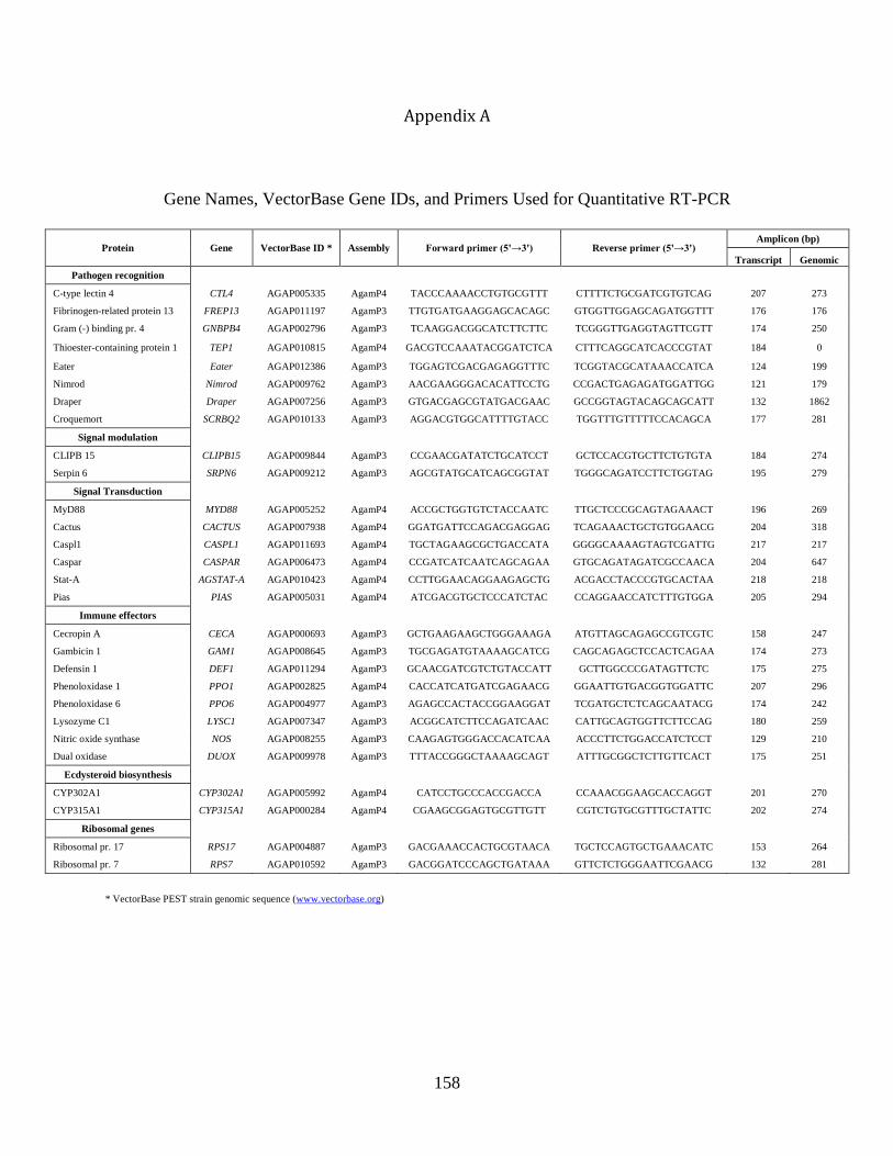
158
Appendix A
Gene Names, VectorBase Gene IDs, and Primers Used for Quantitative RT-PCR
Protein Gene VectorBase ID * Assembly Forward primer (5'→3') Reverse primer (5'→3') Amplicon (bp)
Transcript Genomic
Pathogen recognition
C-type lectin 4 CTL4 AGAP005335 AgamP4 TACCCAAAACCTGTGCGTTT CTTTTCTGCGATCGTGTCAG 207 273
Fibrinogen-related protein 13 FREP13 AGAP011197 AgamP3 TTGTGATGAAGGAGCACAGC GTGGTTGGAGCAGATGGTTT 176 176
Gram (-) binding pr. 4 GNBPB4 AGAP002796 AgamP3 TCAAGGACGGCATCTTCTTC TCGGGTTGAGGTAGTTCGTT 174 250
Thioester-containing protein 1 TEP1 AGAP010815 AgamP4 GACGTCCAAATACGGATCTCA CTTTCAGGCATCACCCGTAT 184 0
Eater Eater AGAP012386 AgamP3 TGGAGTCGACGAGAGGTTTC TCGGTACGCATAAACCATCA 124 199
Nimrod Nimrod AGAP009762 AgamP3 AACGAAGGGACACATTCCTG CCGACTGAGAGATGGATTGG 121 179
Draper Draper AGAP007256 AgamP3 GTGACGAGCGTATGACGAAC GCCGGTAGTACAGCAGCATT 132 1862
Croquemort SCRBQ2 AGAP010133 AgamP3 AGGACGTGGCATTTTGTACC TGGTTTGTTTTTCCACAGCA 177 281
Signal modulation
CLIPB 15 CLIPB15 AGAP009844 AgamP3 CCGAACGATATCTGCATCCT GCTCCACGTGCTTCTGTGTA 184 274
Serpin 6 SRPN6 AGAP009212 AgamP3 AGCGTATGCATCAGCGGTAT TGGGCAGATCCTTCTGGTAG 195 279
Signal Transduction
MyD88 MYD88 AGAP005252 AgamP4 ACCGCTGGTGTCTACCAATC TTGCTCCCGCAGTAGAAACT 196 269
Cactus CACTUS AGAP007938 AgamP4 GGATGATTCCAGACGAGGAG TCAGAAACTGCTGTGGAACG 204 318
Caspl1 CASPL1 AGAP011693 AgamP4 TGCTAGAAGCGCTGACCATA GGGGCAAAAGTAGTCGATTG 217 217
Caspar CASPAR AGAP006473 AgamP4 CCGATCATCAATCAGCAGAA GTGCAGATAGATCGCCAACA 204 647
Stat-A AGSTAT-A AGAP010423 AgamP4 CCTTGGAACAGGAAGAGCTG ACGACCTACCCGTGCACTAA 218 218
Pias PIAS AGAP005031 AgamP4 ATCGACGTGCTCCCATCTAC CCAGGAACCATCTTTGTGGA 205 294
Immune effectors
Cecropin A CECA AGAP000693 AgamP3 GCTGAAGAAGCTGGGAAAGA ATGTTAGCAGAGCCGTCGTC 158 247
Gambicin 1 GAM1 AGAP008645 AgamP3 TGCGAGATGTAAAAGCATCG CAGCAGAGCTCCACTCAGAA 174 273
Defensin 1 DEF1 AGAP011294 AgamP3 GCAACGATCGTCTGTACCATT GCTTGGCCCGATAGTTCTC 175 275
Phenoloxidase 1 PPO1 AGAP002825 AgamP4 CACCATCATGATCGAGAACG GGAATTGTGACGGTGGATTC 207 296
Phenoloxidase 6 PPO6 AGAP004977 AgamP3 AGAGCCACTACCGGAAGGAT TCGATGCTCTCAGCAATACG 174 242
Lysozyme C1 LYSC1 AGAP007347 AgamP3 ACGGCATCTTCCAGATCAAC CATTGCAGTGGTTCTTCCAG 180 259
Nitric oxide synthase NOS AGAP008255 AgamP3 CAAGAGTGGGACCACATCAA ACCCTTCTGGACCATCTCCT 129 210
Dual oxidase DUOX AGAP009978 AgamP3 TTTACCGGGCTAAAAGCAGT ATTTGCGGCTCTTGTTCACT 175 251
Ecdysteroid biosynthesis
CYP302A1 CYP302A1 AGAP005992 AgamP4 CATCCTGCCCACCGACCA CCAAACGGAAGCACCAGGT 201 270
CYP315A1 CYP315A1 AGAP000284 AgamP4 CGAAGCGGAGTGCGTTGTT CGTCTGTGCGTTTGCTATTC 202 274
Ribosomal genes
Ribosomal pr. 17 RPS17 AGAP004887 AgamP3 GACGAAACCACTGCGTAACA TGCTCCAGTGCTGAAACATC 153 264
Ribosomal pr. 7 RPS7 AGAP010592 AgamP3 GACGGATCCCAGCTGATAAA GTTCTCTGGGAATTCGAACG 132 281
* VectorBase PEST strain genomic sequence (www.vectorbase.org)
159
Appendix B
Relative mRNA Expression Levels of Individual Immunity Genes
The following are graphs of the relative induction of individual immunity genes in naïve,
injured, and E. coli-infected larvae, 1-day-old adults, and 5-day-old adults assayed in the qPCR
analysis presented in Figure 11 of Chapter IV. Graphs show the average relative mRNA fold
change of pathogen recognition (A), signal transduction (B), signal modulation (C), immune
effector (D), ecdysteroid biosynthesis (E) and ribosomal (F) genes relative to the naïve group of
a given life stage or adult age. Whiskers denote the SEM. Asterisks denote the significant
regulation of mRNA levels relative to the naïve group as determined by the Kruskal-Wallis test,
followed by Dunn’s post hoc test (*, P<0.05; **, P<0.01; ***, P<0.001; ****, P<0.0001).
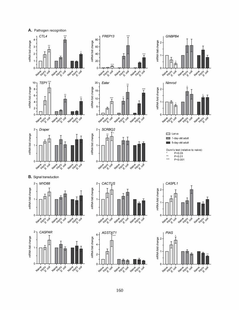
160
161