Embed Size (px)
Citation preview
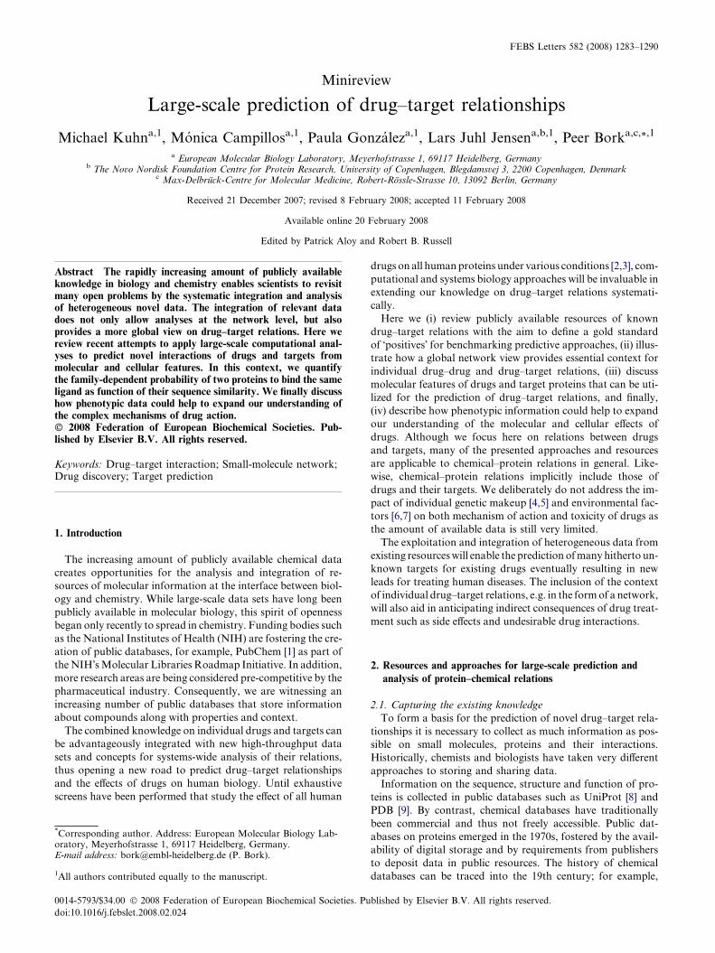
FEBS Letters 582 (2008) 1283–1290
Minireview
Large-scale prediction of drug–target relationships
Michael Kuhna,1, Monica Campillosa,1, Paula Gonzaleza,1, Lars Juhl Jensena,b,1, Peer Borka,c,*,1
a European Molecular Biology Laboratory, Meyerhofstrasse 1, 69117 Heidelberg, Germanyb The Novo Nordisk Foundation Centre for Protein Research, University of Copenhagen, Blegdamsvej 3, 2200 Copenhagen, Denmark
c Max-Delbruck-Centre for Molecular Medicine, Robert-Rossle-Strasse 10, 13092 Berlin, Germany
Received 21 December 2007; revised 8 February 2008; accepted 11 February 2008
Available online 20 February 2008
Edited by Patrick Aloy and Robert B. Russell
Abstract The rapidly increasing amount of publicly availableknowledge in biology and chemistry enables scientists to revisitmany open problems by the systematic integration and analysisof heterogeneous novel data. The integration of relevant datadoes not only allow analyses at the network level, but alsoprovides a more global view on drug–target relations. Here wereview recent attempts to apply large-scale computational anal-yses to predict novel interactions of drugs and targets frommolecular and cellular features. In this context, we quantifythe family-dependent probability of two proteins to bind the sameligand as function of their sequence similarity. We finally discusshow phenotypic data could help to expand our understanding ofthe complex mechanisms of drug action.� 2008 Federation of European Biochemical Societies. Pub-lished by Elsevier B.V. All rights reserved.
Keywords: Drug–target interaction; Small-molecule network;Drug discovery; Target prediction
1. Introduction
The increasing amount of publicly available chemical data
creates opportunities for the analysis and integration of re-
sources of molecular information at the interface between biol-
ogy and chemistry. While large-scale data sets have long been
publicly available in molecular biology, this spirit of openness
began only recently to spread in chemistry. Funding bodies such
as the National Institutes of Health (NIH) are fostering the cre-
ation of public databases, for example, PubChem [1] as part of
the NIH�s Molecular Libraries Roadmap Initiative. In addition,
more research areas are being considered pre-competitive by the
pharmaceutical industry. Consequently, we are witnessing an
increasing number of public databases that store information
about compounds along with properties and context.
The combined knowledge on individual drugs and targets can
be advantageously integrated with new high-throughput data
sets and concepts for systems-wide analysis of their relations,
thus opening a new road to predict drug–target relationships
and the effects of drugs on human biology. Until exhaustive
screens have been performed that study the effect of all human
*Corresponding author. Address: European Molecular Biology Lab-oratory, Meyerhofstrasse 1, 69117 Heidelberg, Germany.E-mail address: [email protected] (P. Bork).
1All authors contributed equally to the manuscript.
0014-5793/$34.00 � 2008 Federation of European Biochemical Societies. Pu
doi:10.1016/j.febslet.2008.02.024
drugs on all human proteins under various conditions [2,3], com-
putational and systems biology approaches will be invaluable in
extending our knowledge on drug–target relations systemati-
cally.
Here we (i) review publicly available resources of known
drug–target relations with the aim to define a gold standard
of �positives� for benchmarking predictive approaches, (ii) illus-
trate how a global network view provides essential context for
individual drug–drug and drug–target relations, (iii) discuss
molecular features of drugs and target proteins that can be uti-
lized for the prediction of drug–target relations, and finally,
(iv) describe how phenotypic information could help to expand
our understanding of the molecular and cellular effects of
drugs. Although we focus here on relations between drugs
and targets, many of the presented approaches and resources
are applicable to chemical–protein relations in general. Like-
wise, chemical–protein relations implicitly include those of
drugs and their targets. We deliberately do not address the im-
pact of individual genetic makeup [4,5] and environmental fac-
tors [6,7] on both mechanism of action and toxicity of drugs as
the amount of available data is still very limited.
The exploitation and integration of heterogeneous data from
existing resources will enable the prediction of many hitherto un-
known targets for existing drugs eventually resulting in new
leads for treating human diseases. The inclusion of the context
of individual drug–target relations, e.g. in the form of a network,
will also aid in anticipating indirect consequences of drug treat-
ment such as side effects and undesirable drug interactions.
2. Resources and approaches for large-scale prediction and
analysis of protein–chemical relations
2.1. Capturing the existing knowledge
To form a basis for the prediction of novel drug–target rela-
tionships it is necessary to collect as much information as pos-
sible on small molecules, proteins and their interactions.
Historically, chemists and biologists have taken very different
approaches to storing and sharing data.
Information on the sequence, structure and function of pro-
teins is collected in public databases such as UniProt [8] and
PDB [9]. By contrast, chemical databases have traditionally
been commercial and thus not freely accessible. Public dat-
abases on proteins emerged in the 1970s, fostered by the avail-
ability of digital storage and by requirements from publishers
to deposit data in public resources. The history of chemical
databases can be traced into the 19th century; for example,
blished by Elsevier B.V. All rights reserved.

1284 M. Kuhn et al. / FEBS Letters 582 (2008) 1283–1290
the Beilstein Handbook of Organic Chemistry has been pub-
lished since 1881. The distribution of data in the form of books
and the economic success of the chemical industry have lead to
a tradition of commercial databases on chemical structures
and their properties such as the Chemical Abstracts Registry.
Only in the past decade several public alternatives have been
created, including repositories like PubChem [1], ChEBI [10]
and ChemDB [11] that contain information on chemicals
and their physicochemical properties. Other databases such
as ZINC [12] have been designed as resources for virtual
screening applications. These emerging public databases allow
access to useful parts lists of proteins and chemicals.
For understanding higher-order processes these parts list
have to be connected by determining how the parts interact
within biological systems. For proteins, several public reposito-
ries for experimentally determined interactions have been
established (e.g. BioGRID [13], IntAct [14] and MINT [15])
using a common standard, PSI-MI [16]. Notably, publishers en-
force that new experimental evidence is openly accessible to the
research community, for example data from high-throughput
screens for physical [17,18] and genetic interactions [19,20].
This provides a foundation for the construction of tools that
integrate such interactions with other data types (e.g. the
STRING database [21] and other resources reviewed in [22]).
The corresponding databases for the relationships of
chemicals have not yet reached a comparable state. Although
large-scale screens of chemicals in cell-based assays have been
performed and are available from repositories such as Chem-
Bank [23] and PubChem BioAssay, deposition of data from
chemical screens in standardized repositories is not being en-
forced. The difficulties involved in obtaining and combining
the data has hampered the development of methods for pre-
dicting relationships between drugs; to our knowledge cur-
rently only one public tool exists that combines data from
chemical screens and other sources to infer relationships for
chemicals, namely STITCH [21] (Fig. 1).
Databases that centre on drug–target relations are also
emerging in the public sector: the Therapeutic Target Database
TTD [24], DrugBank [25], SuperTarget [26] and Matador [26]
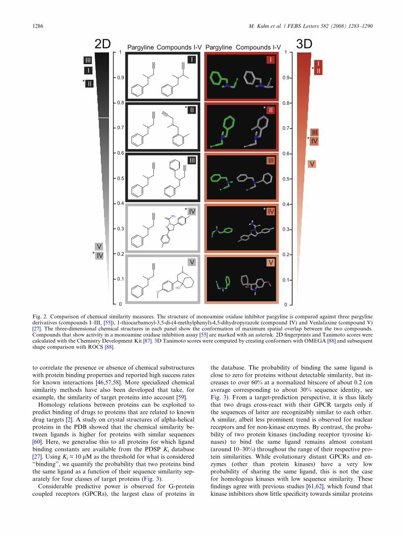
Fig. 1. Network context of drugs and targets. Proteins are shown as sphercapsules. Connecting lines (edges) depict known or predicted associations.STITCH resource [21] from which both examples are taken. (a) Drug–targetagonist cisapride also binds to the cardiac ion channel hERG (KNCH2), whithe interaction of cisapride with metabolizing Cytochrome P450 enzymessources, as depicted by the colored lines: experiments (magenta), databases (cserotonin receptors and serotonin transporter inhibitors. Compounds with sconnected by cyan lines and form two distinct groups. The serotonin receptoinhibit (red line) the serotonin receptor HTR2A. By contrast, the second-genknown to be more promiscuous and inhibit both the serotonin transporter S
all collect direct drug–target interactions. In addition, Mata-
dor [26] includes indirect drug–target interactions that capture
more distant effects of drugs on the human protein network.
Resources like the PDSP Ki database [27] and BindingDB
[28] provide in vitro binding affinities that add knowledge
about potential lead molecules; for example, Roth and collab-
orators discovered that Salvinorin A, the main active ingredi-
ent of the hallucinogenic plant Salvia divinorum, is a potent
kappa opioid agonist by screening it on a collection of recep-
tors [29]. The accumulated content of these databases (summa-
rized in Table 1) constitutes a gold standard. Such a standard
is crucial for the development of prediction methods, for exam-
ple, in the context of proper benchmarking protocols.
All the databases described above contain experimental data
related to individual proteins, chemicals or binary interactions.
To obtain a global picture of their interplay, the data therein
can be integrated with a variety of existing molecular, cellular
and organismal data such as microarray experiments (e.g.
GEO [30] or ArrayExpress [31]) and pathways (e.g. Reactome
[32], KEGG [33] or MetaCyc [34]). By bringing together these
heterogeneous data types, it is possible to construct a network
that captures many aspects of how drugs and other small-mol-
ecules function in a cellular context; for an example see Fig. 1
created using the STITCH database and its visualization capa-
bilities [21].
2.2. Context and its visualization
Systems biology approaches are increasingly being applied
to investigate the relationships between proteins, utilizing the
biological context of a protein to gather more information
about its function [22]. Similarly, the context of a drug needs
also to be considered as drugs usually do not only affect the ac-
tion of isolated targets, but influence entire pathways. Thus the
introduction of systems biology concepts into drug discovery is
being foreseen [35,36]. While there are many specialized tools
to visualize the context of proteins (e.g. [37–40]), chemists
mostly have to resort to general purpose tools such as Cyto-
scape [41,42] to view networks involving chemical compounds,
although first visualization tools are emerging [21].
es (with representative PDB structures, if available) and chemicals asEdge representation depends on query and visualization mode of therelationships of cisapride. The serotonin receptor (HTR4 and HTR2A)ch leads to arrhythmias as a side effect. In addition, the network shows(CYP3A4 and CYP2D6). The interactions are derived from variousyan), text mining (yellow) and homology (lavender). (b) Antagonists ofimilar MeSH (Medical Subject Headings) pharmacological action are
r antagonists ketanserin, clozapine and risperidone bind (blue line) anderation antidepressive agents fluoxetine, nefazodone and trazodone areLC6A4 and the serotonin receptor HTR2A.
Table 1Databases freely available for academic research that contain information on drug–target interactions
Database Number of chemicals Content
Ligand–target databasesDrugBank
http://redpoll.pharmacy.ualberta.ca/drugbank/
�1000 FDA-approved drugs,and �3000 experimental drugs
6000 drug–targets relationships; chemical, pharmacologicaland pharmaceutical data
Matadorhttp://matadorembl.de/ �770 drugs �7000 direct and �5000 indirect drug–target relationships;
links to literature sources for interactions
SuperTargethttp://insilico.charite.de/supertarget/ �1500 drugs 7300 drug–target relations
TTDhttp://bidd.nus.edu.sg/group/cjttd/TTD_ns.asp �2100 drugs drug–target relationships with 1535 targets
PDSP Ki
http://pdsp.med.unc.edu/pdsp.php �6800 chemicals �46,000 Ki values
BindingDBhttp://www.bindingdb.org/ �18000 chemicals �30000 records with Ki, IC50, or thermodynamic data
Cellular assaysPubChem BioAssay
http://pubchem.ncbi.nlm.nih.gov/ �560000 chemicals �600 single compound and high-throughputscreening assays
ChemBankhttp://chembank.broad.harvard.edu/ �1.2 million chemicals 2500 high-throughput biological assays from
188 screening projects
Note: Ki: inhibition constant; IC50: concentration of an inhibitor that is required for 50% inhibition of its target.Database content was recorded on November 1, 2007.
M. Kuhn et al. / FEBS Letters 582 (2008) 1283–1290 1285
Networks have been used recently to analyse topological and
global properties of chemical–protein interactions such as poly-
pharmacology (a term usually used to describe multiple actions
for the same drug [43], for examples, see Fig. 1) and drug target–
disease relationships [44,45]. Paolini and co-workers [46] pro-
vided an overview of the existing polypharmacology relations
by integrating data from several proprietary and public chemi-
cal screening sources. The authors presented a protein network
in which two proteins are connected if chemicals are known to
bind both of them with similar affinity. In this network a highly
family-dependent degree of promiscuity of targets was ob-
served, both within the same family and across families.
A similar level of target promiscuity was observed by
Yıldırım and co-workers [45] in a network of known drug–
target relationships obtained from DrugBank [25]. Within a
interaction network of human proteins derived from yeast
two-hybrid screens [47,48], the authors also explored the
distribution of distances between drug targets and disease
genes described in OMIM [49]. Although some drugs were
found to target disease genes or their direct network neigh-
bours, the distance distribution otherwise matched that of
the random control. This suggests that most drugs in fact alle-
viate the symptoms (being palliative drugs) rather than target
directly the actual cause of the disease [45].
Although such networks attempt to offer a global view on
the relations of proteins and chemicals, our knowledge of
drug–target relations is far from complete and needs to be ex-
panded in order to increase our understanding of the actions of
drugs. One promising avenue in this regard is the accurate pre-
diction of drug–target relations followed by directed experi-
mental validations.
3. Concepts for large-scale drug–target predictions
3.1. Predicting relations based on molecular features of
chemicals and proteins
Exploiting similarities between chemical structures is a
common way to infer the activity of compounds. The most
prevalent approach for comparing compounds is to convert
the two-dimensional representation of each compound into a
fingerprint either by using a defined list of substructures or
by encoding (hashing) all the encountered substructures up
to a certain size. This results in fixed-length bit vectors for
which the Tanimoto (or Jacquard) similarity measure is com-
puted by dividing the size of intersection of the set bits by
the size of the union [50]. Alternatively, chemical similarity
can be determined by aligning three-dimensional models of
the compounds [51–53]. To illustrate these similarity measures,
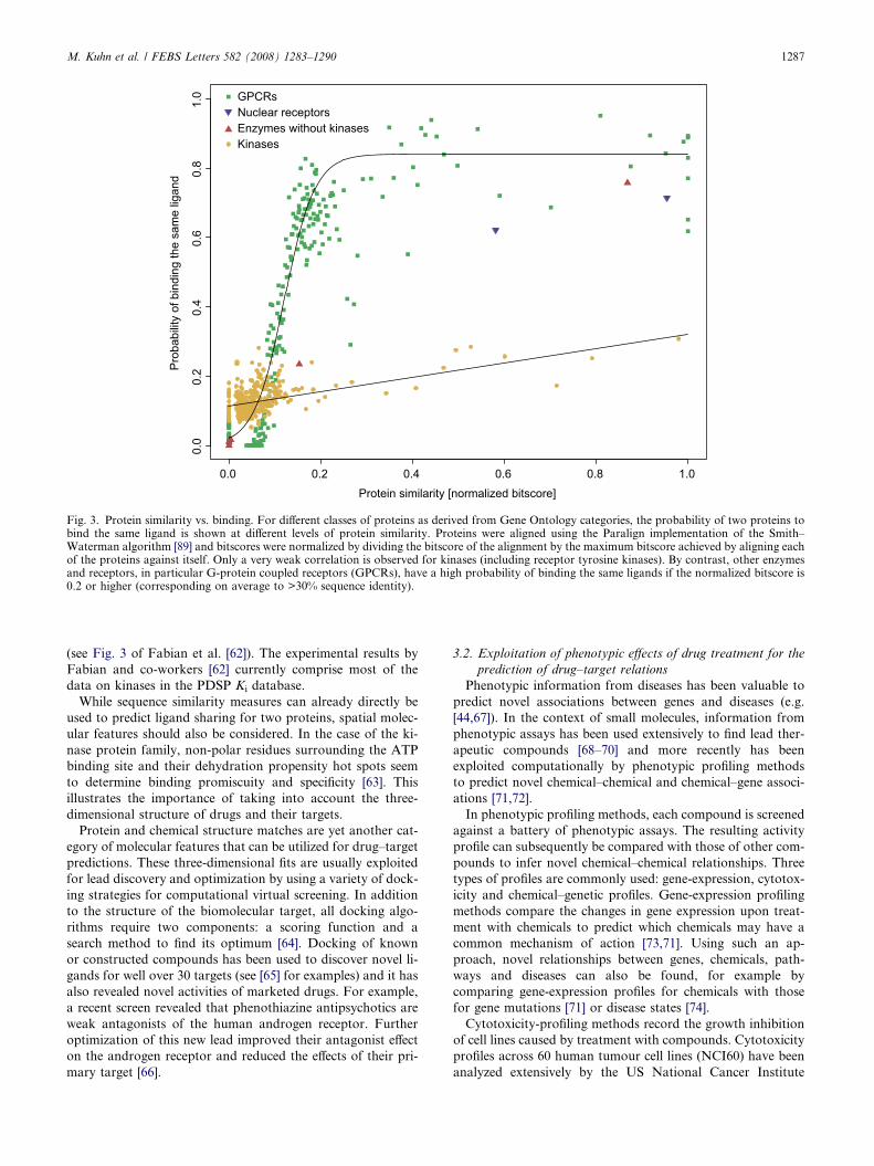
we show two- and three-dimensional structure comparisons of
the monoamine oxidase inhibitor pargyline with five other
compounds (Fig. 2).
Initial optimistic results [54] on the relationship between
chemical similarity and activity were put into perspective by
the analysis of more unbiased chemical libraries. For these,
there is only a 30% chance of binding the same compound at
the similarity level previously thought to warrant >80% chance
[55]. For example, only one of the compounds in Fig. 2 with
high similarity to pargyline also inhibits monoamine oxidase.
To overcome the limited predictive power of pairwise chemical
structure comparison, Keiser and co-workers developed a sta-
tistical model to detect remote, yet significant similarities be-
tween groups of drugs and used it to predict novel drug–
target relations [56]. Other groups used Bayesian classifiers
VN
OOH
N
III
I
V
V
IIII
0.9
0.8
0.7
0.6
0.5
0.4
0.3
0.2
0.1
1
0
II*
II*0.9
0.8
0.7
0.6
0.5
0.4
0.3
0.2
0.1
1
0
IV*
III
I
Pargyline Compounds I-V Pargyline Compounds I-V
II*
N
N
N
N
N
N
N
IV
V
III
II
II
IV*
IV*
*
*
Fig. 2. Comparison of chemical similarity measures. The structure of monoamine oxidase inhibitor pargyline is compared against three pargylinederivatives (compounds I–III, [55]), 1-thiocarbamoyl-3,5-di-(4-methylphenyl)-4,5-dihydropyrazole (compound IV) and Venlafaxine (compound V)[27]. The three-dimensional chemical structures in each panel show the conformation of maximum spatial overlap between the two compounds.Compounds that show activity in a monoamine oxidase inhibition assay [55] are marked with an asterisk. 2D fingerprints and Tanimoto scores werecalculated with the Chemistry Development Kit [87]. 3D Tanimoto scores were computed by creating conformers with OMEGA [88] and subsequentshape comparison with ROCS [88].
1286 M. Kuhn et al. / FEBS Letters 582 (2008) 1283–1290
to correlate the presence or absence of chemical substructures
with protein binding properties and reported high success rates
for known interactions [46,57,58]. More specialized chemical
similarity methods have also been developed that take, for
example, the similarity of target proteins into account [59].
Homology relations between proteins can be exploited to
predict binding of drugs to proteins that are related to known
drug targets [2]. A study on crystal structures of alpha-helical
proteins in the PDB showed that the chemical similarity be-
tween ligands is higher for proteins with similar sequences
[60]. Here, we generalise this to all proteins for which ligand
binding constants are available from the PDSP Ki database
[27]. Using Ki = 10 lM as the threshold for what is considered
‘‘binding’’, we quantify the probability that two proteins bind
the same ligand as a function of their sequence similarity sep-
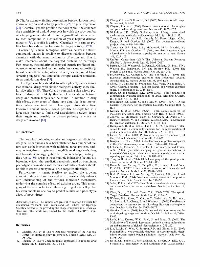
arately for four classes of target proteins (Fig. 3).
Considerable predictive power is observed for G-protein
coupled receptors (GPCRs), the largest class of proteins in
the database. The probability of binding the same ligand is
close to zero for proteins without detectable similarity, but in-
creases to over 60% at a normalized bitscore of about 0.2 (on
average corresponding to about 30% sequence identity, see
Fig. 3). From a target-prediction perspective, it is thus likely
that two drugs cross-react with their GPCR targets only if
the sequences of latter are recognizably similar to each other.
A similar, albeit less prominent trend is observed for nuclear
receptors and for non-kinase enzymes. By contrast, the proba-
bility of two protein kinases (including receptor tyrosine ki-
nases) to bind the same ligand remains almost constant
(around 10–30%) throughout the range of their respective pro-
tein similarities. While evolutionary distant GPCRs and en-
zymes (other than protein kinases) have a very low
probability of sharing the same ligand, this is not the case
for homologous kinases with low sequence similarity. These
findings agree with previous studies [61,62], which found that
kinase inhibitors show little specificity towards similar proteins
0.0 0.2 0.4 0.6 0.8 1.0
0.0
0.2
0.4
0.6
0.8
1.0
Protein similarity [normalized bitscore]
Prob
abilit
y of
bin
ding
the
sam
e lig
and
Enzymes without kinasesNuclear receptors
Kinases
GPCRs
Fig. 3. Protein similarity vs. binding. For different classes of proteins as derived from Gene Ontology categories, the probability of two proteins tobind the same ligand is shown at different levels of protein similarity. Proteins were aligned using the Paralign implementation of the Smith–Waterman algorithm [89] and bitscores were normalized by dividing the bitscore of the alignment by the maximum bitscore achieved by aligning eachof the proteins against itself. Only a very weak correlation is observed for kinases (including receptor tyrosine kinases). By contrast, other enzymesand receptors, in particular G-protein coupled receptors (GPCRs), have a high probability of binding the same ligands if the normalized bitscore is0.2 or higher (corresponding on average to >30% sequence identity).
M. Kuhn et al. / FEBS Letters 582 (2008) 1283–1290 1287
(see Fig. 3 of Fabian et al. [62]). The experimental results by
Fabian and co-workers [62] currently comprise most of the
data on kinases in the PDSP Ki database.
While sequence similarity measures can already directly be
used to predict ligand sharing for two proteins, spatial molec-
ular features should also be considered. In the case of the ki-
nase protein family, non-polar residues surrounding the ATP
binding site and their dehydration propensity hot spots seem
to determine binding promiscuity and specificity [63]. This
illustrates the importance of taking into account the three-
dimensional structure of drugs and their targets.
Protein and chemical structure matches are yet another cat-
egory of molecular features that can be utilized for drug–target
predictions. These three-dimensional fits are usually exploited
for lead discovery and optimization by using a variety of dock-
ing strategies for computational virtual screening. In addition
to the structure of the biomolecular target, all docking algo-
rithms require two components: a scoring function and a
search method to find its optimum [64]. Docking of known
or constructed compounds has been used to discover novel li-
gands for well over 30 targets (see [65] for examples) and it has
also revealed novel activities of marketed drugs. For example,
a recent screen revealed that phenothiazine antipsychotics are
weak antagonists of the human androgen receptor. Further
optimization of this new lead improved their antagonist effect
on the androgen receptor and reduced the effects of their pri-
mary target [66].
3.2. Exploitation of phenotypic effects of drug treatment for the
prediction of drug–target relations
Phenotypic information from diseases has been valuable to
predict novel associations between genes and diseases (e.g.
[44,67]). In the context of small molecules, information from
phenotypic assays has been used extensively to find lead ther-
apeutic compounds [68–70] and more recently has been
exploited computationally by phenotypic profiling methods
to predict novel chemical–chemical and chemical–gene associ-
ations [71,72].
In phenotypic profiling methods, each compound is screened
against a battery of phenotypic assays. The resulting activity
profile can subsequently be compared with those of other com-
pounds to infer novel chemical–chemical relationships. Three
types of profiles are commonly used: gene-expression, cytotox-
icity and chemical–genetic profiles. Gene-expression profiling
methods compare the changes in gene expression upon treat-
ment with chemicals to predict which chemicals may have a
common mechanism of action [73,71]. Using such an ap-
proach, novel relationships between genes, chemicals, path-
ways and diseases can also be found, for example by
comparing gene-expression profiles for chemicals with those
for gene mutations [71] or disease states [74].
Cytotoxicity-profiling methods record the growth inhibition
of cell lines caused by treatment with compounds. Cytotoxicity
profiles across 60 human tumour cell lines (NCI60) have been
analyzed extensively by the US National Cancer Institute
1288 M. Kuhn et al. / FEBS Letters 582 (2008) 1283–1290
(NCI), for example, finding correlations between known mech-
anism of action and activity profiles [72] or gene expression
[75]. Chemical–genetic profiling methods exploit the enhanced
drug sensitivity of diploid yeast cells in which the copy number
of a target gene is reduced. From the growth inhibition caused
by each compound in a collection of yeast haploid deletion
mutants [76], a profile is derived. Chemicals with similar pro-
files have been shown to have similar target activity [77,78].
Correlating similar biological activities between different
compounds makes it possible to discover relations between
chemicals with the same mechanism of action and thus to
make inferences about the targeted proteins or pathways.
For instance, the similarity of chemical–genetic profiles of ami-
odarone (an antianginal and antiarrhythmic) and tamoxifen (a
breast cancer therapeutic) observed in a yeast haploid deletion
screening suggests that tamoxifen disrupts calcium homeosta-
sis as amiodarone does [79].
This logic can be extended to other phenotypic measures.
For example, drugs with similar biological activity show simi-
lar side effects [80]. Therefore, by comparing side effects pro-
files of drugs, it is likely that novel associations between
drugs and protein targets can be found. In addition to drug
side effects, other types of phenotypic data like drug interac-
tions, when combined with phenotypic information from
knockout animal models, could possibly be exploited in a
large-scale manner to find novel associations between drugs,
their targets and possibly the disease pathway in which the
drugs are involved [81].
4. Conclusions
The complex molecular, cellular and organismal effects that
drugs cause in humans have been attributed to a number of fac-
tors such as the interaction with additional target proteins, path-
way context, drug–drug interactions, different dosage levels, drug
metabolization and aggregation or irreversible target binding of
the drug [82–86]. Despite these multiple influencing factors, it is
becoming evident that prediction methods based on combining
phenotypic information with known molecular activities should
be able to generate many novel drug–target relationships.
Furthermore, it seems feasible to exploit the growing
amount of data we have reviewed here to considerably enhance
our understanding of the various molecular mechanisms
underlying the complex effects of existing drugs. This untan-
gling of the various factors influencing drug effects will proba-
bly even enable us one day to predict cellular and phenotypic
effect of novel drugs.
Acknowledgements: The authors are grateful to Konrad Forstner fordiscussions. We thank Paul Hawkins and Bob Tolbert from OpenEyeScientific Software for providing the shape comparisons of chemicalstructures. This work was funded by the BMBF QuantPro Grant(0313831D).
References
[1] Wheeler, D.L. et al. (2007) Database resources of the NationalCenter for Biotechnology Information. Nucleic Acids Res. 35,D5–D12.
[2] Rognan, D. (2007) Chemogenomic approaches to rational drugdesign. Br. J. Pharmacol. 152, 38–52.
[3] Chong, C.R. and Sullivan Jr., D.J. (2007) New uses for old drugs.Nature 448, 645–646.
[4] Clayton, T.A. et al. (2006) Pharmaco-metabonomic phenotypingand personalized drug treatment. Nature 440, 1073–1077.
[5] Nicholson, J.K. (2006) Global systems biology, personalizedmedicine and molecular epidemiology. Mol. Syst. Biol. 2, 52.
[6] Turnbaugh, P.J., Ley, R.E., Hamady, M., Fraser-Liggett, C.M.,Knight, R. and Gordon, J.I. (2007) The human microbiomeproject. Nature 449, 804–810.
[7] Turnbaugh, P.J., Ley, R.E., Mahowald, M.A., Magrini, V.,Mardis, E.R. and Gordon, J.I. (2006) An obesity-associated gutmicrobiome with increased capacity for energy harvest. Nature444, 1027–1031.
[8] UniProt Consortium (2007) The Universal Protein Resource(UniProt). Nucleic Acids Res. 35, D193–D197.
[9] Berman, H.M., Westbrook, J., Feng, Z., Gilliland, G., Bhat, T.N.,Weissig, H., Shindyalov, I.N. and Bourne, P.E. (2000) TheProtein Data Bank. Nucleic Acids Res. 28, 235–242.
[10] Brooksbank, C., Cameron, G. and Thornton, J. (2005) TheEuropean Bioinformatics Institute�s data resources: towardssystems biology. Nucleic Acids Res. 33, D46–D53.
[11] Chen, J.H., Linstead, E., Swamidass, S.J., Wang, D. and Baldi, P.(2007) ChemDB update – full-text search and virtual chemicalspace. Bioinformatics 23, 2348–2351.
[12] Irwin, J.J. and Shoichet, B.K. (2005) ZINC – a free database ofcommercially available compounds for virtual screening. J. Chem.Inf. Model. 45, 177–182.
[13] Breitkreutz, B.J., Stark, C. and Tyers, M. (2003) The GRID: theGeneral Repository for Interaction Datasets. Genome Biol. 4,R23.
[14] Kerrien, S. et al. (2007) IntAct – open source resource formolecular interaction data. Nucleic Acids Res. 35, D561–D565.
[15] Zanzoni, A., Montecchi-Palazzi, L., Quondam, M., Ausiello, G.,Helmer-Citterich, M. and Cesareni, G. (2002) MINT: a MolecularINTeraction database. FEBS Lett. 513, 135–140.
[16] Hermjakob, H. et al. (2004) The HUPO PSI�s molecular inter-action format – a community standard for the representation ofprotein interaction data. Nat. Biotechnol. 22, 177–183.
[17] Gavin, A.C. et al. (2006) Proteome survey reveals modularity ofthe yeast cell machinery. Nature 440, 631–636.
[18] Krogan, N.J. et al. (2006) Global landscape of protein complexesin the yeast Saccharomyces cerevisiae. Nature 440, 637–643.
[19] Lehner, B., Crombie, C., Tischler, J., Fortunato, A. and Fraser,A.G. (2006) Systematic mapping of genetic interactions inCaenorhabditis elegans identifies common modifiers of diversesignaling pathways. Nat. Genet. 38, 896–903.
[20] Tong, A.H. et al. (2004) Global mapping of the yeast geneticinteraction network. Science 303, 808–813.
[21] Kuhn, M., von Mering, C., Campillos, M., Jensen, L.J. and Bork,P. (2008) STITCH: interaction networks of chemicals andproteins. Nucleic Acids Res. 36, D684–D688.
[22] Bork, P., Jensen, L.J., von Mering, C., Ramani, A.K., Lee, I. andMarcotte, E.M. (2004) Protein interaction networks from yeast tohuman. Curr. Opin. Struct. Biol. 14, 292–299.
[23] Seiler, K.P. et al. (2007) ChemBank: a small-molecule screeningand cheminformatics resource database. Nucleic Acids Res. 18,18.
[24] Chen, X., Ji, Z.L. and Chen, Y.Z. (2002) TTD: TherapeuticTarget Database. Nucleic Acids Res. 30, 412–415.
[25] Wishart, D.S., Knox, C., Guo, A.C., Shrivastava, S., Hassanali,M., Stothard, P., Chang, Z. and Woolsey, J. (2006) DrugBank: acomprehensive resource for in silico drug discovery and explora-tion. Nucleic Acids Res. 34, D668–D672.
[26] Gunther, S. et al. (2008) SuperTarget and Matador: resources forexploring drug–target relationships. Nucleic Acids Res. 36, D919–D922.
[27] Roth, B.L., Kroeze, W.K., Patel, S. and lopez, E. (2000) TheMultiplicity of Serotonin Receptors: uselessly diverse molecules oran embarrasment of riches? Neuroscientist 6, 252–262.
[28] Liu, T., Lin, Y., Wen, X., Jorissen, R.N. and Gilson, M.K. (2007)BindingDB: a web-accessible database of experimentally deter-mined protein–ligand binding affinities. Nucleic Acids Res. 35,D198–D201.
[29] Roth, B.L., Baner, K., Westkaemper, R., Siebert, D., Rice, K.C.,Steinberg, S., Ernsberger, P. and Rothman, R.B. (2002) Salvino-
M. Kuhn et al. / FEBS Letters 582 (2008) 1283–1290 1289
rin A: a potent naturally occurring nonnitrogenous kappa opioidselective agonist. Proc. Natl. Acad. Sci. USA 99, 11934–11939.
[30] Edgar, R., Domrachev, M. and Lash, A.E. (2002) Gene Expres-sion Omnibus: NCBI gene expression and hybridization arraydata repository. Nucleic Acids Res. 30, 207–210.
[31] Parkinson, H. et al. (2007) ArrayExpress – a public database ofmicroarray experiments and gene expression profiles. NucleicAcids Res. 35, D747–D750.
[32] Vastrik, I. et al. (2007) Reactome: a knowledge base of biologicpathways and processes. Genome Biol. 8, R39.
[33] Kanehisa, M. et al. (2006) From genomics to chemical genomics:new developments in KEGG. Nucleic Acids Res. 34, D354–D357.
[34] Caspi, R. et al. (2006) MetaCyc: a multiorganism database ofmetabolic pathways and enzymes. Nucleic Acids Res. 34, D511–D516.
[35] Apic, G., Ignjatovic, T., Boyer, S. and Russell, R.B. (2005)Illuminating drug discovery with biological pathways. FEBS Lett.579, 1872–1877.
[36] Rajasethupathy, P., Vayttaden, S.J. and Bhalla, U.S. (2005)Systems modeling: a pathway to drug discovery. Curr. Opin.Chem. Biol. 9, 400–406.
[37] Breitkreutz, B.J. et al. (2008) The BioGRID Interaction Data-base: 2008 update. Nucleic Acids Res. 36, D637–D640.
[38] Overbeek, R. et al. (2005) The subsystems approach to genomeannotation and its use in the project to annotate 1000 genomes.Nucleic Acids Res. 33, 5691–5702 (Print 2005).
[39] Suderman, M. and Hallett, M. (2007) Tools for visually exploringbiological networks. Bioinformatics 23, 2651–2659.
[40] von Mering, C., Jensen, L.J., Kuhn, M., Chaffron, S., Doerks, T.,Kruger, B., Snel, B. and Bork, P. (2007) STRING 7 – recentdevelopments in the integration and prediction of proteininteractions. Nucleic Acids Res. 35, D358–D362.
[41] Cline, M.S. et al. (2007) Integration of biological networksand gene expression data using Cytoscape. Nat. Protoc. 2, 2366–2382.
[42] Shannon, P. et al. (2003) Cytoscape: a software environment forintegrated models of biomolecular interaction networks. GenomeRes. 13, 2498–2504.
[43] Hopkins, A.L., Mason, J.S. and Overington, J.P. (2006) Can werationally design promiscuous drugs? Curr. Opin. Struct. Biol. 16,127–136.
[44] Lage, K. et al. (2007) A human phenome–interactome network ofprotein complexes implicated in genetic disorders. Nat. Biotech-nol. 25, 309–316.
[45] Yildirim, M.A., Goh, K.I., Cusick, M.E., Barabasi, A.L. andVidal, M. (2007) Drug–target network. Nat. Biotechnol. 25, 1119–1126.
[46] Paolini, G.V., Shapland, R.H., van Hoorn, W.P., Mason, J.S. andHopkins, A.L. (2006) Global mapping of pharmacological space.Nat. Biotechnol. 24, 805–815.
[47] Stelzl, U. et al. (2005) A human protein–protein interactionnetwork: a resource for annotating the proteome. Cell 122, 957–968.
[48] Rual, J.F. et al. (2005) Towards a proteome-scale map of thehuman protein–protein interaction network. Nature 437, 1173–1178.
[49] Hamosh, A., Scott, A.F., Amberger, J.S., Bocchini, C.A. andMcKusick, V.A. (2005) Online Mendelian Inheritance in Man(OMIM), a knowledgebase of human genes and genetic disorders.Nucleic Acids Res. 33, D514–D517.
[50] Willett, P., Barnard, J. and Downs, G. (1998) Chemical similaritysearching. J. Chem. Inform. Comp. Sci. 38, 983–996.
[51] Lemmen, C. and Lengauer, T. (2000) Computational methods forthe structural alignment of molecules. J. Comp. Aided Mol. Des.14, 215–232.
[52] Nettles, J.H., Jenkins, J.L., Bender, A., Deng, Z., Davies, J.W.and Glick, M. (2006) Bridging chemical and biological space:‘‘target fishing’’ using 2D and 3D molecular descriptors. J. Med.Chem. 49, 6802–6810.
[53] Thimm, M., Goede, A., Hougardy, S. and Preissner, R. (2004)Comparison of 2D similarity and 3D superposition. Applicationto searching a conformational drug database. J. Chem. Inform.Comp. Sci. 44, 1816–1822.
[54] Matter, H. (1997) Selecting optimally diverse compounds fromstructure databases: a validation study of two-dimensional and
three-dimensional molecular descriptors. J. Med. Chem. 40, 1219–1229.
[55] Martin, Y.C., Kofron, J.L. and Traphagen, L.M. (2002) Dostructurally similar molecules have similar biological activity? J.Med. Chem. 45, 4350–4358.
[56] Keiser, M.J., Roth, B.L., Armbruster, B.N., Ernsberger, P., Irwin,J.J. and Shoichet, B.K. (2007) Relating protein pharmacology byligand chemistry. Nat. Biotechnol. 25, 197–206.
[57] Bender, A. et al. (2007) Analysis of pharmacology data and theprediction of adverse drug reactions and off-target effects fromchemical structure. ChemMedChem 2, 861–873.
[58] Xia, X., Maliski, E.G., Gallant, P. and Rogers, D. (2004)Classification of kinase inhibitors using a Bayesian model. J.Med. Chem. 47, 4463–4470.
[59] Schuffenhauer, A., Floersheim, P., Acklin, P. and Jacoby, E.(2003) Similarity metrics for ligands reflecting the similarity of thetarget proteins. J. Chem. Inform. Comp. Sci. 43, 391–405.
[60] Mitchell, J.B. (2001) The relationship between the sequenceidentities of alpha helical proteins in the PDB and the molecularsimilarities of their ligands. J. Chem. Inform. Comp. Sci. 41,1617–1622.
[61] Fedorov, O. et al. (2007) A systematic interaction map ofvalidated kinase inhibitors with Ser/Thr kinases. Proc. Natl.Acad. Sci. USA 104, 20523–20528.
[62] Fabian, M.A. et al. (2005) A small molecule–kinase interactionmap for clinical kinase inhibitors. Nat. Biotechnol. 23, 329–336.
[63] Chen, J., Zhang, X. and Fernandez, A. (2007) Molecular basis forspecificity in the druggable kinome: sequence-based analysis.Bioinformatics 23, 563–572.
[64] Laird, E.R. and Blake, J.F. (2004) Structure-based generation ofviable leads from small combinatorial libraries. Curr. Opin. DrugDiscov. Dev. 7, 354–359.
[65] Shoichet, B.K., McGovern, S.L., Wei, B. and Irwin, J.J. (2002)Lead discovery using molecular docking. Curr. Opin. Chem. Biol.6, 439–446.
[66] Bisson, W.H. et al. (2007) Discovery of antiandrogen activity ofnonsteroidal scaffolds of marketed drugs. Proc. Natl. Acad. Sci.USA. 104, 11927–11932.
[67] Perez-Iratxeta, C., Bork, P. and Andrade, M.A. (2002) Associ-ation of genes to genetically inherited diseases using data mining.Nat. Genet. 31, 316–319.
[68] Peterson, R.T., Shaw, S.Y., Peterson, T.A., Milan, D.J., Zhong,T.P., Schreiber, S.L., MacRae, C.A. and Fishman, M.C. (2004)Chemical suppression of a genetic mutation in a zebrafish modelof aortic coarctation. Nat. Biotechnol. 22, 595–599.
[69] Zon, L.I. and Peterson, R.T. (2005) In vivo drug discovery in thezebrafish. Nat. Rev. Drug Discov. 4, 35–44.
[70] Stern, H.M. and Zon, L.I. (2003) Cancer genetics and drugdiscovery in the zebrafish. Nat. Rev. Cancer 3, 533–539.
[71] Hughes, T.R. et al. (2000) Functional discovery via a compen-dium of expression profiles. Cell 102, 109–126.
[72] Weinstein, J.N. et al. (1997) An information-intensive approachto the molecular pharmacology of cancer. Science 275, 343–349.
[73] Waring, J.F. et al. (2001) Clustering of hepatotoxins based onmechanism of toxicity using gene expression profiles. Toxicol.Appl. Pharmacol. 175, 28–42.
[74] Lamb, J. et al. (2006) The Connectivity Map: using gene-expression signatures to connect small molecules, genes, anddisease. Science 313, 1929–1935.
[75] Covell, D.G., Wallqvist, A., Huang, R., Thanki, N., Rabow, A.A.and Lu, X.J. (2005) Linking tumor cell cytotoxicity to mechanismof drug action: an integrated analysis of gene expression, small-molecule screening and structural databases. Proteins 59, 403–433.
[76] Lum, P.Y. et al. (2004) Discovering modes of action fortherapeutic compounds using a genome-wide screen of yeastheterozygotes. Cell 116, 121–137.
[77] Brown, J.A. et al. (2006) Global analysis of gene function in yeastby quantitative phenotypic profiling. Mol. Syst. Biol. 2,2006.0001.
[78] Lee, W. et al. (2005) Genome-wide requirements for resistance tofunctionally distinct DNA-damaging agents. PLoS Genet. 1, e24.
[79] Parsons, A.B. et al. (2006) Exploring the mode-of-action ofbioactive compounds by chemical–genetic profiling in yeast. Cell126, 611–625.
1290 M. Kuhn et al. / FEBS Letters 582 (2008) 1283–1290
[80] Fliri, A.F., Loging, W.T., Thadeio, P.F. and Volkmann, R.A.(2005) Analysis of drug-induced effect patterns to link structureand side effects of medicines. Nat. Chem. Biol. 1, 389–397.
[81] Zambrowicz, B.P. and Sands, A.T. (2003) Knockouts model the100 best-selling drugs – will they model the next 100? Nat. Rev.Drug Discov. 2, 38–51.
[82] Liebler, D.C. and Guengerich, F.P. (2005) Elucidating mecha-nisms of drug-induced toxicity. Nat. Rev. Drug Discov. 4, 410–420.
[83] Demoly, P. and Hillaire-Buys, D. (2004) Classification andepidemiology of hypersensitivity drug reactions. Immunol. Allerg.Clin. N. Am. 24, 345–356, v.
[84] Evans, D.C., Watt, A.P., Nicoll-Griffith, D.A. and Baillie, T.A.(2004) Drug–protein adducts: an industry perspective on mini-mizing the potential for drug bioactivation in drug discovery anddevelopment. Chem. Res. Toxicol. 17, 3–16.
[85] Honig, P.K., Woosley, R.L., Zamani, K., Conner, D.P. andCantilena Jr., L.R. (1992) Changes in the pharmacokinetics and
electrocardiographic pharmacodynamics of terfenadine with con-comitant administration of erythromycin. Clin. Pharmacol. Ther.52, 231–238.
[86] Waring, J.F. and Anderson, M.G. (2005) Idiosyncratic toxicity:mechanistic insights gained from analysis of prior compounds.Curr. Opin. Drug Discov. Dev. 8, 59–65.
[87] Steinbeck, C., Hoppe, C., Kuhn, S., Floris, M., Guha, R. andWillighagen, E.L. (2006) Recent developments of the chemistrydevelopment kit (CDK) – an open-source java library for chemo-and bioinformatics. Curr. Pharm. Des. 12, 2111–2120.
[88] Rush 3rd, T.S., Grant, J.A., Mosyak, L. and Nicholls, A. (2005)A shape-based 3-D scaffold hopping method and its application toa bacterial protein–protein interaction. J. Med. Chem. 48, 1489–1495.
[89] Rognes, T. and Seeberg, E. (2000) Six-fold speed-up ofSmith–Waterman sequence database searches using parallelprocessing on common microprocessors. Bioinformatics 16,699–706.