Embed Size (px)
Citation preview
Lanthanide-doped Upconversion Nanoparticles forEmerging Applications in Drug Delivery and
TherapeuticsElisabeth Duijnstee*
Zernike Institute for Advanced Materials, University of GroningenSupervisor: Dr. A. Salvati†, Second supervisor: Prof. A. Herrmann‡
March 29, 2017
Abstract
Lanthanide-doped upconversion nanoparticles, in which the sequential absorption of two or more low-energy photons leads tothe emission of a higher-energy photon under excitation by near-infrared light, have showed an impressive array of potentialapplications in many different fields, including biomedicine. The upconverted luminescence exhibits improved tissue penetrationdepth, higher photochemical stability, sharp and tunable emission peaks, high resistance to photobleaching and minimizedbackground autofluoresence, compared to traditional downconversion luminescent biolabels. Moreover, surface syntheses andmodifications can provide functional groups to enrich the properties of the upconversion nanoparticles. The unique upconversionprocess of lanthanide-doped upconversion nanoparticles can be utilized for drug delivery and photoactivated release of therapeuticagents, as well as for applications in therapy for cancer treatment. This review will first describe mechanisms for upconversionemission and properties of upconversion nanoparticles. Then, it will focus on the composition, synthesis and bioconjugation ofthe upconversion nanoparticles, and the applications for drug delivery and therapy are examined. Finally, the recent advancesand future prospects in this emerging field are discussed. It is found that thorough development on several aspects like quantumyield, toxicity and reproducible synthetic approaches would allow the applications of upconversion nanoparticles to potentially goto clinical use.
Contents
I Introduction 1
II Mechanisms for upconversion emission 2
III Properties of upconversion nanoparticles 3i Composition . . . . . . . . . . . . . . . . . 3ii Optical properties . . . . . . . . . . . . . . 4iii Quantum yield . . . . . . . . . . . . . . . 5
IV Synthesis, surface modification and bioconju-gation of upconversion nanoparticles 6i Synthesis . . . . . . . . . . . . . . . . . . . 6ii Surface modification . . . . . . . . . . . . 6iii Bioconjugation . . . . . . . . . . . . . . . . 7
V Upconversion nanoparticles for biomedicalapplications 7i Drug delivery . . . . . . . . . . . . . . . . 7
i In vitro and in vivo photoactivation 9ii Phototherapy . . . . . . . . . . . . . . . . 9
i Photodynamic therapy . . . . . . . 9
*Email: [email protected], Studentnumber: S2542846†Department of Pharmacokinetics, Toxicology and Targeting,
University of Groningen‡Zernike Institute for Advanced Materials, University of Gronin-
gen
ii Photothermal therapy . . . . . . . 10iii Lanthanide nanocomplexes in other ther-
apy forms . . . . . . . . . . . . . . . . . . 12
VI Remaining challenges for lanthenide-dopedupconversion nanoparticles 13i Quantum yield and extinction coefficient 13ii Toxicity and biodegradability . . . . . . . 14
VII Summary and future directions 15
VIIIAcknowledgements 15
References 15
I. Introduction
Photon upconversion is a nonlinear optical phe-nomenon which was proposed as a theoreticalconcept by Bloembergen in 1959.1 It is an anti-
stokes emission where the sequential absorption of twoor more low-energy photons via excited states absorp-tion (ESA) leads to the luminescence emission of ahigh-energy photon.2 Despite his pioneering work innonlinear optics, he was not able to validate his idea asa consequence of the lack of coherent pumping sources.
1
Lanthanide-doped Upconversion Nanoparticles for Emerging Applications in Drug Delivery and Therapeutics
Francois Auzel was the first to establish the validity in1966, by reporting energy transfer upconversion (ETU).3
Conventional fluorphores like quantum dots, organicfluorphores and fluorescent proteins, which usuallyemit a lower-energy photon after absorption of a high-energy photon, have been widely exploited for biomed-ical applications such as sensing, labeling, detection,imaging and therapy. Yet, they are associated with sev-eral drawbacks like autofluoresence, high toxicity andlow chemical stability.4
Recently, the upconversion phenomena have gainedincreasing attention because of the widespread researchon nanomaterial synthesis, in conjunction with thepressing demand in the interdisciplinary fields of biol-ogy and nanoscience. Lanthanide (Ln3+)-doped upcon-version nanoparticles (UCNPs), which can emit high-energy photons in the ultraviolet/visible after absorb-ing two or more low-energy photons, demonstrated tohave excellent properties for biomedical applicationsdue to the sharp emission peaks,5 absence of autoflu-oresence, photobleaching and chemical degradationunder near-infrared (NIR) radiation,6,7 great signal-to-background ratio,8 long luminescence lifetime andlarge stokes-shift.9 Also the low-cost NIR excitationlight and deep penetration depth favours the use ofUCNPs.9
As there are several comprehensive reviews pub-lished about lanthanide-doped UCNPs for bioimag-ing and biosensing applications, this review will fo-cus on the recent applications in drug delivery andtherapeutics.9–12 First, a brief introduction to the funda-mental aspects of upconversion will be given, followedby the composition and optical properties of upcon-version nanoparticles. Then a description of the syn-thesis, functionalization and bioconjugation of UCNPsis given. Next, emphasis is placed on the applicationof UCNPs in drug delivery and therapy in biomedi-cal applications. The following section highlights theremaining challenges for improving the properties ofUCNPs. Finally, a summary and a personal view offuture perspectives for research in this area will begiven.
II. Mechanisms for upconversion
emission
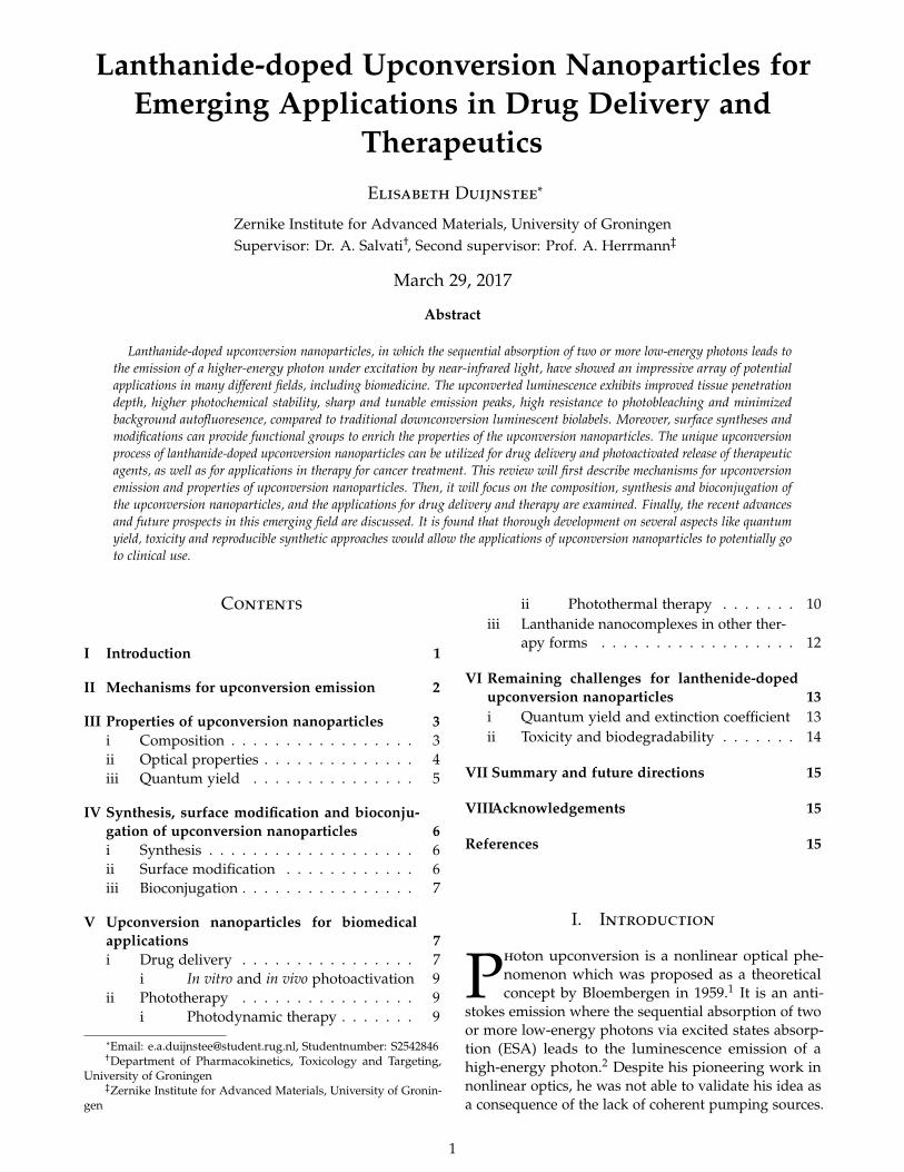
The upconversion (UC) mechanisms can roughly be cat-egorized into four classes according to recent advances:excited-state absorption (ESA),3 energy transfer upcon-version (ETU),3 photon avalanche (PA),14 and energymigration-mediated upconversion (EMU),13 which wasmore recently developed.12 The different upconversionprocesses are shown in figure 1. In figure 1a, b, andc, I and II are the sensitizer and activator, respectively.In figure 1d, I, II III & IV, are the sensitizer, accumula-tor, migrator and activator, respectively. All processes
are based on the sequential absorption of two or morephotons by metastable, long-lived energy states. Thesequential absorption then leads to the upconversionemission from a highly excited state.15 It should benoted that the upconversion emission is strongly dom-inated by the ETU process and that ETU and EMUare mostly used for applications in drug delivery andtherapeutics, as shown in section 5.
In a typical ESA process (figure 1a), the absorptionof a low-energy photon leads to the electron transi-tion from the ground state to a metastable intermedi-ate state.16 A second pump photon then leads to theexcitation of the excited electrons to a higher excitedstate. When the electrons radiatively fall back into theirground state, UC emission occurs.
ETU (figure 1b), is different from ESA as ETU in-volves two neighboring ions in stead of one lanthanideion, like in ESA. So, in ETU there are two types onluminescent centres: the sensitizer and the activator,both embedded in a host matrix. Upon excitation oflow-energy photons, the sensitizer is excited from theground state to its metastable energy level. Afterwards,it transfers its harvested energy to the activator. As aconsequence of the non-radiative energy transfer fromthe sensitizer to the activator, there is sequential absorp-tion of the activator to the higher excited state, whilethe sensitizer relaxes back to the ground state. Radia-tive emission in the activator back to the ground statecan then be observed as UC emission.16 The efficiencyof this UC process is determined by the distance be-tween the sensitizer and the activator (rs-a), which isdetermined by the concentration of the dopants, andthe overlap of the spectra.17 In addition, for efficientESA and ETU, a ladder-like arrangement of the energystates is required to minimize relaxation losses and tofacilitate absorption. Of course, high absorption at theemission wavelength of the sensitizer is required.
Under certain conditions, there is a unique UC pro-cess that is based on the combination of the above twomechanism called photon avalanche (PA), as shown infigure 1c. In order for PA to occur, the pumping energymust be larger than the absorption energy of the inter-mediate level (E1) (Epump > E1 – G) for the populationof E1 trough non-resonant transitions, and the differ-ence in energy between the excited state (E2) and theintermediate energy levels (E1) must be resonant withthe energy of the pumping system (Epump = E2 – E1).Also, there must be an efficient cross-relaxation process,where the activator transfers part of its excited energyto the sensitizer. Above a certain pump threshold, thereis an increase in the number of active ions that populatethe intermediate metastable energy level of the activa-tor, and then the photon avalanche process may start.The competition between intensity gain and intensitylosses gives rise to an enhancement of the upconvertedemission from the excited level. Again, the efficiency
2

Lanthanide-doped Upconversion Nanoparticles for Emerging Applications in Drug Delivery and Therapeutics
Figure 1: Principal UC processes for lanthanide-based UCNPs: (a) ESA, (b) ETU, (c) PA, (d) EMU. Note that core and shell regions arehighlighted with different background colors. The red arrows represent photon excitation and energy transfer. The blue arrows represent theemission processes.13
of this process is strongly related to the concentrationsof the dopants. Nevertheless, PA is rarely observed inUCNPs, as it requires a pump threshold and a longtime (seconds) to build up.18
The fourth process, EMU, is a quite complicated pro-cess proposed by Wang et al.13 In the EMU process(figure 1d), a sensitizer is used to harvest the pumpphotons and then promote a neighbouring accumu-lator to the excited state. This excitation energy isthen extracted from high-lying energy states of theaccumulator. Afterwards, there is random energy hop-ping through the migrator ion sublattice and trappingof the migrating energy by an activator. The sensi-tizer/accumulator and the activator are spatially con-fined in different layers of a core-shell structure toregulate the energy exchange interaction between theaccumulator and the activator. Several migrator ionsthrough the core-shell interface are required for an ef-ficient EMU process to bridge the transfer of energyfrom the accumulator to the activator.13
III. Properties of upconversion
nanoparticles
For efficient upconversion to proceed, the compositionand optical properties of UCNPs should be well under-stood.
i. Composition
As explained, luminescent materials featuring f- andd-ions and containing more than one intermediate en-ergy level, can in principle be used for generation ofUC luminescence. However, efficient UC processesmostly occur in lathanide-doped UCNPs because oftheir extremely long-lived intermediate energy states.11
Lathanides are rare earth elements, with an electronic
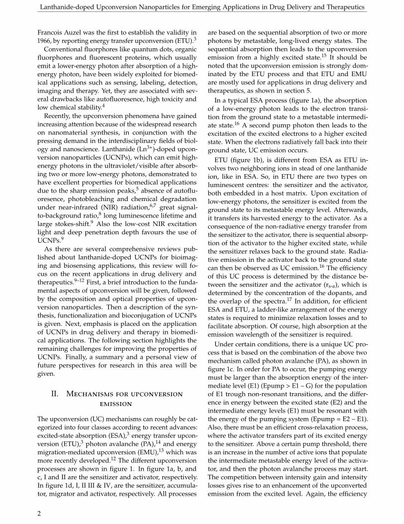
structure that has the ability to emit with higher en-ergy than the excitation energy. The lanthanides 4fn
(n=0-14) electron configurations are split by electronicrepulsion and spin-orbit coupling as shown in figure2. This leads to a splitted energy level pattern, wherethe energy levels can act as energy reservoirs, which isrequired for efficient UC.19
As mentioned before, in the dominated ETU process,Ln3+-doped UCNPs are composed of three differentcomponents, namely a host matrix, a sensitizer and anactivator, where the sensitizer can effectively be excitedby the incident light source and can transfer its energyto the activator, which in its turn emits the radiation.3
Optical emissions under low pump power densitiesare only generated by using Er3+, Tm3+ and Ho3+ asactivator, as they have equally spaced energy levels,which minimize irradiative losses and facilitate absorp-tion and energy transfer.8,20 An often used sensitizeris Yb3+, as it contains a large absorption cross sectionin the NIR. Other sensitizers may be used to enhanceof quench specific emission bands. The sensitizer andactivator are added to the host lattice in low concen-trations (about 20% for the sensitizer and less than 2%for the activator), as this allows for a good distancebetween the activator and the sensitizer without havingcross-relaxations but with enough absorption for thesensitizer to be efficient.10
For the realization of an efficient UC process, the hostmaterial plays an important role in terms of crystalstructure and optical properties. The host materialshould desirably have low phonon energy to suppressvibrational relaxation and to increase the lifetime ofthe intermediate states, adequate transparency in theinterested energy range to minimize scattering losses,and it should be chemically stable. The crystal field ofthe host material has a large effect on the UC efficiencyof the lanthanide-doped nanoparticles. Depending on
3
Lanthanide-doped Upconversion Nanoparticles for Emerging Applications in Drug Delivery and Therapeutics
Figure 2: Electronic energy level diagrams of Ln3+ ions in relation to upconversion processes. The 4fn electronic configurations splitsinto many energy sublevels due to the strong effects of the Coulombic interaction and spin-orbit coupling as well as weak crystal-fieldperturbation. The energy levels are represented by the symbol 2S+1LJ (where S, L, J are the total spin, orbital and angular momentumquantum number of the electron, respectively), and optical transitions among these energy levels lead to abundant emission bands. Inprinciple, a Ln3+ ion residing in an intermediate level |2> can be further promoted to an upper emitting level |3> through absorption ofanother photon Eex (ESA process), or through cooperative energy transfer between a pair of excited ions with one ion being non-radiativelydecayed to the ground state |1> (ETU process). The right inset panel shows that the realization of an efficient ETU process requiresconsideration of the sensitizer-activator distance (rS−A), and the spectral overlap between sensitizer emission ( f S
em (E)) and activatorabsorption ( f A
abs(E)) profiles.19
the symmetry of the crystal field, the optical propertiesare influenced.11 In order to achieve high doping levels,the host lattice should have close lattice matches todopant ions to readily incorporate Ln3+ dopant ions.10
Fluoride based lattices, such as LaF4, YF4, NaYF4 andBaYF4, have been employed extensively as host latticeas they meet many of the above named criteria.9,21
To date, the most efficient mono dispersed core-shellUCNPs which produce green and blue emissions areYb3+/Er3+ and Yb3+/Tm3+ doped in hexagonal phaseNaYF4 (β-NaYF4).22,23 The greater UC emission of β-NaYF4 made this composition more preferable thanthe cubic form α-NaYF4 which is the composition atlower temperatures. Thermal treatment at ca. 400-600 oC transforms the cubic phase to the hexagonalphase.24 α-NaYF4 was the first composition used forthe demonstration of tunable color UC luminescencefrom green to red.25 It was proven that a less symmetriccrystal phase is more favourable for the UC efficiency,as it allows for intermixing of the lanthanide ion its fstates with higher electronic configurations.11,25,26
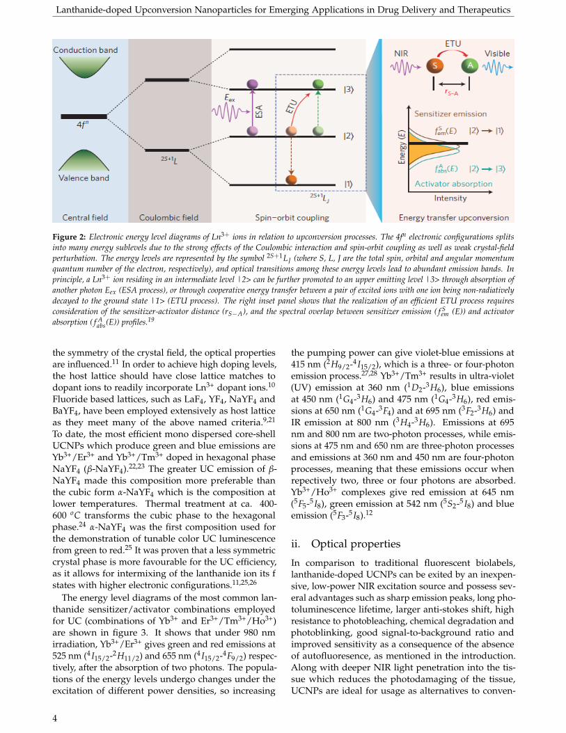
The energy level diagrams of the most common lan-thanide sensitizer/activator combinations employedfor UC (combinations of Yb3+ and Er3+/Tm3+/Ho3+)are shown in figure 3. It shows that under 980 nmirradiation, Yb3+/Er3+ gives green and red emissions at525 nm (4 I15/2-2H11/2) and 655 nm (4 I15/2-4F9/2) respec-tively, after the absorption of two photons. The popula-tions of the energy levels undergo changes under theexcitation of different power densities, so increasing
the pumping power can give violet-blue emissions at415 nm (2H9/2-4 I15/2), which is a three- or four-photonemission process.27,28 Yb3+/Tm3+ results in ultra-violet(UV) emission at 360 nm (1D2-3H6), blue emissionsat 450 nm (1G4-3H6) and 475 nm (1G4-3H6), red emis-sions at 650 nm (1G4-3F4) and at 695 nm (3F2-3H6) andIR emission at 800 nm (3H4-3H6). Emissions at 695nm and 800 nm are two-photon processes, while emis-sions at 475 nm and 650 nm are three-photon processesand emissions at 360 nm and 450 nm are four-photonprocesses, meaning that these emissions occur whenrepectively two, three or four photons are absorbed.Yb3+/Ho3+ complexes give red emission at 645 nm(5F5-5 I8), green emission at 542 nm (5S2-5 I8) and blueemission (5F3-5 I8).12
ii. Optical properties
In comparison to traditional fluorescent biolabels,lanthanide-doped UCNPs can be exited by an inexpen-sive, low-power NIR excitation source and possess sev-eral advantages such as sharp emission peaks, long pho-toluminescence lifetime, larger anti-stokes shift, highresistance to photobleaching, chemical degradation andphotoblinking, good signal-to-background ratio andimproved sensitivity as a consequence of the absenceof autofluoresence, as mentioned in the introduction.Along with deeper NIR light penetration into the tis-sue which reduces the photodamaging of the tissue,UCNPs are ideal for usage as alternatives to conven-
4
Lanthanide-doped Upconversion Nanoparticles for Emerging Applications in Drug Delivery and Therapeutics
Figure 3: Energy level diagrams and energy transfer process in Yb3+, Er3+/Tm3+/Ho3+.12
tional biolabels. This section will focus on the opticalproperties of lanthanide-doped UCNPs.
The shielding of the partially filled 4f orbitals by theouter 5s and 5p shells of the lanthanide ions, results in adistinct set of sharp emission peaks, which makes accu-rate interpretation of the spectra possible.5,11,29 Tuningthe host/dopant combinations, dopant concentration,particle size, surface ligands and crystallinity enablesemission manipulation.11 Each lanthanide ion can pro-duce distinct sharp emission peaks. So a selection ofdifferent lanthanide ions, or combinations of them, en-ables the production of multicolour emission and theirrelative emission intensities.11,30 Changing the pump-ing power density of the laser adjusts the population ofthe energy levels and can cause a diverse set of possibleemission wavelengths.28
The spin forbidden 4f-4f transition, due to quan-tum mechanical selection rules, is relaxed due to localcrystal-field inducing mixing of the f states with higherelectronic configurations.29 The primary forbidden na-ture of the f-f transition causes the lifetime lumines-cence to be long (up to tens of milliseconds), whichfavours sequential excitations in the excited state inthe ESA process and also allows for favourable ion-ioninteractions in the excited state states for permittingenergy transfers between two or more Ln3+ ions inthe ETU process, it also makes time-resolved lumines-cence techniques possible because of minimization ofbackground fluorescence.8,11
The sequential absorption of multiple photonsthrough the use of real ladder-like intermediate en-ergy levels, with long lifetime, provides a large anti-stokes shift, which allows for easy separation of theemission wavelength from the excitation wavelength.8
The narrow absorption profile limits the amount ofpossible excitation sources. However, the inexpensiveInGaAs diode laser excites at 980 nm and matches well
with the absorption spectrum of the lanthanide-dopedUCNPs. Since the upconverted emission is based onexisting intermediate energy levels, it can be inducedby a low power laser. Because the excitation is in theNIR region, which is within the optical transparency re-gion, UCNPs allow for much deeper penetration depthand reduce the photodamaging of the tissue. In addi-tion, this optical transparency window provides muchhigher signal-to-background ratio due to the absence ofautofluoresence and reduced light scattering, comparedto conventional biolabels.9
Since the upconverted emission from the 4f-4f transi-tions of Ln3+ does not involve chemical bond breaking,the UCNPs are stable against photobleaching and pho-tochemical degradation, which implies that the UCNPsremain unaltered after irradiation by NIR lasers forhours.8,31 Furthermore, UCNPs, which usually containmany lanthanide dopants ions, possess non-blinkingemission under a NIR excitation source.31
iii. Quantum yield
An import parameter that describes the emission effi-ciency of the UCNPs is defined as the quantum yield(QY) (equation 1). The nonlinear nature of the UCprocess indicates that the efficiency of this process isstrongly dependent on the excitation power density.High efficiencies are required for biomedical applica-tions to improve the limit of detection, to obtain highersignal-to-background ratio for bioimaging and for in-creased therapy effects.
QY =number of emitted photons
number of absorbed photons(1)
Unfortunately, the QY in most UCNPs barely exceeds1%. Currently, the highest reported QY developed byHuang et al., is 7.6% for LiLuF4:Yb3+/Tm3+@LiLuF4
5
Lanthanide-doped Upconversion Nanoparticles for Emerging Applications in Drug Delivery and Therapeutics
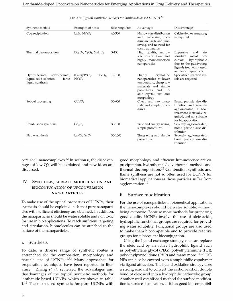
Table 1: Typical synthetic methods for lanthanide-based UCNPs.12
Synthetic method Examples of hosts Size range/nm Advantages Disadvantages
Co-precipitation LaF3, NaYF4 40-500 Narrow size distributionand tunable size, proce-dure are facile and time-saving, and no need forcostly apparatus
Calcination or annealingis required
Thermal decomposition Dy2O3, Y2O3, NaLnF4 3-150 High quality, narrowsize distribution andhighly monodispersednanoparticles
Expensive and air-sensitive metal pre-cursors, hydrophobicdue to the passivatingligands frequently used,and toxic byproducts
Hydrothermal, solvothermal,liquid-solid-solution, ionicliquid synthesis
(La-Dy)VO4, YVO4,NaYF4
10-1000 Highly crystallinenanoparticles at lowertemperature, cheap rawmaterials and simpleprocedures, and tun-able crystal size andmorphology
Specialized reaction ves-sels are required
Sol-gel processing GdVO4 30-600 Cheap and raw mate-rials and simple proce-dures
Broad particle size dis-tribution and severelyagglomerated, a heattreatment is usually re-quired, and not suitablefor bioapplication
Combustion synthesis Gd2O3 30-150 Time and energy saving,simple procedures
Severely agglomerated,broad particle size dis-tribution
Flame synthesis La2O3, Y2O3 30-1000 Timesaving and simpleprocedures
Severely agglomerated,broad particle size dis-tribution
core-shell nanocomplexes.32 In section 6, the disadvan-tages of low QY will be explained and new ideas arediscussed.
IV. Synthesis, surface modification and
bioconjugation of upconversion
nanoparticles
To make use of the optical properties of UCNPs, theirsynthesis should be exploited such that pure nanoparti-cles with sufficient efficiency are obtained. In addition,the nanoparticles should be water soluble and non toxicfor use in bio applications. To reach sufficient targetingand circulation, biomolecules can be attached to thesurface of the nanoparticles.
i. Synthesis
To date, a diverse range of synthetic routes isentrenched for the composition, morphology andparticle size of UCNPs.19,33 Many approaches forpreparation techniques have been reported in liter-ature. Zhang et al, reviewed the advantages anddisadvantages of the typical synthetic methods forlanthanide-based UCNPs, which are shown in table1.12 The most used synthesis for pure UCNPs with
good morphology and efficient luminescence are co-precipitation, hydrothemal/solvothermal methods andthermal decomposition.12 Combustion synthesis andflame synthesis are not so often used for UCNPs forbiomedical applications as those particles suffer fromagglomeration.12
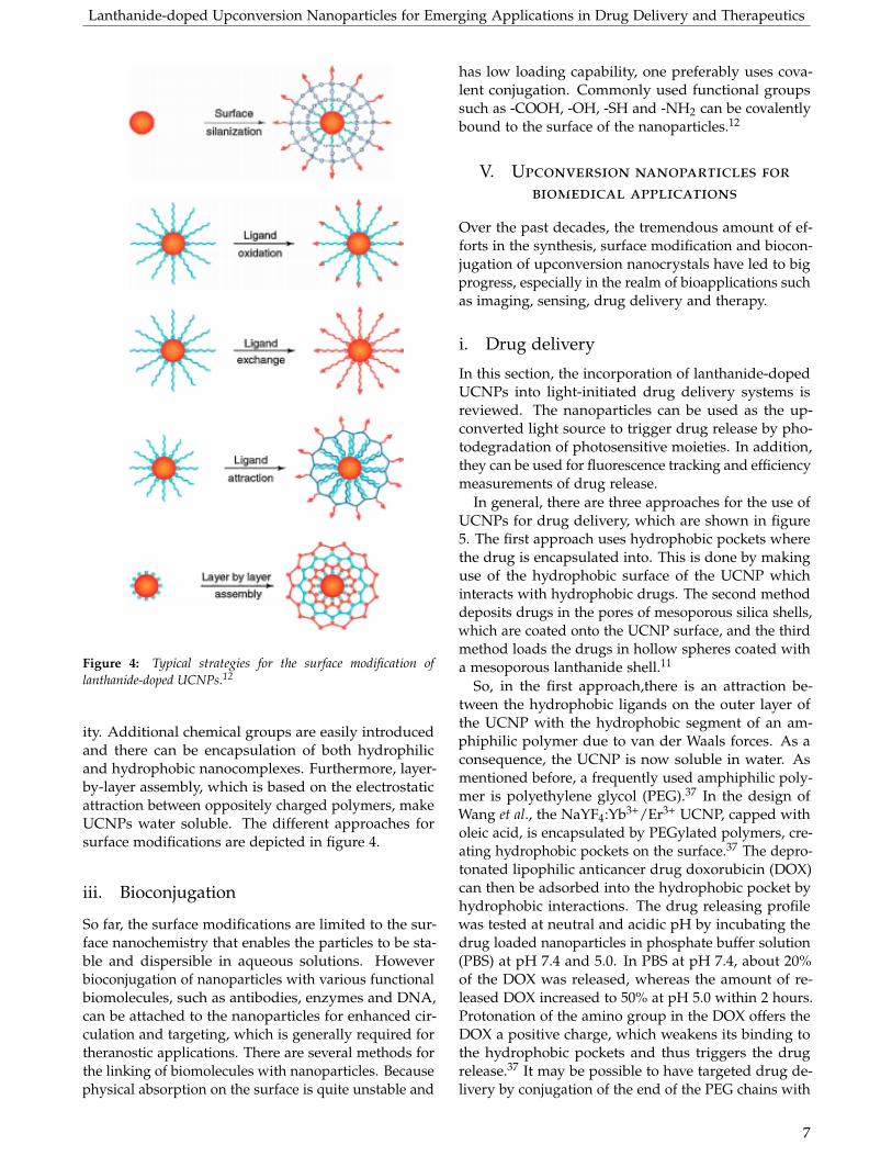
ii. Surface modification
For the use of nanoparticles in biomedical applications,the nanocomplexes should be water soluble, withoutbeing cytotoxic. Because most methods for preparinggood quality UCNPs involve the use of oleic acids,hydrophilic functional groups are required for provid-ing water solubility. Functional groups are also usedto make them biocompatible and to provide reactivegroups for subsequent bioconjugation.
Using the ligand exchange strategy, one can replacethe oleic acid by an active hydrophilic ligand suchas polyethylene glycol (PEG), polyethylenimine (PEI),polyvinylpyrriolidone (PVP) and many more.34–36 UC-NPs can also be covered with a amphiphilic copolymervia ligand attraction. The ligand oxidation strategy usesa strong oxidant to convert the carbon-carbon doublebond of oleic acid into a hydrophilic carboxylic group.Another well-established method for surface modifica-tion is surface silanization, as it has good biocompatibil-
6
Lanthanide-doped Upconversion Nanoparticles for Emerging Applications in Drug Delivery and Therapeutics
Figure 4: Typical strategies for the surface modification oflanthanide-doped UCNPs.12
ity. Additional chemical groups are easily introducedand there can be encapsulation of both hydrophilicand hydrophobic nanocomplexes. Furthermore, layer-by-layer assembly, which is based on the electrostaticattraction between oppositely charged polymers, makeUCNPs water soluble. The different approaches forsurface modifications are depicted in figure 4.
iii. Bioconjugation
So far, the surface modifications are limited to the sur-face nanochemistry that enables the particles to be sta-ble and dispersible in aqueous solutions. Howeverbioconjugation of nanoparticles with various functionalbiomolecules, such as antibodies, enzymes and DNA,can be attached to the nanoparticles for enhanced cir-culation and targeting, which is generally required fortheranostic applications. There are several methods forthe linking of biomolecules with nanoparticles. Becausephysical absorption on the surface is quite unstable and
has low loading capability, one preferably uses cova-lent conjugation. Commonly used functional groupssuch as -COOH, -OH, -SH and -NH2 can be covalentlybound to the surface of the nanoparticles.12
V. Upconversion nanoparticles for
biomedical applications
Over the past decades, the tremendous amount of ef-forts in the synthesis, surface modification and biocon-jugation of upconversion nanocrystals have led to bigprogress, especially in the realm of bioapplications suchas imaging, sensing, drug delivery and therapy.
i. Drug delivery
In this section, the incorporation of lanthanide-dopedUCNPs into light-initiated drug delivery systems isreviewed. The nanoparticles can be used as the up-converted light source to trigger drug release by pho-todegradation of photosensitive moieties. In addition,they can be used for fluorescence tracking and efficiencymeasurements of drug release.
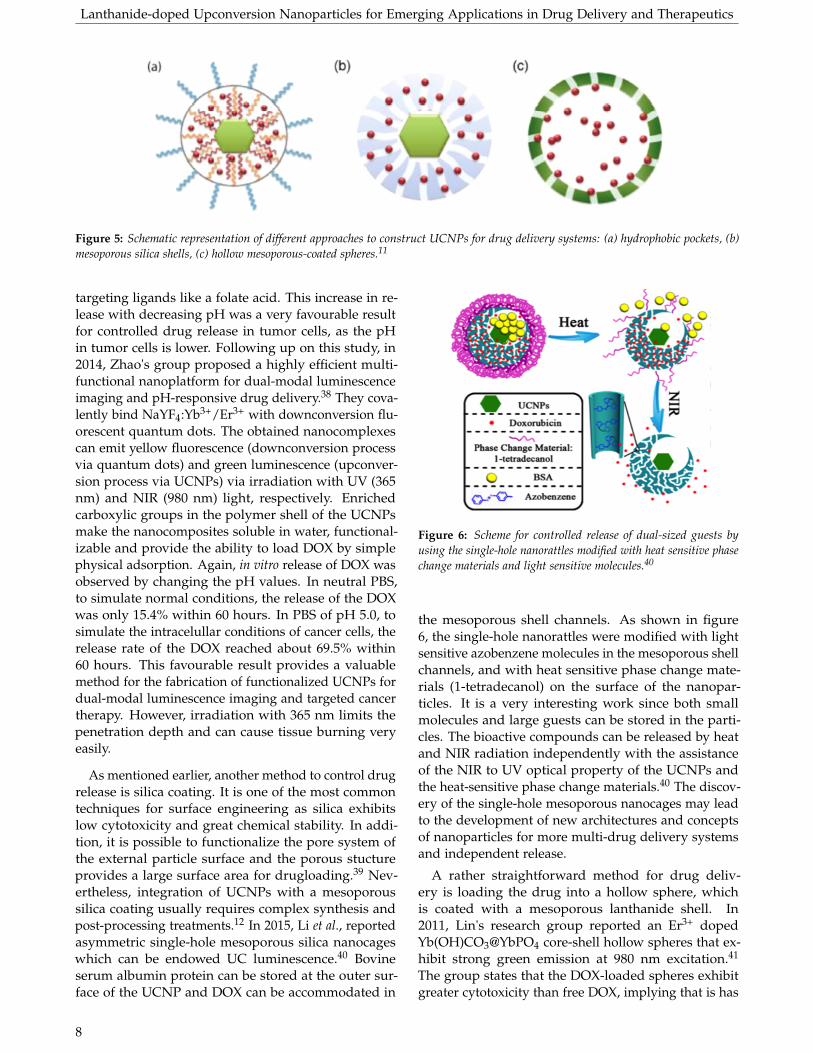
In general, there are three approaches for the use ofUCNPs for drug delivery, which are shown in figure5. The first approach uses hydrophobic pockets wherethe drug is encapsulated into. This is done by makinguse of the hydrophobic surface of the UCNP whichinteracts with hydrophobic drugs. The second methoddeposits drugs in the pores of mesoporous silica shells,which are coated onto the UCNP surface, and the thirdmethod loads the drugs in hollow spheres coated witha mesoporous lanthanide shell.11
So, in the first approach,there is an attraction be-tween the hydrophobic ligands on the outer layer ofthe UCNP with the hydrophobic segment of an am-phiphilic polymer due to van der Waals forces. As aconsequence, the UCNP is now soluble in water. Asmentioned before, a frequently used amphiphilic poly-mer is polyethylene glycol (PEG).37 In the design ofWang et al., the NaYF4:Yb3+/Er3+ UCNP, capped witholeic acid, is encapsulated by PEGylated polymers, cre-ating hydrophobic pockets on the surface.37 The depro-tonated lipophilic anticancer drug doxorubicin (DOX)can then be adsorbed into the hydrophobic pocket byhydrophobic interactions. The drug releasing profilewas tested at neutral and acidic pH by incubating thedrug loaded nanoparticles in phosphate buffer solution(PBS) at pH 7.4 and 5.0. In PBS at pH 7.4, about 20%of the DOX was released, whereas the amount of re-leased DOX increased to 50% at pH 5.0 within 2 hours.Protonation of the amino group in the DOX offers theDOX a positive charge, which weakens its binding tothe hydrophobic pockets and thus triggers the drugrelease.37 It may be possible to have targeted drug de-livery by conjugation of the end of the PEG chains with
7
Lanthanide-doped Upconversion Nanoparticles for Emerging Applications in Drug Delivery and Therapeutics
Figure 5: Schematic representation of different approaches to construct UCNPs for drug delivery systems: (a) hydrophobic pockets, (b)mesoporous silica shells, (c) hollow mesoporous-coated spheres.11
targeting ligands like a folate acid. This increase in re-lease with decreasing pH was a very favourable resultfor controlled drug release in tumor cells, as the pHin tumor cells is lower. Following up on this study, in2014, Zhao's group proposed a highly efficient multi-functional nanoplatform for dual-modal luminescenceimaging and pH-responsive drug delivery.38 They cova-lently bind NaYF4:Yb3+/Er3+ with downconversion flu-orescent quantum dots. The obtained nanocomplexescan emit yellow fluorescence (downconversion processvia quantum dots) and green luminescence (upconver-sion process via UCNPs) via irradiation with UV (365nm) and NIR (980 nm) light, respectively. Enrichedcarboxylic groups in the polymer shell of the UCNPsmake the nanocomposites soluble in water, functional-izable and provide the ability to load DOX by simplephysical adsorption. Again, in vitro release of DOX wasobserved by changing the pH values. In neutral PBS,to simulate normal conditions, the release of the DOXwas only 15.4% within 60 hours. In PBS of pH 5.0, tosimulate the intracelullar conditions of cancer cells, therelease rate of the DOX reached about 69.5% within60 hours. This favourable result provides a valuablemethod for the fabrication of functionalized UCNPs fordual-modal luminescence imaging and targeted cancertherapy. However, irradiation with 365 nm limits thepenetration depth and can cause tissue burning veryeasily.
As mentioned earlier, another method to control drugrelease is silica coating. It is one of the most commontechniques for surface engineering as silica exhibitslow cytotoxicity and great chemical stability. In addi-tion, it is possible to functionalize the pore system ofthe external particle surface and the porous stuctureprovides a large surface area for drugloading.39 Nev-ertheless, integration of UCNPs with a mesoporoussilica coating usually requires complex synthesis andpost-processing treatments.12 In 2015, Li et al., reportedasymmetric single-hole mesoporous silica nanocageswhich can be endowed UC luminescence.40 Bovineserum albumin protein can be stored at the outer sur-face of the UCNP and DOX can be accommodated in
Figure 6: Scheme for controlled release of dual-sized guests byusing the single-hole nanorattles modified with heat sensitive phasechange materials and light sensitive molecules.40
the mesoporous shell channels. As shown in figure6, the single-hole nanorattles were modified with lightsensitive azobenzene molecules in the mesoporous shellchannels, and with heat sensitive phase change mate-rials (1-tetradecanol) on the surface of the nanopar-ticles. It is a very interesting work since both smallmolecules and large guests can be stored in the parti-cles. The bioactive compounds can be released by heatand NIR radiation independently with the assistanceof the NIR to UV optical property of the UCNPs andthe heat-sensitive phase change materials.40 The discov-ery of the single-hole mesoporous nanocages may leadto the development of new architectures and conceptsof nanoparticles for more multi-drug delivery systemsand independent release.
A rather straightforward method for drug deliv-ery is loading the drug into a hollow sphere, whichis coated with a mesoporous lanthanide shell. In2011, Lin's research group reported an Er3+ dopedYb(OH)CO3@YbPO4 core-shell hollow spheres that ex-hibit strong green emission at 980 nm excitation.41
The group states that the DOX-loaded spheres exhibitgreater cytotoxicity than free DOX, implying that is has
8

Lanthanide-doped Upconversion Nanoparticles for Emerging Applications in Drug Delivery and Therapeutics
the potential to be used for drug loading and deliveryfor inducing cancer cell death. In this context, Lu etal. employed a novel UC luminescent nanorattle by in-corporation of lanthanide-doped fluorides into hollowmesoporous silica.42 The nanorattles have high capacitydue to hydrophobic interactions between the UCNPsand DOX. And again, the DOX showed fast release inpH 7.4, compared to the PBS solution of pH 5.0, causedby the enhanced electrostatic repulsion between DOXand the aminopropyl group of the mesoporous silica aswell as by the repulsion between the DOX molecules.Therefore, these systems can be used as smart carriersfor drug release by varying the pH from 7.4 to an acidicenvironment owing to endocytosis.
i In vitro and in vivo photoactivation
UV irradiation-mediated photochemical reactions are ofimportance for drug delivery systems. The UV photonshave the ability to manipulate functions of biomoleculesand can mediate on-demand drug release via photoac-tivation. Nevertheless, UV lasers and lamps have manydrawbacks such as phototoxicity, a large radiation areaand limited penetration depth. NIR-to-UV Ln3+-dopedUCNPs can play a vital role in drug release mecha-nisms because of their deep penetration depth andability to precisely light control drug release, as de-scribed in section 3.2 and 5.1. In addition, the radiationarea is minimized to the nanometer regime.11 For ex-ample, a direction for in vitro and in vivo photoactiva-tion is NIR light-induced photoswitching of molecularswitches to switch back and forth between differentisomers. Capobianco and coworkers invented a NIRlight photoswitching method by grafting bis-spiropyranonto the surface of LiYF4:Yb3+/Tm3+ UCNPs via ligandexchange.43 The UCNPs are excited at 980 nm and fluo-rescence resonance energy transfer from the UCNPs tothe bis-spiropyran molecules on the surface can triggerthe transformation of the ring-closed bis-spiropyranform to the ring-open bis-merocyanine form, whichwas achieved by irradiation at 365 nm. The reversephotocyclization can be achieved by radiation with vis-ible light with a wavelength larger than 500 nm. Theswitched absorption peak may also be utilized for themanipulation of the color emission of the UCNPs insidethe cell by changing the irradiation wavelength.
ii. Phototherapy
The impressive array of surface characteristics oflanthanide-doped UCNPs made them not only use-ful as vehicles for drug delivery and release but also fortherapeutics. In this section, the different forms of ther-apy involving lanthanide-doped UCNPs are discussed.Phototherapies like photodynamic therapy (PDT) andphotothermal therapy (PTT) garnered tremendous inter-est over the last years within biomedical applications.
i Photodynamic therapy
PDT involves the selective uptake and localization ofa photosensitizer into specific tumor cells and tissuetypes. The photosensitizers can be activated by irradia-tion of a predetermined dose of light and can generatethe cytotoxic reactive oxygen species (ROS), 1O2, whichcan kill cancer cells without affecting the surroundingtissue (see figure 7).44,45 The penetration depth of PDTis, however, only about one centimeter, which impliesthat only tumors just below the skin can be treatedwith PDT. Also destroying large tumors is very difficultwith PDT. Currently, PDT is used for the treatment ofprostate, lung, neck, head, and skin cancers.46
The explained deep penetration depth of UCNPscaused large interest in using them for cancer therapy.The emitted light from the UCNPs can excite photo-sensitizers, which then generate the ROS, in order todestroy the tumor cells, and drawbacks of existing PDTcan be overcome. This idea was firstly proposed byPrasad et al., and was realized by Zhang et al..47,48 Theyencapsulated merocyanine 540 as photosensitizer intoa silica layer which was coated on Y2O3:Yb3+/Er3+ UC-NPs. An antibody, specific to antigens on the targetcell surface, was covalently attached to the UCNPs. Ir-ridation with NIR leads to the upconverted emission,which is then absorbed by the photosensitizers. Subse-quently, excited photosensitizers interact with ground-state molecular oxygen, and generate ROS, which even-tually leads to cancer cell death in vitro.
Liu and coworkers reported the pioneering workto show the effectiveness of upconversion for in vivoPDT.49 They used the porphyrin derivative chlorine 6(Ce6) as photosensitizer which was non covalently in-corporated into PEGylated coated β-NaYF4:Yb3+/Er3+.The direct injection of the photosensitized UCNPs intothe tumor showed 70% tumor regression after 30 min-utes under 980 nm light irradiation. The tissue penetra-tion abilities between UCNP-based PDT and traditionalPDT in pork tissue was tested. Direct exposure of Ce6solution with 660 nm excitation source generated muchmore ROS compared to the generation of ROS in UC-NPs sensitized with Ce6, which was exposed with a980 nm excitation source. However, ROS formationwas completely eliminated when the 660 nm light wasblocked by a 8 mm tissue, implying that the penetrationdepth is much deeper for UCNP-Ce6 nanocomplexes,because for those nanocomplexes, only 50% of the lightwas blocked by 8 mm pork tissue and still ROS wasgenerated. The selectivity of the UCNP-Ce6 into tu-mor sites was not reported here, which is important forin vivo photodynamic therapy. Modification of activetargeting ligands to the UCNPs is required for the in-crease of local concentration in tumors and to avoid sideeffects, and even if there is no control of where the UC-NPs go, they should only be activated where needed.
In 2012, Zhang and coworkers reported a novel
9

Lanthanide-doped Upconversion Nanoparticles for Emerging Applications in Drug Delivery and Therapeutics
Figure 7: Schematic illustration of the UCNPs-based PDTtreatment.50
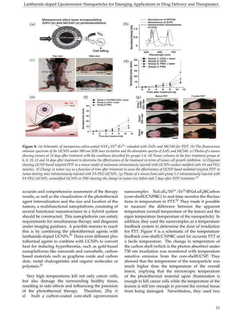
way to amplify therapeutic effects by the multicolor-emission capability of UCNPs at a single 980 nm excita-tion wavelength for the simultaneous activation of twosensitizers.53 They prepared the NaYF4 UCNPs by uni-formly coating them with mesoporous silica (figure 8a).The multicolor-emission capability of the UCNPs wasused to activate two photosensitizers. The two emis-sions, green and red, from the UCNPs matched wellwith the absorption of the photosensitizers, merocya-nine 540 (MC540) and zinc(II)phthalocyanine (ZnPc),respectively (figure 8b). The dual-photosensitizer ap-proach was used as it showed enhanced generationof ROS and reduced cell viability. They first irradi-ated B16-F0 cells labeled with each of the differentiallyloaded UCNPs under the skin of mice. Irradiation by a980 nm laser showed an increase in ROS, and the greenfluorescence intensity was more intense than that ofcells treated with either of the sensitizers. Secondly,they showed the efficacy of the UCNPs in vivo in mice.They intratumorally injected coloaded UCNPs into B16-F0 cells in mice and shined on the tumor with the NIRlaser. As controls, other mice received intratumoralinjection of PBS (the vehicle in which the UCNPs weresuspended). The growth of the tumor cells with injectedcoloaded UCNPs was significantly inhibited (figure 8cand figure 8d). Extension of the study showed targetedPDT efficacy of folic acid (FA) conjugated PEGylatedUCNPs (figure 8e). The B16-F0 tumors in mice whereinjected intravenously with coloaded FA-PEG-UCNPsand irradiated with a 980 nm laser. The tumor growthwas reduced compared to the mice treated with PBS(figure 8f). The photosensitizers may be prematurelyreleased from the nanoparticles, which can cause a re-duction in efficiency of the cancer treatment, as theyare solely physically entrapped inside the mesoporoussilica. If they would be covalently bound at the sur-face of the nanoparticles, the premature release may beovercome.
Very recently, Lin and coworkers designeda core-shell structured NaGdF4:Yb3+/Tm3+
@NaGdF4:Yb3+@NaNdF4:Yb3+@NaGdF4@mSiO2@TiO2nanocomplex by coating a layer of TiO2 photosensitizeron an effective 808 nm upconversion luminescentcore to achieve simultaneous bioimaging and efficientPDT.54 Thus far, most reported light-controlledtherapies are based on NIR light excitation. This wave-length can directly overlap with the water absorbtionspectrum and can thus cause tissue heating (see section6.2). The excitation wavelength of 880 nm has muchless overlap with the absorption spectrum of water andtherefore greatly minimizes heating of the tissue. Theconstructed design limits the reverse energy transferfrom activator to sensitizer and thus improves the UCemission efficiency. The high surface area of the silicacoated nanoparticle makes it very stable and enableshigh loading possibilities. In vivo results indicate muchhigher efficacy due to deeper penetration depth whenexciting with a high wavelength. The nanocomplexitself can simultaneously be used as an imaging probe.For nanoplatforms with multifunctional propertiesfor simultaneous therapy and imaging, the designshould take into account that the absorption of thephotosensitizer should have minimal mismatch withthe upconverted emission to posses high UC efficiencyand thus larger ROS production.
In addition to cancer therapy, UCNPs for photody-namic therapy have been extended to the reductionof viral infections, which provides promising antivi-ral approaches for treatment of viral infections.11 PEI-NaYF4:Yb3+/Er3+ loaded with a ZnPc photosensitizerwas used for intra and extracellular inactivation of ade-noviruses and dengue viruses as a consequence of theproduction of ROS.55 Reduction in the infectious virusin vitro was shown in a murine model under NIR excita-tion source. The conjugation of antibodies or enzymeson the UCNPs provide the ability to specifically local-ize the UCNPs to the virus-infected cells only. It isquite an unexplored area, but yet shows potential fornanomedicine.
ii Photothermal therapy
The basic principle of photothermal therapy (PTT) isthat normal cells and tumor cells have different sensi-tivity to heat. Healthy cells are more resistant to heatand have faster recovery when exposed to heat or radi-ation. Under NIR radiation, photothermal agents canconvert light into heat to induce hyperthermia (higherthan 42 oC) resulting in thermal ablation of the tumorcells, with minimal effect on the normal cells.56 In orderfor effective cell death of tumor cells, the photothermalagent should have a high absorption coefficient to NIRradiation, well-engineered surface modifications andgood biodegradability to dodge toxicity effects.57 For an
10
Lanthanide-doped Upconversion Nanoparticles for Emerging Applications in Drug Delivery and Therapeutics
Figure 8: (a) Schematic of mesoporous-silica-coated NYF4:Yb3+/Er3+ coloaded with ZnPc and MC540 for PDT. (b) The fluorescenceemission spectrum of the UCNPs under 980-nm NIR laser excitation and the absorption spectra of ZnPc and MC540. (c) Photos of a mouseshowing tumors at 14 days after treatment with the conditions described for groups 1-4. (d) Tumor volumes in the four treatment groups at6, 8, 10, 12 and 14 days after treatment to determine the effectiveness of the treatment in terms of tumor cell growth inhibition. (e) Diagramshowing UCNP-based targeted PDT in a mouse model of melanoma intravenously injected with UCNPs surface modified with FA and PEGmoieties. (f) Change in tumor size as a function of time after treatment to asses the effectiveness of UCNP-based mediated targeted PDT intumor-bearing mice intraveneously injected with FA-PEG-UCNPs. (g) Photos of a mouse from each group 1-3 intraveneously injected withFA-PEG-UCNPs, unmodified UCNPs or PBS showing the change in tumor size before and 7 days after PDT treatment.12
accurate and comprehensive assessment of the therapyresults, as well as the visualization of the photothermalagent internalization and the size and location of thetumors, a multifunctional nanoplatform, consisting ofseveral functional nanostructures in a hybrid systemshould be constructed. This nanoplatform can satisfyrequirements for simultaneous therapy and diagnosisunder imaging guidance. A possible manner to reachthis is by combining the photothermal agents withlanthanide-doped UCNPs.57 There exist different pho-tothermal agents to combine with UCNPs to convertheat for inducing hyperthermia, such as gold-basednanoplatforms like nanorods and nanoshells, carbon-based materials such as graphene oxide and carbondots, metal chalcogenides and organic molecules orpolymers.57
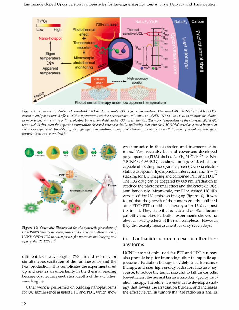
Very high temperatures kill not only cancer cells,but also damage the surrounding healthy tissue,resulting in side effects and influencing the precisionof the photothermal therapy. Therefore, Zhy etal. built a carbon-coated core-shell upconversion
nanocomplex NaLuF4:Yb3+/Er3+@NaLuF4@Carbon(core-shellUCNP@C) to real-time monitor the fluctua-tions in temperature in PTT.51 They made it possibleto measure the difference between the apparenttemperature (overall temperature of the lesion) and theeigen temperature (temperature of the nanoparticle). Inaddition, they used the nanocomplex as a temperature-feedback system to determine the dose of irradiationfor PTT. Figure 9 is a schematic of the temperature-feedback core-shellUCNP@C used for accurate PTT ata facile temperature. The change in temperature ofthe carbon shell (which is the photon absorber) under730 nm irradiation was monitored with temperaturesensitive emission from the core-shellUCNP. Theyshowed that the temperature of the nanoparticle wasmuch higher than the temperature of the overalllesion, implying that the microscopic temperatureof the photothermal material upon illumination isenough to kill cancer cells while the temperature of thelesions is still low enough to prevent the normal tissuefrom being damaged. Nevertheless, they used two
11
Lanthanide-doped Upconversion Nanoparticles for Emerging Applications in Drug Delivery and Therapeutics
Figure 9: Schematic illustration of core-shellUCNP@C for accurate PTT at facile temperature. The core-shellUCNP@C exhibit both UCLemission and photothermal effect. With temperature-sensitive upconversion emission, core-shellUCNP@C was used to monitor the changein microscopic temperature of the photoabsorber (carbon shell) under 730 nm irradiation. The eigen temperature of the core-shellUCNP@Cwas much higher than the apparant temperature observed macroscopically, indicating that core-shellUCNP@C acted as a nano-hotspot atthe microscopic level. By utilizing the high eigen temperature during photothermal process, accurate PTT, which prevent the damage tonormal tissue can be realized.51
Figure 10: Schematic illustration for the synthetic procedure ofUCNPs@PDA-ICG nanocomposites and a schematic illustration ofUCNPs@PDA-ICG nanocomposites for upconversion imaging andsynergistic PDT/PTT.52
different laser wavelengths, 730 nm and 980 nm, forsimultaneous excitation of the luminescence and theheat production. This complicates the experimental setup and creates an uncertainty in the thermal readingbecause of unequal penetration depths of the excitationwavelengths.
Other work is performed on building nanoplatformsfor UC luminesence assisted PTT and PDT, which show
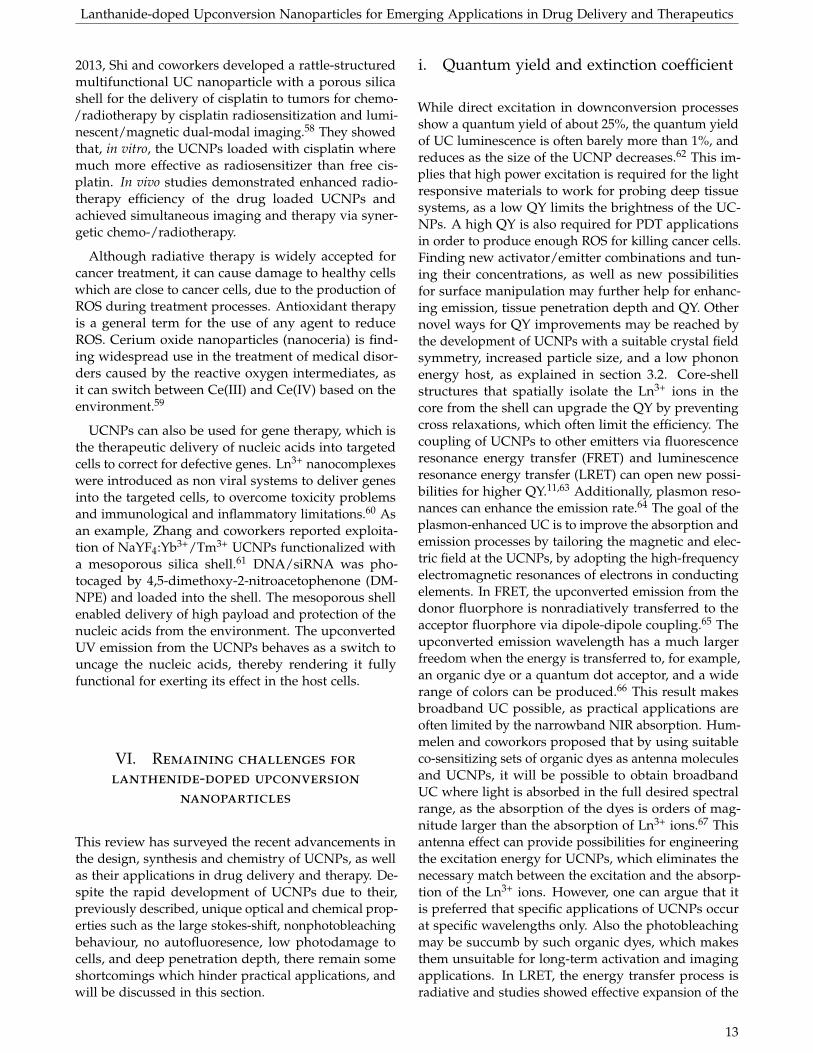
great promise in the detection and treatment of tu-mors. Very recently, Lin and coworkers developedpolydopamine (PDA)-shelled NaYF4:Yb3+/Er3+ UCNPs(UCNPs@PDA-ICG), as shown in figure 10, which arecapable of loading indocyanine green (ICG) via electro-static adsorption, hydrophobic interaction and π − πstacking for UC imaging and combined PTT and PDT.52
The ICG drug can be triggered by 808 nm irradiation toproduce the photothermal effect and the cytotoxic ROSsimultaneously. Meanwhile, the PDA-coated UCNPswere used for UC emission imaging (figure 10). It wasfound that the growth of the tumors greatly inhibitedafter PDT/PTT combined therapy after 13 days posttreatment. They state that in vivo and in vitro biocom-patibility and bio-distribution experiments showed noobvious toxicity effects of the nanocomplexes. However,they did toxicity measurement for only seven days.
iii. Lanthanide nanocomplexes in other ther-apy forms
UCNPs are not only used for PTT and PDT but mayalso provide help for improving other therapeutic ap-proaches. Radiation therapy is widely used for cancertherapy, and uses high-energy radiation, like an x-raysource, to reduce the tumor size and to kill cancer cells.Nevertheless, the normal tissue is also damaged by radi-ation therapy. Therefore, it is essential to develop a strat-egy that lowers the irradiation burden, and increasesthe efficacy even, in tumors that are radio-resistant. In
12
Lanthanide-doped Upconversion Nanoparticles for Emerging Applications in Drug Delivery and Therapeutics
2013, Shi and coworkers developed a rattle-structuredmultifunctional UC nanoparticle with a porous silicashell for the delivery of cisplatin to tumors for chemo-/radiotherapy by cisplatin radiosensitization and lumi-nescent/magnetic dual-modal imaging.58 They showedthat, in vitro, the UCNPs loaded with cisplatin wheremuch more effective as radiosensitizer than free cis-platin. In vivo studies demonstrated enhanced radio-therapy efficiency of the drug loaded UCNPs andachieved simultaneous imaging and therapy via syner-getic chemo-/radiotherapy.
Although radiative therapy is widely accepted forcancer treatment, it can cause damage to healthy cellswhich are close to cancer cells, due to the production ofROS during treatment processes. Antioxidant therapyis a general term for the use of any agent to reduceROS. Cerium oxide nanoparticles (nanoceria) is find-ing widespread use in the treatment of medical disor-ders caused by the reactive oxygen intermediates, asit can switch between Ce(III) and Ce(IV) based on theenvironment.59
UCNPs can also be used for gene therapy, which isthe therapeutic delivery of nucleic acids into targetedcells to correct for defective genes. Ln3+ nanocomplexeswere introduced as non viral systems to deliver genesinto the targeted cells, to overcome toxicity problemsand immunological and inflammatory limitations.60 Asan example, Zhang and coworkers reported exploita-tion of NaYF4:Yb3+/Tm3+ UCNPs functionalized witha mesoporous silica shell.61 DNA/siRNA was pho-tocaged by 4,5-dimethoxy-2-nitroacetophenone (DM-NPE) and loaded into the shell. The mesoporous shellenabled delivery of high payload and protection of thenucleic acids from the environment. The upconvertedUV emission from the UCNPs behaves as a switch touncage the nucleic acids, thereby rendering it fullyfunctional for exerting its effect in the host cells.
VI. Remaining challenges for
lanthenide-doped upconversion
nanoparticles
This review has surveyed the recent advancements inthe design, synthesis and chemistry of UCNPs, as wellas their applications in drug delivery and therapy. De-spite the rapid development of UCNPs due to their,previously described, unique optical and chemical prop-erties such as the large stokes-shift, nonphotobleachingbehaviour, no autofluoresence, low photodamage tocells, and deep penetration depth, there remain someshortcomings which hinder practical applications, andwill be discussed in this section.
i. Quantum yield and extinction coefficient
While direct excitation in downconversion processesshow a quantum yield of about 25%, the quantum yieldof UC luminescence is often barely more than 1%, andreduces as the size of the UCNP decreases.62 This im-plies that high power excitation is required for the lightresponsive materials to work for probing deep tissuesystems, as a low QY limits the brightness of the UC-NPs. A high QY is also required for PDT applicationsin order to produce enough ROS for killing cancer cells.Finding new activator/emitter combinations and tun-ing their concentrations, as well as new possibilitiesfor surface manipulation may further help for enhanc-ing emission, tissue penetration depth and QY. Othernovel ways for QY improvements may be reached bythe development of UCNPs with a suitable crystal fieldsymmetry, increased particle size, and a low phononenergy host, as explained in section 3.2. Core-shellstructures that spatially isolate the Ln3+ ions in thecore from the shell can upgrade the QY by preventingcross relaxations, which often limit the efficiency. Thecoupling of UCNPs to other emitters via fluorescenceresonance energy transfer (FRET) and luminescenceresonance energy transfer (LRET) can open new possi-bilities for higher QY.11,63 Additionally, plasmon reso-nances can enhance the emission rate.64 The goal of theplasmon-enhanced UC is to improve the absorption andemission processes by tailoring the magnetic and elec-tric field at the UCNPs, by adopting the high-frequencyelectromagnetic resonances of electrons in conductingelements. In FRET, the upconverted emission from thedonor fluorphore is nonradiatively transferred to theacceptor fluorphore via dipole-dipole coupling.65 Theupconverted emission wavelength has a much largerfreedom when the energy is transferred to, for example,an organic dye or a quantum dot acceptor, and a widerange of colors can be produced.66 This result makesbroadband UC possible, as practical applications areoften limited by the narrowband NIR absorption. Hum-melen and coworkors proposed that by using suitableco-sensitizing sets of organic dyes as antenna moleculesand UCNPs, it will be possible to obtain broadbandUC where light is absorbed in the full desired spectralrange, as the absorption of the dyes is orders of mag-nitude larger than the absorption of Ln3+ ions.67 Thisantenna effect can provide possibilities for engineeringthe excitation energy for UCNPs, which eliminates thenecessary match between the excitation and the absorp-tion of the Ln3+ ions. However, one can argue that itis preferred that specific applications of UCNPs occurat specific wavelengths only. Also the photobleachingmay be succumb by such organic dyes, which makesthem unsuitable for long-term activation and imagingapplications. In LRET, the energy transfer process isradiative and studies showed effective expansion of the
13
Lanthanide-doped Upconversion Nanoparticles for Emerging Applications in Drug Delivery and Therapeutics
UC luminescent emissions.63
So, the prohibitively low efficiencies of current ma-terials makes integration of UCNPs into biomedicalapplications difficult and development of UCNPs withhigher QY is therefore considerably important.
ii. Toxicity and biodegradability
In addition, there exist a general concern of the effectof nanoparticles on health and environment. Predictingthe potential toxicity is a significant challenge as thebiotoxicity depends on many different factors like size,dosage, surface modifications and functionalization,material type, surface charge, cell types, cell incuba-tion time, local chemical and physical environment,etc.10,11,68,69 This implies that nanotoxicology shouldtake many more interdependent parameters into ac-count than conventional toxicological studies, whichsolely focus on concentration, composition and expo-sure time. In order to understand the toxicity of theLn3+-doped UCNPs, the chemical characterisation ofthe UCNPs and their reactivity when the surface is incontact with living cells should be known.
The majority of in vitro studies have shown thatlanthanide-doped UCNPs are tested to have negligi-ble or low toxicity in a certain concentration range andfor a limited incubation period, implying that their ap-plications remain undoubtedly highly promising andcompetitive to traditional approaches.11,12,69 Althoughthe chemical elements of lanthanide-doped UCNPs maybe non-toxic, and secondary toxicity effects through in-teractions of degradation products with the cellularbiochemical environment are not observed, the chem-ical and physical properties of the nanocomplexes asa whole may cause various effects.69 Also, extensiveresearch should be performed to test the long-term (i.e.a few animal generation) toxicity effects of the UCNPsand the reagents and ligands used in the synthesis,on and with the immune system, possible interferencewith the reproductive system and affection of the nextgeneration, before going into clinical use.70 In addition,the use of UCNPs for in vivo applications is question-able. This is mostly due to the lack of knowledge con-sidering primary and secondary toxicity effects of thenanoparticles on the human body and the environment.
Another need is the understanding of where and hownanoparticles accumulate in the body, and what timeis indispensable to get the nanoparticles from the dif-ferent organs, like liver, lungs and spleen. Nowadays,most nanoparticles accumulate in reticuloendothelialsystems, such as the liver and spleen.68 For improve-ments of pharmacokinetics, tumor targeting and tox-icity, there is need for the optimization of the surfacecoating and the size of UCNPs, as well as for directeddiffusion and incorporation of selective and precise tar-gets on the cellular level. Nanocomplexes usually do
not degrade into biological compounds, and therefore,may contribute to secondary toxicity effects. In orderfor the nanoparticles to degrade, cells have to containenzymes which are capable of breaking the bonds of thenanomaterials, and in most cases, these enzymes arenot existing in the cells. In order to efficiently excretethe nanoparticles via the urinary system, sub-10 nmparticles should be prepared.71 Small nanoparticles arealso much more efficient taken in by endocytosis. Re-ports show that nanocomplexes with a diameter largerthan 20 nm are slowly excreted by the urinary systemen can circulate in the body for longer than a week,which is sufficient for the UCNPs to interact with thebiological environment and other nanoparticles andto cause toxic effects.69,72 Paradoxically, the objectivefor using nanoparticles is for facilitating diffusion andincorporation of very selective and precise targetingpossibilities at the subcelullar level.
As mentioned in section 5.1, the excitation wave-length of 980 nm overlaps with the absorption spectrumof water, causing undesirable tissue heating under NIRradiation. Using a shorter excitation wavelength cangreatly minimize the tissue heating and investigationsin core-shell structures or multiple sensitizers can causea blue shift in the excitation wavelength.
Another issue for nanoparticles in the body is thenanoparticle-protein corona, which is a protein adsorp-tion layer that forms on the surface of colloidal nanopar-ticles when they enter a biological fluid. Proteins aremajor constituents of biological fluids and generallyadsorb on the surface of materials. The compositionof the corona is dependent on both the particle andthe protein. This corona can change the properties ofthe modified nanoparticles with bioactive moleculesand capping agents. In order to prolong the stabilityof the nanoparticles and to reach longer circulation inthe blood for in vivo targeted drug delivery, studiesshould be performed on surface engineering to avoidthe adsorption of proteins on the material surface.
From many reviews and papers, it can be concludedthat knowledge on the general impact of nanoparti-cles on human health is not well established. Even themost comprehensive EU chemical legislation, REACH,does not refer to nanoparticles in special. The currenttoxicity evaluations lack uniformity in the nanopar-ticle parameters, like chemical composition (of thenanoparticles itself), physical parameters (i.e. structure,size distribution, agglomeration, distribution), surfacestate (i.e. surface chare/porosity/functionality/hydro-/-lipophilicity/phobicity), local chemical and phys-ical environmental factors (i.e. pH, temperature,protein-corona formation), presence of other chemi-cals/biocomponents or electromagnetic fields, etc. Theknowledge of these parameters is indispensable for theunderstanding of the toxicity of the nanoparticles. Thelack of standardized protocols for the assessment of
14
Lanthanide-doped Upconversion Nanoparticles for Emerging Applications in Drug Delivery and Therapeutics
toxicity is a very important issue in the determinationof the toxicity of nanoparticles for clinical trails, as itis difficult to compare results from various studies asthey are measured under different conditions (time ofexposure and dose) using particles with varying mor-phology, size, surface charge, chemical compositionand functional groups.72 All in all, knowledge of theinteractions with the body is paramount in order toknow the potential effects that nanoparticles may haveas nanotoxicological studies should take many moreinterdependent parameters into account than conven-tional studies on toxicity determination.
VII. Summary and future directions
I have endeavored to review the recent developmentsof lanthanide-doped upconversion nanoparticles, andto offer unique insights into their diverse and multipur-pose applications in the field of drug delivery and ther-apy for nanomedicine. This review describes the funda-mentals of the upconversion principles and the chem-ical aspects of lanthanide-doped UCNPs, as well astheir synthesis, surface modification and bioconjugationstrategies. The distinct properties of lanthanide-dopedupconversion nanoparticles, like long-lived intermedi-ate energy states, large anti-stokes shift, sharp emis-sion peaks, high resistance to photobleaching, chemi-cal degradation and photoblinking, deep penetrationdepth and absence of autofluoresence, have led to therapid development of many bioapplications over thepast few years. A wide set of promising demonstra-tions for biomedical applications like targeted drugdelivery, photodynamic therapy, photothermal therapy,and other therapy forms, is reported and UCNPs haveshown to be promising for this class of materials. Nev-ertheless, there remain some some issues, which areaddressed in section 6, before they can go into clinicaluse. One of the crucial issues is their low QY. New acti-vator/emitter combinations in various concentrationsand manipulation of the ligands on the particles mayhigher the QY, which is especially required for deeptissue imaging. Other concerns like biodegradation,biodistribution, long-term toxicity and stability andbiocompatibility of nanoparticles in general, should bethoroughly evaluated for UCNPs to be used in biomed-ical applications. Current evaluations on toxicity lackuniformity in parameters of the nanocomplexes. Inaddition, it should be noted that Wilhelm et al., did ananalysis of nanoparticle delivery to tumours and foundthat only 0.7% (median) of the administered nanopar-ticle dose is found to be delivered to a solid tumour,whereas the remaining particles circulate in the body.73
Years of development have brought the upconver-sion nanoparticles for biomedicine from cell level todifferent animals, with great advantages on diagnosis,drug delivery, therapy and imaging. However, most of
the demonstrated results have been obtained in vitroand more work should be performed to demonstratethe in vivo use of UCNPs. In addition, a higher pay-load and improved targeted release of drugs shouldbe employed for the use of UCNPs in drug delivery.Also the apparatus for the optical characterization ofthe nanoparticles is laboratory customized, and mostcommercial instruments used in clinics are based ondown-conversion probes.
Future applications in the exciting field of UCNPsrequire the synergistic efforts of multidisciplinary col-laboration between scientists from material science andclinical medicine to further push forward the potentialin clinical applications and to open new promises innanomedicine for Ln3+-doped UCNPs. Studies shouldfocus on the development of a general synthetic ap-proach, which is easily reproducible for large scale, andcontains multifunctional properties for simultaneousdiagnosis, therapy and imaging, which can be of highpotential for the inclusion of lanthanide-doped upcon-version nanoparticles in biomedicine. It is envisionedthat lanthanide-based UCNPs will continue to be ar-dently studied as one of the platforms for the evolutionof biomedicine based on nanocomplexes.
VIII. Acknowledgements
I would like to thank my supervisor Dr. A. Salvati forthe useful and extensive feedback she gave. I appreciatethe fact that she was willing to invest time and effort inguiding me through the process of writing this review.I also thank Prof. A. Herrmann for being my secondsupervisor and Dr. M.S. Pshenichnikov, Prof. Dr. R.C.Chiechi and Prof. T. Banerjee for the useful lectures onpaper writing and scientific integrity.
References
1. Bloembergen, N. Solid state infrared quantumcounters. Physical Review Letters 2, 84 (1959).
2. Liu, X., Yan, C.-H. & Capobianco, J. A. Photon up-conversion nanomaterials. Chemical Society Reviews44, 1299–1301 (2015).
3. Auzel, F. Upconversion and anti-stokes processeswith f and d ions in solids. Chemical reviews 104,139–174 (2004).
4. Wang, F. & Liu, X. Upconversion multicolor fine-tuning: visible to near-infrared emission fromlanthanide-doped NaYF4 nanoparticles. Journal ofthe American Chemical Society 130, 5642–5643 (2008).
5. Heer, S., Kömpe, K., Güdel, H.-U. & Haase, M.Highly efficient multicolour upconversion emis-sion in transparent colloids of lanthanide-dopedNaYF4 nanocrystals. Advanced Materials 16, 2102–2105 (2004).
15
Lanthanide-doped Upconversion Nanoparticles for Emerging Applications in Drug Delivery and Therapeutics
6. Chatterjee, D. K. & Yong, Z. Upconvertingnanoparticles as nanotransducers for photody-namic therapy in cancer cells. Nanomedicine 3, 73–82 (2008).
7. Idris, N. M. et al. Tracking transplanted cells in liveanimal using upconversion fluorescent nanoparti-cles. Biomaterials 30, 5104–5113 (2009).
8. Wang, F., Banerjee, D., Liu, Y., Chen, X. & Liu,X. Upconversion nanoparticles in biological label-ing, imaging, and therapy. Analyst 135, 1839–1854(2010).
9. Liu, Y., Tu, D., Zhu, H. & Chen, X. Lanthanide-doped luminescent nanoprobes: controlled syn-thesis, optical spectroscopy, and bioapplications.Chemical Society Reviews 42, 6924–6958 (2013).
10. Wang, M., Abbineni, G., Clevenger, A., Mao, C.& Xu, S. Upconversion nanoparticles: synthesis,surface modification and biological applications.Nanomedicine: Nanotechnology, Biology and Medicine7, 710–729 (2011).
11. Chen, G., Qiu, H., Prasad, P. N. & Chen, X. Upcon-version nanoparticles: design, nanochemistry, andapplications in theranostics. Chemical reviews 114,5161–5214 (2014).
12. Zhang, Y., Wei, W., Das, G. K. & Tan,T. T. Y. Engineering lanthanide-based materialsfor nanomedicine. Journal of Photochemistry andPhotobiology C: Photochemistry Reviews 20, 71–96(2014).
13. Wang, F. et al. Tuning upconversion through en-ergy migration in core–shell nanoparticles. NatureMaterials 10, 968–973 (2011).
14. Chivian, J. S., Case, W. & Eden, D. The photonavalanche: A new phenomenon in Pr3+-based in-frared quantum counters. Applied Physics Letters35, 124–125 (1979).
15. Haase, M. & Schäfer, H. Upconverting nanopar-ticles. Angewandte Chemie International Edition 50,5808–5829 (2011).
16. Dong, H., Sun, L.-D. & Yan, C.-H. Basic under-standing of the lanthanide related upconversionemissions. Nanoscale 5, 5703–5714 (2013).
17. Dexter, D. L. A theory of sensitized luminescencein solids. The Journal of Chemical Physics 21, 836–850 (1953).
18. Joubert, M.-F. Photon avalanche upconversion inrare earth laser materials. Optical materials 11, 181–203 (1999).
19. Zhou, B., Shi, B., Jin, D. & Liu, X. Controlling up-conversion nanocrystals for emerging applications.Nature nanotechnology 10, 924–936 (2015).
20. Zhou, J., Liu, Q., Feng, W., Sun, Y. & Li, F. Up-conversion luminescent materials: advances andapplications. Chemical reviews 115, 395–465 (2014).
21. Liu, Y. et al. A strategy to achieve efficient dual-mode luminescence of Eu3+ in lanthanides dopedmultifunctional NaGdF4 nanocrystals. AdvancedMaterials 22, 3266–3271 (2010).
22. Page, R. H. et al. Upconversion-pumped lumines-cence efficiency of rare-earth-doped hosts sensi-tized with trivalent ytterbium. JOSA B 15, 996–1008 (1998).
23. Wang, M. et al. Two-phase solvothermal synthesisof rare-earth doped NaYF 4 upconversion fluo-rescent nanocrystals. Materials Letters 63, 325–327(2009).
24. Shan, S.-N., Wang, X.-Y. & Jia, N.-Q. Synthesisof NaYF 4: Yb 3+, Er 3+ upconversion nanoparti-cles in normal microemulsions. Nanoscale researchletters 6, 539 (2011).
25. Chen, G., Liu, H., Somesfalean, G., Liang, H. &Zhang, Z. Upconversion emission tuning fromgreen to red in Yb3+/Ho3+-codoped NaYF4nanocrystals by tridoping with Ce3+ ions. Nan-otechnology 20, 385704 (2009).
26. Krämer, K. W. et al. Hexagonal sodium yttriumfluoride based green and blue emitting upconver-sion phosphors. Chemistry of Materials 16, 1244–1251 (2004).
27. Zhang, C., Lingdong, S., Zhang, Y. & Chunhua, Y.Rare earth upconversion nanophosphors: synthe-sis, functionalization and application as biolabelsand energy transfer donors. Journal of Rare Earths28, 807–819 (2010).
28. Xue, X. et al. Laser power density dependent en-ergy transfer between Tm 3+ and Tb 3+: tunableupconversion emissions in NaYF 4: Tm 3+, Tb3+, Yb 3+ microcrystals. Optics Express 24, 26307–26321 (2016).
29. Judd, B. Optical absorption intensities of rare-earth ions. Physical Review 127, 750 (1962).
30. Wang, F. & Liu, X. Multicolor tuning of lanthanide-doped nanoparticles by single wavelength excita-tion. Accounts of chemical research 47, 1378–1385(2014).
31. Wu, S. et al. Non-blinking and photostable upcon-verted luminescence from single lanthanide-dopednanocrystals. Proceedings of the National Academy ofSciences 106, 10917–10921 (2009).
32. Huang, P. et al. Lanthanide-Doped LiLuF4 Upcon-version Nanoprobes for the Detection of DiseaseBiomarkers. Angewandte Chemie International Edi-tion 53, 1252–1257 (2014).
16
Lanthanide-doped Upconversion Nanoparticles for Emerging Applications in Drug Delivery and Therapeutics
33. Yang, Y., Velmurugan, B., Liu, X. & Xing, B.NIR photoresponsive crosslinked upconvertingnanocarriers toward selective intracellular drugrelease. Small 9, 2937–2944 (2013).
34. Jin, J. et al. Polymer-coated NaYF4: Yb3+, Er3+upconversion nanoparticles for charge-dependentcellular imaging. ACS nano 5, 7838–7847 (2011).
35. Chien, Y.-H. et al. Near-infrared light photocon-trolled targeting, bioimaging, and chemotherapywith caged upconversion nanoparticles in vitroand in vivo. Acs Nano 7, 8516–8528 (2013).
36. Li, Z. & Zhang, Y. Monodisperse silica-coatedpolyvinylpyrrolidone/NaYF4 nanocrystals withmulticolor upconversion fluorescence emission.Angewandte Chemie 118, 7896–7899 (2006).
37. Wang, C., Cheng, L. & Liu, Z. Drug delivery withupconversion nanoparticles for multi-functionaltargeted cancer cell imaging and therapy. Biomate-rials 32, 1110–1120 (2011).
38. Zhao, P. et al. A novel strategy for the aqueoussynthesis of down-/up-conversion nanocompos-ites for dual-modal cell imaging and drug delivery.Journal of Materials Chemistry B 2, 8372–8377 (2014).
39. Bagheri, A., Arandiyan, H., Boyer, C. & Lim, M.Lanthanide-Doped Upconversion Nanoparticles:Emerging Intelligent Light-Activated Drug Deliv-ery Systems. Advanced Science 3 (2016).
40. Li, X. et al. Anisotropic encapsulation-inducedsynthesis of asymmetric single-hole mesoporousnanocages. Journal of the American Chemical Society137, 5903–5906 (2015).
41. Xu, Z. et al. Monodisperse core–shell structuredup-conversion Yb (OH) CO 3@ YbPO 4: Er 3+hollow spheres as drug carriers. Biomaterials 32,4161–4173 (2011).
42. Lu, S., Tu, D., Li, X., Li, R. & Chen, X. A facile ship-in-a-bottle approach to construct nanorattles basedon upconverting lanthanide-doped fluorides. NanoResearch 9, 187–197 (2016).
43. Zhang, B. F., Frigoli, M., Angiuli, F., Vetrone,F. & Capobianco, J. A. Photoswitching of bis-spiropyran using near-infrared excited upconvert-ing nanoparticles. Chemical Communications 48,7244–7246 (2012).
44. Dolmans, D. E., Fukumura, D. & Jain, R. K. Photo-dynamic therapy for cancer. Nature reviews cancer3, 380–387 (2003).
45. Castano, A. P., Mroz, P. & Hamblin, M. R. Photody-namic therapy and anti-tumour immunity. NatureReviews Cancer 6, 535–545 (2006).
46. Prasad, P. N. Introduction to nanomedicine andnanobioengineering (John Wiley & Sons, 2012).
47. Roy, I. et al. Ceramic-based nanoparticles entrap-ping water-insoluble photosensitizing anticancerdrugs: A novel drug- carrier system for photo-dynamic therapy. Journal of the American ChemicalSociety 125, 7860–7865 (2003).
48. Zhang, P., Steelant, W., Kumar, M. & Scholfield, M.Versatile photosensitizers for photodynamic ther-apy at infrared excitation. Journal of the AmericanChemical Society 129, 4526–4527 (2007).
49. Wang, C., Tao, H., Cheng, L. & Liu, Z. Near-infrared light induced in vivo photodynamic ther-apy of cancer based on upconversion nanoparti-cles. Biomaterials 32, 6145–6154 (2011).
50. Liu, J. et al. Magnetic and fluorescent Gd2O3:Yb3+/Ln3+ nanoparticles for simultaneous upcon-version luminescence/MR dual modal imagingand NIR-induced photodynamic therapy. Interna-tional Journal of Nanomedicine 12, 1 (2017).
51. Zhu, X. et al. Temperature-feedback upconversionnanocomposite for accurate photothermal ther-apy at facile temperature. Nature communications 7(2016).
52. Liu, B., Li, C., Xing, B., Yang, P. & Lin, J. Multi-functional UCNPs@ PDA-ICG nanocomposites forupconversion imaging and combined photother-mal/photodynamic therapy with enhanced anti-tumor efficacy. Journal of Materials Chemistry B 4,4884–4894 (2016).
53. Idris, N. M. et al. In vivo photodynamic ther-apy using upconversion nanoparticles as remote-controlled nanotransducers. Nature medicine 18,1580–1585 (2012).
54. Yang, G. et al. A Single 808 nm Near-Infrared Light-Mediated Multiple Imaging and PhotodynamicTherapy Based on Titania Coupled UpconversionNanoparticles. Chemistry of Materials 27, 7957–7968(2015).
55. Lim, M. E., Lee, Y.-l., Zhang, Y. & Chu, J. J. H.Photodynamic inactivation of viruses using upcon-version nanoparticles. Biomaterials 33, 1912–1920(2012).
56. Nikfarjam, M., Muralidharan, V. & Christophi,C. Mechanisms of focal heat destruction of livertumors. Journal of Surgical Research 127, 208–223(2005).
57. Wang, D. et al. New Advances on the Marryingof UCNPs and Photothermal Agents for Imaging-Guided Diagnosis and Therapy of Tumors. Journalof Materials Chemistry B (2017).
17
Lanthanide-doped Upconversion Nanoparticles for Emerging Applications in Drug Delivery and Therapeutics
58. Fan, W. et al. Rattle-structured multifunctional nan-otheranostics for synergetic chemo-/radiotherapyand simultaneous magnetic/luminescent dual-mode imaging. Journal of the American ChemicalSociety 135, 6494–6503 (2013).
59. Karakoti, A. et al. Nanoceria as antioxidant: syn-thesis and biomedical applications. JOM Journal ofthe Minerals, Metals and Materials Society 60, 33–37(2008).
60. Young, L. S., Searle, P. F., Onion, D. & Mautner, V.Viral gene therapy strategies: from basic scienceto clinical application. The Journal of pathology 208,299–318 (2006).
61. Jayakumar, M. K. G., Idris, N. M. & Zhang, Y. Re-mote activation of biomolecules in deep tissues us-ing near-infrared-to-UV upconversion nanotrans-ducers. Proceedings of the National Academy of Sci-ences 109, 8483–8488 (2012).
62. Boyer, J.-C. & Van Veggel, F. C. Absolute quan-tum yield measurements of colloidal NaYF 4:Er3+, Yb3+ upconverting nanoparticles. Nanoscale2, 1417–1419 (2010).
63. Cheng, L., Yang, K., Shao, M., Lee, S.-T. & Liu,Z. Multicolor in vivo imaging of upconversionnanoparticles with emissions tuned by lumines-cence resonance energy transfer. The Journal ofPhysical Chemistry C 115, 2686–2692 (2011).
64. Wu, D. M., García-Etxarri, A., Salleo, A. & Dionne,J. A. Plasmon-enhanced upconversion. The journalof physical chemistry letters 5, 4020–4031 (2014).
65. Medintz, I. L., Uyeda, H. T., Goldman, E. R. & Mat-toussi, H. Quantum dot bioconjugates for imaging,labelling and sensing. Nature materials 4, 435–446(2005).
66. Li, Z., Zhang, Y. & Jiang, S. Multicolor core/shell-structured upconversion fluorescent nanoparticles.Advanced Materials 20, 4765–4769 (2008).
67. Zou, W., Visser, C., Maduro, J. A., Pshenich-nikov, M. S. & Hummelen, J. C. Broadband dye-sensitized upconversion of near-infrared light. Na-ture Photonics 6, 560–564 (2012).
68. Cheng, L., Yang, K., Shao, M., Lu, X. & Liu, Z. Invivo pharmacokinetics, long-term biodistributionand toxicology study of functionalized upconver-sion nanoparticles in mice. Nanomedicine 6, 1327–1340 (2011).
69. Gnach, A., Lipinski, T., Bednarkiewicz, A., Rybka,J. & Capobianco, J. A. Upconverting nanoparticles:assessing the toxicity. Chemical Society Reviews 44,1561–1584 (2015).
70. Altavilla, C. Upconverting Nanomaterials: Perspec-tives, Synthesis, and Applications (CRC Press, 2016).
71. Chen, C., Li, C. & Shi, Z. Current Advances inLanthanide-Doped Upconversion Nanostructuresfor Detection and Bioapplication. Advanced Science3 (2016).
72. Chithrani, B. D., Ghazani, A. A. & Chan, W. C.Determining the size and shape dependence ofgold nanoparticle uptake into mammalian cells.Nano letters 6, 662–668 (2006).
73. Wilhelm, S. et al. Analysis of nanoparticle deliv-ery to tumours. Nature Reviews Materials 1, 16014(2016).
18


![Recent advances in upconversion nanocrystals: Expanding ......Application a b s t r a c t ... [1,2]. Since then, tremen-dous research efforts have been paid to Ln3+-doped upconversion](https://img.pdfslide.us/doc/110x75/5f3990d1d7761748f40f90bf/recent-advances-in-upconversion-nanocrystals-expanding-application-a-b.jpg)