Embed Size (px)
Citation preview

Language specificity of lexical-phonological therapy in
bilingual aphasia: A clinical and electrophysiological
study
Narges Radman1, Lucas Spierer1, Marina Laganaro2,Jean-Marie Annoni1, and Francoise Colombo3
1Neurology Unit, Department of Medicine, Faculty of Sciences, University
of Fribourg, Fribourg, Switzerland2Faculty of Psychology and Educational Sciences, University of Geneva,
Geneva, Switzerland3Neuropsychology Unit, Hopital fribourgeois, Fribourg, Switzerland
Based on findings for overlapping representations of bilingual people’s first(L1) and second (L2) languages, unilingual therapies of bilingual aphasiahave been proposed to benefit the untrained language. However, the generalis-ation patterns of intra- and cross-language and phonological therapy and theirneural bases remain unclear. We tested whether the effects of an intensivelexical-phonological training (LPT) in L2 transferred to L1 word productionin a Persian-French bilingual stroke patient with Broca’s aphasia. Languageperformance was assessed using the Bilingual Aphasia Test, a 144-itempicture naming (PN) task and a word–picture verification (WPV) task. Electro-encephalography (EEG) was recorded during PN and WPV in both languagesbefore and after an LPT in French on a wordlist from the PN task. After thetherapy, naming improved only for the treated L2 items. The naming perform-ance improved neither in the untrained L2 items nor in the corresponding itemsin L1. EEG analyses revealed a Language x Session topographic interaction at540 ms post-stimulus, driven by a modification of the electrophysiologicalresponse to the treated L2 but not L1 items. These results indicate that LPTmodified the brain networks engaged in the phonological-phonetic processingduring naming only in the trained language for the trained items.
Correspondence should be addressed to Narges Radman, Neurology, Department of Medi-
cine, University of Fribourg, Chemin du Musee 5, CH-1700 Fribourg, Switzerland. E-mail:
1

Keywords: Bilingual aphasia; Lexical-phonological therapy; Cross languagegeneralisation; Brain lesion.
INTRODUCTION
The term “bilingual” refers to speakers being proficient in at least twolanguages. Clinical and neuroimaging data indicate that in bilinguals, partlyshared brain structures support lexical and morphosyntactic representationsof the first (L1) and the second language (L2) (Goral, Levy, & Kastl, 2007;Kroll & Stewart, 1994), with the degree of overlap mostly depending on pro-ficiency in the second language.
Because of the partial overlap between L1 and L2 representations, brainlesions in bilingual speakers may alter the two languages in parallel or differ-ently (Lucas, McKhann, & Ojemann, 2004). Importantly, post-lesional pat-terns of language recovery may also vary (Paradis, 1998), with (1) bothimpaired languages improving to a similar extent and concurrently (parallelrecovery); (2) one language recovering better (differential recovery, usuallythe first language); (3) one language remaining impaired while the otherrecovers (selective recovery); and (4) the complete recovery of one languagepreceding the recovery of the other language (successive recovery). Otherclinical reports coming from bilingual aphasics using languages with substan-tial structural differences suggest that type and degree of language mastery aswell as many cultural and language-specific factors influence the recovery ofone or the other language (Aglioti, Beltramello, Girardi, & Fabbro, 1996; Gil& Goral, 2004; Nilipour, 1988).
Since the majority of the world’s population is becoming bilingual, thenumber of bilingual aphasic patients is rapidly growing (Faroqi-Shah,Frymark, Mullen, & Wang, 2010). However, the optimal language rehabilita-tion strategy for bilingual aphasic patients remains unclear. Empirical dataenabling the establishment of principled bases upon which to decidewhether therapies of bilingual aphasic patients should focus on the rehabilita-tion of one or both languages, and whether the first or the second languageshould be rehabilitated, are lacking (Ansaldo & Saidi, 2014).
Despite the fact that conducting therapy in both languages has beenadvanced to facilitate language recovery (Ansaldo, Marcotte, Scherer, &Raboyeau, 2008; Kohnert, 2004), the general trend in aphasia rehabilitationstill favours “monolingual” therapies (i.e., a therapy in one language) forthe following reasons: (1) bilingual therapy has been argued to confuse thepatient and lead to an increase in code mixing or code switching or thatimprovement occurs in only one of the treated languages (Edmonds &Kiran, 2006; Kiran, Sandberg, Gray, Ascenso, & Kester, 2013); (2) bilingual
2

therapy can often not be provided due to practical limitations, and (3) basedon evidence that in bilinguals the two languages usually share the samelexical and morphosyntactic representations (Gollan, Montoya, Fennema-Notestine, & Morris, 2005), unilingual therapy may be the optimal approachto improve both languages in bilingual aphasic patients because cross-language treatment generalisation effects (CLG) should occur (Faroqi-Shahet al., 2010; Kohnert, 2009).
CLG effects are reported to depend on several variables: similaritiesbetween languages and words, premorbid language proficiencies, treatedlanguage, cultural aspects, intensity of therapy, etc. Intra-language generalis-ation has already been observed in studies on naming therapy for within-language anomia. In these studies, semantic therapies (a word retrievaltreatment relying on the semantic attributes of objects) have been shown togeneralise to untreated items, whereas the effects of phonological therapy(i.e., a word-retrieval treatment strategy based on phonological cueing)have generally been reported to be item-specific (Howard, Patterson, Frank-lin, Orchard-Lisle, & Morton, 1985).
Concerning cross-language generalisation patterns induced by semantic orphonological therapies, semantic therapy has been reported to induce CLG instudies with bilingual aphasic patients (Croft, Marshall, Pring, & Hardwick,2011; Edmonds & Kiran, 2006; Kiran et al., 2013; Kohnert, 2004; Miertsch,Meisela, & Isel, 2009). In contrast, although Hinckley (2003) and Marangoloet al. (Marangolo, Rizzi, Peran, Piras, & Sabatini, 2009) showed some level ofCLG with mixed semantic-phonological therapy or using phonologicaltherapy in L2, CLG was not found after phonological therapy in Meinzeret al. (Meinzer, Obleser, Flaisch, Eulitz, & Rockstroh, 2007), or transferwas limited to phonologically similar words (cognates) (Kohnert, 2004;Pillon & de Partz, 2005). CLG is also influenced by the lexico-semanticorganisation in bilingual speakers, which depends on the level of convergencebetween language representations (Abutalebi & Green, 2007) and lexical pro-cessing (Parker Jones et al., 2012).
Although specific CLG patterns have been reported in the literaturereviewed above, whether and how lexical-phonological therapy in L2would induce intra- and cross-language generalisation and the effects ofsuch therapy on the brain mechanisms involved in naming in L1 and L2,remain largely unresolved. Specific predictions on CLG after lexical-phono-logical therapy in L2 could however be made based on current models of thebilingual lexical organisation and access.
Regarding the organisation of the bilingual lexicon, we will first summar-ise word production models (mainly based on picture naming studies). Suchmodels agree that the following processing stages are involved in word pro-duction: semantic system activation from a concept, activation of the corre-sponding lexical nodes (the lemma in some models; Levelt, Roelofs, &
3

Meyer, 1999), retrieval/activation of the phonological representations, andfinally motor planning for articulation.
According to a majority of models of bilingual lexical organisation, thetwo languages share the same semantic system (Costa, Colome, & Cara-mazza, 2000). Differences appear between models mostly at the lexical/pho-nological level. Some models propose that the activation of this sharedsemantic system spreads only to the lexical representations of the languagesin use (language-specific lexical selection; Costa et al., 2000; Roelofs, 1998),while other theories suggest that the activation of this shared semantic systemspreads to the two lexicons, i.e., parallel activation of the two languages(Language non-specific lexical selection; De Bot, 1992; Hermans, Bongaerts,de Bot, & Schreuder, 1998). In the phonological retrieval stage, two differentviews have been discussed (see details in Costa et al., 2000): According to thediscrete view, only the phonological segments of the selected word are acti-vated and the activation of non-selected lexical representations does notspread to their phonological segments. In contrast, the cascaded view statesthat activation spreads from all lexical nodes (selected and non-selected) totheir phonological representations. In this regard, some authors (Costaet al., 2000; Peterson & Savoy, 1998) suggest that the activation of lexicalrepresentations of the non-target language spreads to their phonological rep-resentations. Accordingly, language-specific lexical selection predicts notransfer of the effect of therapy to the untreated language, while languagenon-specific lexical selection stands for a transfer of the effect of therapy tothe untreated language (because of the flow of activation of the semanticsystem to the lexical representations in both languages). More specifically,if the “discrete view” of phonological retrieval is correct, we should notexpect the transfer of the effect of therapy to untrained items. In contrast,the “cascade view” predicts the transfer of the effect of therapy to untraineditems.
Among the cascade view models, two make specific predictions on CLG.The Bilingual Interactive Activation + model (BIA+) predicts importantCLG effects with phonological therapy (Dijkstra & van Heuven, 2002).This model indeed suggests that an integrated lexicon stores all the wordsacross languages and is accessed in a language non-selective way (parallelactivation of both languages) and, thus, modifying the lexicon by trainingphonology in one language should impact on the untrained language,especially when the two languages are phonologically similar. The RevisedHierarchical model enables even more specific predictions on the CLG,induced by unilingual phonological therapy: since the different languageshave different although interconnected lexicons with stronger associationsfrom L2 to L1, a phonological therapy in L2 is more likely to generalise toL1 compared to therapy in L1. In addition, since the lexicon interacts withsemantic representations, lexico-semantic processing should also be
4

improved with phonological therapy and in turn improve the global score ofclinical aphasia assessment (Faroqi-Shah et al., 2010; Kroll & Stewart, 1994).
To test these hypotheses, the present study investigates the behavioural andelectrophysiological cross-language generalisation of the effects induced by anintensive lexical-phonological therapy in L2 in a bilingual patient (L1: Persian,L2: French), who suffered from initial global aphasia with evolution to Broca’saphasia following left fronto-temporo-parietal ischaemic stroke.We tested theeffect of therapy specifically on naming, but also on the word–picture match-ing as a semantic control task. The rationale for choosing the type and languageof therapywere: (1) the patient had aBroca’s aphasia with important apraxia ofspeech and anomia but only mild comprehension deficits, and therefore herword-finding difficulty was considered to be related to post-semantic levels;(2) the patient was professionally active in an L2 environment before thestroke and the main language in her everyday life was French (L2). Based onthe language-specific lexical selection model, the effect of therapy shouldnot transfer to the untreated language, while the language non-specificlexical selection model predicts generalisation to the untreated language.However, given the limited lexical similarity between Persian and French,two languages with few cognates, we predict that lexical-phonologicaltherapy in L2 should not transfer to the untreated language. We used event-related potential (ERP) analyses during the word–picture matching and thepicture naming tasks to identifywhether and how the therapy impacted the tem-poral dynamic of lexical-phonological processing of our patient. ERP analysescan indeed provide detailed temporal information on themodifications of brainprocessing induced by the therapy. Based on currentmodels of the dynamics ofpicture naming (e.g., Indefrey, 2011), the latency of the ERPmodifications fol-lowing therapy can help determinewhether visuo-perceptual, lexical-semanticand/or phonological-phonetic stages were modified.
METHODS
Case report
Patient’s history of bilingualism
The patient KJ was born in Iran and had Iranian parents. Her maternallanguage (L1) was Persian (Farsi), and she had started architectural studiesin Iran, although she had to quit her university studies after 1.5 years. Shestarted to learn French at the age of 26 when she moved to a French-speakingpart of Switzerland. She had worked in a French-speaking environment forabout 20 years before the stroke. Her language use was mainly in French;she spoke in French at work (100%), 50% French, 50% Persian with her
5

children and 100% in Persian with her siblings and parents. She followed TVand radio programmes in French. However, her readingwasmainly in Persian;she read newspapers in French (30%), and books and journals in Persian (70%).
The patient had also learnt English at school at the age of 12 and she hadused English at University (for some courses). According to the patient, herEnglish performance was poor and she used English very rarely. For thisreason, although not congruent with the temporal order of learning Frenchand English, we considered French (the main language of daily use) as hersecond language (L2) and English as her third language (L3). The perform-ance in L3 was not evaluated (Grosjean, 2004).
Clinical history
Patient KJ is a 52-year-old right-handed bilingual well-educated womanworking as a healthcare aid. She had no significant past medical historyand was admitted at Lausanne University Hospital (CHUV) mute and withright sensorimotor hemi-syndrome. The initial cerebral CT scan suggesteda left sylvian ischaemic stroke due to the occlusion of the first segment ofthe left middle cerebral artery (M1). A rapid clinical language examinationin the emergency room revealed a global aphasia without any oral expression,reduced written abilities, and comprehension impairment. Two hours after theacute event, she received intravenous thrombolysis and a mechanical throm-bectomy. However, the placement of a stent failed because of an importantvasospasm. The patient was then transferred to Fribourg Cantonal Hospital.
She spent the first week in the stroke unit where she benefited from stan-dard medical procedures for cerebrovascular accident management, as well asspeech and language therapy, physiotherapy and occupational therapy. Shewas then transferred to the neurorehabilitation unit where the initial languageevaluation showed a persistent severe Broca’s aphasia; she could onlyproduce some isolated speech sounds, presented a complete failure inpicture naming, and there was no automatic-voluntary dissociation. Auditoryand written comprehension was only slightly disrupted. She also presentedagraphia and oro-facial and speech apraxia. Brain MRI performed threemonths after the stroke confirmed the sequel of the left sylvian ischaemicstroke without any sign of bleeding in the stroke site nor new ischaemicstroke (Figure 1). Two weeks after the stroke, an evaluation was performedin French. A general rehabilitation programme was started; the patient under-went 12 weeks of rehabilitation for a total of 43 × 45-minute sessions withglobal multimodal therapy in French (L2), including semantic classification,naming, cueing, reading and writing tasks. During this period, comprehensionand pragmatic communication improved, and she started to express shortwords but was still impaired by her speech apraxia and word finding difficul-ties. Table 1 shows language performances over three evaluations: at the
6

acute phase (week 2, French), T1 (week 16, before the start of therapy, bothlanguages) and T2 (after the therapy, both languages). The acute phase evalu-ation was performed only in French.
Experimental session
We investigated the behavioural and electrophysiological effects of a lexical-phonological intensive computer-assisted therapy conducted in L2 on L1 andL2. The first session of this study consists of the evaluation of “picturenaming” performance 15 weeks after the stroke (T0). The complete behav-ioural assessments and the EEG recording were performed before (T1: 16weeks after the stroke) and after a four-week intensive phonological speechtherapy programme (T2) (Figure 2).
Evaluation of languages pre- and post-phonological therapysessions
Behavioural language assessment at T0. Picture naming was assessedusing a 144-item task composed of matched sets of items for French
Figure 1. T2-FLAIR MRI sequences at the chronic stage: Brain MRI performed 3 months after the
stroke confirmed the sequelae of the left sylvian ischaemic stroke (with the involvement of left fronto-
temporo-parietal regions) without any sign of bleeding in the stroke site nor new ischaemic stroke.
7

(D144) (Laganaro, Di Pietro, & Schnider, 2003, 2006) (see section on Exper-imental stimuli and tasks, below). As the main focus of the present study is toevaluate the possible cross-language generalisation of a lexical-phonologicaltherapy, assessments for the target picture-naming task were done twice atbaseline before therapy (T0 and T1) and once after therapy (T2), in L1 andL2 in different sessions two days apart. EEG acquisition was performedduring D144 picture-naming and word–picture verification tasks at T1 andT2 (see below).
Behavioural language assessment at T1 and T2. The patient’s languagefunctions were assessed using some subtests of the Bilingual Aphasia Test(BAT, part B) in Persian (L1) and French (L2) on separate non-consecutivedays, i.e., with one day in between, for different language modalities.These evaluations were performed by native Persian and French speakers.
TABLE 1Language performance at week 2, T1 and T2, Bilingual Aphasia Test (BAT)
French
only Persian (L1) French (L2)
Week 2 T1 T2
p-
value T1 T2
p-
value
Verbal comprehensiona 6/40 32/38 40/41 0.031∗ 37/40 38/43 1
Syntactic
comprehension
6/10 27/47 44/47 .000∗ 29/43 35/47 .031∗
Reading comprehension 2/5 12/20 16/20 .125 12/20 12/20 1
Semantic category - 5/5 5/5 1 5/5 5/5 1
Grammatical judgement - 10/10 10/10 1 7/10 5/10 .5
Semantic acceptability - 10/10 10/10 1 10/10 10/10 1
Word non-word
repetition
0/4 17/30 29/30 .000∗ 19/30 23/30 .125
Word non-word
judgement
- 20/30 30/30 .002∗ 26/30 27/30 1
Series Unable 0/2 1/2 .24 0/2 1/2 .24
Mental arithmetic No
response
4/6 6/7 .5 4/6 2/7 .5
Reading - 7/10 9/10 .5 0/10 5/10 .063
Copying Her name 5/5 5/5 1 5/5 5/5 1
Dictation 0/2 1/5 1/5 1 0/3 0/5 1
Global score 159/218 206/222 .000∗ 154/214 168/224 .250
aVerbal comprehension tests consisted of: Pointing, simple and semicomplex orders, complex
orders and verbal auditory discrimination. Evaluation of week 2 was performed only in French and
according to the patient’s performances.∗p , .05 considered significant.
8

Scoring was performed based on the BAT scoring guidelines (Paradis &Libben, 1987).
The BAT is a comprehensive language test designed to assess the mainlanguage functions and to compare patients’ performance in their spokenlanguages (e.g., spontaneous speech, oral production, comprehension). TheBAT is a “criterion-referenced” test: based on the test design, the success cri-terion score should be as close as possible to 100% correct for each subtest(Paradis & Libben, 1987).
A picture-naming task (D144) (Laganaro et al., 2003, 2006) and word–picture verification task were performed before (T1) and after (T2) an inten-sive phonological language therapy in L2 (French). EEG acquisition wasperformed during D144 picture-naming and word–picture verification tasksat two phases.
EEG study
Experimental stimuli and tasks.
. Picture naming: The 144 naming task consisted of two parallel lists of 72items selected from previous studies (Laganaro et al., 2003, 2006); one
Figure 2. Study design: Both languages (L1; L2) were evaluated behaviourally and with EEG
recording before (T1) and after (T2) an intensive lexical-phonological therapy in L2.
9
list was selected to be treated and the other as a control list. The items inthe two lists were matched for name agreement, lexical frequency, wordlength and syllabic structure in French. In the absence of lexical valuesfor Persian words, a Persian version was created by translation of theFrench stimuli by a native Persian speaker. The translation was con-trolled by five native Persian speakers and further adapted based onthe results of testing of five age- and education-matched Persian speak-ers. The D144 list consisted of 20 cognate words. The number of pho-nemes was significantly higher in Persian (5.75 + 2.3) than in French(4.1 + 1.5), p , .001. However, the words were longer in French(word length: 6.02 + 1.8) than in Persian (word length: 4.61 + 1.6),p , .001).
. Word–picture verification task: The stimuli consisted of 96 image/wordpairs. The images were selected from the same database as for thepicture-naming task. Half of the image/word pairs were matched, theother half were unmatched. Half of the total word/image pairs wereselected from the treated list and the other pairs were selected fromthe untreated list.
Procedure and task.
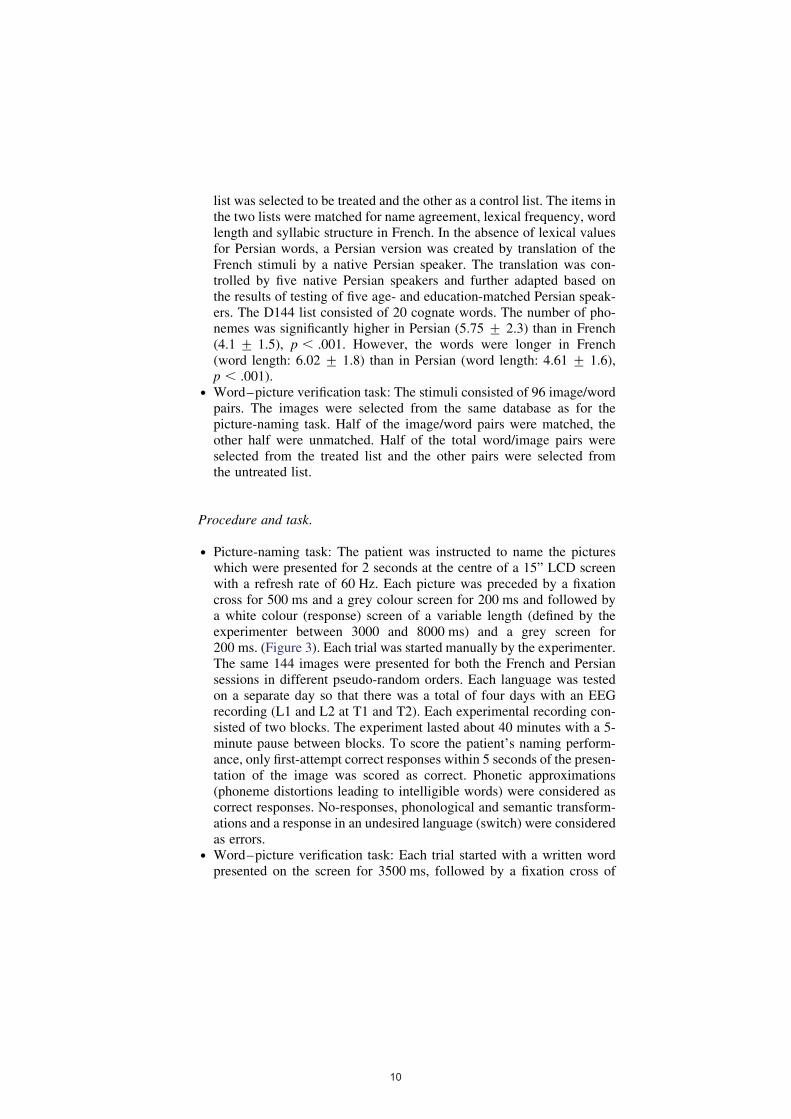
. Picture-naming task: The patient was instructed to name the pictureswhich were presented for 2 seconds at the centre of a 15” LCD screenwith a refresh rate of 60 Hz. Each picture was preceded by a fixationcross for 500 ms and a grey colour screen for 200 ms and followed bya white colour (response) screen of a variable length (defined by theexperimenter between 3000 and 8000 ms) and a grey screen for200 ms. (Figure 3). Each trial was started manually by the experimenter.The same 144 images were presented for both the French and Persiansessions in different pseudo-random orders. Each language was testedon a separate day so that there was a total of four days with an EEGrecording (L1 and L2 at T1 and T2). Each experimental recording con-sisted of two blocks. The experiment lasted about 40 minutes with a 5-minute pause between blocks. To score the patient’s naming perform-ance, only first-attempt correct responses within 5 seconds of the presen-tation of the image was scored as correct. Phonetic approximations(phoneme distortions leading to intelligible words) were considered ascorrect responses. No-responses, phonological and semantic transform-ations and a response in an undesired language (switch) were consideredas errors.
. Word–picture verification task: Each trial started with a written wordpresented on the screen for 3500 ms, followed by a fixation cross of
10
500 ms, a grey screen for 200 ms, a target picture for 2000ms, and ablank page for 200 ms. The patient had to press a button to respond assoon as the picture appeared on the screen. The responses were recordedif they were performed up to 2200 ms after the onset of the stimuluspresentation. In 48 trials, picture and image were matched and in theother 48 trials they were not matched. The patient was asked to pressthe button “Y” when the picture and word matched and to press “N”where they did not match. The same paradigm was performed inFrench and Persian in the corresponding picture-naming session.
During the EEG recording in each language session, the two experimenterstalked only the target language to prevent the participant from being in “bilin-gual” mode (speaking only in French on the French testing days and inPersian on the Persian testing days; Grosjean, 2004).
The entire sessions were audio-recorded using a digital voice recorder.Picture-naming scoring was done online and subsequently double-checkedbased on the audio-recording. The word–picture verification task wasscored offline using E-prime outputs. The E-Prime 2.0 software (PsychologySoftware Tools, Sharpsburg, PA) was used to deliver stimulations and collectmanual responses.
Lexical-phonological computer-assisted therapy
Sixteen weeks after the stroke, the day after our second pretest baseline, thepatient received an intensive lexical-phonological computer-assisted therapy
Figure 3. Picture-naming paradigm: Picture-naming task. Each picture was preceded by a fixation
cross and a grey colour screen and followed by a white colour (response) screen of a variable
length, defined by the experimenter, and finally a grey screen. The timing is indicated in the figure.
Each trial was started manually by the experimenter.
11

in L2 (French). A period of intensive (4 weeks, 5 days per week) phonologicaltherapy of naming in L2 was conducted using a computerised aphasia therapy(CAT) for anomia. Of note, this protocol has already been used with mono-lingual French-speaking patients (Laganaro et al., 2006).
The lexical-phonological therapy consisted of 5 x 1-hour sessions in thefirst week, and 15 x 45-minute sessions afterwards (based on the patient’srequest to decrease the duration of each session). The patient received atotal of 16 hours of therapy.
During this therapy, only one list of 72 French words was treated. Thetreated words were selected among the items of the D144 picture-namingtask (see section on stimuli above). Two tasks were used, involving limitedcomputer skills (the patient had to use the keyboard for writing or copyingand the mouse for selecting a button on the screen). In the first programme,a spoken–written picture-naming task, the patient had to write the word cor-responding to a picture which appeared on the screen. Two help buttons wereavailable: one provided the pronounced word (phonological cue), the secondprovided help on each letter of the word (orthographic cue). The second taskconsisted of a spoken naming with first grapheme help as well as the pro-nounced word.
Behavioural data analyses
In order to test the interaction between the factors Language and Session andthe main effect of these factors on picture-naming and word–picture verifica-tion for the treated and untreated items, the scores were subjected to separategeneralised linear mixed effect regression (GLMER) analyses for binomialdata using R (Team, 2008), lme4 (Bates, Maechler, Bolker, & Walker,2014) and contrast (Kuhn, Weston, Wing, Forester, & Thaler, 2013)packages. For the fixed effects, we included Language and Session as categ-orical variables, an interaction term, and we included the items as randomeffect.
EEG analyses
EEG acquisition and pre-processing
The EEG was recorded with a sampling rate of 512 Hz from 64 electrodes(64-channel ActiveTwo system from Biosemi, Inc., Amsterdam, The Nether-lands). The ground and reference electrodes were placed at the inion andvertex (“Cz”), respectively. The data were analysed off-line using theCartool software (http://brainmapping.unige.ch/Cartool.php) (Brunet,Murray, & Michel, 2011) as well as the Ragu software (Koenig, Kottlow,Stein, & Melie-Garcia, 2011). Epochs from –100 ms before the presentationof the stimuli to 600 ms post-stimulus were extracted from the raw EEG data
12

and filtered between 0.1 and 30 Hz with the addition of a 50 Hz Notch filter toremove AC noise. Epochs with eye-blinks or other artefacts (as determined byamplitude changes exceeding 80 mV on at least one electrode during theepoch) were rejected before epoch averaging. For both paradigms, onlyepochs corresponding to the treated items (independently of the patient’sresponse), accepted in all four conditions were considered, leading to theinclusion of 36 out of 144 epochs for picture naming and 33 out of 96epochs for word–picture verification. The epochs were then averaged separ-ately for each of the experimental conditions and recomputed against theaverage reference.
Event-related potentials
A first level of analysis was conducted by comparing the ERPs in the L1/T1; L1/T2, L2/T1 and L2/T2 conditions using a time-frame wise 2 × 2within-subject ANOVA with factors Language (L2; L1) and Session (T1;T2) at each scalp electrode as a function of peri-stimulus time. The resultsof this ERP waveform analysis are presented as a plot (Figure 5 b) depictingthe time frames showing a significant (p , .01) Language × Session inter-action as a function of peri-stimulus time and electrodes. While highly sensi-tive, the statistical results of the ERP analyses are dependent on the choice ofthe reference electrode. We therefore base our interpretations on reference-independent global analyses of the topography of the electric field at the scalp.
Global dissimilarity analysis
Topographic modulations were analysed using randomisation statisticsapplied to Global Map Dissimilarity measures (GMD; Lehmann & Skrandies,1980) using a 2 × 2 within-subject ANOVA with factors Language (L1; L2)and Session (T1; T2). GMD is calculated as the root mean square of the differ-ence between the strength-normalised voltage potentials across the electrodemontage. We analysed GMD values as a function of peri-stimulus time as forthe ERP analyses (Koenig et al., 2011; Koenig & Melie-Garcia, 2010;Murray, Brunet, &Michel, 2008). Correction was made for temporal autocor-relation through the application of a . 11 contiguous data points temporalcriterion for the persistence of significant effects (Guthrie & Buchwald,1991). In addition to the independence on the choice of the reference elec-trode, the GMD global analyses of the shape of the electric field over localelectrode analyses allows for neurophysiological interpretation of theobserved effects. Because topographic modulations necessarily follow frommodifications in the configuration of the underlying neural generators,GMD modulations indicate that qualitative changes in the brainnetworks engaged across conditions (Lehmann, Ozaki, & Pal, 1987). As
13

strength-normalised maps are used in the calculation on the GMD, expla-nation of our results in terms of pure amplitude modulation can be ruled out.
RESULTS
Behavioural results
General language assessment
The patient was evaluated using the L2 (French) and L1 (Persian) versionsof BAT, before and after the language phonological therapy (T1; T2). Table 1shows the detailed result of the patient’s performance.
At T1, the overall pattern based on the BAT results showed no differencebetween L1 and L2 (McNemar’s chi-squared p ¼ 1.0). Before the therapy atT1, the major impairment for L1 was found in the following subtests: semi-complex orders, verbal–auditory discrimination, syntactic comprehension,repetition, series, mental calculation, word reading, dictation and words andphrases comprehension.
At the same phase (T1), the patient was below the limits of the norm in L2for complex orders, syntactic comprehension, acceptability judgement, wordand nonword repetition and judgement, series, naming, mental calculation,word reading, dictation and word and phrase comprehension.
After the therapy (at T2), in the BAT, her global score of L1 improved.Specifically, the scores in verbal comprehension and word and nonword rep-etition and judgement in L1 significantly improved at T2. She also performedbetter in syntactic comprehension in both languages at T2. However, herglobal score in L2 function did not improve after the therapy (Table 1).
D144 picture naming
Pre-treatment picture-naming scores (at T0 and T1, i.e., one week apart)were analysed to check for the amount of variability of language performanceimmediately before starting the therapy. A GLMER model was applied toanalyse the interaction between the factors Session (T0; T1), Language (L1untreated; L2 treated) and List (treated items; untreated items). The resultsshowed a main effect of factor Language (z ¼ 22.70, p ¼ .006), a maineffect of Session (z ¼ 22.5, p ¼ .012) (T0 . T1), but there was no maineffect of List (z ¼ 0.32, p ¼ .74). No interaction was found between thefactors Session and List (z ¼ 21.03, p ¼ .30), nor between Language andList (z ¼ 0.31, p ¼ .75). However, an interaction was found betweenLanguage and Session (z ¼ 2.74, p ¼ .006). This interaction was drivenby a “trend” to decrease in L1 naming score from T0 to T1, z ¼ 22.01,
14

p¼ .08 (Bonferroni corrected) but no change in L2 naming fromT0 toT1, z ¼20.611, p ¼ 1.0 (Bonferroni corrected). See Figure 4(a).
A GLMER model was applied to analyse the interaction between thefactors Session (T0; T1) and Language (L1 untreated; L2 treated) for thetreated and untreated items separately.
Regarding the treated items, there was no main effect of Session (z ¼ 0.20,p ¼ .84), but there was a main effect of Language (z ¼ 4.50, p , .001), andan interaction between the factors Language and Session (z ¼ 4.79, p ¼ .03).The interaction was driven by an improvement in the treated L2, z ¼ 22.77,p ¼ .01 (Bonferroni corrected), Cohen’s d ¼ 4.71, but not in the untreated L1language, z ¼ 0.19, p ¼ 1.0 (Bonferroni corrected), Cohen’s d ¼ 20.79.The effect size (Cohen’s d) of the therapy on all items was d ¼ 5.42 forthe treated language (L2) and d ¼ 20.87 for the untreated language (L1).
Testing the untreated items, there were no main effects of factors Session(z ¼ 0.37, p ¼ .71) and Language (z ¼ 20.19, p ¼ .85). Moreover, no inter-action between the factors Language and Session was found (z ¼ 20.39, p ¼.69). See Figure 4(a).
The number of semantic errors (7% of errors in L1 and 2% in L2), phonolo-gical errors (24% of errors in L1 and 25% in L2), and switches (4% of errors inL1 and 1% in L2) as well as the other types (including no responses and lateresponses) in picture naming was similar at T1 in both languages. Similarly,the number of semantic errors (6% of errors in L1 and 3% in L2), and switches(4%of errors inL1 and1%inL2)were similar atT2 in both languages, and therewas a larger number of phonological errors (although not statistically signifi-cant) in L2 (35% of errors in L2) than and in L1 (25% of errors in L1) p ¼ .37.
Word–picture verification task
The same GLMER model as for the picture-naming task was applied toanalyse the interaction between the factors Session (T1; T2) and Language(L1 untreated; L2 treated) for the treated and untreated items separately.
Regarding the treated items, there was no main effect of Session (z ¼20.37, p ¼ .70) or Language (z ¼ 0.70, p ¼ .48). There was also no inter-action between the factors Language and Session (z ¼ 20.43, p ¼ .66).
For the untreated items, there was no main effect of Session (z ¼ 20.85,p ¼ .39) or Language (z ¼ 1.14, p ¼ .25). There was also no interactionbetween the factors Language and Session (z ¼ 20.44, p ¼ .66).
EEG results
ERP waveforms analysis
The time-wise 2 × 2 waveform analyses on all electrodes with the factorsSession and Language for the picture-naming task showed a Language ×
15

Figure 4. (a) Picture naming results at T0, T1 and T2: After the therapy (T2), the improvement in
naming performances was found only for treated items in the treated language (L2). Detailed
scores are as follow: L1 treated list: T0 ¼ 35, T1 ¼ 19, T2 ¼ 18; L1 untreated list: T0 ¼ 35, T1
¼ 25, T2 ¼ 23; L2 treated list: T0 ¼ 22, T1 ¼ 28, T2 ¼ 45; L2 untreated list: T0 ¼ 21, T1 ¼ 21,
T2 ¼ 24. (b) Word–picture verification results at T1 and T2: In testing treated and untreated items
separately, there is no main effect of Session and Language nor the interaction between Session
and Language. Detailed scores are as follow: L1 treated list: T1 ¼ 20, T2 ¼ 27; L1 untreated list:
T1 ¼ 12, T2 ¼ 14; L2 treated list: T1 ¼ 12, T2 ¼ 18; L2 untreated list: T1 ¼ 6, T2 ¼ 10.
16

Session interaction at left frontal electrodes from 200 to 260 ms and 550 to600 ms. See Figure 5(a) and (b).
The 2 × 2 waveform analyses of all electrodes for the word–picture ver-ification task showed a Language × Session interaction at the frontal electro-des from 2100 to 280 ms (before the image presentation) and 310 to 360 msafter the image presentation.
The interpretation of the EEG data will however be based on the global dis-similarity analysis reported below.
Global dissimilarity analysis
. Picture naming: The 2 × 2 timeframe-wise analysis of global dissimilar-ity with factors Session (T1; T2) and Language (L1 and L2) showed amain effect of Session at 300–350 ms after the stimulus presentation.Critically, there was an interaction between the two factors at 540–600 ms after the stimulus presentation. See Figure 5(c).
Post-hoc analyses were conducted over the period showing the significantinteraction 540 to 600 ms after the stimulus presentation to investigate theeffect of session on each language separately. Over the period of interest,there was a significant effect of Session on the treated L2, p , .001, seeFigure 5(c), and no significant effect of session for equivalent list in untreatedL1, p ¼ .18.
. Word–picture verification: The same design Session (T1; T2) ×Language (L1; L2) as for the picture-naming task was used. Therewas a main effect of Session at 320–340 ms and 500–520 ms afterthe stimulus presentation. The main effect of Language was foundfrom 20–120 ms, 300–420 ms and 460–600 ms after the stimulus pres-entation. No interaction was found between the factors Language andSession.
DISCUSSION
We examined the cross-language generalisation of intensive anomia lexical-phonological therapy on naming abilities in the second language (L2) of abilingual Broca’s aphasic patient presenting with severe word-finding diffi-culties and apraxia of speech.
Comparison between T1 and T2 shows an improvement in naming in thetreated L2 but not in the untreated L1 language after intensive lexical-phono-logical therapy. Electrophysiological recording during naming corroboratedthese results by showing a topographic interaction between Session andLanguage at 540–600 ms, driven by an effect of session (before and after
17

Figure 5. EEG analyses of picture-naming task: Waveforms (a) and (b) and topographic analyses (c):
(a) Exemplar ERP waveforms of left and right frontal electrodes. (b) The time-wise 2 × 2 waveform
analyses on all electrodes with the factors Session and Language for the picture naming task showing
an interaction at left frontal electrodes from 200 to 260 ms and 550–600 ms. (c) The 2 × 2 time-frame
wise analysis of global dissimilarity with factors Session and Language showing an interaction
between the two factors at 540–600 ms after stimulus presentation.
18

the therapy) on the ERP topography to the pictures in the treated L2 but not inthe corresponding L1 stimuli. The absence of cross-language generalisationwas confirmed by behavioural and EEG analyses. While a main effect ofsession in behavioural analysis could have been due to mere test-retesteffects (habituation, modification in the attentional state between recordings,etc.) or caused by spontaneous recovery, the interpretation of the Language xSession interaction term is unequivocal: the response to therapy in the treatedL2 but not in the corresponding untreated L1 rules out that global, languageunspecific effects accounted for our finding.
By contrast to the picture-naming task, the performance in the semanticword–picture verification task (a control semantic task) did not change atT2 either in the treated or untreated language. Consistently, electrophysio-logical topographic analyses of the word–picture verification task showedno interaction between the factors Session and Language. However, accord-ing to several models of lexical access (Costa et al., 2000) the semantic, butnot the lexical system, is shared between both languages. Since therapy wasfocused at the lexical-phonological level, we did not expect that the effects oflexical-phonological therapy would transfer to the semantic system. Thisresult should, however, be interpreted with caution since we cannot excludetype 2 error due to data insensitivity. Hence, a global ERP pattern modulationspecific to treated items was observed only in the picture-naming task in thelate 540–600 ms time-window.
According to previous estimates in healthy speakers (Indefrey, 2011; Inde-frey & Levelt, 2004) such a late time-window in the picture-naming task hasbeen associated with phonetic encoding. Phonetic encoding is the stage ofword production when articulatory gestures are generated from an abstractphonological code. In brain-damaged speakers, the phonological-phoneticencoding process, during which an abstract phonological code is transformedto a phonetic plan, has been suggested to be involved from around 400 ms afterthe picture presentation (Laganaro, Python, & Toepel, 2013). Laganaro et al.(2013) found that in aphasic patients who produced specifically phonologicaland/or phonetic errors, ERPs to picture naming diverged from healthy controlsubjects after 400 ms post-picture onset. In another paper, by comparing theERP to picture naming in two groups of anomic patients (with, respectively,predominantly semantic or phonological errors) with a healthy controlgroup, Laganaro et al. (Laganaro, Morand, & Schnider, 2009) showed differ-ent waveform amplitude and topographic maps at � 100–250 ms only in thegroup with semantic errors, and different waveform amplitude and topo-graphic maps at � 300–450 ms only in the group with phonological errors;the waveforms and topographic maps were comparable to a healthy groupout of these time windows. This finding accounts for the same language-processing pathway in aphasic patients with brain lesions and healthy subjects.However, in these previous studies, the time-course of divergent ERPs was
19

compared to that of healthy speakers, therefore reflecting the dynamics of thecontrols, while aphasic patients had much longer production latencies.Accordingly, because of longer latencies in our patient’s speaking onset, itis also probable that the effects that we found at the latency of 540–600 ms,i.e., 100 ms later than in previous estimates, were also related to phonologi-cal-phonetic processes.
Because ERP topographic modulations necessarily follow from modifi-cations in the configuration of neural generators, our results indicate thatthe lexical-phonological therapy modified qualitatively the brain networksengaged in phonetic encoding (and probably the phonological-phoneticprocess). More specifically, the therapy modified the brain networksengaged during naming in the treated but not in the untreated language.ERP change patterns after aphasia therapy have been rarely mentioned inthe literature. We are aware of two studies: in four monolingual aphasicpatients, Laganaro et al. (Laganaro, Morand, Schwitter, Zimmermann, &Schnider, 2008) showed that post-treatment increased abnormal amplitudeof ERP to picture naming, and different topographic map distribution (incomparison to the control group) occurs in the time windows correspondingto the impaired process, i.e., lexical-phonologic or lexical-semantic. Theauthors address these changes as “re-learning” of the process. However,after the treatment, in time windows corresponding to unimpaired processes,the ERP changed towards normalisation. In addition, Pulvermuller et al. (Pul-vermuller, Hauk, Zohsel, Neininger, & Mohr, 2005) suggested a post-treat-ment enhanced negativity in the ERP to words around 250–300 ms afterword presentation in monolingual aphasic patients as an index of recoveryfrom aphasia. They found that the ERP to pseudowords did not changeafter therapy, suggesting that change at this time window corresponds tolexico-semantic processes.
The absence of transfer of the effect of therapy to the untreated items(item-specificity of lexical-phonological therapy) is compatible with the dis-crete view which states that in the process of picture naming only the phono-logical segments of the selected word are activated and the activation ofnon-selected lexical representations does not spread to their phonological seg-ments. In addition, the absence of CLG could be explained by the model ofCosta et al. (2000) on language-specific lexical selection, which proposesthat the activation of the shared semantic system spreads only to the lexicalrepresentations of the languages in use. Alternatively, the absence of CLGcan also be accounted for by the fact that (1) the patient had a high proficiencyin L2 and her main language exposure was in her L2 prior to the stroke, and(2) the etymological roots of the words in the two languages were mostlydistinct.
Based on the Revised Hierarchical model, the link between lexicons isstronger from L2 to L1 than from L1 to L2 (Kroll & Stewart, 1994); thus,
20

in low proficient bilinguals, L2 is more dependent on borrowings from L1,while L2 in high proficient bilinguals is relatively independent of L1. Inthis regard, since our patient was highly proficient in L2, the therapy in L2did not increase the access to L1 because the highly mastered L2 was quiteindependent from L1. Supporting this hypothesis, some authors havesuggested that the patients with low L2 proficiency benefit from CLG aftertherapy in the less dominant L2 (Edmonds & Kiran, 2006; Gil & Goral,2004). In contrast, another group of studies report no generalisation fromthe low proficient treated language to the non-treated high proficient L1:Miertsch et al. (2009) have found no generalisation from their patient’streated L3 to the stronger non-treated L1, while a CLG from the treated L3to the non-treated L2 was found. The authors proposed that the absence ofCLG to the untreated L1 (which was the main language of the environment)followed from the performance of the L1 being already high in the first assess-ment. Goral et al. (Goral, Rosas, Conner, Maul, & Obler, 2012) examined acase of a multilingual aphasic patient (L1: Spanish, L2: German, L3:French and L4: English) and used a therapy in L1 and L4. They found no gen-eralisation from L1 (again, the main language of the environment) to the otherlanguages. In addition, after therapy in the less proficient language, L4, theyobserved no improvement in picture naming in untreated languages.
The absence of generalisation of the lexical-phonological therapy to theuntreated language could also be explained by the fact that the patient’stwo languages (Persian and French) were different from each other at thelevel of the phonology, morphology, lexis and syntax. CLG has indeedbeen shown to be facilitated by a high degree of linguistic structure similaritybetween the treated and untreated languages. It has been suggested that trans-fer of the effect of therapy is expected at the level of shared linguistic struc-tures in the two languages (Paradis, 1993) and thus CLG cannot take placebetween languages which do not share common structures (Kurland &Falcon, 2011).
In contrast to our results for an absence of CLG to untreated L1, Marangoloet al. (2009) used a 6-month phonological therapy in L2 in a bilingual(Flemish/Italian) aphasic patient and found that CLG resulted in a parallelrecovery of both languages after the therapy. This positive result possibly fol-lowed from the fact that the authors used a long duration therapy (6 months),whereas the intervention was much shorter in our study (4 weeks). FunctionalMRI recordings before and after two weeks of therapy in L2 confirmed theirbehavioural results by showing that the same brain regions were functioningfor both languages before and after two weeks of the therapy.
Although we did not test directly for a difference between the effect oftherapy on the treated vs. untreated items in L1 and L2, we did not findany main effect of Session nor Language x Session interaction for the
21

untreated items. This result suggests that there were no cross-item generalis-ation of the therapy.
The lack of generalisation of the lexical-phonological therapy on thenaming of untreated items in L2 corroborates current evidence that phonolo-gical treatment is item-specific. Hickin, Best, Herbert, Howard, and Osborne(2002) and Lorenz and Zieglerb (2009) found that phonological therapy foranomia in monolingual aphasic patients improved only the treated itemsand did not generalise to untreated items. Hickin et al. (2002) suggestedthat the effects of the therapy were item-specific because phonologicaltherapy focuses on the “output form of the individual word”.
In support of the fact that the phonological therapy improved only phonol-ogy and not semantic processing, there were neither main effects of Languageand Session nor an interaction between Language and Session in the word–picture verification task. The EEG result further supported this finding byshowing no Language x Session topographic interaction. Electrophysiologi-cally, we also found short-lived main effects of Session at 320–340 ms and500–520 ms post-stimulus onset. As discussed above, main effects ofsession could be due to differences between the two recording sessionsunrelated to the therapy and are thus difficult to interpret. Although wecannot exclude that the phonological therapy had a global effect on semanticprocessing in both L1 and L2 (more importantly on L1), the word–pictureverification task relies less on phonological processing (Marshall, Pound,White-Thomson, & Pring, 1990) and this hypothesis is thus unlikely.
Improvement of global score of language function only in the untreatedlanguage (L1) can be explained by different mechanisms: spontaneous recov-ery, generalisation of the effect of therapy, as well as the effect of language ofenvironment. Spontaneous recovery consists of a series of physiologicalchanges in the patient’s brain taking place in the first weeks immediatelyafter the onset of aphasia and generally occurring during the first threemonths after the stroke onset. Although the exact period which can be referredto as spontaneous recovery remains controversial (Gil & Goral, 2004), in thebeginning of the chronic phase, after three months post-lesion, spontaneousrecovery generally slows down. In bilingual aphasic patients, spontaneousrecovery can lead to both parallel and non-parallel recovery (Nilipour &Ashayeri, 1989). Non-parallel spontaneous recovery of languages can becaused by differences in lexical systems of these languages in addition tocomplete or partial inhibition of one language during the activation of otherlanguages (Green & Price, 2001). Differentiation between spontaneous recov-ery and therapy-induced improvement is not well studied because eliminatingtherapy programmes after aphasia onset in order to study spontaneous recov-ery is not ethically acceptable (Basso et al., 2011). However, as stated before,spontaneous recovery decreases gradually around three months after thestroke. Although the treatment programme for this patient was performed
22

after three months post-onset of aphasia, one cannot draw definite conclusionson the role of spontaneous recovery in our patient’s improvement of globalscore of clinical aphasia assessment in L1. On the other hand, becausethere was no improvement in word–picture verification, the improvementof global score should be interpreted with caution. Therefore, in our patientit is difficult to determine to what extent the recovery of global score of theuntreated language is related to phonological therapy in L2. In this regard,multiple evaluations after the therapy could have helped clarify the originof this improvement. Unfortunately, the patient did not participate tofurther evaluations after the therapy.
We would further note that, before the therapy, two baseline tests of picturenaming (T0 and T1, one week apart) were conducted. These comparisonsrevealed that, at the beginning of the training, the naming performance wasrather unstable; however, given that there was only one week between T0and T1 and that the amplitude of the variations were small, we interpretthese fluctuations as being primarily due to test-retest effects rather than asa consequence of spontaneous recovery. In addition, as detailed, the signifi-cant change in performance was specific to the treated L2, and accompaniedby specific electrophysiological modulations only for treated items.
CONCLUSION
Our behavioural and EEG results suggest that intensive lexical-phonologicaltherapy in L2 in a highly proficient bilingual aphasic patient with two etymo-logically different languages might be language-specific. The present resultsshould however be considered as preliminary since the study focused on onlya single case. A control condition in our patient (which would be to use thesame therapy in L1) was not possible due to fatigability. Further studies arenecessary to interpret patterns of generalisation in bilingual aphasic patients.
REFERENCES
Abutalebi, J., & Green, D. W. (2007). Bilingual language production: The neurocognition of
language representation and control. Journal of Neurolinguistics, 20, 242–275.
Aglioti, S., Beltramello, A., Girardi, F., & Fabbro, F. (1996). Neurolinguistic and follow-up
study of an unusual pattern of recovery from bilingual subcortical aphasia. Brain,
119(Pt 5), 1551–1564.
Ansaldo, A. I., Marcotte, K., Scherer, L., & Raboyeau, G. (2008). Language therapy and bilin-
gual aphasia: Clinical implications of psycholinguistic and neuroimaging research. Journal
of Neurolinguistics, 21, 539–557.
Ansaldo, A. I., & Saidi, L. G. (2014). Aphasia therapy in the age of globalization: Cross-linguis-
tic therapy effects in bilingual aphasia. Behav Neurol, 2014, 603085.
23

Basso, A., Cattaneo, S., Girelli, L., Luzzatti, C., Miozzo, A., Modena, L., & Monti, A. (2011).
Treatment efficacy of language and calculation disorders and speech apraxia: A review of
the literature. Eur J Phys Rehabil Med, 47, 101–121.
Bates, D., Maechler, M., Bolker, B., & Walker, S. (2014). lme4: Linear mixed-effects models
using Eigen and S4. In (1.1–6 ed.).
Brunet, D., Murray, M. M., & Michel, C. M. (2011). Spatiotemporal analysis of multichannel
EEG: CARTOOL. Comput Intell Neurosci, 2011, 813870.
Costa, A., Colome, A., & Caramazza, A. (2000). Lexical access in speech production: The bilin-
gual case. Psicologica, 21, 403–437.
Croft, S., Marshall, J., Pring, T., & Hardwick, M. (2011). Therapy for naming difficulties in
bilingual aphasia: Which language benefits?. Int J Lang Commun Disord, 46, 48–62.
De Bot, K. (1992). A bilingual production model: Levelt’s speaking model adapted. Applied
Linguistics, 13, 1–24.
Dijkstra, T., & van Heuven, W. J. B. (2002). The architecture of the bilingual word recognition
system: From identification to decision. Bilingualism-Language and Cognition, 5, 175–197.
Edmonds, L. A., & Kiran, S. (2006). Effect of semantic naming treatment on crosslinguistic
generalization in bilingual aphasia. Journal of Speech Language and Hearing Research,
49, 729–748.
Faroqi-Shah, Y., Frymark, T., Mullen, R., & Wang, B. (2010). Effect of treatment for bilingual
individuals with aphasia: A systematic review of the evidence. Journal of Neurolinguistics,
23, 319–341.
Gil, M., & Goral, M. (2004). Nonparallel recovery in bilingual aphasia: Effects of language
choice, language proficiency, and treatment. International journal of bilingualism, 8,
191–219.
Gollan, K. H., Montoya, R. I., Fennema-Notestine, C., & Morris, S. K. (2005). Bilingualism
affects picture naming but not picture classification. Memory & Cognition, 33, 1220–1234.
Goral, M., Levy, E. S., & Kastl, R. (2007). Cross-language treatment generalisation: A case of
trilingual aphasia. Aphasiology, 103, 203–204.
Goral, M., Rosas, J., Conner, P. S., Maul, K. K., & Obler, L. K. (2012). Effects of language pro-
ficiency and language of the environment on aphasia therapy in a multilingual. J Neurolin-
guistics, 25, 538–551.
Green, D., & Price, C. (2001). Functional imaging in the study of recovery patterns in bilingual
aphasia. Bilingualism: Language and Cognition, 4, 191–201.
Grosjean, F. (2004). Studying bilinguals: Methodological and conceptual issues. In The hand-
book of bilingualism (pp. 32–63). Oxford: Blackwell Publishing Ltd.
Guthrie, D., & Buchwald, J. S. (1991). Significance testing of different potentials. Psychophy-
siology, 28, 240–244.
Hermans, D., Bongaerts, T., de Bot, K., & Schreuder, R. (1998). Producing words in a foreign
language: Can speakers prevent interference from their first language? Bilingualism:
Language and Cognition, 1, 213–229.
Hickin, J., Best, W., Herbert, R., Howard, D., & Osborne, F. (2002). Phonological therapy for
word-finding difficulties: A re-evaluation. Aphasiology, 16, 981–999.
Hinckley, J. (2003). Picture naming treatment in aphasia yields greater improvement in L1.
Brain and Language, 87, 171–172.
Howard, D., Patterson, K., Franklin, S., Orchard-Lisle, V., & Morton, J. (1985). Treatment of
word retrieval deficits in aphasia. A comparison of two therapy methods. Brain, 108(Pt 4),
817–829.
Indefrey, P. (2011). The spatial and temporal signatures of word production components: A
critical update. Frontiers in Psychology, 2, 255. doi:10.3389/fpsyg.2011.00255
Indefrey, P., & Levelt, W. J. M. (2004). The spatial and temporal signatures of word production
components. Cognition, 92, 101–144.
24

Kiran, S., Sandberg, C., Gray, T., Ascenso, E., & Kester, E. (2013). Rehabilitation in bilingual
aphasia: Evidence for within and between-language generalization. Am J Speech Lang
Pathol, 22, S298–S309.
Koenig, T., Kottlow, M., Stein, M., &Melie-Garcia, L. (2011). Ragu: a free tool for the analysis
of EEG and MEG event-related scalp field data using global randomization statistics.
Comput Intell Neurosci, 2011, 938925.
Koenig, T., & Melie-Garcia, L. (2010). A method to determine the presence of averaged event-
related fields using randomization tests. Brain Topography, 23, 233–242.
Kohnert, K. (2004). Cognitive and cognate-based treatments for bilingual aphasia: A case study.
Brain and Language, 91, 294–302.
Kohnert, K. (2009). Cross-language generalization following treatment in bilingual speakers
with aphasia: A review. Seminars in Speech and Language, 30, 174–186.
Kroll, J., & Stewart, E. (1994). Category interference in translation and picture naming: Evi-
dence for asymmetric connections between bilingual memory representations. Journal of
Memory and Language, 33, 149–174.
Kuhn, M., Weston, S., Wing, J., Forester, J., & Thaler, T. (2013). Contrast: A collection of con-
trast methods. In (0.19 ed.).
Kurland, J., & Falcon, M. (2011). Effects of cognate status and language of therapy during
intensive semantic naming treatment in a case of severe nonfluent bilingual aphasia. Clinical
Linguistics & Phonetics, 25, 584–600.
Laganaro, M., Di Pietro, M., & Schnider, A. (2003). Computerised treatment of anomia in
chronic and acute aphasia: An exploratory study. Aphasiology, 17, 709–721.
Laganaro, M., Di Pietro, M., & Schnider, A. (2006). Computerised treatment of anomia in acute
aphasia: Treatment intensity and training size. Neuropsychological Rehabilitation, 16,
630–640.
Laganaro, M., Morand, S., & Schnider, A. (2009). Time course of evoked-potential changes in
different forms of anomia in aphasia. Journal of Cognitive Neuroscience, 21, 1499–1510.
Laganaro, M., Morand, S., Schwitter, V., Zimmermann, C., & Schnider, A. (2008). Normalisa-
tion and increase of abnormal ERP patterns accompany recovery from aphasia in the post-
acute stage. Neuropsychologia, 46, 2265–2273.
Laganaro, M., Python, G., & Toepel, U. (2013). Dynamics of phonological-phonetic encoding
in word production: Evidence from diverging ERPs between stroke patients and controls.
Brain and Language, 126, 123–132.
Lehmann, D., Ozaki, H., & Pal, I. (1987). EEG alpha map series: Brain micro-states by space-
oriented adaptive segmentation. Electroencephalography and Clinical Neurophysiology, 67,
271–288.
Lehmann, D., & Skrandies, W. (1980). Reference-free identification of components of checker-
board-evoked multichannel potential fields. Electroencephalography and Clinical Neuro-
physiology, 48, 609–621.
Levelt, W., Roelofs, A., & Meyer, A. (1999). A theory of lexical access in speech production.
Behavioral and Brain Sciences, 22.01, 1–38.
Lorenz, A., & Zieglerb, W. (2009). Semantic vs. word-form specific techniques in anomia treat-
ment: A multiple single-case study. Journal of Neurolinguistics, 22, 515–537.
Lucas, T. H., 2nd, McKhann, G. M., 2nd, & Ojemann, G. A. (2004). Functional separation of
languages in the bilingual brain: A comparison of electrical stimulation language mapping in
25 bilingual patients and 117 monolingual control patients. Journal of Neurosurgery, 101,
449–457.
Marangolo, P., Rizzi, C., Peran, P., Piras, F., & Sabatini, U. (2009). Parallel recovery in a bilin-
gual aphasic: A neurolinguistic and fMRI study. Neuropsychology, 23, 405–409.
Marshall, J., Pound, C., White-thomson, M., & Pring, T. (1990). The use of picture/word match-
ing tasks to assist word retrieval in aphasic patients. Aphasiology, 4, 167–184.
25

Meinzer, M., Obleser, J., Flaisch, T., Eulitz, C., & Rockstroh, B. (2007). Recovery from aphasia
as a function of language therapy in an early bilingual patient demonstrated by fMRI. Neu-
ropsychologia, 45, 1247–1256.
Miertsch, B., Meisela, J. M., & Isel, F. (2009). Non-treated languages in aphasia therapy of
polyglots benefit from improvement in the treated language. Journal of Neurolinguistics,
22, 135–150.
Murray, M. M., Brunet, D., & Michel, C. M. (2008). Topographic ERP analyses: A step-by-step
tutorial review. Brain Topography, 20, 249–264.
Nilipour, R. (1988). Bilingual aphasia in Iran: A preliminary report. Journal of Neurolinguistics,
3, 185–232.
Nilipour, R., & Ashayeri, H. (1989). Alternating antagonism between two languages with suc-
cessive recovery of a third in a trilingual aphasic patient. Bilingualism and Neurolinguistics,
36, 23–48.
Paradis, M. (1993). Bilingual aphasia rehabilitation. In Foundations of aphasia rehabilitation
(pp. 413–419). Oxford: Pergamon Press.
Paradis, M. (1998). Language and communication in multilinguals. In Handbook of neurolin-
guistics (pp. 417–430). San Diego, CA: Academic Press.
Paradis, M., & Libben, G. (1987). The assessment of bilingual aphasia. Hillsdale, N.J: Lawr-
ence Erlbaum Associates.
Parker Jones, O., Green, D. W., Grogan, A., Pliatsikas, C., Filippopolitis, K., Ali, N., . . . Price,
C. J. (2012). Where, when and why brain activation differs for bilinguals and monolinguals
during picture naming and reading aloud. Cerebral Cortex, 22, 892–902.
Peterson, R., & Savoy, P. (1998). Lexical selection and phonological encoding during language
production: Evidence for cascaded processing. Journal of Experimental Psychology: Learn-
ing, Memory, and Cognition, 24, 539–557.
Pillon, D., & de Partz, M. P. (2005). A direct processing route to translate words from the first to
the second language: Evidence from a case of a bilingual aphasic. Brain Lang, 95, 4041.
Pulvermuller, F., Hauk, O., Zohsel, K., Neininger, B., & Mohr, B. (2005). Therapy-related reor-
ganization of language in both hemispheres of patients with chronic aphasia. Neuroimage,
28, 481–489.
Roelofs, A. (1998). Lemma selection without inhibition of languages in bilingual speakers.
Bilingualism: Language and Cognition, 1, 94–95.
Team, R. D. C. (2008). R: A language and environment for statistical computing. Vienna,
Austria: R Foundation for Statistical Computing.
26