Embed Size (px)
Citation preview

880 www.thelancet.com/oncology Vol 12 September 2011
Articles
Lancet Oncol 2011; 12: 880–90
Published OnlineAugust 23, 2011
DOI:10.1016/S1470-2045(11)70188-7
See Comment page 831
American Society for Clinical Pathology Institute,
Washington, DC, USA (P E Castle PhD); University of
Virginia Health System, Charlottesville, VA, USA
(Prof M H Stoler MD); Department of Pathology,
Columbia University School of Medicine, New York, NY, USA
(Prof T C Wright Jr MD); and Roche Molecular Systems,
Pleasanton, CA, USA (A Sharma PhD, T L Wright MD,
C M Behrens MD)
Correspondence to:Dr Philip E Castle, American
Society for Clinical Pathology Institute, 1225 New York
Avenue NW, Suite 250, Washington, DC 20005, USA
Performance of carcinogenic human papillomavirus (HPV) testing and HPV16 or HPV18 genotyping for cervical cancer screening of women aged 25 years and older: a subanalysis of the ATHENA studyPhilip E Castle, Mark H Stoler, Thomas C Wright Jr, Abha Sharma, Teresa L Wright, Catherine M Behrens
SummaryBackground The ATHENA study was designed to assess the performance of carcinogenic human papillomavirus (HPV) testing and HPV16 or HPV18 genotyping compared with liquid-based cytology for cervical cancer screening in a large US population aged 21 years and older. We did a subanalysis of this population to compare the screening performance of the cobas HPV test versus liquid-based cytology in women aged 25 years and older, and assess management strategies for HPV-positive women.
Methods Women aged 25 years or older who were attending routine cervical screening were enrolled from 61 clinical centres in 23 US states. Cervical specimens were obtained for liquid-based cytology and HPV DNA testing with two fi rst-generation assays (Amplicor HPV test and Linear Array HPV genotyping test) and the second-generation cobas HPV test (with individual HPV16 and HPV18 detection). Colposcopy and diagnostic biopsies were done on women with atypical squamous cells of undetermined signifi cance (ASC-US) or worse cytology, those who tested positive with either fi rst-generation HPV test, and a random sample of women who tested negative for HPV and cytology. All women not selected for colposcopy received their results and exited the study. Participants and colposcopists were masked to cytology and HPV test results until the colposcopy visit was completed. The primary endpoint for this substudy was histologically confi rmed cervical intraepithelial neoplasia grade 3 (CIN3) or worse. This study is registered with ClinicalTrials.gov, number NCT00709891; the study is in the follow-up phase, which is due to be completed in December, 2012.
Findings From May 27, 2008, to Aug 27, 2009, 47 208 women were enrolled, of whom 41 955 met our eligibility criteria. Valid cobas HPV and liquid-based cytology test results were available for 40 901 women (97%), who were included in this analysis. Of these, 4275 women (10%) tested cobas HPV positive and 2617 (6%) had abnormal cytology. 431 women were diagnosed with CIN2 or worse and 274 with CIN3 or worse. In women who had colposcopy, the cobas HPV test was more sensitive than liquid-based cytology for detection of CIN3 or worse (252/274 [92·0%, 95% CI 88·1–94·6] vs 146/274 [53·3%, 95% CI 47·4–59·1]; diff erence 38·7%, 95% CI 31·9–45·5; p<0·0001). Addition of liquid-based cytology to HPV testing increased sensitivity for CIN3 or worse to 96·7% (265/274, 95% CI 93·9–98·3), but increased the number of screen positives by 35·2% (5783/40 901 vs 4275/40 901) compared with HPV testing alone. As a triage test to identify CIN3 or worse in HPV-positive women, detection of HPV16, HPV18, or both alone was equivalent to detection of ASC-US or worse alone in terms of sensitivity (150/252 [59·5%] vs 133/252 [52·8%]; p=0·11) and positive predictive value (PPV) (150/966 [15·5%] vs 133/940 [14·1%]; p=0·20). Among HPV-positive women, detection of HPV16, HPV18, or both or low-grade squamous intraepithelial lesion or worse cytology had better sensitivity (182/252 [72·2%]; p<0·0001) and similar PPV (182/1314 [13·9%]; p=0·70) for detection of CIN3 or worse than ASC-US or worse cytology alone. Furthermore, detection of HPV16, HPV18, or both or high-grade squamous intraepithelial lesion or worse cytology had higher sensitivity (165/252 [65·5%]; p=0·0011) and PPV (165/1013 [16·3%]; p=0·031) for detection of CIN3 or worse than ASC-US or worse cytology alone.
Interpretation HPV testing with separate HPV16 and HPV18 detection could provide an alternative, more sensitive, and effi cient strategy for cervical cancer screening than do methods based solely on cytology.
Funding Roche Molecular Systems.
IntroductionCervical cytology, fi rst with the Papanicolaou (Pap) smear and now with liquid-based cytology, has been the traditional method for cervical cancer screening in developed countries. Since cytology-based programmes were
intro duced in the mid-20th century, rates of cervical cancer have decreased substantially wherever these screening programmes have been successfully implemented.1 In the USA, cancer of the cervix is fairly uncommon, with an estimated 12 200 new cases and 4210 related deaths

Articles
www.thelancet.com/oncology Vol 12 September 2011 881
occurring in 2010.2 Despite their success, cytology-based screening programmes are now widely recognised as ineffi cient because of the low sensitivity of one screen and, therefore, many repeat screens are needed during a lifetime to achieve programmatic sensitivity. The US screening and prevention programme costs about US$4 billion annually.3
As a consequence, more effi cient screening methods are desirable from a comparative performance and cost-eff ectiveness perspective. Human papillomavirus (HPV) DNA testing, because of its greater sensitivity for cervical precancer and cancer than cytology with one screen, provides lead-time detection of precancerous lesions. In turn, early detection reduces the future risk of cervical cancer4–6 and related mortality,7 thereby providing greater reassurance for screen-negative women than can be off ered by cytology; as such, HPV testing permits a safe extension of screening intervals.8–10 The increased sensitivity of HPV testing over cytology also applies to the detection of glandular cancer (adenocarcinoma) and its precursor (adenocarcinoma in situ),11 which is increasingly important because of the rise in adeno-carcinoma rates in the USA,12 Canada,13 and Europe.14 Thus, a switch to HPV testing, either alone or in conjunction with cytology (co-testing), could provide a safer, more effi cient screening programme.
A crucial consideration in the use of HPV testing, either as a co-test with cytology or as the primary screening test in cervical cancer screening, is the management of HPV-positive women. According to US guidelines, HPV-positive women who are aged 30 years or older and have normal cervical cytology (negative for intraepithelial lesion or malignancy [NILM]) should be rescreened in 1 year.15 This approach might be less than optimum because some HPV-positive women with a negative cytology will be lost to follow-up16 and a small fraction might develop invasive cancer during the rescreening interval.17 To address these limitations of HPV testing, additional stratifi cation of HPV-positive women with NILM cytology could be used to identify those at greatest risk for cervical precancerous lesions who warrant immediate colposcopy referral. Genotyping for HPV16, HPV18, or both has been proposed as a triage technique, because these two HPV genotypes are associated with about 70% of all invasive cervical carcinomas,18 but few clinical trial data supporting the clinical performance of genotyping for HPV16, HPV18, or both are available.
Three DNA tests for carcinogenic or high-risk HPV have been approved by the US Food and Drug Administration (FDA): Hybrid Capture 2 (Qiagen, Gaithersburg, MD, USA; 2003), Cervista HPV HR (Hologic, Bedford, MA, USA; 2009), and the cobas HPV test (Roche Molecular Systems, Pleasanton, CA, USA; 2011). The cobas HPV test is a fully automated HPV DNA test that detects in three separate channels: HPV16 individually, HPV18 individually, and a pool of 12 other
HPV genotypes (11 defi nite high-risk genotypes plus one possibly high-risk genotype).19 Hereafter, HPV refers to carcinogenic or high-risk HPV genotypes.
The ATHENA (Addressing THE Need for Advanced HPV diagnostics) study is the largest clinical trial so far to assess HPV DNA tests and liquid-based cytology for cervical cancer screening in the USA. The study was done in three protocol-specifi ed populations: (1) women aged 21 years or older with atypical squamous cells of undetermined signifi cance (ASC-US) cytology to assess the performance of the assay for the intended use of ASC-US triage to colposcopy, which has been reported previously;20 (2) women aged 30 years or older with NILM cytology to assess the performance of the assay for the intended use as an adjunctive test with cytology;21 (3) women aged 25 years and older with any cytology result (ie, independent of cytology) to assess the performance of HPV testing with genotyping as a primary screening test. Here, we present data for the cobas HPV test in the third of these populations.
MethodsStudy population Women presenting for routine cervical cancer screening were enrolled into the ATHENA study at 61 clinical centres in 23 US states (webappendix p 1). Eligible women were aged 21 years or older and were not pregnant. Other study inclusion and exclusion criteria have been described previously20—briefl y, eligible women had an intact uterus, had not received treatment for CIN with 12 months of enrolment, and had no present or planned participation in a clinical trial for HPV treatment. For this subanalysis, the population was restricted to women aged 25 years and older.
The study protocol was approved by the institutional review boards of all study sites, and all women provided written informed consent before undergoing any study procedures. Institutional review board project number MWP-HPV-159 was obtained on March 13, 2008.
ProceduresTwo liquid-based cervical cytology samples were obtained from each participant at study visit one (enrolment visit), placed in PreservCyt (Hologic, Bedford, MA, USA), and processed with ThinPrep (Hologic). One sample was used for liquid-based cytology and for HPV testing with three assays (Roche Molecular Systems, Pleasanton, CA, USA): the fi rst-generation Amplicor HPV test, the fi rst-generation Linear Array HPV genotyping test, and the second-generation cobas HPV test. The second sample was reserved for additional testing. For the purposes of this analysis, a positive result for genotype 16, genotype 18, or both with the cobas HPV test was regarded as positive for HPV16, HPV18, or both, even if the sample was also positive for 12 other high-risk HPV types. Cytology was reported by use of the 2001 Bethesda System nomenclature.22
See Online for webappendix

Articles
882 www.thelancet.com/oncology Vol 12 September 2011
Sample processing and testing were done at four clinical laboratories in the USA, and a fi fth laboratory, also in the USA, did cobas HPV testing only; testing was done as described previously.20 The assignment of specimens to a clinical testing laboratory was made on the basis of the order and volume of patients enrolled at the clinical site, and the capacity of the laboratory; assignment was not based on geographical location.
A randomisation centre identifi ed participants for colposcopy on the basis of the results of liquid-based cytology and fi rst-generation HPV testing at study visit one. Women were eligible for colposcopy if they had ASC-US or worse (positive) cervical cytology, or tested HPV positive by any test and had NILM cervical cytology. A randomly selected subset of women who were HPV negative and had NILM cervical cytology was also referred to colposcopy; randomisation of this subset was done with a block size of 35 by use of SAS software (version 9.1.3). Women not selected for colposcopy received their results and exited the study.
Colposcopy with biopsy or endocervical curettage, or both, was done within 12 weeks of the enrolment visit (at study visit two), according to a standardised protocol.20 Participants and colposcopists were masked to cytology and HPV test results until the colposcopy visit was completed. Biopsy and endocervical curettage results were reviewed by a panel of three pathologists (central pathology review) who were masked to cytology and HPV test results, and diagnosed according to standard criteria and cervical intraepithelial neoplasia (CIN) terminology.23 The primary endpoint for the ATHENA study was biopsy-confi rmed CIN grade 2 (CIN2) or worse.24 Women reaching this endpoint exited the study after the colposcopy visit and were referred for treatment; those who did not reach CIN2 were eligible for the longitudinal follow-up phase of the trial, expected to be completed in December, 2012. However, for this subanalysis, we used the primary endpoint of CIN3 or worse because CIN3 is a more certain, rigorous histological diagnosis of precancer than CIN2 or worse. Data are also presented for CIN2 or worse because CIN2 is the standard threshold for treatment,25 but this endpoint was judged to be secondary because CIN2 is an equivocal diagnosis of precancer, representing an admixture of CIN1 and CIN3.26–28 CIN2 is poorly reproducible, can be caused by non-carcinogenic HPV genotypes, and is much more likely to regress than CIN3.
Statistical analysisIn accordance with the sample sizes in similar registration trials for the use of HPV testing as a refl ex test for ASC-US (ie, an HPV test done on a sample of cervical cells after an ASC-US Pap test result),29,30 we calculated that about 70 women with ASC-US cytology who had been diagnosed with histological CIN2 or worse would be needed to validate the cobas HPV test for this indication. Together with published rates of ASC-US cytology31 and HPV
positivity30 in the overall population, we calculated that a sample size of about 45 000 women would be needed. The sample size of about 880 women in the NILM population aged 30 years and older was estimated a priori to have about 95% power to detect a diff erence in disease (CIN2 or worse) between women who were positive for HPV16, HPV18, or both and those who were HPV negative at baseline, and about 98% power to detect a diff erence in disease between women who were HPV positive and those who were HPV negative across 3 years.
Several characteristics were used to assess detection of cervical precancer and cancer by HPV testing and liquid-based cytology: sensitivity, specifi city, positive predictive value (PPV) and negative predictive value (NPV), positive likelihood ratio (PLR) and negative likelihood ratio (NLR), and relative risk (RR) and, for comparison, cytology. All results are presented for the cobas HPV test, not the fi rst-generation Amplicor or Linear Array HPV tests. Estimates of performance, both crude and those adjusted for sampling fractions to correct for possible verifi cation bias, with 95% CIs, were calculated.32 Crude estimates were based on results from women with valid results for disease verifi cation. To adjust for verifi cation bias, we calculated how many women had CIN3 or worse (or CIN2 or worse) of those who underwent verifi cation by colposcopy and projected these estimates onto the corresponding women who had no verifi cation. We tested diff erences in crude sensitivity and specifi city by use of McNemar’s χ² test, and diff erences in PPV and NPV with the method described by Leisenring and colleagues.33 For estimates adjusted for verifi cation bias, 95% CIs were estimated by bootstrapping (1000 times).34 All analyses were done with SAS software (version 9.1.3).
Combinations of genotyping for HPV16, HPV18, or both provided by the cobas HPV test and diff erent thresholds of cytological interpretations (ASC-US or worse, low-grade squamous intraepithelial lesion [LSIL] or worse, or high-grade squamous intraepithelial lesion [HSIL] or worse) were also assessed to identify potentially better algorithms for triage to immediate colposcopy, thereby avoiding problems created by deferred management. To address the main concerns of clinicians, we assessed sensitivity from the proportion of disease detected and PPV from the proportion of screen positives with disease. As a post-hoc analysis, we stratifi ed these data by the four laboratories producing the HPV and liquid-based cytology results, excluding the one laboratory doing HPV testing alone, as a qualitative assessment of the triage method’s reliability, which is another important feature of test performance.
The ATHENA study is registered with ClinicalTrials.gov, number NCT00709891; the study is in the follow-up phase, which is due to be completed in December, 2012.
Role of the funding sourceThe sponsor designed the study in consultation with the other investigators (PEC, MHS, and TCWJr) and was

Articles
www.thelancet.com/oncology Vol 12 September 2011 883
responsible for the study conduct and data collection. Data analysis was done by PEC with the assistance of the sponsor (AS, Guili Zhang, TLW, and CMB). The sponsor participated in the interpretation of the data. The sponsor (AS, TLW, and CMB) assisted in editing and review of the report. PEC had full access to all data in this subanalysis,
designed the analytic approach, and had fi nal responsibility for the decision to submit the report for publication.
Results From May 27, 2008, to Aug 27, 2009, 47 208 women aged 21 years or older were enrolled in the ATHENA study.
47 208 enrolled women aged ≥21 years
4999 aged <25 years
42 209 aged ≥25 years
254 not eligible to participate in the study*
41 955 eligible for analysis
929 exited study because of invalid or missing test results 755 missing liquid-based cytology test results 56 missing first-generation HPV test results 118 missing liquid-based cytology and first-generation HPV test results
2609 had ASC-US or worse 1105 positive cobas HPV test result 1498 negative cobas HPV test result 6 invalid cobas HPV test result
14 had ASC-US or worse 4 positive cobas HPV test result 10 negative cobas HPV test result
87 had NILM cytology and positive first-generation HPV test results 18 positive cobas HPV test result 68 negative cobas HPV test result 1 invalid cobas test result
101 exited because of error in selection and randomisation process
5726 had NILM cytology and positive first-generation HPV test results 3065 positive cobas HPV test result 2647 negative cobas HPV test result 14 invalid cobas HPV test result
31 549 had NILM cytology and negative first-generation HPV test results 78 positive cobas HPV test result 31 370 negative cobas HPV test result 101 invalid cobas HPV test result
1041 had NILM cytology and negative first-generation HPV test results† 5 positive cobas HPV test result 1033 negative cobas HPV test result 3 invalid cobas HPV test result
2247 proceeded to study visit two 966 positive cobas HPV test result 1276 negative cobas HPV test result 5 invalid cobas HPV test result
73 no sample at study visit two 171 indeterminate result from central pathology review 274 reached endpoint of CIN3 or worse 157 reached endpoint of CIN2 7410 did not reach endpoint of CIN2 according to central pathology review
1291 exited study before study visit two 72 withdrew authorisation 545 no longer wanted to participate 2 withdrawn by clinician 29 pregnant at study visit two 372 lost to follow-up 71 protocol deviations 200 other reasons
4943 proceeded to study visit two 2640 positive cobas HPV test result 2293 negative cobas HPV test result 10 invalid cobas HPV test result
895 proceeded to study visit two 4 positive cobas HPV test result 888 negative cobas HPV test result 3 invalid cobas HPV test result
41 026 had valid results from Papanicolaou and first-generation HPV tests
31 650 exited after study visit one
9376 selected to attend study visit two
Figure: Study profi leFirst-generation HPV tests were Amplicor and Linear-Array HPV tests. NILM=negative for intraepithelial lesion or malignancy. ASC-US=atypical squamous cells of undetermined signifi cance. CIN2 or CIN3=cervical intraepithelial neoplasia grade 2 or 3. *165 did not meet inclusion or exclusion criteria, 82 enrolled into the study more than once, and seven withdrew authorisation. †Women randomly selected to attend study visit two.
Articles
884 www.thelancet.com/oncology Vol 12 September 2011
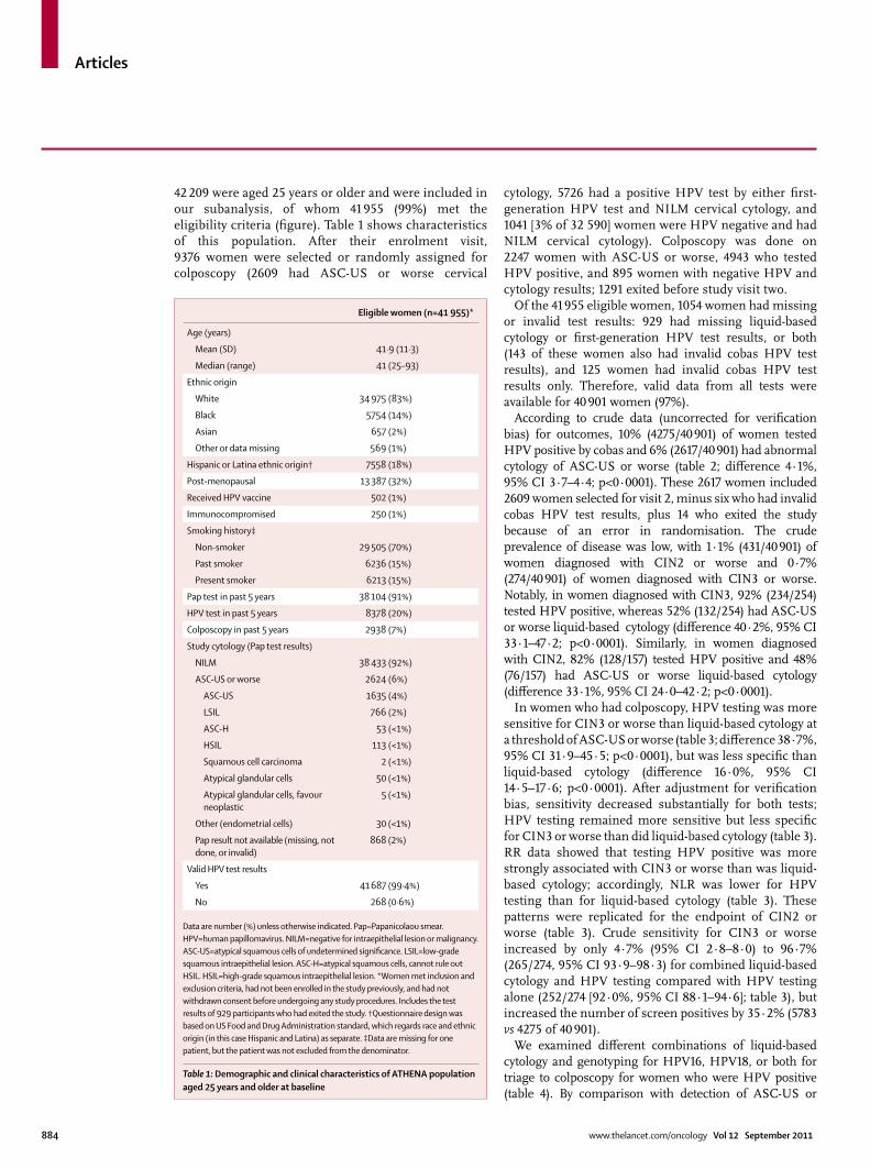
42 209 were aged 25 years or older and were included in our subanalysis, of whom 41 955 (99%) met the eligibility criteria (fi gure). Table 1 shows characteristics of this population. After their enrolment visit, 9376 women were selected or randomly assigned for colposcopy (2609 had ASC-US or worse cervical
cytology, 5726 had a positive HPV test by either fi rst-generation HPV test and NILM cervical cytology, and 1041 [3% of 32 590] women were HPV negative and had NILM cervical cytology). Colposcopy was done on 2247 women with ASC-US or worse, 4943 who tested HPV positive, and 895 women with negative HPV and cytology results; 1291 exited before study visit two.
Of the 41 955 eligible women, 1054 women had missing or invalid test results: 929 had missing liquid-based cytology or fi rst-generation HPV test results, or both (143 of these women also had invalid cobas HPV test results), and 125 women had invalid cobas HPV test results only. Therefore, valid data from all tests were available for 40 901 women (97%).
According to crude data (uncorrected for verifi cation bias) for outcomes, 10% (4275/40 901) of women tested HPV positive by cobas and 6% (2617/40 901) had abnormal cytology of ASC-US or worse (table 2; diff erence 4·1%, 95% CI 3·7–4·4; p<0·0001). These 2617 women included 2609 women selected for visit 2, minus six who had invalid cobas HPV test results, plus 14 who exited the study because of an error in randomisation. The crude prevalence of disease was low, with 1·1% (431/40 901) of women diagnosed with CIN2 or worse and 0·7% (274/40 901) of women diagnosed with CIN3 or worse. Notably, in women diagnosed with CIN3, 92% (234/254) tested HPV positive, whereas 52% (132/254) had ASC-US or worse liquid-based cytology (diff erence 40·2%, 95% CI 33·1–47·2; p<0·0001). Similarly, in women diagnosed with CIN2, 82% (128/157) tested HPV positive and 48% (76/157) had ASC-US or worse liquid-based cytology (diff erence 33·1%, 95% CI 24·0–42·2; p<0·0001).
In women who had colposcopy, HPV testing was more sensitive for CIN3 or worse than liquid-based cytology at a threshold of ASC-US or worse (table 3; diff erence 38·7%, 95% CI 31·9–45·5; p<0·0001), but was less specifi c than liquid-based cytology (diff erence 16·0%, 95% CI 14·5–17·6; p<0·0001). After adjustment for verifi cation bias, sensitivity decreased substantially for both tests; HPV testing remained more sensitive but less specifi c for CIN3 or worse than did liquid-based cytology (table 3). RR data showed that testing HPV positive was more strongly associated with CIN3 or worse than was liquid-based cytology; accordingly, NLR was lower for HPV testing than for liquid-based cytology (table 3). These patterns were replicated for the endpoint of CIN2 or worse (table 3). Crude sensitivity for CIN3 or worse increased by only 4·7% (95% CI 2·8–8·0) to 96·7% (265/274, 95% CI 93·9–98·3) for combined liquid-based cytology and HPV testing compared with HPV testing alone (252/274 [92·0%, 95% CI 88·1–94·6]; table 3), but increased the number of screen positives by 35·2% (5783 vs 4275 of 40 901).
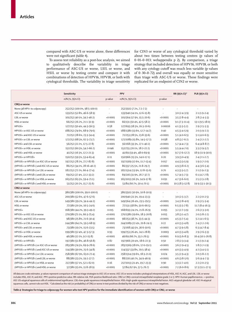
We examined diff erent combinations of liquid-based cytology and genotyping for HPV16, HPV18, or both for triage to colposcopy for women who were HPV positive (table 4). By comparison with detection of ASC-US or
Eligible women (n=41 955)*
Age (years)
Mean (SD) 41·9 (11·3)
Median (range) 41 (25–93)
Ethnic origin
White 34 975 (83%)
Black 5754 (14%)
Asian 657 (2%)
Other or data missing 569 (1%)
Hispanic or Latina ethnic origin† 7558 (18%)
Post-menopausal 13 387 (32%)
Received HPV vaccine 502 (1%)
Immunocompromised 250 (1%)
Smoking history‡
Non-smoker 29 505 (70%)
Past smoker 6236 (15%)
Present smoker 6213 (15%)
Pap test in past 5 years 38 104 (91%)
HPV test in past 5 years 8378 (20%)
Colposcopy in past 5 years 2938 (7%)
Study cytology (Pap test results)
NILM 38 433 (92%)
ASC-US or worse 2624 (6%)
ASC-US 1635 (4%)
LSIL 766 (2%)
ASC-H 53 (<1%)
HSIL 113 (<1%)
Squamous cell carcinoma 2 (<1%)
Atypical glandular cells 50 (<1%)
Atypical glandular cells, favour neoplastic
5 (<1%)
Other (endometrial cells) 30 (<1%)
Pap result not available (missing, not done, or invalid)
868 (2%)
Valid HPV test results
Yes 41 687 (99·4%)
No 268 (0·6%)
Data are number (%) unless otherwise indicated. Pap=Papanicolaou smear. HPV=human papillomavirus. NILM=negative for intraepithelial lesion or malignancy. ASC-US=atypical squamous cells of undetermined signifi cance. LSIL=low-grade squamous intraepithelial lesion. ASC-H=atypical squamous cells, cannot rule out HSIL. HSIL=high-grade squamous intraepithelial lesion. *Women met inclusion and exclusion criteria, had not been enrolled in the study previously, and had not withdrawn consent before undergoing any study procedures. Includes the test results of 929 participants who had exited the study. †Questionnaire design was based on US Food and Drug Administration standard, which regards race and ethnic origin (in this case Hispanic and Latina) as separate. ‡Data are missing for one patient, but the patient was not excluded from the denominator.
Table 1: Demographic and clinical characteristics of ATHENA population aged 25 years and older at baseline

Articles
www.thelancet.com/oncology Vol 12 September 2011 885
worse alone, the only available method for triage to colposcopy, detection of HPV16, HPV18, or both was more sensitive and had a greater PPV, but neither diff erence was signifi cant (table 4). Sensitivity was further increased and PPV was decreased by use of detection of HPV16, HPV18, or both as an additional or complementary triage strategy to ASC-US or worse (table 4). Notably, testing positive for HPV16, HPV18, or both had a sensitivity of 53·8% (64/119, 95% CI 44·9–62·5) and PPV of 10·2% (64/629, 95% CI 8·1–12·8) for CIN3 or worse in women aged 25 years or older who were HPV positive and had NILM cytology. Use of a threshold of LSIL or worse with
HPV16, HPV18, or both was more sensitive than detection of ASC-US or worse alone, with similar PPV (table 4). Furthermore, detection of HSIL or worse with HPV16, HPV18, or both had a higher sensitivity and PPV than did ASC-US or worse alone. Similar patterns were recorded for the endpoint of CIN2 or worse, with the exception that detection of HPV16, HPV18, or both did not have higher sensitivity or PPV than detection of ASC-US or worse alone, although these diff erences were not signifi cant. Furthermore, although detection of HSIL or worse as an additional or alternative triage strategy to detection of HPV16, HPV18, or both increased sensitivity and PPV
Crude data Adjusted data*
Liquid-based cytology Cobas HPV test p value Liquid-based cytology Cobas HPV test
CIN3 or worse
Sensitivity 146/274 (53·3%, 47·4–59·1) 252/274 (92·0%, 88·1–94·6) <0·0001 174/403 (43·2%, 32·1–55·9) 302/402 (75·1%, 58·4–94·1)
Specifi city 5509/7549 (73·0%, 72·0–74·0) 4299/7549 (56·9%, 55·8–58·1) <0·0001 38 055/40 498 (94·0%, 93·7–94·2) 36 526/40 499 (90·2%, 89·9–90·5)
PPV 146/2186 (6·7%, 6·0–7·4) 252/3502 (7·2%, 6·9–7·5) 0·20 174/2617 (6·6%, 5·5–7·6) 302/4275 (7·1%, 6·2–7·8)
NPV 5509/5637 (97·7%, 97·4–98·0) 4299/4321 (99·5%, 99·2–99·7) <0·0001 38 055/38 284 (99·4%, 99·1–99·6) 36 526/36 626 (99·7%, 99·4–100·0)
PLR 1·97 (1·75–2·22) 2·14 (2·05–2·23) ·· 7·16 (5·30–9·25) 7·66 (5·89–9·62)
NLR 0·64 (0·56–0·73) 0·14 (0·09–0·21) ·· 0·60 (0·47–0·72) 0·28 (0·07–0·46)
RR 2·9 (2·3–3·7) 14·1 (9·2–21·8) ·· 11·1 (9·2–13·5) 25·9 (20·7–32·4)
CIN2 or worse
Sensitivity 222/431 (51·5%, 46·8–56·2) 380/431 (88·2%, 84·8–90·9) <0·0001 265/738 (35·9%, 27·5–46·6) 455/738 (61·7%, 48·0–78·9)
Specifi city 5428/7392 (73·4%, 72·4–74·4) 4270/7392 (57·8%, 56·6–58·9) <0·0001 37 811/40 163 (94·1%, 93·9–94·4) 36 343/40 163 (90·5%, 90·2–90·8)
PPV 222/2186 (10·2%, 9·3–11·1) 380/3502 (10·9%, 10·4–11·3) 0·17 265/2617 (10·1%, 8·7–11·3) 455/4275 (10·6%, 9·6–11·6)
NPV 5428/5637 (96·3%, 95·9–96·6) 4270/4321 (98·8%, 98·5–99·1) <0·0001 37 811/38 284 (98·8%, 98·3–99·2) 36 343/36 626 (99·2%, 98·7–99·7)
PLR 1·94 (1·76–2·14) 2·09 (2·00–2·18) ·· 6·13 (4·64–8·02) 6·48 (5·00–8·38)
NLR 0·66 (0·60–0·73) 0·20 (0·16–0·27) ·· 0·68 (0·57–0·77) 0·42 (0·23–0·58)
RR 2·7 (2·3–3·3) 9·2 (6·9–12·3) ·· 8·2 (7·1–9·5) 13·8 (11·9–15·9)
Data are n/N (%, 95% CI) or point estimate (95% CI). Adjusted p values are two-sided; it is not possible to derive p values for verifi cation bias adjusted data. HPV=human papillomavirus. CIN2 or CIN3=cervical intraepithelial neoplasia grade 2 or 3. PPV=positive predictive value. NPV=negative predictive value. PLR=positive likelihood ratio. NLR=negative likelihood ratio. RR=relative risk. Pap=Papanicolaou smear. *Data are adjusted for verifi cation bias; fractions are projected numbers in the overall population, not actual numbers.
Table 3: Clinical performance of Pap and HPV testing for identifying women with a biopsy diagnosis of CIN3 or CIN2, or worse
LBC+ HPV+ LBC– and HPV– LBC– and HPV+ LBC+ and HPV– LBC+ and HPV+
No colposcopy (n=32 834) 375 (1%) 665 (2%) 31 937 (97%) 522 (2%)* 232 (1%) 143 (<1%)
Out of time window (n=60)† 18 (30%) 27 (45%) 23 (38%) 19 (32%) 10 (17%) 8 (13%)
Colposcopy or no biopsy (n=184)‡ 38 (21%) 81 (44%) 83 (45%) 63 (34%)* 20 (11%) 18 (10%)
Negative biopsy (n=6802) 1704 (25%) 2756 (41%) 2922 (43%) 2176 (32%)* 1124 (17%) 580 (9%)
CIN1 (n=590) 260 (44%) 366 (62%) 124 (21%) 206 (35%)* 100 (17%) 160 (27%)
CIN2 (n=157) 76 (48%) 128 (82%) 20 (13%) 61 (39%)* 9 (6%) 67 (43%)
CIN3 (n=254) 132 (52%) 234 (92%) 9 (4%) 113 (44%)* 11 (4%) 121 (48%)
Adenocarcinoma in situ (n=16) 10 (63%) 14 (88%) 0 6 (38%) 2 (13%) 8 (50%)
Squamous cell carcinoma (n=3) 3 (100%) 3 (100%) 0 0 0 3 (100%)
ADC or ASC (n=1) 1 (100%) 1 (100%) 0 0 0 1 (100%)
Total (n=40 901) 2617§ (6%) 4275 (10%) 35 118 (86%) 3166 (8%) 1508 (4%) 1109 (3%)
LBC=liquid-based cytology. +=positive. HPV=human papillomavirus. –=negative. CIN1, CIN2, or CIN3=cervical intraepithelial neoplasia grade 1, 2, or 3. ADC=adenocarcinoma. ASC=adenosquamous carcinoma. *p<0·05 for LBC vs HPV with McNemar’s χ² test. †Colposcopy visit happened after at least 12 months. ‡Includes inadequate biopsy for diagnosis and no biopsy taken for various reasons. §Includes 2609 women selected for visit 2, minus six who had invalid cobas HPV test results, plus 14 who exited the study because of an error in randomisation.
Table 2: Diagnosis of biopsy by central pathology review panel, stratifi ed by liquid-based cytology and HPV test results
Articles
886 www.thelancet.com/oncology Vol 12 September 2011
compared with ASC-US or worse alone, these diff erences were not signifi cant (table 4).
To assess test reliability as a post-hoc analysis, we aimed to qualitatively describe the variability in triage performance of ASC-US or worse, LSIL or worse, and HSIL or worse by testing centre and compare it with combinations of detection of HPV16, HPV18, or both with cytological thresholds. The variability in triage sensitivity
for CIN3 or worse of any cytological threshold varied by about two times between testing centres (p values of 0·01–0·013; webappendix p 2). By comparison, a triage strategy that included detection of HPV16, HPV18, or both with any cytology cutoff was much less variable (p values of 0·30–0·72) and overall was equally or more sensitive than triage with ASC-US or worse. These fi ndings were replicated for an endpoint of CIN2 or worse.
Sensitivity PPV RR (95% CI)* PLR (95% CI)
n/N (%, 95% CI) p value n/N (%, 95% CI) p value
CIN3 or worse
None (all HPV+ to colposcopy) 252/252 (100·0%, 98·5–100·0) ·· 252/3502 (7·2%, 7·2–7·2) ·· ·· ··
ASC-US or worse 133/252 (52·8%, 46·6–58·9) ·· 133/940 (14·1%, 12·6–15·8) ·· 3·0 (2·4–3·9) 2·1 (1·9–2·4)
LSIL or worse 101/252 (40·1%, 34·2–46·2) <0·0001 101/564 (17·9%, 15·5–20·6) <0·0001 3·5 (2·8–4·4) 2·8 (2·4–3·3)
HSIL or worse 66/252 (26·2%, 21·1–31·9) <0·0001 66/131 (50·4%, 42·5–58·2) <0·0001 9·1 (7·3–11·4) 13·1 (9·5–18·0)
HPV16+ 127/252 (50·4%, 44·3–56·5) 0·58 127/693 (18·3%, 16·3–20·6) 0·00016 4·1 (3·3–5·2) 2·9 (2·5–3·3)
HPV16+ or ASC-US or worse 188/252 (74·6%, 68·9–79·6) <0·0001 188/1380 (13·6%, 12·7–14·7) 0·40 4·5 (3·4–5·9) 2·0 (1·9–2·2)
HPV16+ and ASC-US or worse 72/252 (28·6%, 23·3–34·4) <0·0001 72/253 (28·5%, 23·8–33·6) <0·0001 5·1 (4·0–6·5) 5·1 (4·0–6·5)
HPV16+ or LSIL or worse 172/252 (68·3%, 62·3–73·7) <0·0001 172/1089 (15·8%, 14·5–17·2) 0·038 4·8 (3·7–6·2) 2·4 (2·2–2·7)
HPV16+ and LSIL or worse 56/252 (22·2%, 17·5–27·8) <0·0001 56/168 (33·3%, 27·1–40·2) <0·0001 5·7 (4·4–7·3) 6·4 (4·8–8·7)
HPV16+ or HSIL or worse 152/252 (60·3%, 54·2–66·2) 0·046 152/755 (20·1%, 18·2–22·2) <0·0001 5·5 (4·4–7·0) 3·3 (2·9–3·7)
HPV16+ and HSIL or worse 41/252 (16·3%, 12·2–21·3) <0·0001 41/69 (59·4%, 48·0–69·9) <0·0001 9·7 (7·6–12·2) 18·9 (11·9–30·0)
HPV16+ or HPV18+ 150/252 (59·5%, 53·4–65·4) 0·11 150/966 (15·5%, 14·0–17·1) 0·20 3·9 (3·0–4·9) 2·4 (2·1–2·7)
(HPV16+ or HPV18+) or ASC-US or worse 197/252 (78·2%, 72·7–82·8) <0·0001 197/1569 (12·6%, 11·7–13·4) 0·017 4·4 (3·3–5·9) 1·9 (1·7–2·0)
(HPV16+ or HPV18+) and ASC-US or worse 86/252 (34·1%, 28·16–40·2) <0·0001 86/337 (25·5%, 21·8–29·7) <0·0001 4·9 (3·8–6·2) 4·4 (3·6–5·4)
(HPV16+ or HPV18+) or LSIL or worse 182/252 (72·2%, 66·4–77·4) <0·0001 182/1314 (13·9%, 12·8–15·0) 0·70 4·3 (3·3–5·7) 2·1 (1·9–2·3)
(HPV16+ or HPV18+) and LSIL or worse 69/252 (27·4%, 22·2–33·2) <0·0001 69/216 (31·9%, 26·7–37·7) <0·0001 5·7 (4·5–7·3) 6·1 (4·7–7·8)
(HPV16+ or HPV18+) or HSIL or worse 165/252 (65·5%, 59·4–71·1) 0·0011 165/1013 (16·3%, 14·9–17·8) 0·031 4·7 (3·6–6·0) 2·5 (2·3–2·8)
(HPV16+ or HPV18+) and HSIL or worse 51/252 (20·2%, 15·7–25·6) <0·0001 51/84 (60·7%, 50·4–70·1) <0·0001 10·3 (8·3–12·8) 19·9 (13·1–30·3)
CIN2 or worse
None (all HPV+ to colposcopy) 380/380 (100·0%, 99·0–100·0) ·· 380/3502 (10·9%, 10·8–10·9) ·· ·· ··
ASC-US or worse 200/380 (52·6%, 47·6–57·6) ·· 200/940 (21·3%, 19·4–23·3) ·· 3·0 (2·5–3·7) 2·2 (2·0–2·5)
LSIL or worse 149/380 (39·2%, 34·4–44·2) <0·0001 149/564 (26·4%, 23·5–29·5) <0·0001 3·4 (2·8–4·0) 2·9 (2·5–3·4)
HSIL or worse 77/380 (20·3%, 16·5–24·6) <0·0001 77/131 (58·8%, 50·6–66·5) <0·0001 6·5 (5·5–7·8) 11·7 (8·4–16·3)
HPV16+ 168/380 (44·2%, 39·3–49·2) 0·015 168/693 (24·2%, 21·8–26·9) 0·055 3·2 (2·7–3·9) 2·6 (2·3–3·0)
HPV16+ or ASC-US or worse 270/380 (71·1%, 66·3–75·4) <0·0001 270/1380 (19·6%, 18·3–20·8) 0·015 3·8 (3·1–4·7) 2·0 (1·8–2·2)
HPV16+ and ASC-US or worse 98/380 (25·8%, 21·6–30·4) <0·0001 98/253 (38·7%, 33·5–44·3) <0·0001 4·5 (3·7–5·4) 5·2 (4·1–6·5)
HPV16+ or LSIL or worse 244/380 (64·2%, 59·3–68·9) <0·0001 244/1089 (22·4%, 20·8–24·1) 0·22 4·0 (3·3–4·8) 2·4 (2·2–2·6)
HPV16+ and LSIL or worse 73/380 (19·2%, 15·6–23·5) <0·0001 73/168 (43·5%, 36·6–50·6) <0·0001 4·7 (3·9–5·8) 6·3 (4·7–8·4)
HPV16+ or HSIL or worse 199/380 (52·4%, 47·3–57·3) 0·93 199/753 (26·4%, 24·1–28·8) 0·0003 4·0 (3·3–4·8) 2·9 (2·6–3·3)
HPV16+ and HSIL or worse 46/380 (12·1%, 9·2–15·8) <0·0001 46/69 (66·7%, 55·1–76·5) <0·0001 6·9 (5·6–8·3) 16·4 (10·1–26·8)
HPV16+ or HPV18+ 197/380 (51·8%, 46·8–56·8) 0·82 197/966 (20·4%, 18·6–22·3) 0·50 2·8 (2·3–3·4) 2·1 (1·9–2·4)
(HPV16+ or HPV18+) or ASC-US or worse 283/380 (74·5%, 69·9–78·6) <0·0001 283/1569 (18·0%, 17·0–19·1) <0·0001 3·6 (2·9–4·5) 1·8 (1·7–1·9)
(HPV16+ or HPV18+) and ASC-US or worse 114/380 (30·0%, 25·6–34·8) <0·0001 114/337 (33·8%, 29·5–38·4) <0·0001 4·0 (3·3–4·9) 4·2 (3·4–5·1)
(HPV16+ or HPV18+) or LSIL or worse 258/380 (67·9%, 63·0–72·4) <0·0001 258/1314 (19·6%, 18·3–21·0) 0·074 3·5 (2·9–4·3) 2·0 (1·8–2·2)
(HPV16+ or HPV18+) and LSIL or worse 88/380 (23·2%, 19·2–27·7) <0·0001 88/216 (40·7%, 34·9–46·9) <0·0001 4·6 (3·8–5·6) 5·6 (4·4–7·3)
(HPV16+ or HPV18+) or HSIL or worse 217/380 (57·1%, 52·1–62·0) 0·16 217/1013 (21·4%, 19·7–23·3) 0·90 3·3 (2·7–4·0) 2·2 (2·0–2·5)
(HPV16+ or HPV18+) and HSIL or worse 57/380 (15·0%, 11·8–18·9) <0·0001 57/84 (67·9%, 57·5–76·7) <0·0001 7·2 (6·0–8·6) 17·3 (11·1–27·1)
All data are crude estimates. p values represent comparison of various triage strategies to ASC-US or worse. ASC-US or worse includes cytological interpretations of HSIL, ASC-H, AGC, and LSIL. LSIL or worse includes HSIL, ASC-H, and AGC. PPV=positive predictive value. RR=relative risk. PLR=positive likelihood ratio. CIN2 or CIN3=cervical intraepithelial neoplasia grade 2 or 3. HPV=human papillomavirus. +=positive. ASC-US=atypical squamous cells of undetermined signifi cance. LSIL=low-grade squamous intraepithelial lesion. HSIL=high-grade squamous intraepithelial lesion. AGC=atypical glandular cell. ASC-H=atypical squamous cells, cannot rule out HSIL. *Calculated as the risk (or probability) of CIN3 or worse in test positives divided by the risk of CIN3 or worse in test negatives.
Table 4: Strategies for triage to colposcopy for women who test HPV positive for the immediate identifi cation of women with CIN3 or CIN2, or worse

Articles
www.thelancet.com/oncology Vol 12 September 2011 887
DiscussionIn our comparison of HPV testing with liquid-based cytology, we showed that HPV testing was more sensitive, albeit less specifi c, than liquid-based cytology for identifi cation of women with CIN3 or worse, confi rming the fi ndings of previous clinical trials in Europe4–6,35 and Canada36 (panel).
Combination of liquid-based cytology and HPV testing for CIN3 or worse increased sensitivity by less than 5% and increased the number of screen positives by more than a third compared with HPV testing alone. By contrast, use of HPV16 or HPV18 detection as an additional or alternative triage strategy to reproducible cytological abnormalities (LSIL or worse, or HSIL or worse) resulted in increased, more reliable (inter-laboratory) performance for identifi cation of women with CIN3 or worse compared with the use of ASC-US or worse cytology alone.
In view of our observations, use of HPV testing as the primary screening test to rule out disease, and use of a specifi c test, like liquid-based cytology, to decide which women need immediate colposcopy, seems to be a rational approach. Use of either HPV testing or cytology in HPV-positive women sends almost the same group of women to immediate colposcopy as would the existing strategies of HPV and cytology co-testing and cytology-based screening—ie, women who are HPV positive and have ASC-US cytology or those with cytology of LSIL or worse. Most women with LSIL or worse are HPV positive except for those with the rare atypical glandular cell diagnosis, which, when HPV negative, is related mostly to endometrial rather than cervical abnormalities.39
Our fi ndings support the premise that co-testing has little benefi t over HPV testing alone for clinical performance. Addition of cytology to HPV testing increased safety (NPV) slightly compared with HPV testing alone for identifi cation of CIN3 or worse (3066/3075, 99·7%, 95% CI 99·4–99·9 vs 4299/4321, 99·5%, 95% CI 99·2–99·7; p=0·0137). However, until clinicians become comfortable with use of HPV as a fi rst-line test, they might initially favour co-testing, and so co-testing could have an underlying merit that is diffi cult to quantify.40 Since none of the methods will be perfect for prevention of cervical cancer, the decision to switch from co-testing to HPV testing alone, and the intervals between screenings, will ultimately depend on clinicians’ perceptions of acceptable risks.41,42
The management of HPV-positive women with negative cytology results remains a clinical dilemma.43 Although some guidelines recommend rescreening of such women in 1 year,15,44 this strategy has substantial drawbacks. First, some women will already have CIN3 or worse, which includes a small but appreciable number of women with invasive cervical cancer.17 Second, loss to follow-up in this population can be high (about 50%)16,45 and, as such, can off set the gain in sensitivity (but not
NPV) of use of HPV testing for primary screening. In view of these issues, an immediate triage strategy is needed for this group.
Findings from an epidemiological study46 indicate that detection of HPV16, HPV18, or both might be used in HPV-positive women to identify those at increased risk of CIN3 or worse who need immediate colposcopy.15,47 In our study, we showed that detection of HPV16, HPV18, or both provided at least equivalent and more reliable clinical performance than did detection of ASC-US or worse with liquid-based cytology for triage to immediate colposcopy for all HPV-positive women. ASC-US is the most common abnormal cytological interpretation15,48 and, as an equivocal result, the least reproducible. Thus, use of liquid-based cytology to detect ASC-US or worse (most cases of which are ASC-US in general) as a triage strategy for HPV-positive women could result in substantial variation in performance because of well-documented inter-laboratory diff erences in diagnosing ASC-US.49 The variability in the performance of cytology might,
Panel: Research in context
Systematic reviewThe comparative performance of HPV DNA testing versus conventional cytology in primary cervical cancer screening has been documented,5,6,36–38 but no studies have reported the performance of HPV DNA testing with individual genotyping for HPV16, HPV18, or both in a large screening population. In a review of North American (Canada and USA) and European screening studies, HPV DNA testing was more sensitive than cytology for detection of CIN2 or worse (96·1% vs 53·0%) but was less specifi c (90·7% vs 96·3%).37 The screening studies were done in populations that were similar in age to the ATHENA population, and HPV DNA testing was generally done with Hybrid Capture 2.37 Similarly, in a Canadian study of more than 10 000 women, HPV DNA testing was more sensitive than cytology for detection of CIN2 or worse (94·6% vs 55·4%) but was less specifi c (94·1% vs 96·8%).36 As in the ATHENA trial, all women with ASC-US cytology or worse, all women who had normal cytology but were high-risk HPV positive, and a subset of women negative for both, were referred to colposcopy, and similar adjustments were made for verifi cation bias.36 In studies of algorithms incorporating HPV DNA testing for screening of cervical cancer, primary HPV DNA testing detected disease earlier than did conventional cytology.5,6 Furthermore, triage with primary HPV DNA testing and cytology, and repeat HPV DNA testing of women with negative cytology, was shown to be a realistic strategy for cervical cancer screening.38
InterpretationATHENA is the largest US registration trial to assess the performance of HPV DNA testing with individual genotyping for HPV16, HPV18, or both compared with liquid-based cytology for cervical cancer screening. Our fi ndings show that HPV DNA testing has higher sensitivity than cytology; detection of HPV16, HPV18, or both alone has similar sensitivity to ASC-US or worse cytology; and detection of HPV16, HPV18, or both in combination with low-grade squamous intraepithelial lesion or worse cytology has better sensitivity than ASC-US or worse cytology. HPV testing with individual genotyping for HPV16, HPV18, or both could provide a more effi cient strategy for cervical cancer screening than do existing programmes based on cytology.
HPV=human papillomavirus. CIN2 or CIN3=cervical intraepithelial neoplasia grade 2 or 3. ASC-US=atypical squamous cells of undetermined signifi cance.

Articles
888 www.thelancet.com/oncology Vol 12 September 2011
however, be lower in places such as Europe, where testing is more centralised and might have better quality control than in the USA. Nevertheless, detection of HPV16 and HPV18 provides an objective measurement of risk and is already provided by the cobas HPV test, making this indicator easy to integrate into management algorithms.
One such algorithm, combining ASC-US or worse with detection of HPV16, HPV18, or both to triage women to colposcopy, would use the same criteria for referral as would use of HPV16 or HPV18 detection to triage HPV-positive women and negative cytology,15,47 and would result in higher, more reliable sensitivity but with a lower PPV than ASC-US or worse alone. Furthermore, use of combinations of the most specifi c and severe cytological interpretations of abnormality (LSIL or worse, or HSIL or worse) with detection of HPV16, HPV18, or both provides an even more sensitive method to triage HPV-positive women than does either HPV16 or HPV18 detection, or ASC-US or worse alone, while retaining good PPV. The tradeoff s in sensitivity for immediate detection of CIN3 or worse versus the over-referral and poor PPV, together with the added costs to maintain cytology as part of this algorithm, will have to be considered by professional medical groups, who will ultimately decide the preferred or acceptable methods to triage women who are HPV positive. Nevertheless, on the basis of our fi ndings, we suggest that detection of HPV16, HPV18, or both combined with a raised threshold of abnormal cervical cytology (LSIL or worse) might be preferable to the existing recommendations for management of HPV-positive women.
We acknowledge that the imperfect sensitivity of any triage strategies will result in some women with CIN3 or worse failing to get immediate colposcopy and being deferred to a 1-year follow-up. For example, if HPV16 or HPV18 detection or LSIL or worse was the strategy used for triage, 27·8% of HPV-positive women with 13·9% risk of CIN3 or worse would be deferred to a 1-year follow-up, compared with 47·2% of HPV-positive women with 14·1% risk of CIN3 or worse if ASC-US or worse was used for triage (table 4). Although sending all HPV-positive women to immediate colposcopy would obviate this concern, doing so comes at a cost of excessive referral to colposcopy, and detection and treatment of some CIN2 that might have otherwise regressed.6 Each strategy has a tradeoff between programmatic sensitivity and specifi city.
We also acknowledge that this cross-sectional analysis did not allow measurement of any benefi t of lead-time detection by HPV testing of CIN3 or adenocarcinoma in situ for reduction of subsequent cancer incidence. However, because immediate detection and treatment of all HPV-positive precancerous lesions reduces the subsequent incidence of cancer6 and cancer-related mortality,7 it seems logical that increasing the proportion of CIN3 or adenocarcinoma in situ that is immediately
detected and treated will ultimately reduce cancer risk. However, in view of the low incidence of CIN3 or adenocarcinoma in situ, such a benefi t might only be observable in large, organised programmes.11
We noted that the clinical performance of HPV testing and liquid-based cytology was reduced after correction for verifi cation bias due to identifi cation of some CIN3 in the HPV-negative, cytology-negative subgroup. In the clinical trial of HPV and Pap testing in Canada, a similar reduction in the estimated overall performance occurred after adjustment for verifi cation bias.36 The most likely explanation is that these rare but true cases of CIN3 or worse are missed by both HPV and cytology testing, resulting in double false negatives, because of poor sampling.
Alternatively, at least some of these cases might have morphological changes that mimic the appearance of precancerous lesions, resulting in misclassifi cation as CIN3 or worse.50 These lookalike lesions are not related to cervical cancer risk, but have to be included in our endpoints because we cannot accurately diff erentiate them from true precancerous lesions. Of the nine cases of CIN3 diagnosed in women who were negative by both cobas HPV test and liquid-based cytology, four were negative for p16 immunohistochemistry (mtm laboratories, Heidelberg, Germany),51 two of whom were also negative by Linear Array and Amplicor HPV tests. One p16-positive case tested positive for HPV82 by Linear Array HPV test, an HPV genotype that rarely if ever causes cervical cancer52 and therefore is not targeted by any FDA-approved tests. Thus, about half of these cases were possibly falsely diagnosed or caused by an HPV genotype that is not targeted by the cobas HPV test. Cases that are negative for both HPV and CIN3 or worse according to liquid-based cytology do not present a substantial risk of cervical cancer, because well screened populations are at a very low risk of cervical cancer. Even with the inclusion of the falsely diagnosed cases, the sensitivity of HPV testing was still better than liquid-based cytology.
In summary, on the basis of our fi ndings and other published data, we propose that rational use of HPV testing (and genotyping for HPV16, HPV18, or both) with or without liquid-based cytology can provide potentially cost-eff ective53 and safe cervical cancer screening. Because HPV16 and HPV18 readouts for the cobas HPV test are provided concurrently with the pooled detection of other carcinogenic HPV genotypes, testing for HPV16 and HPV18 to triage HPV-positive women could be very effi cient and reduce manpower requirements in clinical laboratories compared with cytology. We have also shown that cytology could be applied refl exively to women who are HPV positive without the HPV16 or HPV18 genotype, with women referred to colposcopy only if they have LSIL or HSIL, or worse. This strategy would increase the sensitivity for detection of CIN3 or worse in HPV-positive women above that provided by detection of HPV16,

Articles
www.thelancet.com/oncology Vol 12 September 2011 889
HPV18, or both, while maintaining good PPV. Future studies will need to assess the comparative performance and cost-eff ectiveness of the diff erent cervical cancer strategies to identify best practices.
ContributorsPEC designed and did data analyses and wrote the report. MHS
participated in the study analysis, data interpretation, review of
pathological slides, and editing of the report. TCWJr participated in
study design and analysis, data interpretation, review of pathological
slides, and writing and reviewing the report. AS directed data analysis.
TLW designed the study, supervised study execution, supervised data
analyses, reviewed the report and made comments, and reviewed the
fi nal draft. CMB participated in the study execution, data analysis, and
writing and editing of the report.
Confl icts of interestPEC has a non-disclosure agreement to work with Roche on the analysis
of their clinical trial but receives no fi nancial compensation. MHS has
received consulting fees and honoraria from Roche Molecular Systems;
and has been a paid consultant for BD, Qiagen, Gen-Probe, Ventana,
and Merck. TCWJr has received consultancy fees and honoraria, received
payment for lectures, and fi nancial support for travel to meetings by
Roche Molecular Systems, mtm laboratories, BD Diagnostics,
Gen-Probe, and Merck. AS, TLW, and CMB are employees of Roche
Molecular Systems; and TLW has stock and stock options in Roche
Molecular Systems.
AcknowledgmentsThis research was funded by Roche Molecular Systems, Pleasanton, CA,
USA. Guili Zhang assisted in data analysis. Victoria Tomlinson (Health
Interactions, London, UK) provided editorial assistance for formatting
and proofi ng of the fi nal draft of the report.
References1 International Agency for Research on Cancer. Cervical cancer
screening. Lyon: International Agency for Research on Cancer Press, 2005.
2 Jemal A, Siegel R, Xu J, Ward E. Cancer statistics, 2010. CA Cancer J Clin 2010; 60: 277–300.
3 Insinga RP, Dasbach EJ, Elbasha EH. Assessing the annual economic burden of preventing and treating anogenital human papillomavirus-related disease in the US: analytic framework and review of the literature. Pharmacoeconomics 2005; 23: 1107–22.
4 Anttila A, Kotaniemi-Talonen L, Leinonen M, et al. Rate of cervical cancer, severe intraepithelial neoplasia, and adenocarcinoma in situ in primary HPV DNA screening with cytology triage: randomised study within organised screening programme. BMJ 2010; 340: c1804.
5 Bulkmans NWJ, Berkhof J, Rozendaal L, et al. Human papillomavirus DNA testing for the detection of cervical intraepithelial neoplasia grade 3 and cancer: 5-year follow-up of a randomised controlled implementation trial. Lancet 2007; 370: 1764–72.
6 Ronco G, Giorgi-Rossi P, Carozzi F, et al, and the New Technologies for Cervical Cancer screening (NTCC) Working Group. Effi cacy of human papillomavirus testing for the detection of invasive cervical cancers and cervical intraepithelial neoplasia: a randomised controlled trial. Lancet Oncol 2010; 11: 249–57.
7 Sankaranarayanan R, Nene BM, Shastri SS, et al. HPV screening for cervical cancer in rural India. N Engl J Med 2009; 360: 1385–94.
8 Dillner J, Rebolj M, Birembaut P, et al. Long term predictive values of cytology and human papillomavirus testing in cervical cancer screening: joint European cohort study. BMJ 2008; 337: a1754.
9 Kjaer SK, Frederiksen K, Munk C, Iftner T. Long-term absolute risk of cervical intraepithelial neoplasia grade 3 or worse following human papillomavirus infection: role of persistence. J Natl Cancer Inst 2010; 102: 1478–88.
10 Sherman ME, Lorincz AT, Scott DR, et al. Baseline cytology, human papillomavirus testing, and risk for cervical neoplasia: a 10-year cohort analysis. J Natl Cancer Inst 2003; 95: 46–52.
11 Katki HA, Kinney WK, Fetterman B, et al. Cervical cancer risk for women undergoing concurrent testing for human papillomavirus and cervical cytology: a population-based study in routine clinical practice. Lancet Oncol 2011; 12: 663–72.
12 Wang SS, Sherman ME, Hildesheim A, Lacey JV Jr, Devesa S. Cervical adenocarcinoma and squamous cell carcinoma incidence trends among white women and black women in the United States for 1976–2000. Cancer 2004; 100: 1035–44.
13 Liu S, Semenciw R, Probert A, Mao Y. Cervical cancer in Canada: changing patterns in incidence and mortality. Int J Gynecol Cancer 2001; 11: 24–31.
14 Bray F, Carstensen B, Moller H, et al. Incidence trends of adenocarcinoma of the cervix in 13 European countries. Cancer Epidemiol Biomarkers Prev 2005; 14: 2191–99.
15 Wright TC Jr, Massad LS, Dunton CJ, Spitzer M, Wilkinson EJ, Solomon D. 2006 consensus guidelines for the management of women with abnormal cervical cancer screening tests. Am J Obstet Gynecol 2007; 197: 346–55.
16 Kitchener HC, Almonte M, Thomson C, et al. HPV testing in combination with liquid-based cytology in primary cervical screening (ARTISTIC): a randomised controlled trial. Lancet Oncol 2009; 10: 672–82.
17 Kinney W, Fetterman B, Cox JT, Lorey T, Flanagan T, Castle PE. Characteristics of 44 cervical cancers diagnosed following Pap-negative, high risk HPV-positive screening in routine clinical practice. Gynecol Oncol 2011; 121: 309–13.
18 de Sanjose S, Quint WGV, Alemany L, et al, on behalf of the Retrospective International Survey and HPV Time Trends Study Group. Human papillomavirus genotype attribution in invasive cervical cancer: a retrospective cross-sectional worldwide study. Lancet Oncol 2010; 11: 1048–56.
19 Castle PE, Sadorra M, Lau T, Aldrich C, Garcia FA, Kornegay J. Evaluation of a prototype real-time PCR assay for carcinogenic human papillomavirus (HPV) detection and simultaneous HPV genotype 16 (HPV16) and HPV18 genotyping. J Clin Microbiol 2009; 47: 3344–47.
20 Stoler MH, Wright TC Jr, Sharma A, Apple R, Gutekunst K, Wright TL. High-risk human papillomavirus testing in women with ASC-US cytology: results from the ATHENA HPV study. Am J Clin Pathol 2011; 135: 468–75.
21 Wright TC Jr, Stoler MH, Sharma A, Zhang G, Behrens CM, Wright TL. Evaluation of HPV-16 and HPV-18 genotyping for the triage of high-risk HPV-positive cytology-negative women. Am J Clin Pathol (in press).
22 Solomon D, Davey D, Kurman R, et al. The 2001 Bethesda System: terminology for reporting results of cervical cytology. JAMA 2002; 287: 2114–19.
23 Kurman RJ, Ellenson LH, Ronnett BM. Blaustein’s pathology of the female genital tract, 5th edn. New York, NY: Springer, 2002.
24 US Department of Health and Human Services, Food and Drug Administration, Center for Device and Radiological Health, 2009. Establishing the performance characteristics of in vitro diagnostic devices for the detection or detection and diff erentiation of human papillomaviruses (draft guidance). http://www.fda.gov/downloads/MedicalDevices/DeviceRegulationandGuidance/GuidanceDocuments/UCM181511.pdf (accessed Aug 4, 2011).
25 Wright TC Jr, Massad LS, Dunton CJ, Spitzer M, Wilkinson EJ, Solomon D. 2006 consensus guidelines for the management of women with cervical intraepithelial neoplasia or adenocarcinoma in situ. J Low Genit Tract Dis 2007; 11: 223–39.
26 Castle PE, Stoler MH, Solomon D, Schiff man M. The relationship of community biopsy-diagnosed cervical intraepithelial neoplasia grade 2 to the quality control pathology-reviewed diagnoses: an ALTS report. Am J Clin Pathol 2007; 127: 805–15.
27 Castle PE, Schiff man M, Wheeler CM, Solomon D. Evidence for frequent regression of cervical intraepithelial neoplasia-grade 2. Obstet Gynecol 2009; 113: 18–25.
28 Carreon JD, Sherman ME, Guillen D, et al. CIN2 is a much less reproducible and less valid diagnosis than CIN3: results from a histological review of population-based cervical samples. Int J Gynecol Pathol 2007; 26: 441–46.
29 Third Wave Technologies. Cervista HPV HR test instructions for use. Madison, WI: Third Wave Technologies, 2009.
30 Qiagen. HC2 high-risk HPV DNA test (package insert). Gaithersburg, MD: Qiagen, 2007.
31 ALTS Group. Results of a randomized trial on the management of cytology interpretations of atypical squamous cells of undetermined signifi cance. Am J Obstet Gynecol 2003; 188: 1383–92.

Articles
890 www.thelancet.com/oncology Vol 12 September 2011
32 Zhou XH, Obuchowski NA, Obuchowski DM. Statistical methods in diagnostic medicine. New York, NY: John Wiley and Sons, 2002.
33 Leisenring W, Alonzo T, Pepe MS. Comparisons of predictive values of binary medical diagnostic tests for paired designs. Biometrics 2000; 56: 345–51.
34 Chemick M. Bootstrapping methods: a practitioner’s guide. New York, NY: Wiley Interscience, 1999.
35 Naucler P, Ryd W, Tornberg S, et al. Human papillomavirus and Papanicolaou tests to screen for cervical cancer. N Engl J Med 2007; 357: 1589–97.
36 Mayrand MH, Duarte-Franco E, Rodrigues I, et al. Human papillomavirus DNA versus Papanicolaou screening tests for cervical cancer. N Engl J Med 2007; 357: 1579–88.
37 Cuzick J, Clavel C, Petry KU, et al. Overview of the European and North American studies on HPV testing in primary cervical cancer screening. Int J Cancer 2006; 119: 1095–101.
38 Naucler P, Ryd W, Tornberg S, et al. Effi cacy of HPV DNA testing with cytology triage and/or repeat HPV DNA testing in primary cervical cancer screening. J Natl Cancer Inst 2009; 101: 88–99.
39 Castle PE, Fetterman B, Lorey T, Poitras N, Kinney W. Relationship of atypical glandular cell cytology, age, and human papillomavirus detection to cervical and endometrial cancer risks. Obstet Gynecol 2010; 115: 243–48.
40 Cox JT. History of the use of HPV testing in cervical screening and in the management of abnormal cervical screening results. J Clin Virol 2009; 45 (suppl 1): S3–12.
41 Castle PE, Sideri M, Jeronimo J, Solomon D, Schiff man M. Risk assessment to guide the prevention of cervical cancer. Am J Obstet Gynecol 2007; 197: 356e1–6.
42 Katki HA, Wacholder S, Solomon D, Castle PE, Schiff man M. Risk estimation for the next generation of prevention programmes for cervical cancer. Lancet Oncol 2009; 10: 1022–23.
43 Schiff man M, Wentzensen N, Wacholder S, Kinney W, Gage JC, Castle PE. Human papillomavirus testing in the prevention of cervical cancer. J Natl Cancer Inst 2011; 103: 368–83.
44 Wright TC Jr, Schiff man M, Solomon D, et al. Interim guidance for the use of human papillomavirus DNA testing as an adjunct to cervical cytology for screening. Obstet Gynecol 2004; 103: 304–09.
45 Sasieni P, Castle PE, Cuzick J. Further analysis of the ARTISTIC trial. Lancet Oncol 2009; 10: 841–42.
46 Khan MJ, Castle PE, Lorincz AT, et al. The elevated 10-year risk of cervical precancer and cancer in women with human papillomavirus (HPV) type 16 or 18 and the possible utility of type-specifi c HPV testing in clinical practice. J Natl Cancer Inst 2005; 97: 1072–79.
47 American Society for Colposcopy and Cervical Pathology. HPV genotyping clinical update 2009. http://www.asccp.org/ConsensusGuidelines/HPVGenotypingClinicalUpdate/tabid/5963/Default.aspx (accessed Aug 4, 2011).
48 Castle PE, Fetterman B, Thomas Cox J, et al. The age-specifi c relationships of abnormal cytology and human papillomavirus DNA results to the risk of cervical precancer and cancer. Obstet Gynecol 2010; 116: 76–84.
49 Stoler MH, Schiff man M. Interobserver reproducibility of cervical cytologic and histologic interpretations: realistic estimates from the ASCUS-LSIL Triage Study. JAMA 2001; 285: 1500–05.
50 Castle PE, Cox JT, Jeronimo J, et al. An analysis of high-risk human papillomavirus DNA-negative cervical precancers in the ASCUS-LSIL Triage Study (ALTS). Obstet Gynecol 2008; 111: 847–56.
51 Galgano MT, Castle PE, Atkins KA, Brix WK, Nassau SR, Stoler MH. Using biomarkers as objective standards in the diagnosis of cervical biopsies. Am J Surg Pathol 2010; 34: 1077–87.
52 Bouvard V, Baan R, Straif K, et al, on behalf of the WHO International Agency for Research on Cancer Monograph Working Group. A review of human carcinogens—Part B: biological agents. Lancet Oncol 2009; 10: 321–22.
53 Vijayaraghavan A, Efrusy MB, Goodman KA, Santas CC, Huh WK. Cost-eff ectiveness of using human papillomavirus 16/18 genotype triage in cervical cancer screening. Gynecol Oncol 2010; 119: 237–42.

Reproduced with permission of the copyright owner. Further reproduction prohibited without permission.