Embed Size (px)
Citation preview
RESEARCH ARTICLE2734
Development 140, 2734-2745 (2013) doi:10.1242/dev.097618© 2013. Published by The Company of Biologists Ltd
INTRODUCTIONRather than displaying bilateral symmetry, most non-paired andvisceral organs, such as the heart, liver and pancreas, reside on aparticular side of the midline. Establishment of left-right (L-R)asymmetry is an important aspect of organogenesis and involves asequence of molecular and morphogenetic events. L-R asymmetryinitiates at gastrula stages via activity of motile cilia in theorganizer/node, or Kupffer’s vesicle (KV) in zebrafish (Capdevilaet al., 2000; Levin, 2005), and its maintenance depends on theintegrity of midline structures such as the notochord. Organ chiralityultimately emerges as the morphological outcome of this sequenceof signaling cues (Danos and Yost, 1996; Ryan et al., 1998;Bisgrove et al., 2000; Chin et al., 2000; Horne-Badovinac et al.,2003; Long et al., 2003). Disruption of these events results incongenital conditions such as situs inversus and heterotaxy.
Fluid flow generated by nodal ciliary movements initiates L-Rasymmetry (Nonaka et al., 1998) that is transferred to the lateralplate mesoderm (LPM), as revealed by Nodal expression in the leftLPM (Levin et al., 1995; Collignon et al., 1996; Lowe et al., 1996;Lustig et al., 1996; Long et al., 2003). Expression of the Nodal-related gene southpaw (Long et al., 2003), and of the Nodal-inducedgenes pitx2 (Logan et al., 1998; Piedra et al., 1998; Ryan et al.,1998; Yoshioka et al., 1998; Campione et al., 1999; Essner et al.,2000) and lefty1 (Meno et al., 1996; Bisgrove et al., 1999; Branford
et al., 2000), is restricted to the left LPM. The notochord not onlyserves as a midline physical barrier (Danos and Yost, 1996), but alsoplays an active role in the consolidation of the asymmetricexpression of Nodal in the LPM via midline expression of theinhibitor factor Lefty (Nakamura et al., 2006; Tabin, 2006), therebyensuring organ chirality.
The downstream cellular mechanisms underlying asymmetricorgan morphogenesis take place substantially later in development.In amniotes, expression of Pitx2 is restricted to the left dorsalmesentery that connects the midgut to the body wall (Danos andYost, 1996; Hecksher-Sørensen et al., 2004; Davis et al., 2008;Kurpios et al., 2008), where it locally translates the L-R signals intothe differential cellular organization between the left and right LPM,resulting in leftward tilting of the gut tube. Similarly, in zebrafish,asymmetric migration of the LPM displaces the underlying gutendoderm to adopt an asymmetric configuration (Horne-Badovinacet al., 2003). Little is known about the effector mechanismsultimately connecting early asymmetric signals with later tissuemorphogenesis.
There is increasing evidence supporting the importance of theextracellular matrix (ECM) during the establishment of L-Rasymmetry. For example, disruption of integrinα5β1 or fibronectinduring gastrulation affects the development of the node/KV inzebrafish and Xenopus, resulting in defective L-R asymmetry(Ablooglu et al., 2010; Pulina et al., 2011). Studies in mouse andXenopus have shown that proteoglycans facilitate and providedirectional cues for rapid propagation of Nodal and Lefty signalingfrom the node to the left LPM (Oki et al., 2007), and subsequentlyfrom the left to the right LPM (Oki et al., 2007; Marjoram andWright, 2011). ECM proteins also play crucial roles in downstreammorphogenetic events underlying organ asymmetry. In silicoanalysis of the formation of gut asymmetry in amniotes predicts thata synergy between epithelial cell-cell adhesion and ECM depositionbetween left and right sides leads to asymmetric cell compactionand movement in the dorsal mesentery (Kurpios et al., 2008). Inzebrafish, the LPM cells degrade the basement membrane at theLPM-gut boundary. Such LPM-ECM interaction is necessary for
1Department of Biochemistry and Biophysics, Programs in Developmental and StemCell Biology, Genetics and Human Genetics, Liver Center and Diabetes Center,Institute for Regeneration Medicine, University of California, San Francisco, CA94158, USA. 2Division of Biology, California Institute of Technology, Pasadena, CA91125, USA.
*These authors contributed equally to this work‡Present address: Division of Gastroenterology, Hepatology and Nutrition, CincinnatiChildren’s Hospital Medical Center, Cincinnati, OH 45229, USA§Present address: Department of Developmental Genetics, Max Planck Institute forHeart and Lung Research, Bad Nauheim, Hessen D-61231, Germany¶Authors for correspondence ([email protected]; [email protected])
Accepted 22 April 2013
SUMMARYVisceral organs, including the liver and pancreas, adopt asymmetric positions to ensure proper function. Yet the molecular and cellularmechanisms controlling organ laterality are not well understood. We identified a mutation affecting zebrafish laminin β1a (lamb1a)that disrupts left-right asymmetry of the liver and pancreas. In these mutants, the liver spans the midline and the ventral pancreaticbud remains split into bilateral structures. We show that lamb1a regulates asymmetric left-right gene expression in the lateral platemesoderm (LPM). In particular, lamb1a functions in Kupffer’s vesicle (KV), a ciliated organ analogous to the mouse node, to controlthe length and function of the KV cilia. Later during gut-looping stages, dynamic expression of Lamb1a is required for the bilayeredorganization and asymmetric migration of the LPM. Loss of Lamb1a function also results in aberrant protrusion of LPM cells into thegut. Collectively, our results provide cellular and molecular mechanisms by which extracellular matrix proteins regulate left-rightorgan morphogenesis.
KEY WORDS: Asymmetry, Laminin, Organogenesis
Laminin β1a controls distinct steps during the establishmentof digestive organ lateralityTatiana Hochgreb-Hägele1,2,*,¶, Chunyue Yin1,‡,*, Daniel E. S. Koo2, Marianne E. Bronner2 and Didier Y. R. Stainier1,§,¶
DEVELO
PMENT
2735RESEARCH ARTICLELaminin and organ laterality
the asymmetric migration of the LPM and gut-loopingmorphogenesis (Yin et al., 2010).
Here, we report that laminin plays an essential role in theestablishment of L-R asymmetry of the zebrafish liver and pancreas.Laminins are large heterotrimeric glycoproteins, comprising of α, βand γ chains assembled into a cross-shaped molecule with a longarm and three short globular arms (Engel et al., 1981; Miner, 2008).We present a novel mutant allele of the laminin β1a (lamb1a) gene,which encodes a subunit of the ECM protein laminin 1. In thismutant, the liver spans the midline, and the ventral pancreatic budremains split into bilateral structures. We find that laminin isnecessary for controlling cilia length and fluid flow in the KV, andfor restricting southpaw expression to the left LPM. Subsequently,lamb1a deficiency disrupts the dynamic deposition of laminin 1 inthe LPM epithelium, resulting in a severely disorganized epitheliumwith abnormal protrusions into the gut, and a failure of asymmetricgut looping. Thus, laminin 1 participates in sequential events thatare important for the establishment of L-R asymmetry of visceralorgans, including an early role in the KV and a later role in the LPM.
MATERIALS AND METHODSAnimalsAdult fish and embryos were maintained as described previously(Westerfield, 1995). We used the following lines: Tg(XlEef1a:GFP)s854
(Field et al., 2003) and Tg(hand2:EGFP)pd24 (Kikuchi et al., 2011). Thegrumpy/lamβ1m189 mutant line (Parsons et al., 2002) was used to testcomplementation. The lamb1as804 mutant was identified in an ENUmutagenesis screen (Ober et al., 2006).
Cloning and genotyping of lamb1as804
Mapping showed that the s804 mutation was on LG25 near lamb1a. Theknown lamb1a mutant allele grumpym189 failed to complement the s804allele. The lamb1a gene was amplified by PCR for sequencing: block A(5�-GTTACAACTCGCAGCCCTTT-3�, 5�-ACTTGGCTTCCTCTGC -TTCA-3�), block B (5�-TACCGGAAGGAACTGTGACC-3�, 5�-GCCGTAATAGCCAAGTCTGC-3�), block C (5�-TTTTGTGTCTG-CAACCAAGG-3�, 5�-GCGCTGATAATCTCCAGGTC-3�) and block D(5�-TGGCTGGACAGCTAGAGACA-3�, 5�-TGTGCTGTAGACGGTC -ACTTT-3�). We PCR amplified each block from cDNAs obtained frompooled s804 mutant embryos, or from their wild-type/heterozygous siblings.
The alternative splice variants of lamb1a generated by the s804 mutationwere detected by PCR amplification of Block C. Identity of different splicevariants was confirmed by direct sequencing.
A G-to-T mutation in the splice donor site of exon 24 of lamb1a wasidentified in s804 mutant embryos, resulting in the loss of a SnaBIrestriction site. Embryos were genotyped by PCR-RFLP: a 286 bp genomicregion spanning exons 24-25 is PCR-amplified (5�-CGAAAACA -GTGCCAACTCAA-3�, 5�-TGAGCGACTTCAGTTTGGTG-3�), andsubsequent digestion with SnaBI produces two bands in wild type (93 bpand 193 bp), while the mutant fragment (286 bp) is not cleaved.
Morpholino injectionsSingle-cell embryos were injected with 3.8 ng of lamb1a splice-blockingmorpholino (lamb1a MO: 5�-TAAATCCGTTGACTGCTTTACCTTC-3�)or 2 ng of ntl or flh morpholino (Nasevicius and Ekker, 2000; Clanton et al.,2013) in Tris (pH 8.0) and Phenol Red. For midblastula stage injections,lamb1a MO was co-injected with 1:1000 rhodamine dextran as a tracer intothe yolk of Tg(XlEef1a:GFP)s854 embryos at 3 hpf (Amack and Yost, 2004).Embryos were sorted at 6-8 hpf for homogeneous distribution of rhodaminethroughout the yolk, and analyzed at 52 hpf. Although MO injections at theone-cell stage recapitulated the lamb1as804 mutant phenotypes, such asshortened tail and flattened somites, injections at 3 hpf did not cause obviousbody phenotypes (data not shown).
In situ hybridization and immunohistochemistryWhole-mount in situ hybridization was performed as described previously(Thisse et al., 1993) using probes for southpaw/spaw (Long et al., 2003), no
tail (Schulte-Merker et al., 1992), foxa3 (Chen et al., 2001), myl7 (Yelon etal., 1999) and insulin (Biemar et al., 2001).
Immunohistochemistry was performed on 150 μm vibratome sections asdescribed (Trinh and Stainier, 2004; Yin et al., 2010). Embryos were fixedin 4% paraformaldehyde in PBS and embedded in 4% low-melting agarosein PBS. Antibodies used were: chick anti-GFP (Aves Labs) at 1:1000,mouse anti-ZO-1 (Invitrogen) at 1:200, mouse anti-islet 1 (DevelopmentalStudies Hybridoma Bank) at 1:10, rabbit anti-laminin (Sigma) at 1:100,rabbit anti-pan-cadherin (Sigma) at 1:100, mouse anti-acetylated tubulin(Sigma) at 1:500 and phalloidin (Molecular Probes) at 1:100. Sections wereimaged on a Zeiss Pascal confocal microscope.
Cilia measurementsEmbryos fixed at 7-8 ss were immunostained for acetylated tubulin, laminin1 and phalloidin, mounted in agarose for confocal imaging using anLSM510 inverted confocal microscope, and subsequently genotyped byPCR-RFLP. Cilia length was measured as the distance between two pointsintersected with a specific channel for acetylated tubulin using the‘Measurement Points’ tool in Imaris software (Bitplane).
Analysis of LPM migrationExpression of Tg(hand2:EGFP) in the LPM of wild-type and lamb1as804
mutant embryos was used to examine the time-course of LPM migration.Embryos were fixed at 1-hour intervals between 25 and 30 hpf,immunostained for GFP and phalloidin, and vibratome-sectioned forconfocal imaging.
Analysis of KV flowEmbryos at 6-8 ss were mounted in agarose and injected into the KV (Yuanet al., 2013) with ~1.5 nl of deep red PS-Speck fluorescent beads(Invitrogen) diluted in Ringer’s solution. Mounted embryos were imagedwith an LSM510 inverted confocal scope at maximum speed over 5 minutes(1000 frames). Quantification of the fluid flow was performed by analysisusing the Particle Tracking tools in Imaris software.
Statistical analysisDifferences in the numbers of Tg(hand2:EGFP)-expressing cells and non-expressing cells in the LPM of wild-type and mutant embryos wereanalyzed by the Student’s t-test (Fig. 5I). Analysis of differences in thedistribution of phenotypes between mutants/morphants and controls wasperformed by the contingency table χ2 statistical test.
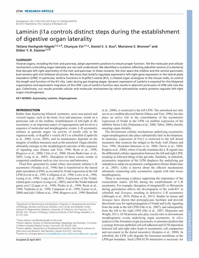
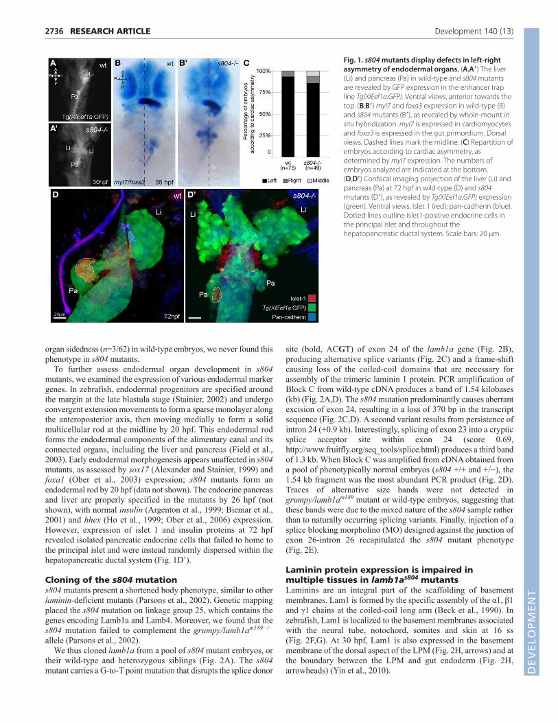
RESULTSCharacterization of s804 mutantsThe s804 mutant was identified in a forward genetic screen usingTg(XlEef1a:GFP)s854, a transgenic zebrafish line expressing greenfluorescent protein (GFP) throughout the developing endoderm(Field et al., 2003; Ober et al., 2006). The mutant exhibits lateralexpansion of the visceral organ region and loss of asymmetric L-Rpositioning of the pancreas and liver. At 30 hours post fertilization(hpf), using expression of Tg(Xla.Eef1a1:GFP) and the gut markerfoxa3 to label the entire gut primordium (Chen et al., 2001), weobserved that the gut loops to the left in wild-type embryos (95%,n=71/75) (Fig. 1A,B), but stays in the midline in s804 mutants(76%, n=37/49) (Fig. 1A�,B�). Although animals with impaired gutlaterality often exhibit defects in cardiac asymmetry (Danos andYost, 1996; Ryan et al., 1998; Bisgrove et al., 2000; Chin et al.,2000; Horne-Badovinac et al., 2003; Long et al., 2003), over 85%of s804 mutants examined showed normal cardiac asymmetry at 35hpf, as revealed by the left-sided expression of the cardiomyocytemarker myl7 (Fig. 1B,B�,C) (Yelon et al., 1999). At 72 hpf, the liverand pancreas are situated on the left and right sides of wild-typelarvae, respectively (92%, n=57/62) (Fig. 1D). In s804 mutants,however, the ventral pancreatic bud splits into bilateral organs (71%,n=26/37) and the liver spans the midline (79%, n=29/37) (Fig. 1D�)(P<0.001). Although we occasionally observed inversion of visceral D
EVELO
PMENT
2736
organ sidedness (n=3/62) in wild-type embryos, we never found thisphenotype in s804 mutants.
To further assess endodermal organ development in s804mutants, we examined the expression of various endodermal markergenes. In zebrafish, endodermal progenitors are specified aroundthe margin at the late blastula stage (Stainier, 2002) and undergoconvergent extension movements to form a sparse monolayer alongthe anteroposterior axis, then moving medially to form a solidmulticellular rod at the midline by 20 hpf. This endodermal rodforms the endodermal components of the alimentary canal and itsconnected organs, including the liver and pancreas (Field et al.,2003). Early endodermal morphogenesis appears unaffected in s804mutants, as assessed by sox17 (Alexander and Stainier, 1999) andfoxa1 (Ober et al., 2003) expression; s804 mutants form anendodermal rod by 20 hpf (data not shown). The endocrine pancreasand liver are properly specified in the mutants by 26 hpf (notshown), with normal insulin (Argenton et al., 1999; Biemar et al.,2001) and hhex (Ho et al., 1999; Ober et al., 2006) expression.However, expression of islet 1 and insulin proteins at 72 hpfrevealed isolated pancreatic endocrine cells that failed to home tothe principal islet and were instead randomly dispersed within thehepatopancreatic ductal system (Fig. 1D�).
Cloning of the s804 mutations804 mutants present a shortened body phenotype, similar to otherlaminin-deficient mutants (Parsons et al., 2002). Genetic mappingplaced the s804 mutation on linkage group 25, which contains thegenes encoding Lamb1a and Lamb4. Moreover, we found that thes804 mutation failed to complement the grumpy/lamb1am189−/−
allele (Parsons et al., 2002).We thus cloned lamb1a from a pool of s804 mutant embryos, or
their wild-type and heterozygous siblings (Fig. 2A). The s804mutant carries a G-to-T point mutation that disrupts the splice donor
RESEARCH ARTICLE Development 140 (13)
site (bold, ACGT) of exon 24 of the lamb1a gene (Fig. 2B),producing alternative splice variants (Fig. 2C) and a frame-shiftcausing loss of the coiled-coil domains that are necessary forassembly of the trimeric laminin 1 protein. PCR amplification ofBlock C from wild-type cDNA produces a band of 1.54 kilobases(kb) (Fig. 2A,D). The s804 mutation predominantly causes aberrantexcision of exon 24, resulting in a loss of 370 bp in the transcriptsequence (Fig. 2C,D). A second variant results from persistence ofintron 24 (+0.9 kb). Interestingly, splicing of exon 23 into a crypticsplice acceptor site within exon 24 (score 0.69,http://www.fruitfly.org/seq_tools/splice.html) produces a third bandof 1.3 kb. When Block C was amplified from cDNA obtained froma pool of phenotypically normal embryos (s804 +/+ and +/−), the1.54 kb fragment was the most abundant PCR product (Fig. 2D).Traces of alternative size bands were not detected ingrumpy/lamb1am189 mutant or wild-type embryos, suggesting thatthese bands were due to the mixed nature of the s804 sample ratherthan to naturally occurring splicing variants. Finally, injection of asplice blocking morpholino (MO) designed against the junction ofexon 26-intron 26 recapitulated the s804 mutant phenotype(Fig. 2E).
Laminin protein expression is impaired inmultiple tissues in lamb1as804 mutantsLaminins are an integral part of the scaffolding of basementmembranes. Lam1 is formed by the specific assembly of the α1, β1and γ1 chains at the coiled-coil long arm (Beck et al., 1990). Inzebrafish, Lam1 is localized to the basement membranes associatedwith the neural tube, notochord, somites and skin at 16 ss(Fig. 2F,G). At 30 hpf, Lam1 is also expressed in the basementmembrane of the dorsal aspect of the LPM (Fig. 2H, arrows) and atthe boundary between the LPM and gut endoderm (Fig. 2H,arrowheads) (Yin et al., 2010).
Fig. 1. s804 mutants display defects in left-rightasymmetry of endodermal organs. (A,A�) The liver(Li) and pancreas (Pa) in wild-type and s804 mutantsare revealed by GFP expression in the enhancer trapline Tg(XlEef1a:GFP). Ventral views, anterior towards thetop. (B,B�) myl7 and foxa3 expression in wild-type (B)and s804 mutants (B�), as revealed by whole-mount insitu hybridization. myl7 is expressed in cardiomyocytesand foxa3 is expressed in the gut primordium. Dorsalviews. Dashed lines mark the midline. (C) Repartition ofembryos according to cardiac asymmetry, asdetermined by myl7 expression. The numbers ofembryos analyzed are indicated at the bottom. (D,D�) Confocal imaging projection of the liver (Li) andpancreas (Pa) at 72 hpf in wild-type (D) and s804mutants (D�), as revealed by Tg(XlEef1a:GFP) expression(green). Ventral views. Islet 1 (red); pan-cadherin (blue).Dotted lines outline islet1-positive endocrine cells inthe principal islet and throughout thehepatopancreatic ductal system. Scale bars: 20 μm.
DEVELO
PMENT
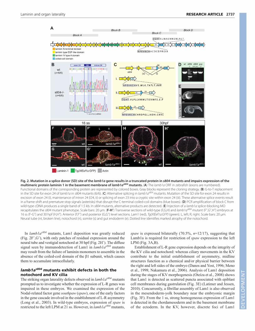
In lamb1as804 mutants, Lam1 deposition was greatly reduced(Fig. 2F�,G�), with only patches of residual expression around theneural tube and vestigial notochord at 30 hpf (Fig. 2H�). The diffusesignal seen by immunodetection of Lam1 in lamb1as804 mutantsmay result from the failure of laminin monomers to assemble in theabsence of the coiled-coil domain of the β1 subunit, which causesthem to accumulate intracellularly.
lamb1as804 mutants exhibit defects in both thenotochord and KV ciliaThe striking organ laterality defects observed in lamb1as804 mutantsprompted us to investigate whether the expression of L-R genes wasimpaired in these embryos. We examined the expression of theNodal-related factor gene southpaw (spaw), one of the early factorsin the gene cascade involved in the establishment of L-R asymmetry(Long et al., 2003). In wild-type embryos, expression of spaw isrestricted to the left LPM at 21 ss. However, in lamb1as804 mutants,
2737RESEARCH ARTICLELaminin and organ laterality
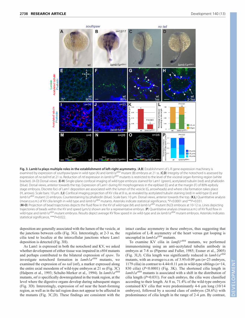
spaw is expressed bilaterally (70.5%, n=12/17), suggesting thatLamb1a is required for restriction of spaw expression to the leftLPM (Fig. 3A,B).
Establishment of L-R gene expression depends on the integrity ofthe KV cilia and notochord: whereas ciliary movements in the KVcontribute to the initial establishment of asymmetry, midlinestructures function as a chemical and/or physical barrier betweenthe right and left sides of the embryo (Danos and Yost, 1996; Menoet al., 1998; Nakamura et al., 2006). Analysis of Lam1 depositionduring the stages of KV morphogenesis (Oteíza et al., 2008) showsthat Lam1 is detected as scattered puncta associated with epiblastcell membranes during gastrulation (Fig. 3E) (Latimer and Jessen,2010). Concurrently, a fibrillar assembly of Lam1 is also observedin the mesendoderm-yolk boundary near the embryonic margin(Fig. 3F). From the 1 ss, strong homogeneous expression of Lam1is detected in the chordamesoderm and in the basement membraneof the ectoderm. In the KV, however, discrete foci of Lam1
Fig. 2. Mutation in a splice donor (SD) site of the lamb1a gene results in a truncated protein in s804 mutants and impairs expression of themultimeric protein laminin 1 in the basement membrane of lamb1as804 mutants. (A) The lamb1a ORF in zebrafish (exons are numbered).Functional domains of the corresponding protein are represented by colored boxes. Gray blocks represent the cloning strategy. (B) G-to-T replacementin the SD site for exon 24 of lamb1a in s804 mutants (6/6). (C) Alternative splicing in lamb1as804 mutants. Mutation of the SD site for exon 24 results inexcision of exon 24 (i), maintenance of intron 24 (I24, ii) or splicing of exon 23 into a cryptic site within exon 24 (iii). These alternative splice events resultin a frame-shift and premature stop signals (asterisks) that disrupt the C-terminal coiled-coil domains (blue boxes). (D) PCR amplification of block C fromwild-type cDNA produces a single band of 1.5 kb. In s804 mutants, alternative products are detected. (E) Injection of a lamb1a splice-blocking MOrecapitulates the s804 mutant phenotype. Scale bars: 20 μm. (F-H�) Transverse sections of wild-type (F,G,H) and lamb1as804 mutant (F�,G�,H�) embryos at16 ss (F-G�) and 30 hpf (H,H�). Anterior (F,F�) and posterior (G,G�) level sections. Lam1 (red), Tg(XIEef1a:GFP) (green). L, left; R, right. Scale bars: 20 μm.Neural tube (nt, broken line), notochord (n), somite (s) and gut endoderm (e). Dotted line identifies marked atrophy of the notochord.
DEVELO
PMENT
2738
deposition are generally associated with the lumen of the vesicle, atthe junctions between cells (Fig. 3G). Interestingly, at 2-3 ss, thecilia tend to localize at the intercellular junctions where Lam1deposition is detected (Fig. 3H).
As Lam1 is expressed in both the notochord and KV, we askedwhether development of either tissue was impaired in s804 mutantsand perhaps contributed to the bilateral expression of spaw. Toinvestigate notochord formation in lamb1as804 mutants, weexamined the expression of no tail (ntl), a marker expressed alongthe entire axial mesoderm of wild-type embryos at 21 ss (Fig. 3C)(Halpern et al., 1993; Schulte-Merker et al., 1994). In lamb1as804
mutants, ntl is specifically downregulated in the trunk region, at thelevel where the digestive organs develop during subsequent stages(Fig. 3D). Interestingly, expression of ntl near the heart-formingregion, as well as in the tail region does not appear to be affected inthe mutants (Fig. 3C,D). These findings are consistent with the
RESEARCH ARTICLE Development 140 (13)
intact cardiac asymmetry in these embryos, thus suggesting thatregulation of L-R asymmetry of the heart versus gut looping isuncoupled in lamb1as804 mutants.
To examine KV cilia in lamβ1s804 mutants, we performedimmunostaining using an anti-acetylated tubulin antibody inembryos at 7-8 ss (Piperno and Fuller, 1985; Essner et al., 2005)(Fig. 3I,J). Cilia length was significantly reduced in lamb1as804
mutants, with an average±s.e.m. of 3.95±0.09 µm (n=23 embryos,1179 cilia) in contrast to 4.44±0.11 µm in wild-type siblings (n=14,830 cilia) (P<0.0001) (Fig. 3K). The shortened cilia length inlamb1as804 mutants is associated with a shift in the distribution ofcilia length (P=0.031). For each embryo, the cilia were classifiedaccording to their length. At 8 ss, 71.4% of the wild-type embryoscontained KV cilia that were predominantly 4-6 µm long (10/14embryos), followed by a second class of embryos (28.6%) withpredominance of cilia length in the range of 2-4 µm. By contrast,
Fig. 3. Lamb1a plays multiple roles in the establishment of left-right asymmetry. (A,B) Establishment of L-R gene expression machinery isexamined by expression of southpaw/spaw in wild-type (A) and lamb1as804 mutant (B) embryos at 21 ss. (C,D) Integrity of the notochord is assessed byexpression of no tail/ntl at 21 ss. Reduction of ntl expression in lamb1as804 mutants is restricted to the level of the visceral organ-forming region (whitebracket). (A-D) Dorsal views. (E-H) Single-plane confocal imaging of wild-type embryos stained for Lam1 (green), acetylated tubulin (red) and phalloidin(blue). Dorsal views, anterior towards the top. Expression of Lam1 during KV morphogenesis in the epiblast (E) and at the margin (F) of 80% epibolystage embryos. Discrete foci of Lam1 deposition are associated with the lumen of the vesicle (G, arrowheads) and where cilia formation takes place (H, arrows). Scale bars: 10 μm. (I,J) Confocal imaging projection of KV cilia at 8 ss, as revealed by acetylated tubulin staining (red) in wild-type (I) andlamb1as804 mutant (J) embryos. Counterstaining by phalloidin (blue). Scale bars: 10 μm. Dorsal views, anterior towards the top. (K,L) Quantitative analysis(mean±s.e.m.) of KV cilia length in wild type and lamb1as804 mutants. Asterisks indicate statistical significance, *P<0.0001 and **P=0.031. (M-O) Projection of bead trajectories depicts the fluid flow in the KV of wild-type (M) and lamb1as804 mutant (N,O) embryos at 10-12 ss. Lines depictingtrajectories of beads within the KV and speed (μm/s) shown are for a representative embryo. (P) Quantitative analysis (mean±s.e.m.) of KV fluid flow inwild-type and lamb1as804 mutant embryos. Results depict average KV flow speed in six wild-type and six lamb1as804 mutant embryos. Asterisks indicatesstatistical significance, ***P=0.022.
DEVELO
PMENT
cilia length was predominantly shorter (2-4 µm) in 65% of thelamb1as804 mutants examined (n=15/23), with the other 35% of theembryos (n=8/23) showing cilia length mainly in the range of 4-6 µm (Fig. 3L). Overall cilia number was not significantly affectedin lamb1as804 mutants (data not shown).
To determine whether this difference in the KV cilia lengthaffected their function in lamb1as804 mutants, we examined thegeneration of fluid flow in the KV of wild-type and mutant embryos.Analysis of the dynamics of fluorescent beads injected into the KVat 8-10 ss showed that deficiency in Lamb1a expression did not haltfluid flow in the KV and that directionality was also largelypreserved in the mutants (data not shown). Instead, we observedvariable defects in the size of the KV and the speed of flow in themutants (Fig. 3M-P). In all the mutants, tracking of the beadsshowed more erratic trajectories and increased tendency to cross thecenter of the vesicle (Fig. 3N), in contrast to the regular pattern offluid flow in the KV of wild-type embryos, where the beads mostlyconcentrated in the periphery of the vesicle (n=5/6) (Fig. 3M). In50% of the lamb1as804 mutant embryos analyzed (n=3/6), the KVappeared smaller, yet the average flow speed (7.99±0.13 µm/s) wasnot significantly affected, when compared with the averagetraveling speed of 7.65±0.11 µm/s (average±s.e.m.) measured inwild-type and heterozygous embryos (n=6). In one-third of themutants analyzed (n=2/6), whereas the KV size was normal, thespeed of fluid flow was reduced, with an average speed of4.58±0.11 µm/s (Fig. 3O). Overall, our analysis of the KV fluid flowindicates that the average flow speed in wild-type embryos rangesbetween 6 and 10 µm/s (n=6). By contrast, in lamb1as804 mutant
2739RESEARCH ARTICLELaminin and organ laterality
embryos, we observed increased prevalence of embryos withreduced flow (average speed of 4-5 µm/s) (P=0.02) (Fig. 3P).
Taken together, our analyses suggest that loss of Lamβ1 functionin lamb1as804 mutants impairs the integrity of the KV cilia andnotochord, both of which are essential for L-R gene expression.
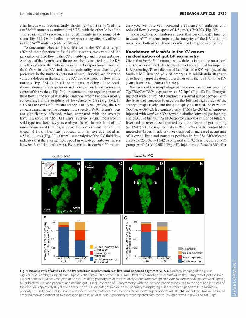
Knockdown of lamb1a in the KV causesrandomization of gut L-R asymmetryGiven that lamba1s804 mutants show defects in both the notochordand KV, we examined which defect directly accounted for impairedL-R patterning. To test the role of Lamb1a in the KV, we injected thelamb1a MO into the yolk of embryos at midblastula stages tospecifically target the dorsal forerunner cells that will form the KV(Amack and Yost, 2004) (Fig. 4A).
We assessed the morphology of the digestive organs based onTg(XlEef1a:GFP) expression at 52 hpf (Fig. 4B-E). Embryosinjected with control MO displayed a normal gut phenotype, withthe liver and pancreas located on the left and right sides of theembryo, respectively, and the gut displaying an S-shape curvature(85.7%, n=36/42). By contrast, only 47.6% (n=20/42) of embryosinjected with lamb1a MO showed a similar leftward gut looping,and 28.6% of the lamb1a MO-injected embryos exhibited bilateralliver and pancreas accompanied by the absence of gut looping(n=12/42) when compared with 4.8% (n=2/42) of the control MO-injected embryos. In addition, we observed an increased occurrenceof inverted liver and pancreas position in lamb1a MO-injectedembryos (23.8%, n=10/42), compared with 9.5% in the control MOgroup (n=4/42) (P=0.001) (Fig. 4F). Injections of lamb1a MO after
Fig. 4. Knockdown of lamb1a in the KV results in randomization of liver and pancreas asymmetry. (A-E) Confocal imaging of the gut inTg(XlEef1a:GFP) embryos injected at 3 hpf (A) with control (B) or lamb1a (C-E) MO. Effect of KV knockdown of lamb1a on the L-R asymmetry of the liver(Li) and pancreas (Pa) was analyzed at 52 hpf. Resulting phenotypes of the liver and pancreas after KV-specific lamb1a knockdown include: wild type (C,blue); bilateral liver and pancreas and midline gut (D, red); inversion of L-R asymmetry, with the liver and pancreas localized to the right and left sides ofthe embryo, respectively (E, yellow). Ventral views. (F) Percentages (mean±s.e.m.) of embryos displaying distinct liver and pancreas L-R asymmetryphenotypes. Forty-two embryos were analyzed for each treatment. Asterisks indicate statistical significance, *P=0.001. (G) Percentages (mean±s.e.m.) ofembryos showing distinct spaw expression patterns at 20 ss. Wild-type embryos were injected with control (n=28) or lamb1a (n=36) MO at 3 hpf. D
EVELO
PMENT
2740
midblastula stages did not significantly affect L-R asymmetry ofthe liver and pancreas.
Knocking down lamb1a in the KV led to randomization of the gutlaterality, rather than failure of gut looping, as in most lamb1as804
mutants. Whereas the majority of lamb1as804 mutants showedbilateral spaw expression during somitogenesis (Fig. 3B), sidednessof spaw expression after lamb1a knockdown in the KV was ratherrandom, with 44% of the embryos showing normal left-sided spawexpression (Fig. 4G). These data suggest that lamb1a deficiency inthe KV alone only partially impairs L-R gene expression and thatlamb1a also functions in other tissues, probably the notochord, toregulate L-R gene expression. Interestingly, lamb1a knockdown inthe KV resulted in a significant increase of inverted liver andpancreas phenotype. However, complete inversion of liver andpancreas sidedness was never observed in lamb1as804 mutants,despite bilateral expression of spaw. The differences betweenlamb1as804 mutants and embryos with lamb1a knockdown in theKV thus suggested that dysregulation of L-R gene expression alonecould not account for the gut-looping defects in the lamb1as804
mutants.
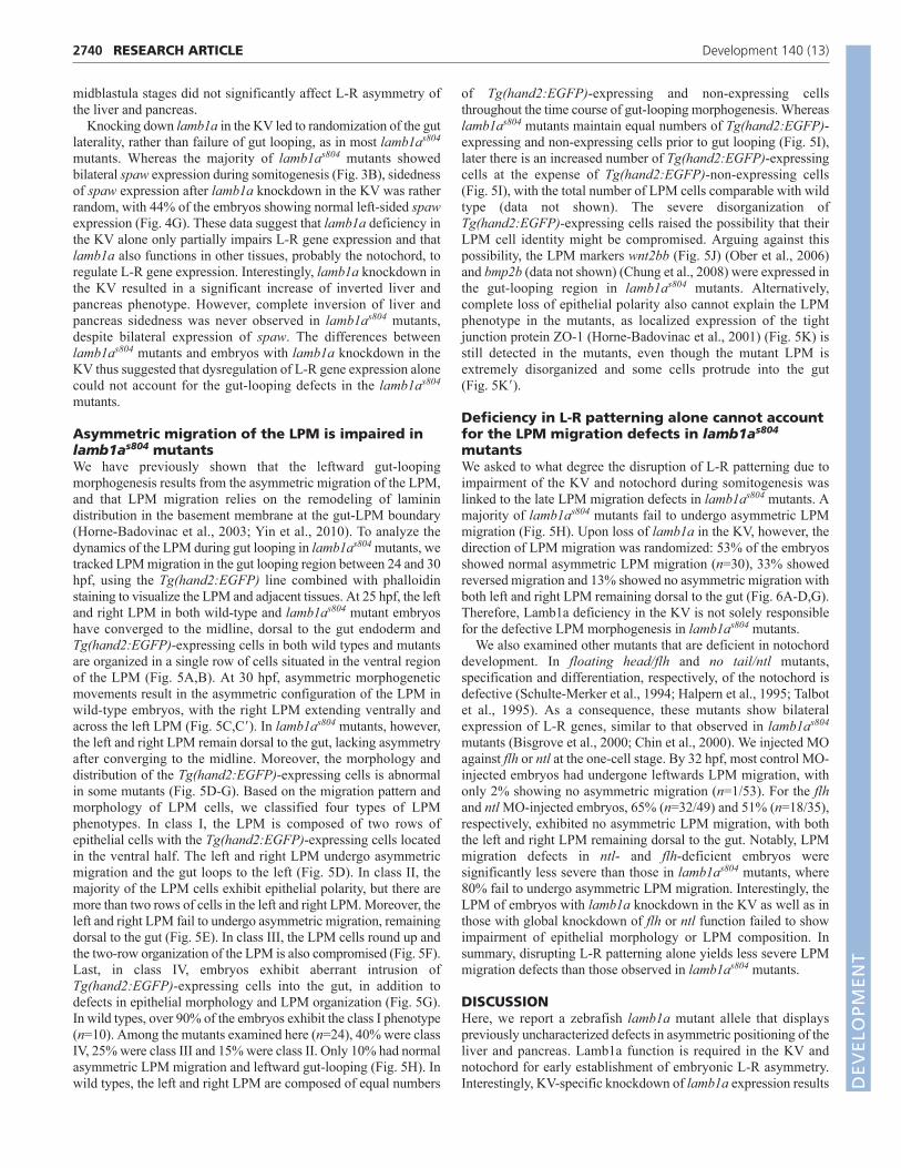
Asymmetric migration of the LPM is impaired inlamb1as804 mutantsWe have previously shown that the leftward gut-loopingmorphogenesis results from the asymmetric migration of the LPM,and that LPM migration relies on the remodeling of laminindistribution in the basement membrane at the gut-LPM boundary(Horne-Badovinac et al., 2003; Yin et al., 2010). To analyze thedynamics of the LPM during gut looping in lamb1as804 mutants, wetracked LPM migration in the gut looping region between 24 and 30hpf, using the Tg(hand2:EGFP) line combined with phalloidinstaining to visualize the LPM and adjacent tissues. At 25 hpf, the leftand right LPM in both wild-type and lamb1as804 mutant embryoshave converged to the midline, dorsal to the gut endoderm andTg(hand2:EGFP)-expressing cells in both wild types and mutantsare organized in a single row of cells situated in the ventral regionof the LPM (Fig. 5A,B). At 30 hpf, asymmetric morphogeneticmovements result in the asymmetric configuration of the LPM inwild-type embryos, with the right LPM extending ventrally andacross the left LPM (Fig. 5C,C�). In lamb1as804 mutants, however,the left and right LPM remain dorsal to the gut, lacking asymmetryafter converging to the midline. Moreover, the morphology anddistribution of the Tg(hand2:EGFP)-expressing cells is abnormalin some mutants (Fig. 5D-G). Based on the migration pattern andmorphology of LPM cells, we classified four types of LPMphenotypes. In class I, the LPM is composed of two rows ofepithelial cells with the Tg(hand2:EGFP)-expressing cells locatedin the ventral half. The left and right LPM undergo asymmetricmigration and the gut loops to the left (Fig. 5D). In class II, themajority of the LPM cells exhibit epithelial polarity, but there aremore than two rows of cells in the left and right LPM. Moreover, theleft and right LPM fail to undergo asymmetric migration, remainingdorsal to the gut (Fig. 5E). In class III, the LPM cells round up andthe two-row organization of the LPM is also compromised (Fig. 5F).Last, in class IV, embryos exhibit aberrant intrusion ofTg(hand2:EGFP)-expressing cells into the gut, in addition todefects in epithelial morphology and LPM organization (Fig. 5G).In wild types, over 90% of the embryos exhibit the class I phenotype(n=10). Among the mutants examined here (n=24), 40% were classIV, 25% were class III and 15% were class II. Only 10% had normalasymmetric LPM migration and leftward gut-looping (Fig. 5H). Inwild types, the left and right LPM are composed of equal numbers
RESEARCH ARTICLE Development 140 (13)
of Tg(hand2:EGFP)-expressing and non-expressing cellsthroughout the time course of gut-looping morphogenesis. Whereaslamb1as804 mutants maintain equal numbers of Tg(hand2:EGFP)-expressing and non-expressing cells prior to gut looping (Fig. 5I),later there is an increased number of Tg(hand2:EGFP)-expressingcells at the expense of Tg(hand2:EGFP)-non-expressing cells(Fig. 5I), with the total number of LPM cells comparable with wildtype (data not shown). The severe disorganization ofTg(hand2:EGFP)-expressing cells raised the possibility that theirLPM cell identity might be compromised. Arguing against thispossibility, the LPM markers wnt2bb (Fig. 5J) (Ober et al., 2006)and bmp2b (data not shown) (Chung et al., 2008) were expressed inthe gut-looping region in lamb1as804 mutants. Alternatively,complete loss of epithelial polarity also cannot explain the LPMphenotype in the mutants, as localized expression of the tightjunction protein ZO-1 (Horne-Badovinac et al., 2001) (Fig. 5K) isstill detected in the mutants, even though the mutant LPM isextremely disorganized and some cells protrude into the gut(Fig. 5K�).
Deficiency in L-R patterning alone cannot accountfor the LPM migration defects in lamb1as804
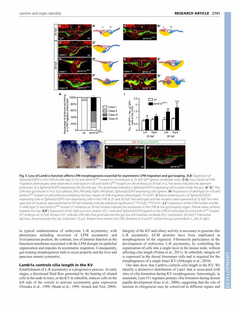
mutantsWe asked to what degree the disruption of L-R patterning due toimpairment of the KV and notochord during somitogenesis waslinked to the late LPM migration defects in lamb1as804 mutants. Amajority of lamb1as804 mutants fail to undergo asymmetric LPMmigration (Fig. 5H). Upon loss of lamb1a in the KV, however, thedirection of LPM migration was randomized: 53% of the embryosshowed normal asymmetric LPM migration (n=30), 33% showedreversed migration and 13% showed no asymmetric migration withboth left and right LPM remaining dorsal to the gut (Fig. 6A-D,G).Therefore, Lamb1a deficiency in the KV is not solely responsiblefor the defective LPM morphogenesis in lamb1as804 mutants.
We also examined other mutants that are deficient in notochorddevelopment. In floating head/flh and no tail/ntl mutants,specification and differentiation, respectively, of the notochord isdefective (Schulte-Merker et al., 1994; Halpern et al., 1995; Talbotet al., 1995). As a consequence, these mutants show bilateralexpression of L-R genes, similar to that observed in lamb1as804
mutants (Bisgrove et al., 2000; Chin et al., 2000). We injected MOagainst flh or ntl at the one-cell stage. By 32 hpf, most control MO-injected embryos had undergone leftwards LPM migration, withonly 2% showing no asymmetric migration (n=1/53). For the flhand ntl MO-injected embryos, 65% (n=32/49) and 51% (n=18/35),respectively, exhibited no asymmetric LPM migration, with boththe left and right LPM remaining dorsal to the gut. Notably, LPMmigration defects in ntl- and flh-deficient embryos weresignificantly less severe than those in lamb1as804 mutants, where80% fail to undergo asymmetric LPM migration. Interestingly, theLPM of embryos with lamb1a knockdown in the KV as well as inthose with global knockdown of flh or ntl function failed to showimpairment of epithelial morphology or LPM composition. Insummary, disrupting L-R patterning alone yields less severe LPMmigration defects than those observed in lamb1as804 mutants.
DISCUSSIONHere, we report a zebrafish lamb1a mutant allele that displayspreviously uncharacterized defects in asymmetric positioning of theliver and pancreas. Lamb1a function is required in the KV andnotochord for early establishment of embryonic L-R asymmetry.Interestingly, KV-specific knockdown of lamb1a expression results D
EVELO
PMENT
in typical randomization of embryonic L-R asymmetry, withphenotypes including inversion of LPM asymmetry andliver/pancreas position. By contrast, loss of laminin function in thebasement membrane associated with the LPM disrupts its epithelialorganization and impedes its asymmetric migration. Consequently,gut-looping morphogenesis fails to occur properly and the liver andpancreas remain symmetric.
Lamb1a controls cilia length in the KVEstablishment of L-R asymmetry is a progressive process. At earlystages, a directional fluid flow generated by the beating of ciliatedcells in the node in mice, or the KV in zebrafish, induces cells on theleft side of the vesicle to activate asymmetric gene expression(Nonaka et al., 1998; Okada et al., 1999; Amack and Yost, 2004).
2741RESEARCH ARTICLELaminin and organ laterality
Integrity of the KV and ciliary activity is necessary to generate thisL-R asymmetry. ECM proteins have been implicated inmorphogenesis of the organizer. Fibronectin participates in thedevelopment of embryonic L-R asymmetry, by controlling theorganization of cells into a single layer in the mouse node, withoutaffecting cilia length (Pulina et al., 2011). In zebrafish, integrin α5is expressed in the dorsal forerunner cells and is required for themorphogenesis of a single intact KV (Ablooglu et al., 2010).
Our data show that Lamb1a controls cilia length in the KV. Weidentify a distinctive distribution of Lam1 that is associated withsites of cilia formation during KV morphogenesis. Interestingly, inmammals, Lam-511 regulates primary cilia formation during dermalpapilla development (Gao et al., 2008), suggesting that the role oflaminin in ciliogenesis may be conserved in different organs and
Fig. 5. Loss of Lamb1a function affects LPM morphogenesis essential for asymmetric LPM migration and gut looping. (A,B) Expression ofTg(hand2:EGFP) in the LPM of wild-type (n=5) and lamb1as804 mutant (n=5) embryos at 25 hpf. GFP (green), phalloidin (red). (C-G) Four classes of LPMmigration phenotypes were observed in wild-type (n=10) and lamb1as804 mutant (n=24) embryos at 30 hpf. In G, the arrow indicates the aberrantprotrusion of a Tg(hand2:EGFP)-expressing cell into the gut. The arrowhead indicates a Tg(hand2:EGFP)-expressing cell located inside the gut. (A�-G�) TheLPM and gut shown in A-G. Gut (yellow); left LPM (red); right LPM (blue); Tg(hand2:EGFP)-expressing cells (green). (H) Proportions of wild-type (n=10) andlamb1as804 mutant (n=24) embryos exhibiting the four classes of LPM migration phenotypes; *P<0.001. (I) Ratios (mean±s.e.m.) of Tg(hand2:EGFP)-expressing cells to Tg(hand2:EGFP)-non-expressing cells in the LPM at 25 and 30 hpf. Five wild-type and five mutants were examined at 25 hpf. Ten wild-type and 24 mutants were examined at 30 hpf. Asterisks indicate statistical significance: **P<0.05; ***P<0.01. (J,J�) Expression of the LPM marker wnt2bbin wild-type (J) and lamb1as804 mutant (J�) embryos at 30 hpf. Arrows indicate the expression in the LPM at the gut-looping region. Dorsal views, anteriortowards the top. (K,K�) Expression of the tight junction protein ZO-1 (red) and Tg(hand2:EGFP) (green) in the LPM of wild-type (K) and lamb1as804 mutant(K�) embryos at 32 hpf. Arrows in K� indicate LPM cells that protrude into the gut but still maintain localized ZO-1 expression. (A-G,K,K�) Transversesections, dorsal towards the top. Scale bars: 10 μm. Broken lines outline the LPM. Shadows in K and K� outline the gut primordium. L, left; R, right.
DEVELO
PMENT
2742
across species. Yet the mechanism by which Lamb1a modulatescilia formation is not clear. Studies of primary cilia of chickchondroblasts indicate that the ECM directly signals to cells viaintegrins α2, α3 and β1, which anchor the cilium to collagen fiberswithin the ECM (Jensen et al., 2004; McGlashan et al., 2006). It ispossible that Lamb1a is involved in establishing the cellularproperty of the ciliated cells essential for cilia formation.
Previous studies in zebrafish have identified signaling moleculesthat regulate KV cilia formation. For example, defects in cilia lengthassociated with deficiency in FGF or Notch signaling pathwayshave been described to affect fluid flow in the KV, whichconsequently results in defects in L-R asymmetry (Albertson andYelick, 2005; Neugebauer et al., 2009; Yamauchi et al., 2009; Lopeset al., 2010). Activation of Wnt/β-catenin signaling results in loss ofcardiac asymmetry, and affects morphology of the KV, with largerKV and increased cilia number. By contrast, reduction of Wnt3 orWnt8 results in shorter cilia and randomization of spaw expression,with effects on both heart and digestive organs sidedness (Nakayaet al., 2005; Lin and Xu, 2009; Caron et al., 2012). In addition, theRho kinase Rock2b controls anterior-posterior asymmetricplacement of ciliated cells in the KV, and subsequently L-Rpatterning of the embryo (Wang et al., 2011). In lamb1as804 mutants,we observed that impaired cilia length and movements partiallycontribute to defects in L-R patterning, resulting in variable defectsin the dynamics or directionality of the fluid flow in the KV. It isintriguing to speculate that Lamb1a and its integrin receptors mightserve as mediators of the signaling pathways described above, tomodulate cilia formation.
Regulation of notochord differentiation byLamb1a is crucial for establishment of L-R geneexpressionAfter initiation of left-sided signaling, the notochord generates amidline barrier that restricts the cascade of gene expression to theleft side of the embryo (Lee and Anderson, 2008). Mutants defective
RESEARCH ARTICLE Development 140 (13)
in notochord formation often exhibit randomization of organlaterality (Danos and Yost, 1996). The notochord of lamb1as804
mutants appears atrophic and collapsed, consistent with the role oflaminins in the formation of this structure. In zebrafish, lamininsubunits β1 and γ1 are essential for establishment of the peri-notochordal basement membrane and organization of the notochordsheath, which provides physical resistance against the pressureexerted by the inflated notochord vacuoles (Parsons et al., 2002).Importantly, differentiation of the notochord is also impaired in thelamb1a and lamg1 mutants, suggesting that there may be signalsemanating from the ECM in the basement membrane to regulatechordamesoderm differentiation (Stemple, 2005).
Analysis of ntl expression showed that the notochord defect inlamb1as804 mutants does not distribute along its full extension, butrather is restricted to the liver and pancreas-forming region.Coincident with the intact ntl expression in the more anterior regionof the notochord, heart sidedness was not affected in the lamb1as804
mutants. The independent phenotypes of the heart and gut lateralityin lamb1as804 mutants suggest that heterotaxy of organ lateralitycaused by midline defects is dependent on distinct domains alongthe anteroposterior axis (Bisgrove et al., 2000; Chin et al., 2000).Moreover, our observation that fewer ntl- and flh-deficient embryosfail to undergo asymmetric LPM migration compared withlamb1as804 mutants indicates that impairment in the notochord alonecannot account for the gut laterality defects seen in lamb1as804
mutants.
Localized expression of Lam1 controls asymmetricmigration of the LPMAsymmetric migration of the left and right LPM is essential forleftward gut-looping morphogenesis (Horne-Badovinac et al.,2003). Lam1 is expressed in a dynamic pattern at the interfacebetween the LPM and gut endoderm during LPM migration, andremodeling of the ECM has been associated with establishment ofproper contacts between these tissues, which are necessary to drive
Fig. 6. lamb1a knock-down in the KV aswell as global knock-down of flh andntl randomized the direction of LPMmigration without altering epithelialpolarity or composition of the LPM.(A-F) Expression of Tg(hand2:EGFP) in theLPM of embryos injected with control (A)or lamb1a (B-D) MO into the KV at 3 hpf, orflh MO (E) or ntl MO (F) at the one-cellstage. Transverse sections, dorsal towardsthe top. Scale bar: 10 μm. Dashed linesoutline the LPM. L, left; R, right. (A�-F�) TheLPM and gut shown in A-D. Gut (yellow),left LPM (red), right LPM (blue),Tg(hand2:EGFP)-expressing cells (green).(G) Proportions of control, lamb1a, flh andntl MO-injected embryos exhibitingdifferent directions of LPM migration.Effect of lamb1a knockdown in the KV onLPM migration is statistically significant,when compared with control, flh and ntlmorphants (*P<0.001). The distribution ofLPM migration phenotype is similar in flhand ntl morphants (P=0.794), butsignificantly different in embryos injectedwith lamb1a MO in the KV or with thecontrol MO.
DEVELO
PMENT
morphogenetic movements (Yin et al., 2010). lamb1as804 mutantslack the coiled-coil domain of the β1 chain, and expression of Lam1protein is detected as intracellular speckles, instead of the discretecontinuous expression observed in the basement membranes ofwild-type embryos. This observation is consistent with the modelproposed for assembly and secretion of Lam1, in which secretion ofthe functional trimeric laminin requires a first step of intracellulardimerization of subunits β1 and γ1, followed by assembly with anα1 chain (Peters et al., 1985; Kammerer et al., 1995; Kumagai etal., 1997; Yurchenco et al., 1997; Goto et al., 2001; Urbano et al.,2009). In the absence of proper laminin deposition, the left and rightLPM fail to undergo asymmetric migration, and remain aligneddorsal to the gut.
By tracking the LPM migration at cellular resolution, weuncovered distinctive phenotypes in the LPM of lamb1as804 mutantsthat may account for the failure of LPM migration. Laminins havebeen implicated in the establishment of cell polarization inembryonic cells (Li et al., 2003). Establishment of epithelial polarityin the LPM is absolutely essential for asymmetric LPM migrationand gut looping (Horne-Badovinac et al., 2003; Yin et al., 2010).Mutant LPM cells are correctly specified and the organization ofthe LPM is unaffected prior to LPM migration. However, as theLPM migrates to the midline, in majority of lamb1as804 mutants theLPM failed to align into two rows of cells and its epithelialarchitecture is compromised. Interestingly, we detected localizedexpression of ZO-1 in mutant LPM cells, indicating that theirepithelial polarity is not completely abolished. In the absence oflaminin in the basement membrane, the mutant LPM cells may bepartially depolarized and undergo epithelial-to-mesenchymaltransition, contributing to the failure of LPM migration.
In addition to the defects in epithelial cell morphology, thecomposition of the LPM is impaired in lamb1as804 mutants, asdemonstrated by an increased number of Tg(hand2:EGFP)-expressing cells at the expense of the non-expressing cells. We alsofound that in some of lamb1as804 mutants, the LPM cells aberrantlyprotruded into the gut. During gut-looping morphogenesis, the LPMcells coordinate with one another and migrate as a coherent sheet.In lamb1as804 mutants, the lack of basement membrane at the LPM-gut boundary allows some LPM cells to escape from the LPM andprotrude into the gut. Such cell behavior could disrupt the normalcommunication between the LPM cells and impede the collectivemigration of the LPM. An intriguing possibility is that the basementmembrane may either sequester or mediate the migration cuessecreted from the LPM or the gut endoderm. In lamb1as804 mutants,such regulation of migration cues was absent due to the deficiencyin the basement membrane. Supporting this notion, a recent reporton L-R asymmetry in Xenopus showed that the ECM is a principalsurface of Nodal and Lefty accumulation and that sulfatedproteoglycans facilitate long distance movement of Nodal(Marjoram and Wright, 2011). Similarly, in Drosophila and mouse,ECM components have been implicated in facilitating transport ofBMPs, FGF and Nodal (García-García and Anderson, 2003;Belenkaya et al., 2004; Oki et al., 2007).
Our results indicate that deficiency of Lamβ1 disrupts both the L-R patterning during somitogenesis and the asymmetric LPMmigration during later development. KV-specific knock-down oflamb1a randomized L-R patterning and the directionality of LPMmigration. However, it did not impair epithelial polarity orcomposition of the LPM. Despite these observations, early and lateroles of Lamb1a in organ laterality cannot be completely uncoupled.Unlike embryos with Lamb1a deficiency in the KV, lamb1as804
mutants show bilateral expression of spaw in the LPM during
2743RESEARCH ARTICLELaminin and organ laterality
somitogenesis. Whether such aberrant expression of spaw has anyimpact on LPM polarity and composition is not yet clear.
Lamb1a is necessary for organization of pancreasand liver architectureIn addition to defects in L-R organ asymmetry, the architecture ofthe liver and pancreas is also affected in lamb1as804 mutants.Diminished expression of laminin affects the adhesion of hepaticand pancreatic cells, as well as the integrity of other endodermal-derived tissues. This phenotype is consistent with a crucial role forbasement membranes in mediating cell adhesion and tissueorganization (Li et al., 2003). Moreover, in the mutant pancreas anincreased number of endocrine cells are associated with thehepatopancreatic duct, suggesting that the lack of laminin mightaffect the homing of newly differentiated endocrine cells. Lamininsand cell surface-associated ECM proteins of the basementmembranes are essential for the survival and in vitro function ofpancreatic islets and liver cells (Stamatoglou and Hughes, 1994;Nagaki et al., 1995; Paraskevas et al., 2000; Schroeder et al., 2006);however, their role in organogenesis is not clear. Understandingtheir normal function in development and their link to signalingpathways is crucial for their future use in a therapeutic context.
AcknowledgementsWe thank Elke Ober, Holly Field, Heather Verkade and other members of theENU screen team, as well as Chantilly (Munson) Apollon for sharing reagentsand expertise in LR organ asymmetry. We thank Mike Parsons for the grumpyallele and Thai Truong for assistance with settings for live imaging of the KVflow. Thanks to Zhaoxia Sun for sharing before publication her protocol for theanalysis of KV flow, and to Pablo Oteíza and Hiroaki Ishikawa for advice.
FundingT.H.-H. was supported by a Pew Latin American Fellowship in the BiomedicalSciences and by a California Institute for Regenerative Medicine Training Grant[T2-00006]. C.Y. is supported by an National Institutes of Health (NIH) K99Award [AA020514], a University of California at San Francisco Liver CenterPilot/Feasibility Award [NIH P30DK026743] and the Cincinnati Children’sHospital Research Foundation. This work was supported in part by grants fromthe NIH [P50 HG004071 to M.E.B., R01DK060322 to D.Y.R.S.] and from thePackard Foundation (to D.Y.R.S.). Deposited in PMC for release after 12months.
Author contributionsT.H.-H. and C.Y. designed and performed experiments, analyzed data andwrote the manuscript. D.E.S.K. performed experiments, analyzed data andedited the manuscript. M.E.B. and D.Y.R.S. supervised the work, includinghelping with experimental design, data, analysis and manuscript preparation.
ReferencesAblooglu, A. J., Tkachenko, E., Kang, J. and Shattil, S. J. (2010). Integrin
alphaV is necessary for gastrulation movements that regulate vertebrate bodyasymmetry. Development 137, 3449-3458.
Albertson, R. C. and Yelick, P. C. (2005). Roles for fgf8 signaling in left-rightpatterning of the visceral organs and craniofacial skeleton. Dev. Biol. 283, 310-321.
Alexander, J. and Stainier, D. Y. (1999). A molecular pathway leading toendoderm formation in zebrafish. Curr. Biol. 9, 1147-1157.
Amack, J. D. and Yost, H. J. (2004). The T box transcription factor no tail inciliated cells controls zebrafish left-right asymmetry. Curr. Biol. 14, 685-690.
Argenton, F., Zecchin, E. and Bortolussi, M. (1999). Early appearance ofpancreatic hormone-expressing cells in the zebrafish embryo. Mech. Dev. 87,217-221.
Beck, K., Hunter, I. and Engel, J. (1990). Structure and function of laminin:anatomy of a multidomain glycoprotein. FASEB J. 4, 148-160.
Belenkaya, T. Y., Han, C., Yan, D., Opoka, R. J., Khodoun, M., Liu, H. and Lin,X. (2004). Drosophila Dpp morphogen movement is independent ofdynamin-mediated endocytosis but regulated by the glypican members ofheparan sulfate proteoglycans. Cell 119, 231-244.
Biemar, F., Argenton, F., Schmidtke, R., Epperlein, S., Peers, B. and Driever,W. (2001). Pancreas development in zebrafish: early dispersed appearance ofendocrine hormone expressing cells and their convergence to form thedefinitive islet. Dev. Biol. 230, 189-203. D
EVELO
PMENT

2744 RESEARCH ARTICLE Development 140 (13)
Bisgrove, B. W., Essner, J. J. and Yost, H. J. (1999). Regulation of midlinedevelopment by antagonism of lefty and nodal signaling. Development 126,3253-3262.
Bisgrove, B. W., Essner, J. J. and Yost, H. J. (2000). Multiple pathways in themidline regulate concordant brain, heart and gut left-right asymmetry.Development 127, 3567-3579.
Branford, W. W., Essner, J. J. and Yost, H. J. (2000). Regulation of gut and heartleft-right asymmetry by context-dependent interactions between xenopuslefty and BMP4 signaling. Dev. Biol. 223, 291-306.
Campione, M., Steinbeisser, H., Schweickert, A., Deissler, K., van Bebber, F.,Lowe, L. A., Nowotschin, S., Viebahn, C., Haffter, P., Kuehn, M. R. et al.(1999). The homeobox gene Pitx2: mediator of asymmetric left-right signalingin vertebrate heart and gut looping. Development 126, 1225-1234.
Capdevila, J., Vogan, K. J., Tabin, C. J. and Izpisúa Belmonte, J. C. (2000).Mechanisms of left-right determination in vertebrates. Cell 101, 9-21.
Caron, A., Xu, X. and Lin, X. (2012). Wnt/β-catenin signaling directly regulatesFoxj1 expression and ciliogenesis in zebrafish Kupffer’s vesicle. Development139, 514-524.
Chen, J. N., van Bebber, F., Goldstein, A. M., Serluca, F. C., Jackson, D.,Childs, S., Serbedzija, G., Warren, K. S., Mably, J. D., Lindahl, P. et al.(2001). Genetic steps to organ laterality in zebrafish. Comp. Funct. Genomics 2,60-68.
Chin, A. J., Tsang, M. and Weinberg, E. S. (2000). Heart and gut chiralities arecontrolled independently from initial heart position in the developingzebrafish. Dev. Biol. 227, 403-421.
Chung, W. S., Shin, C. H. and Stainier, D. Y. (2008). Bmp2 signaling regulatesthe hepatic versus pancreatic fate decision. Dev. Cell 15, 738-748.
Clanton, J. A., Hope, K. D. and Gamse, J. T. (2013). Fgf signaling governs cellfate in the zebrafish pineal complex. Development 140, 323-332.
Collignon, J., Varlet, I. and Robertson, E. J. (1996). Relationship betweenasymmetric nodal expression and the direction of embryonic turning. Nature381, 155-158.
Danos, M. C. and Yost, H. J. (1996). Role of notochord in specification of cardiacleft-right orientation in zebrafish and Xenopus. Dev. Biol. 177, 96-103.
Davis, N. M., Kurpios, N. A., Sun, X., Gros, J., Martin, J. F. and Tabin, C. J.(2008). The chirality of gut rotation derives from left-right asymmetric changesin the architecture of the dorsal mesentery. Dev. Cell 15, 134-145.
Engel, J., Odermatt, E., Engel, A., Madri, J. A., Furthmayr, H., Rohde, H. andTimpl, R. (1981). Shapes, domain organizations and flexibility of laminin andfibronectin, two multifunctional proteins of the extracellular matrix. J. Mol. Biol.150, 97-120.
Essner, J. J., Branford, W. W., Zhang, J. and Yost, H. J. (2000). Mesendodermand left-right brain, heart and gut development are differentially regulated bypitx2 isoforms. Development 127, 1081-1093.
Essner, J. J., Amack, J. D., Nyholm, M. K., Harris, E. B. and Yost, H. J. (2005).Kupffer’s vesicle is a ciliated organ of asymmetry in the zebrafish embryo thatinitiates left-right development of the brain, heart and gut. Development 132,1247-1260.
Field, H. A., Ober, E. A., Roeser, T. and Stainier, D. Y. (2003). Formation of thedigestive system in zebrafish. I. Liver morphogenesis. Dev. Biol. 253, 279-290.
Gao, J., DeRouen, M. C., Chen, C. H., Nguyen, M., Nguyen, N. T., Ido, H.,Harada, K., Sekiguchi, K., Morgan, B. A., Miner, J. H. et al. (2008). Laminin-511 is an epithelial message promoting dermal papilla development andfunction during early hair morphogenesis. Genes Dev. 22, 2111-2124.
García-García, M. J. and Anderson, K. V. (2003). Essential role ofglycosaminoglycans in Fgf signaling during mouse gastrulation. Cell 114, 727-737.
Goto, A., Aoki, M., Ichihara, S. and Kitagawa, Y. (2001). alpha-, beta- orgamma-chain-specific RNA interference of laminin assembly in DrosophilaKc167 cells. Biochem. J. 360, 167-172.
Halpern, M. E., Ho, R. K., Walker, C. and Kimmel, C. B. (1993). Induction ofmuscle pioneers and floor plate is distinguished by the zebrafish no tailmutation. Cell 75, 99-111.
Halpern, M. E., Thisse, C., Ho, R. K., Thisse, B., Riggleman, B., Trevarrow, B.,Weinberg, E. S., Postlethwait, J. H. and Kimmel, C. B. (1995). Cell-autonomous shift from axial to paraxial mesodermal development in zebrafishfloating head mutants. Development 121, 4257-4264.
Hecksher-Sørensen, J., Watson, R. P., Lettice, L. A., Serup, P., Eley, L., DeAngelis, C., Ahlgren, U. and Hill, R. E. (2004). The splanchnic mesodermalplate directs spleen and pancreatic laterality, and is regulated byBapx1/Nkx3.2. Development 131, 4665-4675.
Ho, C. Y., Houart, C., Wilson, S. W. and Stainier, D. Y. (1999). A role for theextraembryonic yolk syncytial layer in patterning the zebrafish embryosuggested by properties of the hex gene. Curr. Biol. 9, 1131-1134.
Horne-Badovinac, S., Lin, D., Waldron, S., Schwarz, M., Mbamalu, G.,Pawson, T., Jan, Y., Stainier, D. Y. and Abdelilah-Seyfried, S. (2001).Positional cloning of heart and soul reveals multiple roles for PKC lambda inzebrafish organogenesis. Curr. Biol. 11, 1492-1502.
Horne-Badovinac, S., Rebagliati, M. and Stainier, D. Y. (2003). A cellularframework for gut-looping morphogenesis in zebrafish. Science 302, 662-665.
Jensen, C. G., Poole, C. A., McGlashan, S. R., Marko, M., Issa, Z. I., Vujcich, K.V. and Bowser, S. S. (2004). Ultrastructural, tomographic and confocalimaging of the chondrocyte primary cilium in situ. Cell Biol. Int. 28, 101-110.
Kammerer, R. A., Antonsson, P., Schulthess, T., Fauser, C. and Engel, J.(1995). Selective chain recognition in the C-terminal α-helical coiled-coilregion of laminin. J. Mol. Biol. 250, 64-73.
Kikuchi, K., Holdway, J. E., Major, R. J., Blum, N., Dahn, R. D., Begemann, G.and Poss, K. D. (2011). Retinoic acid production by endocardium andepicardium is an injury response essential for zebrafish heart regeneration.Dev. Cell 20, 397-404.
Kumagai, C., Kadowaki, T. and Kitagawa, Y. (1997). Disulfide-bondingbetween Drosophila laminin beta and gamma chains is essential for alphachain to form alpha betagamma trimer. FEBS Lett. 412, 211-216.
Kurpios, N. A., Ibañes, M., Davis, N. M., Lui, W., Katz, T., Martin, J. F., IzpisúaBelmonte, J. C. and Tabin, C. J. (2008). The direction of gut looping isestablished by changes in the extracellular matrix and in cell:cell adhesion.Proc. Natl. Acad. Sci. USA 105, 8499-8506.
Latimer, A. and Jessen, J. R. (2010). Extracellular matrix assembly andorganization during zebrafish gastrulation. Matrix Biology 29, 89-96.
Lee, J. D. and Anderson, K. V. (2008). Morphogenesis of the node andnotochord: the cellular basis for the establishment and maintenance of left-right asymmetry in the mouse. Dev. Dyn. 237, 3464-3476.
Levin, M. (2005). Left-right asymmetry in embryonic development: acomprehensive review. Mech. Dev. 122, 3-25.
Levin, M., Johnson, R. L., Stern, C. D., Kuehn, M. and Tabin, C. (1995). Amolecular pathway determining left-right asymmetry in chick embryogenesis.Cell 82, 803-814.
Li, S., Edgar, D., Fässler, R., Wadsworth, W. and Yurchenco, P. D. (2003). Therole of laminin in embryonic cell polarization and tissue organization. Dev. Cell4, 613-624.
Lin, X. and Xu, X. (2009). Distinct functions of Wnt/beta-catenin signaling in KVdevelopment and cardiac asymmetry. Development 136, 207-217.
Logan, M., Pagán-Westphal, S. M., Smith, D. M., Paganessi, L. and Tabin, C.J. (1998). The transcription factor Pitx2 mediates situs-specific morphogenesisin response to left-right asymmetric signals. Cell 94, 307-317.
Long, S., Ahmad, N. and Rebagliati, M. (2003). The zebrafish nodal-relatedgene southpaw is required for visceral and diencephalic left-right asymmetry.Development 130, 2303-2316.
Lopes, S. S., Lourenço, R., Pacheco, L., Moreno, N., Kreiling, J. and Saúde, L.(2010). Notch signalling regulates left-right asymmetry through ciliary lengthcontrol. Development 137, 3625-3632.
Lowe, L. A., Supp, D. M., Sampath, K., Yokoyama, T., Wright, C. V., Potter, S.S., Overbeek, P. and Kuehn, M. R. (1996). Conserved left-right asymmetry of nodal expression and alterations in murine situs inversus. Nature 381, 158-161.
Lustig, K. D., Kroll, K., Sun, E., Ramos, R., Elmendorf, H. and Kirschner, M. W.(1996). A Xenopus nodal-related gene that acts in synergy with noggin toinduce complete secondary axis and notochord formation. Development 122,3275-3282.
Marjoram, L. and Wright, C. (2011). Rapid differential transport of Nodal andLefty on sulfated proteoglycan-rich extracellular matrix regulates left-rightasymmetry in Xenopus. Development 138, 475-485.
McGlashan, S. R., Jensen, C. G. and Poole, C. A. (2006). Localization ofextracellular matrix receptors on the chondrocyte primary cilium. J. Histochem.Cytochem 54, 1005-1014.
Meno, C., Saijoh, Y., Fujii, H., Ikeda, M., Yokoyama, T., Yokoyama, M.,Toyoda, Y. and Hamada, H. (1996). Left-right asymmetric expression of theTGF beta-family member lefty in mouse embryos. Nature 381, 151-155.
Meno, C., Shimono, A., Saijoh, Y., Yashiro, K., Mochida, K., Ohishi, S., Noji, S.,Kondoh, H. and Hamada, H. (1998). lefty-1 is required for left-rightdetermination as a regulator of lefty-2 and nodal. Cell 94, 287-297.
Miner, J. H. (2008). Laminins and their roles in mammals. Microsc. Res. Tech. 71,349-356.
Nagaki, M., Shidoji, Y., Yamada, Y., Sugiyama, A., Tanaka, M., Akaike, T.,Ohnishi, H., Moriwaki, H. and Muto, Y. (1995). Regulation of hepatic genesand liver transcription factors in rat hepatocytes by extracellular matrix.Biochem. Biophys. Res. Commun. 210, 38-43.
Nakamura, T., Mine, N., Nakaguchi, E., Mochizuki, A., Yamamoto, M.,Yashiro, K., Meno, C. and Hamada, H. (2006). Generation of robust left-rightasymmetry in the mouse embryo requires a self-enhancement and lateral-inhibition system. Dev. Cell 11, 495-504.
Nakaya, M. A., Biris, K., Tsukiyama, T., Jaime, S., Rawls, J. A. and Yamaguchi,T. P. (2005). Wnt3a links left-right determination with segmentation andanteroposterior axis elongation. Development 132, 5425-5436.
Nasevicius, A. and Ekker, S. C. (2000). Effective targeted gene ‘knockdown’ inzebrafish. Nat. Genet. 26, 216-220.
Neugebauer, J. M., Amack, J. D., Peterson, A. G., Bisgrove, B. W. and Yost, H.J. (2009). FGF signalling during embryo development regulates cilia length indiverse epithelia. Nature 458, 651-654. D
EVELO
PMENT
2745RESEARCH ARTICLELaminin and organ laterality
Nonaka, S., Tanaka, Y., Okada, Y., Takeda, S., Harada, A., Kanai, Y., Kido, M.and Hirokawa, N. (1998). Randomization of left-right asymmetry due to lossof nodal cilia generating leftward flow of extraembryonic fluid in mice lackingKIF3B motor protein. Cell 95, 829-837.
Ober, E. A., Field, H. A. and Stainier, D. Y. (2003). From endoderm formation toliver and pancreas development in zebrafish. Mech. Dev. 120, 5-18.
Ober, E. A., Verkade, H., Field, H. A. and Stainier, D. Y. (2006). MesodermalWnt2b signalling positively regulates liver specification. Nature 442, 688-691.
Okada, Y., Nonaka, S., Tanaka, Y., Saijoh, Y., Hamada, H. and Hirokawa, N.(1999). Abnormal nodal flow precedes situs inversus in iv and inv mice. Mol.Cell 4, 459-468.
Oki, S., Hashimoto, R., Okui, Y., Shen, M. M., Mekada, E., Otani, H., Saijoh, Y.and Hamada, H. (2007). Sulfated glycosaminoglycans are necessary for Nodalsignal transmission from the node to the left lateral plate in the mouseembryo. Development 134, 3893-3904.
Oteíza, P., Köppen, M., Concha, M. L. and Heisenberg, C. P. (2008). Origin andshaping of the laterality organ in zebrafish. Development 135, 2807-2813.
Paraskevas, S., Maysinger, D., Wang, R., Duguid, T. P. and Rosenberg, L.(2000). Cell loss in isolated human islets occurs by apoptosis. Pancreas 20, 270-276.
Parsons, M. J., Pollard, S. M., Saúde, L., Feldman, B., Coutinho, P., Hirst, E. M.and Stemple, D. L. (2002). Zebrafish mutants identify an essential role forlaminins in notochord formation. Development 129, 3137-3146.
Peters, B. P., Hartle, R. J., Krzesicki, R. F., Kroll, T. G., Perini, F., Balun, J. E.,Goldstein, I. J. and Ruddon, R. W. (1985). The biosynthesis, processing, andsecretion of laminin by human choriocarcinoma cells. J. Biol. Chem. 260,14732-14742.
Piedra, M. E., Icardo, J. M., Albajar, M., Rodriguez-Rey, J. C. and Ros, M. A.(1998). Pitx2 participates in the late phase of the pathway controlling left-rightasymmetry. Cell 94, 319-324.
Piperno, G. and Fuller, M. T. (1985). Monoclonal antibodies specific for anacetylated form of alpha-tubulin recognize the antigen in cilia and flagellafrom a variety of organisms. J. Cell Biol. 101, 2085-2094.
Pulina, M. V., Hou, S. Y., Mittal, A., Julich, D., Whittaker, C. A., Holley, S. A.,Hynes, R. O. and Astrof, S. (2011). Essential roles of fibronectin in thedevelopment of the left-right embryonic body plan. Dev. Biol. 354, 208-220.
Ryan, A. K., Blumberg, B., Rodriguez-Esteban, C., Yonei-Tamura, S., Tamura,K., Tsukui, T., de la Peña, J., Sabbagh, W., Greenwald, J., Choe, S. et al.(1998). Pitx2 determines left-right asymmetry of internal organs in vertebrates.Nature 394, 545-551.
Schroeder, I. S., Rolletschek, A., Blyszczuk, P., Kania, G. and Wobus, A. M.(2006). Differentiation of mouse embryonic stem cells to insulin-producingcells. Nat. Protoc. 1, 495-507.
Schulte-Merker, S., Ho, R. K., Herrmann, B. G. and Nüsslein-Volhard, C.(1992). The protein product of the zebrafish homologue of the mouse T geneis expressed in nuclei of the germ ring and the notochord of the early embryo.Development 116, 1021-1032.
Schulte-Merker, S., van Eeden, F. J., Halpern, M. E., Kimmel, C. B. andNüsslein-Volhard, C. (1994). no tail (ntl) is the zebrafish homologue of themouse T (Brachyury) gene. Development 120, 1009-1015.
Stainier, D. Y. (2002). A glimpse into the molecular entrails of endodermformation. Genes Dev. 16, 893-907.
Stamatoglou, S. C. and Hughes, R. C. (1994). Cell adhesion molecules in liverfunction and pattern formation. FASEB J. 8, 420-427.
Stemple, D. L. (2005). Structure and function of the notochord: an essentialorgan for chordate development. Development 132, 2503-2512.
Tabin, C. J. (2006). The key to left-right asymmetry. Cell 127, 27-32.Talbot, W. S., Trevarrow, B., Halpern, M. E., Melby, A. E., Farr, G.,
Postlethwait, J. H., Jowett, T., Kimmel, C. B. and Kimelman, D. (1995). Ahomeobox gene essential for zebrafish notochord development. Nature 378,150-157.
Thisse, C., Thisse, B., Schilling, T. F. and Postlethwait, J. H. (1993). Structure ofthe zebrafish snail1 gene and its expression in wild-type, spadetail and no tailmutant embryos. Development 119, 1203-1215.
Trinh, L. A. and Stainier, D. Y. (2004). Fibronectin regulates epithelialorganization during myocardial migration in zebrafish. Dev. Cell 6, 371-382.
Urbano, J. M., Torgler, C. N., Molnar, C., Tepass, U., López-Varea, A., Brown,N. H., de Celis, J. F. and Martín-Bermudo, M. D. (2009). Drosophila lamininsact as key regulators of basement membrane assembly and morphogenesis.Development 136, 4165-4176.
Wang, G., Cadwallader, A. B., Jang, D. S., Tsang, M., Yost, H. J. and Amack, J.D. (2011). The Rho kinase Rock2b establishes anteroposterior asymmetry ofthe ciliated Kupffer’s vesicle in zebrafish. Development 138, 45-54.
Westerfield, M. (1995). The Zebrafish Book: A Guide for the Laboratory Use ofZebrafish (Brachydanio rerio). Eugene, OR: University of Oregon Press.
Yamauchi, H., Miyakawa, N., Miyake, A. and Itoh, N. (2009). Fgf4 is requiredfor left-right patterning of visceral organs in zebrafish. Dev. Biol. 332, 177-185.
Yelon, D., Horne, S. A. and Stainier, D. Y. (1999). Restricted expression ofcardiac myosin genes reveals regulated aspects of heart tube assembly inzebrafish. Dev. Biol. 214, 23-37.
Yin, C., Kikuchi, K., Hochgreb, T., Poss, K. D. and Stainier, D. Y. (2010). Hand2regulates extracellular matrix remodeling essential for gut-loopingmorphogenesis in zebrafish. Dev. Cell 18, 973-984.
Yoshioka, H., Meno, C., Koshiba, K., Sugihara, M., Itoh, H., Ishimaru, Y.,Inoue, T., Ohuchi, H., Semina, E. V., Murray, J. C. et al. (1998). Pitx2, a bicoid-type homeobox gene, is involved in a lefty-signaling pathway indetermination of left-right asymmetry. Cell 94, 299-305.
Yuan, S., Zhao, L. and Sun, Z. (2013). Dissecting the Functional InterplayBetween the TOR Pathway and the Cilium in Zebrafish. Methods Enzymol. 525,159-189.
Yurchenco, P. D., Quan, Y., Colognato, H., Mathus, T., Harrison, D., Yamada,Y. and O’Rear, J. J. (1997). The alpha chain of laminin-1 is independentlysecreted and drives secretion of its beta- and gamma-chain partners. Proc.Natl. Acad. Sci. USA 94, 10189-10194.
DEVELO
PMENT


![Laminin-332 and Integrins: Signaling Platform for …Laminin-332 and Integrins 31 laminin globular (LG) subdomains (LG1-5) [2]. The latter is the major interaction sites for cell surface](https://img.pdfslide.us/doc/110x75/5f712e9e3f945d798f112220/laminin-332-and-integrins-signaling-platform-for-laminin-332-and-integrins-31-laminin.jpg)