Embed Size (px)
Citation preview
Laing, Stacey and Suriano, Raffaella and Lamprou, Dimitrios A. and
Smith, Carol-Anne and Dalby, Matthew J. and Mabbott, Samuel and
Faulds, Karen and Graham, Duncan (2016) Thermoresponsive polymer
micropatterns fabricated by dip-pen nanolithography for a highly
controllable substrate with potential cellular applications. ACS Applied
Materials and Interfaces, 8 (37). 24844–24852. ISSN 1944-8244 (In Press) ,
http://dx.doi.org/10.1021/acsami.6b03860
This version is available at https://strathprints.strath.ac.uk/57585/
Strathprints is designed to allow users to access the research output of the University of
Strathclyde. Unless otherwise explicitly stated on the manuscript, Copyright © and Moral Rights
for the papers on this site are retained by the individual authors and/or other copyright owners.
Please check the manuscript for details of any other licences that may have been applied. You
may not engage in further distribution of the material for any profitmaking activities or any
commercial gain. You may freely distribute both the url (https://strathprints.strath.ac.uk/) and the
content of this paper for research or private study, educational, or not-for-profit purposes without
prior permission or charge.
Any correspondence concerning this service should be sent to the Strathprints administrator:
The Strathprints institutional repository (https://strathprints.strath.ac.uk) is a digital archive of University of Strathclyde research
outputs. It has been developed to disseminate open access research outputs, expose data about those outputs, and enable the
management and persistent access to Strathclyde's intellectual output.
1
Thermoresponsive Polymer Micropatterns
Fabricated by Dip-Pen Nanolithography for a Highly
Controllable Substrate with Potential Cellular
Applications
By Stacey Laing,# † Raffaella Suriano,# ‡ Dimitrios A. Lamprou,§ Carol-Anne Smith,ワ Matthew J.
Dalby, ワ Samuel Mabbott,† Karen Faulds† and Duncan Graham*†
# both authors contributed equally to this work
† Centre of Molecular Nanometrology, Technology and Innovation Centre, University of
Strathclyde, 99 George Street, Glasgow G1 1RD, United Kingdom.
‡ Department of Chemistry, Materials and Chemical Engineering “Giulio Natta”, Politecnico di
Milano, Piazza Leonardo da Vinci 32, 20133 Milano, Italy.
§ Strathclyde Institute of Pharmacy and Biomedical Sciences (SIPBS), University of Strathclyde,
161 Cathedral Street, Glasgow G4 0RE, United Kingdom. EPSRC Centre for Innovative
Manufacturing in Continuous Manufacturing and Crystallisation (CMAC), University of
Strathclyde, Technology and Innovation Centre, 99 George Street, Glasgow G1 1RD, United
Kingdom.
2
ワ Centre for Cell Engineering, Institute for Molecular, Cell and Systems Biology, University of
Glasgow, Glasgow G12 8LT, United Kingdom.
KEYWORDS
thermoresponsive polymers, smart hydrogel structures, polymer arrays, dip-pen nanolithography,
atomic force microscopy.
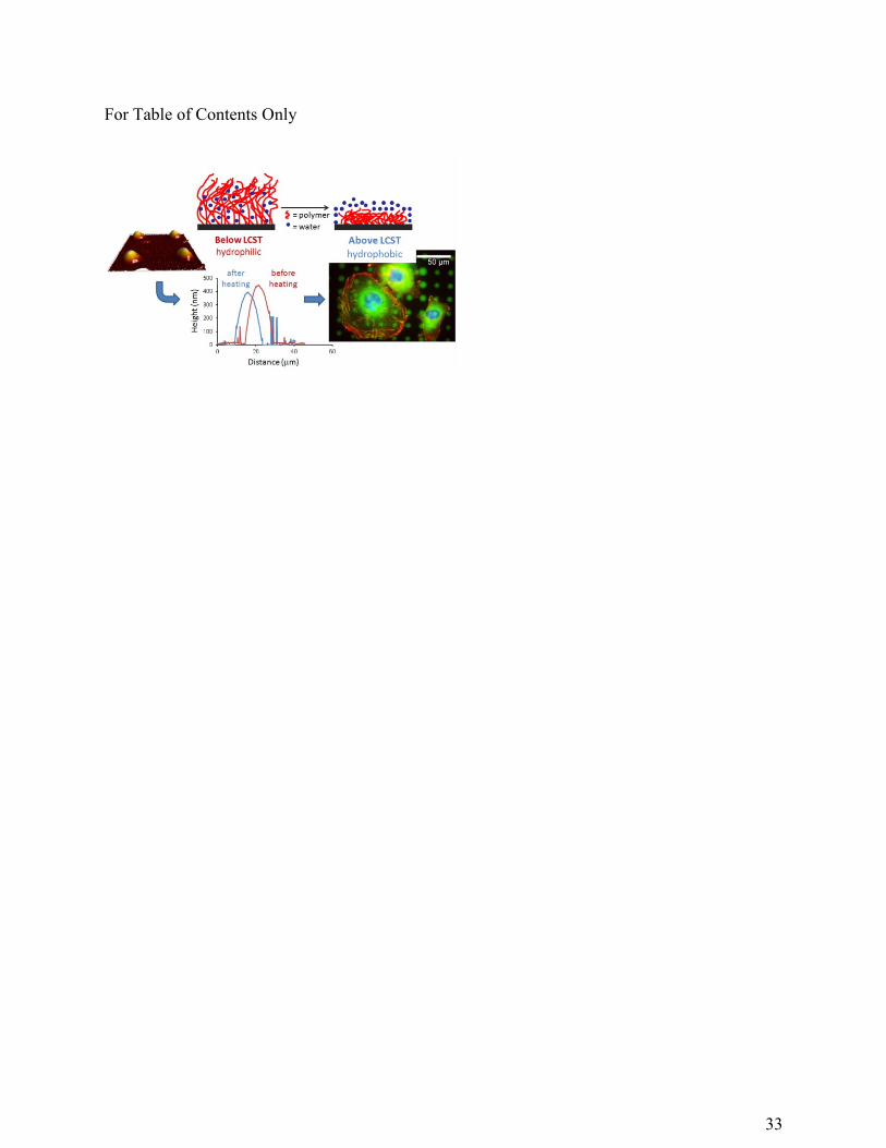
ABSTRACT
We report a novel approach for patterning thermoresponsive hydrogels based on N,N-
diethylacrylamide (DEAAm) and bifunctional Jeffamine ED-600 by dip-pen nanolithography
(DPN). The direct writing of micron-sized thermoresponsive polymer spots was achieved with
efficient control over feature size. A Jeffamine-based ink prepared through the combination of
organic polymers, such as DEAAm, in an inorganic silica network was used to print
thermosensitive arrays on a thiol-silanised silicon oxide substrate. The use of a Jeffamine
hydrogel, acting as a carrier matrix, allowed a reduction in the evaporation of ink molecules with
high volatility, such as DEAAm, and facilitated the transfer of ink from tip to substrate. The
thermoresponsive behaviour of polymer arrays which swell/de-swell in aqueous solution in
response to a change in temperature was successfully characterised by atomic force microscopy
(AFM) and Raman spectroscopy: a thermally-induced change in height and hydration state was
observed, respectively. Finally, we demonstrate that cells can adhere to and interact with these
dynamic features and exhibit a change in behaviour when cultured on the substrates above and
below the transition temperature of the Jeffamine/DEAAm thermoresponsive hydrogels. This
3
demonstrates the potential of these micropatterned hydrogels to act as a controllable surface for
cell growth.
INTRODUCTION
Hydrogels are three-dimensional polymer networks which exhibit a high level of
biocompatibility due to their ability to trap water and biological fluids.1,2 Some hydrogels undergo
changes in swelling or network structure in response to external stimuli such as pH,3
temperature,4,5 light,6,7 ionic strength3 and electric field.8 The development of such materials has
recently received considerable interest due to the potential applications in a vast range of areas.9,10
Of the possible stimuli, temperature is the most widely studied due to the prospective use of
thermoresponsive polymers in biological systems, as well as the relatively simple control of
temperature as an external stimulus.11,12
Thermoresponsive polymers in aqueous solution undergo a phase transition at a certain
temperature, which causes a change in the solubility of linear polymer chains in water. At this
phase transition temperature, a given polymer–water mixture passes from a one-phase system to a
two-phase system or vice versa. Polymers which become soluble upon heating have an upper
critical solution temperature (UCST), while systems whose solubility in water increases upon
cooling have a lower critical solution temperature (LCST).13 The majority of thermoresponsive
systems reported in the literature exhibit a LCST in aqueous solution, such as poly(N-
isopropylacrylamide), polysaccharides, and block copolymers of poly(ethyleneoxide) (PEO) and
poly(propyleneoxide) (PPO).14 The LCST-type transition takes place as a result of a local
structural transition which involves water molecules surrounding macromolecular chains in
4
solution: water-polymer interactions are thermodynamically favoured below the LCST and by
increasing the temperature above the LCST, the hydrophobic backbone and non-polar groups of
the polymer tend to interact and aggregate. Such changes in the hydration state of the polymer
chains are attributed both to the breakdown of polymer-water hydrogen bonding interactions, and
to the “hydrophobic effect” which causes a local ordered structure like a hydrated shell between
the molecules of water surrounding the hydrophobic groups of the polymer.15,16 At the molecular
level, this phase transition also leads to a volume transition from a coiled state of the polymer to
the collapsed or globular state, in which hydrophobic interactions can occur between the polymer
molecules. Accordingly, polymer chains which are completely soluble in water at temperatures
below the LCST undergo a precipitation in aqueous solutions as the temperature is increased
beyond the LCST. In the case of crosslinked hydrogels, in water at temperatures below the LCST,
they cannot be dissolved due to the covalent bonds between polymer chains but they will be
hydrophilic and therefore “swollen”; whereas when the temperature goes above the LCST, the
hydrogel becomes hydrophobic and will thus “de-swell”. By comparison with the corresponding
linear polymer molecules, the temperature sensitivity of these gels similarly occurs due to a
delicate balance of specific interactions between the water molecules and the monomer units and
results in changes of enthalpy and entropy of mixing according to the Flory-Huggins theory.17
These controlled conformational and thermodynamic changes can be exploited for their potential
use in a variety of applications such as cell culture,18-22 thermally controlled drug delivery,23-25
protein separation,26-28 microactuators29-31 and microfluidic devices.32,33 Many of these
applications involve the formation of a switchable substrate, either by coating the entire surface
with a layer of thermoresponsive polymer, or by producing polymer patterns which allow spatially
controlled thermoresponsive features.
5
Previous patterns of thermoresponsive polymers on a surface involve the formation of polymer
films or polymer brushes created by, for example, electron beam lithography,34 microcontact
printing35 or nanografting.36 These approaches involve patterning of an initiator followed by atom
transfer radical polymerisation (ATRP) for the production of polymers. However, direct printing
of polymer patterns onto a surface allows further control of feature size and shape and can provide
a switchable substrate with highly tuneable topography.37
Dip-pen nanolithography (DPN) is a direct-write technique which uses an atomic force
microscope (AFM) tip to “write” molecular “inks” onto a surface with extremely high accuracy
and resolution.38 The patterning of polymers and hydrogels by DPN has been an area of significant
interest over recent years and many useful applications have been investigated.39-42 Lee et. al. used
thermal dip-pen nanolithography (tDPN) to produce nanostructures of a thermoresponsive
polymer, poly(N-isopropylacrylamide) (PNIPAAm), for protein adsorption.37 They printed lines
of PNIPAAm and used adhesion force measurements to characterise the thermoresponse. Whilst
they observed the expected hydrophilic-hydrophobic transition, they observed no change in
topography which could potentially limit the use of their substrates for certain applications.
The work presented in this paper demonstrates the patterning of thermoresponsive polymers
using DPN with significant control over feature size and shape. AFM and Raman spectroscopy
have been utilised for the morphological and chemical characterisation of the printed arrays and
to confirm the thermoresponsive behaviour of the micron-sized features when attached to the thiol-
silanised silicon oxide surfaces. To demonstrate the potential use of these patterned substrates in
biological applications, cells were cultured on the surfaces in order to investigate if they would
respond to the polymer features. The cells adhered to and interacted with the polymer microspots
and a change in behaviour was observed across the transition temperature.
6
EXPERIMENTAL SECTION
Reagents and Materials
Acetone (≥99.5%), ethanol (≥99.8%), isopropyl alcohol (≥99.7%), glacial acetic acid (≥99.7%),
3-glycidoxypropyltrimethoxysilane (GPTMS) (≥98%), O,O‘-Bis(2-aminopropyl) polypropylene
glycol-block-polyethylene glycol-block-polypropylene glycol (Jeffamine ED-600), poly(ethylene
glycol) dimethacrylate, average Mn=550 (PEGDMA), 1-hydroxycyclohexyl phenyl ketone
(photoinitiator) (99%), 3-(trimethoxysilyl)propyl methacrylate (TMSPM) (98%) and (3-
mercaptopropyl)trimethoxysilane (MPTMS) (95%) were purchased from Sigma-Aldrich (Dorset,
UK. N,N-diethylacrylamide (DEAAm) (>98%) was purchased from Tokyo Chemical Industry
UK Ltd (Oxford, UK). All the chemicals were used as received except Jeffamine ED-600 which
was dried under dynamic vacuum for 2 h before use to remove humidity. Silicon dioxide substrates
with addressable registration marks to easily identify locations, 1D M-type pen arrays with 12 tips
and a pitch of 66 たm, and multi-channel inkwell arrays were provided by Nanoink Inc. (Skokie,
IL, USA).
Fabrication of thermoresponsive polymer arrays
Silicon dioxide substrates were cleaned by sonication in acetone, isopropanol and water, for 10
min in each solvent, and blown dry with nitrogen after each sonication bath. Substrates were then
plasma-cleaned for 40 s at 50 % power, 72 cm3 / min in oxygen. To silanise surfaces with MPTMS,
the cleaned substrates were placed in an Erlenmeyer flask with 2 mL of MPTMS in a nitrogen
atmosphere for 30 minutes and then placed in an oven at 100 °C overnight before printing. For
treating substrates with TMSPM,43 50 たL of TMSPM was diluted in 10 mL of ethanol and then
7
0.3 mL of dilute acetic acid (1:10 glacial acetic acid:water) was added just before use. This silane
solution was poured onto the surfaces and allowed to react for 3 min. The excess was poured off
and then substrates were rinsed with ethanol to remove the residual reagent and also dried under a
nitrogen flux.
DPN experiments were performed using a Nanoink NLP 2000 nanolithography instrument. 1D
M-type pen arrays were plasma cleaned for 40 s at 50% power (72 cm3 / minute) prior to use to
remove any organic contamination. All the printing experiments were performed at 22-23 °C and
in a relative humidity range of 25-35 %. DEAAm ink was prepared by mixing 500 mg of DEAAm,
0.5% wt of PEGDMA w.r.t. DEAAm (2.51 mg) and 3 % wt of photoinitiator w.r.t. DEAAm (15.46
mg) in a closed vial protected from light to minimise the activation of photoinitiator before
printing. 0.3 たL of this mixture was then added to each inkwell and the tips were dipped in a 12-
channel microfluidic inkwell. Once printed arrays were created, the substrates were exposed to a
UV lamp for 10 min. For Jeffamine ink, stoichiometric amounts (2:1) of GPTMS (442.4 mg) and
Jeffamine ED-600 (494.2 mg) were mixed in a closed glass vial under magnetic stirring for at least
2 h. 100 たL of distilled water was then added to the mixture and left under stirring for 10 more
minutes. The printed Jeffamine arrays were left in a closed box overnight and then cured at 40°C
for 1 h. The Jeffamine/DEAAm ink (mixed system) was made up by mixing the Jeffamine ink,
prepared as described above, and the DEAAm ink, including also 1 % wt (5.05 mg) of TMSPM
w.r.t. DEAAm, in a 1:1 ratio and then directly used for printing. These arrays were UV-cured for
10 min, left in a closed box overnight and eventually cured at 40 °C for 1 h.
Characterisation and instrumentation
The swelling behaviour of bulk thermoresponsive materials was analysed in duplicate at
temperatures ranging from room temperature (20 °C) to 40 °C. Cured polymer samples were firstly
8
placed in a lidded Petri dish filled with distilled water to swell at ambient temperature. After
swelling for 72 h to reach equilibrium swelling, the hydrogels were blotted free of excess water
with paper filter and their weights were measured using a scale. The samples were then moved in
a beaker full of distilled water at 40 °C for 72 h. The weights at 40 °C were measured as previously
described and the swelling ratio was calculated according the following formula:
where W20°C is the weight of the swollen samples at 20 °C and W40°C at 40 °C. The above process
was repeated for a second time in order to verify the reversibility of swelling/shrinking
characteristics.
AFM topography analysis in air was carried out on a DPN 5000TM nanofabrication system
(Nanoink Inc., Skokie, IL, USA), in close-contact mode using ACT probes purchased from
AppNano (nominal value of spring constant = 40 N/m).
The printed spots were analysed by a Witec Alpha300 R microscope (Ulm, Germany) provided
with a 633 nm laser and a 100x objective. The grating was 600 g mm-1 and coupled to a
thermoelectrically cooled charge-coupled device (CCD). Spectra were collected using 5 x 10 s
accumulations.
AFM images in liquid were obtained by scanning the surface using a PeakForce QNM Scanning
Probe Microscope (Digital Instruments, Santa Barbara, CA, USA). The AFM measurements were
obtained using ScanAsyst-Fluid probes with a spring constant of 0.67 N/m (nominal value of tip
radius = 2 nm).
Cell Culture
LE2 cells (a line of mouse lung capillary endothelia from B10D2 congenic mice, cd 133+)44
were cultured in Hams F10 media (Sigma Aldrich, UK) supplemented with 3% FBS (Sigma
20 40Swelling ratio (SR) C CW W
9
Aldrich, UK), 2% antibiotic mix (60% v/v 200 mM L-glutamine (Sigma Aldrich, UK), 35% v/v
penicillin/streptomycin (Sigma Aldrich, UK), 5% v/v fungizone (Invitrogen, UK)), 5 mL of 7.5%
sodium bicarbonate (Sigma Aldrich, UK) and 10 mL ITS (100x, Life Technologies). Hams was
chosen as it is CO2 independent and thus can be simply cultured in different temperature
environments.
Immunocytochemistry
After 4 days of culture, cells were fixed (10 mL 37% formaldehyde, 2 g sucrose in 90 mL PBS
solution) for 15 minutes. Permeabilising buffer (10.3 g sucrose, 0.292 g NaCl, 0.06 g MgCl2,
0.476 g HEPES, 0.5 mL Triton X, in 100 mL of H2O, at pH 7.2) was then added for 5 mins at 4°C.
To block non-specific binding, samples were next incubated in 1% BSA/PBS for 5 mins at 37°C.
H-vin 1 primary antibody (1:200, monoclonal antihuman raised in mouse (IgG1) Sigma Aldrich
UK, in 1% BSA/PBS) was added for 1 hour along with rhodamine-conjugated phalloidin (1:100,
Sigma Aldrich UK). Substrates were then washed three times in 0.5% Tween 20/PBS (5 minutes
each). Secondary, biotin-conjugated antibody (1:50 in 1% BSA/PBS, antimouse (IgG) raised in
horse, Vector laboratories UK) was added for 1 hour, followed by substrate washing as described
above. FITC-conjugated streptavidin was added (1:50 in 1% BSA/PBS, Vector Laboratories UK)
for 2 hours before samples were given a final wash. Samples were mounted using mounting
medium for fluorescence, with DAPI counterstain (Vector Laboratories), and viewed by
fluorescent microscopy (Zeiss Axiophot).
In-cell Western
10
Cells were fixed and permeabilised as per immunocytochemistry. Afterwards, non-specific
binding sites were blocked with PBS/1% milk protein for 1.5 hours at room temperature with
gentle agitation on a plate shaker. Samples were incubated in PBS/1% milk protein containing
anti-KLF2 antibody H60 (sc-28675, Santa Cruz Biotechnology, CA, USA) primary antibody
(1:50) and GAPDH primary antibody (1:10000, mouse monoclonal antihuman, Sigma Aldrich,
UK) for 2 hours at 37°C. Samples were washed five times in 0.1% PBS/Tween 20 with gentle
agitation (5 minutes each). Samples were then incubated for 1 hour with gentle agitation in
secondary antibodies (donkey anti mouse IR680RD and donkey anti rabbit IR800CW, 1:500, Li-
cor UK) diluted in PBS/1% milk protein containing 0.2% Tween 20. The samples were then
washed five times in 0.1% PBS/Tween, as previously described. Wells were dried and analysed
using the Licor Odyssey Imaging System. Readings at 700 nm and 800 nm were taken and
quantified in excel.
RESULTS AND DISCUSSION
Thermoresponsive ink properties and fabrication of polymer arrays
The thermoresponsive nature of polymers can arise from various structural properties such as
the presence of a thermosensitive polymer backbone45,46 or by thermosensitive units linked on the
polymer chains.47,48 Among these systems exhibiting thermoresponsive behaviour, N-substituted
poly(acrylamides) are one of the most extensively investigated polymers, especially due to their
known biocompatibility.49 In addition, N-alkyl acrylamides can be copolymerised with other
stimuli-responsive monomers to combine characteristics of different monomeric units in a single
system. Recently, a range of commercially available amino-terminated copolymers of ethylene
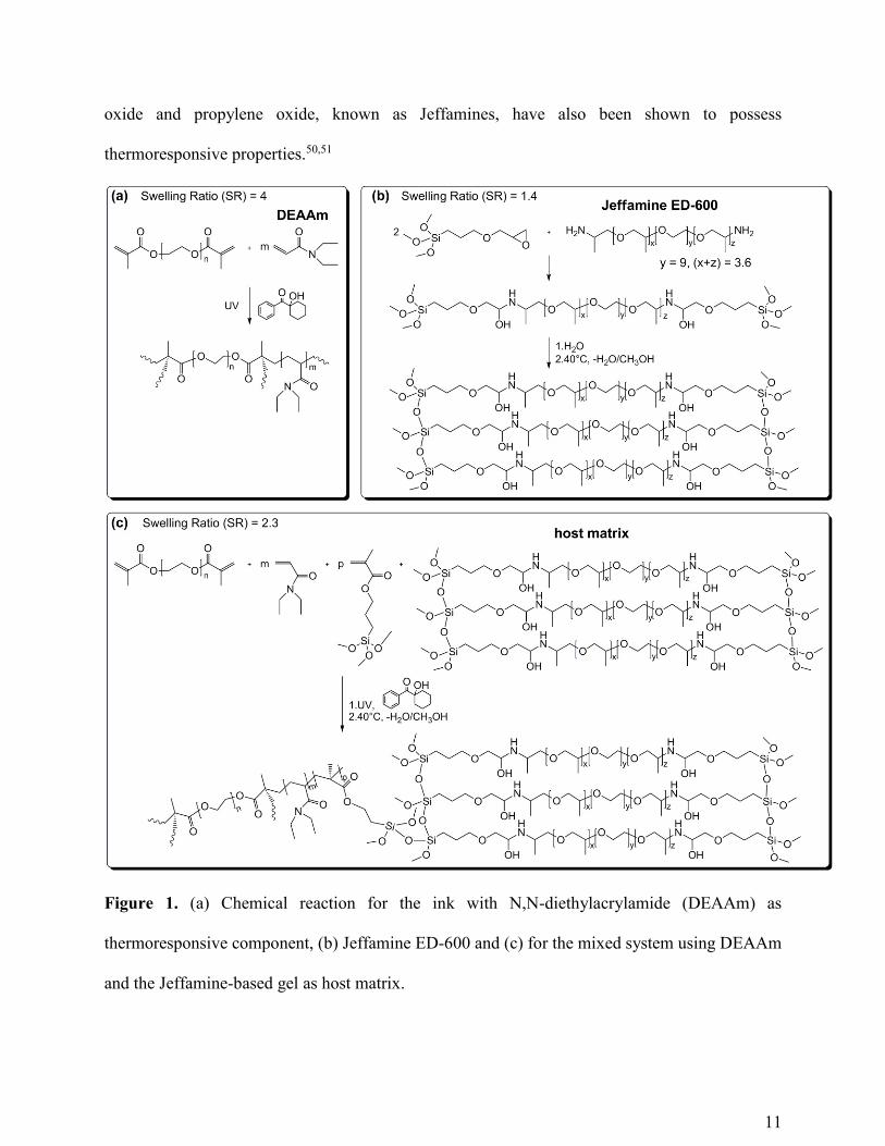
11
oxide and propylene oxide, known as Jeffamines, have also been shown to possess
thermoresponsive properties.50,51
Figure 1. (a) Chemical reaction for the ink with N,N-diethylacrylamide (DEAAm) as
thermoresponsive component, (b) Jeffamine ED-600 and (c) for the mixed system using DEAAm
and the Jeffamine-based gel as host matrix.
12
The development of a suitable thermoresponsive ink for DPN printing was one of the most
crucial phases in our work. We investigated different thermoresponsive inks and compared their
ink transport and writing capabilities. We first studied a UV-curable ink mainly composed of
N,N-diethylacrylamide (DEAAm) acting as a thermoresponsive monomer with a small percentage
of crosslinker and photoinitiator (Figure 1 (a)). To covalently bond the thermoresponsive
structures patterned by DPN to silicon oxide substrates, two different silanes, one with a
methacrylate end functional group and another one with a terminal thiol, were investigated for the
functionalisation of the silicon surface. The transfer of this thermoresponsive ink from the inked
tip onto the two substrates, however, was dominated by the volatility of DEAAm in air. Due to its
high volatility, DEAAm becomes even more volatile at the microscale. UV-cured micron–sized
spots deposited by DPN showed a Raman spectrum very similar to that of the crosslinker, because
the monomer in the ink had evaporated during the loading of the tip and the printing process (See
supporting information, Figure S1). The printing experiments were performed at room
temperature (22-23°C) and at a rather low percentage of relative humidity (25-35%). An increase
of the temperature did not improve ink transport and patterning because it caused an increased
vaporisation of DEAAm. In addition, a higher relative humidity accelerates the process of
evaporation of DEAAm due to the high solubility of DEAAm in water, which causes liquid
DEAAm to convert into a gas more rapidly. To solve this issue, we examined an ink with
Jeffamine ED-600 (Figure 1 (b)). Due to the presence of Jeffamine, the ink exhibited a
hydrophilicity higher than the UV-curable material which resulted in it spreading across surfaces
such as bare and methacrylate-silanised silicon oxide. Reproducible polymer arrays were created
only on thiol-silanised silicon oxide substrates (Figure 2(a)), which turned out to be hydrophobic
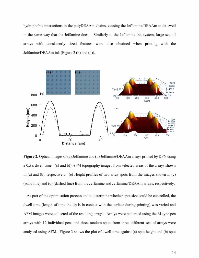
enough to prevent the spreading of the Jeffamine ink. Figure 2 (a) shows a white light image of
13
the printed arrays with Jeffamine ink and (c) displays an AFM topography image of a small area
within the same region. Figure 2 (e) shows the height profile measured across two array spots.
The increased viscosity of the Jeffamine ED-600 (75 cp) allowed for improvement of the kinetics
of ink flow from the tip to the substrate and enabled the printing of large sets of arrays, without
the need to re-ink the tip. The bulk material of Jeffamine ink after heat-curing showed a swelling
ratio of 1.4 which is lower than that of the UV-cured DEAAm bulk sample (≈ 4). Moreover, a
reversible switch was not observed across the transition temperature for the Jeffamine bulk
samples.
To create patterns with a higher degree of swelling and a reversible temperature-induced switch,
the combination of these two systems was also studied. The ink composed of Jeffamine can be
used not only as a thermoresponsive component but also as a carrier matrix. The addition of the
Jeffamine matrix having hydroxyl and secondary amine groups capable of polar interactions and
hydrogen bonding with the amide group of DEAAm, can reduce the evaporation of DEAAm and
improve the ink transport. After preparing the Jeffamine ink, we added the DEAAm ink prepared
as mentioned previously, as well as a silane with a methacrylate terminal group to covalently link
the Jeffamine matrix to the acrylamide network (Figure 1 (c)). This mixed system (from here on
referred to as Jeffamine/DEAAm) exhibited a swelling ratio of 2.3, an intermediate value between
those measured for the other inks investigated. The increased swelling ratio of the mixed system
in comparison to the Jeffamine alone is due to the larger swelling weight of the Jeffamine/DEAAm
at room temperature, which can be attributed to the less packed hydrogel structure of this system
and the fact that the water-polymer interactions in the Jeffamine/DEAAm are more
thermodynamically favoured at 20°C than when the Jeffamine is present alone. Less difference in
swelling weight is observed at 40°C due to the less packed polymerized structure which favours
14
hydrophobic interactions in the polyDEAAm chains, causing the Jeffamine/DEAAm to de-swell
in the same way that the Jeffamine does. Similarly to the Jeffamine ink system, large sets of
arrays with consistently sized features were also obtained when printing with the
Jeffamine/DEAAm ink (Figure 2 (b) and (d)).
Figure 2. Optical images of (a) Jeffamine and (b) Jeffamine/DEAAm arrays printed by DPN using
a 0.5 s dwell time. (c) and (d) AFM topography images from selected areas of the arrays shown
in (a) and (b), respectively. (e) Height profiles of two array spots from the images shown in (c)
(solid line) and (d) (dashed line) from the Jeffamine and Jeffamine/DEAAm arrays, respectively.
As part of the optimisation process and to determine whether spot size could be controlled, the
dwell time (length of time the tip is in contact with the surface during printing) was varied and
AFM images were collected of the resulting arrays. Arrays were patterned using the M-type pen
arrays with 12 individual pens and three random spots from three different sets of arrays were
analysed using AFM. Figure 3 shows the plot of dwell time against (a) spot height and (b) spot
0
200
400
600
800
0 20 40
Heig
ht
(nm
)
Distance (たm)
(a) (b)
(c)
(d)
(e)
15
diameter for both the Jeffamine and Jeffamine/DEAAm inks. It can be observed here that there is
a general increase in both spot height and diameter with increasing dwell time for each of the two
inks. The height of the spots patterned in this study with Jeffamine ink ranged from about 605 nm
up to 720 nm by varying the dwell time from 0.1 s to 1 s. The smallest spot diameter obtained was
11 たm using a 0.1 s dwell time which increased up to nearly 15 たm using a dwell time of 1 s. For
the Jeffamine/DEAAm ink, the printed spots were smaller, ranging from 546 nm to 654 nm in
height and from about 9 たm to around 13 たm in diameter. The observed difference in spot size
could be attributed to a slight change in viscosity between the two inks or the fact that the arrays
were printed using different tips. It is important to note that the size of spots can vary between
different surfaces and tips and therefore a standard size range should be tested to determine a
relationship between dwell time and spot size in individual cases. However, the general increase
with increasing dwell time for both ink systems indicates that feature size can be controlled by the
straightforward adjustment of printing conditions.
16
Figure 3. Plot of dwell time against spot height (a) and spot diameter (b) for the Jeffamine ED-
600 arrays (black) and the Jeffamine/DEAAm arrays (red).
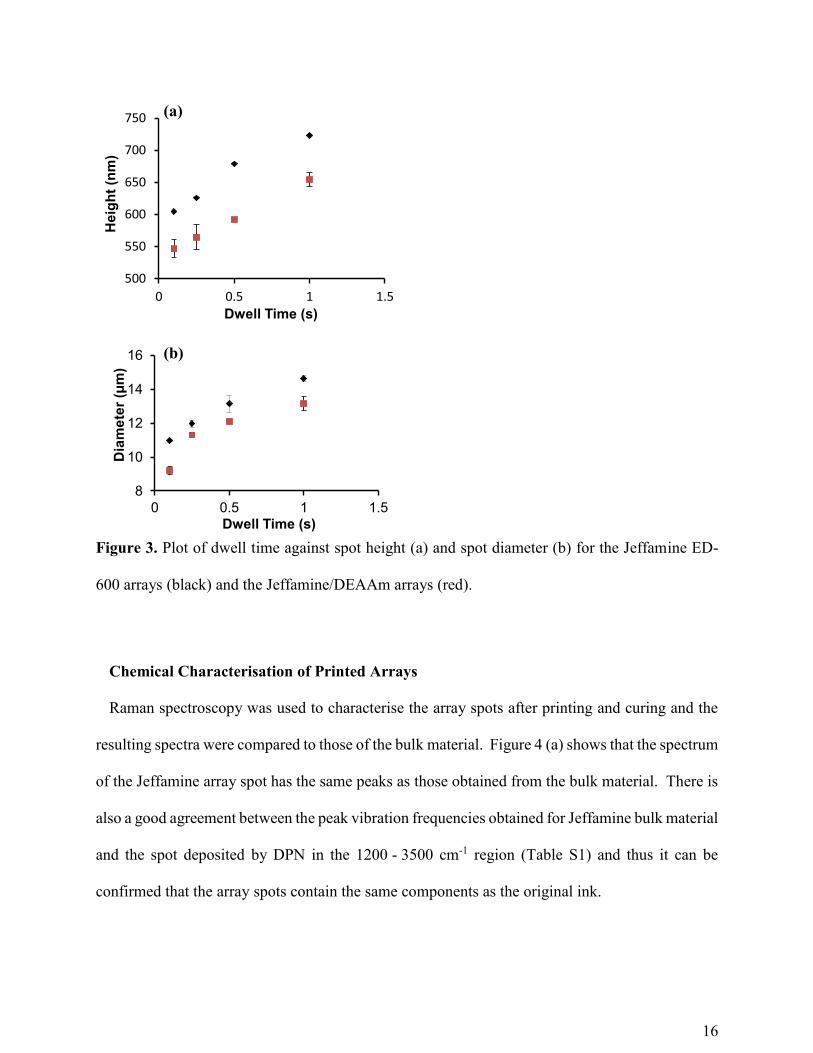
Chemical Characterisation of Printed Arrays
Raman spectroscopy was used to characterise the array spots after printing and curing and the
resulting spectra were compared to those of the bulk material. Figure 4 (a) shows that the spectrum
of the Jeffamine array spot has the same peaks as those obtained from the bulk material. There is
also a good agreement between the peak vibration frequencies obtained for Jeffamine bulk material
and the spot deposited by DPN in the 1200 - 3500 cm-1 region (Table S1) and thus it can be
confirmed that the array spots contain the same components as the original ink.
500
550
600
650
700
750
0 0.5 1 1.5
Heig
ht
(nm
)
Dwell Time (s)
8
10
12
14
16
0 0.5 1 1.5
Dia
me
ter
(ȝm
)
Dwell Time (s)
(a)
(b)
17
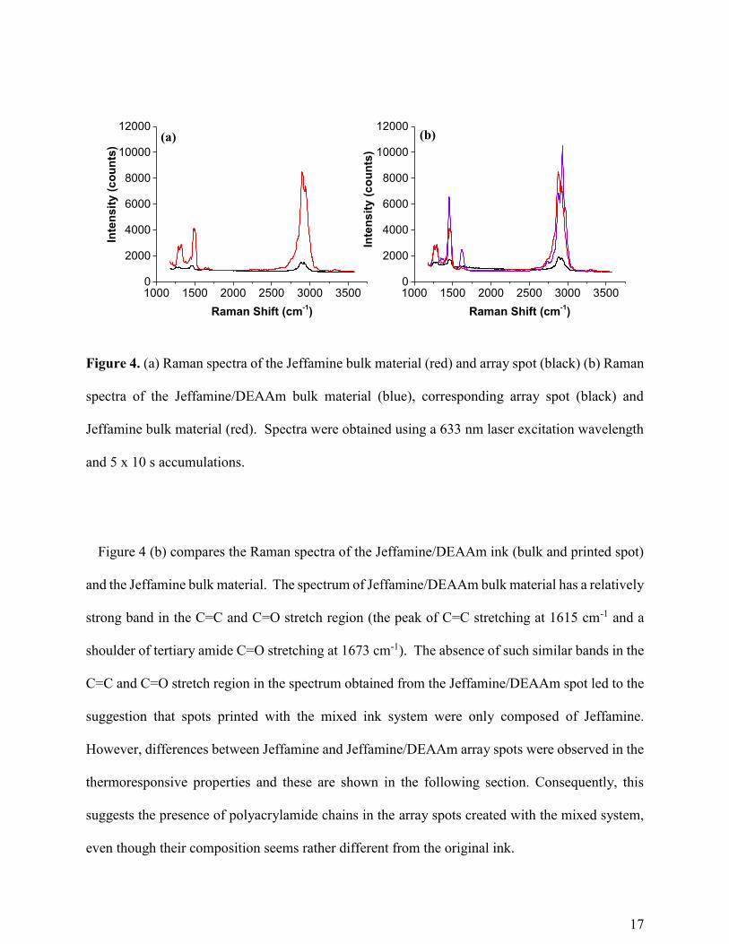
Figure 4. (a) Raman spectra of the Jeffamine bulk material (red) and array spot (black) (b) Raman
spectra of the Jeffamine/DEAAm bulk material (blue), corresponding array spot (black) and
Jeffamine bulk material (red). Spectra were obtained using a 633 nm laser excitation wavelength
and 5 x 10 s accumulations.
Figure 4 (b) compares the Raman spectra of the Jeffamine/DEAAm ink (bulk and printed spot)
and the Jeffamine bulk material. The spectrum of Jeffamine/DEAAm bulk material has a relatively
strong band in the C=C and C=O stretch region (the peak of C=C stretching at 1615 cm-1 and a
shoulder of tertiary amide C=O stretching at 1673 cm-1). The absence of such similar bands in the
C=C and C=O stretch region in the spectrum obtained from the Jeffamine/DEAAm spot led to the
suggestion that spots printed with the mixed ink system were only composed of Jeffamine.
However, differences between Jeffamine and Jeffamine/DEAAm array spots were observed in the
thermoresponsive properties and these are shown in the following section. Consequently, this
suggests the presence of polyacrylamide chains in the array spots created with the mixed system,
even though their composition seems rather different from the original ink.
1000 1500 2000 2500 3000 35000
2000
4000
6000
8000
10000
12000
1000 1500 2000 2500 3000 35000
2000
4000
6000
8000
10000
12000
Inte
ns
ity
(c
ou
nts
)
Raman Shift (cm-1)
(a)
Inte
ns
ity
(c
ou
nts
)
Raman Shift (cm-1)
(b)
18
Thermoresponsive properties of patterns
In order to assess the thermoresponsive behaviour of the polymer arrays, we investigated the
changes in the array spots which occurred in aqueous solution and when heated across the LCST.
Raman scattering was first used to investigate any structural changes taking place as a result of the
interactions between the polymer and water molecules. Previous studies have shown that changes
in Raman spectra of thermoresponsive polymers can be observed across the LCST.52,53 Arrays
printed using a 0.5 s dwell time were analysed dry before being left in water for 72 h to allow
equilibrium swelling to be reached. After collecting the Raman spectra of the arrays in water, the
temperature was increased to 37 °C and the arrays were again left for 72 h to allow the system to
equilibrate. Resulting spectra can be found in Figure 5 (a) and (b) for the Jeffamine and
Jeffamine/DEAAm, respectively. This shows that, as well as the appearance of the OH stretch
between 3000 cm-1 and 3500 cm-1 and the OH bending mode at around 1600 cm-1, there is a change
in the ratio of the symmetric and asymmetric CH stretches when the arrays are in water. The
observed OH stretch is broad and flat which can be attributed to the interaction of the water
molecules with the polymer and thus the restriction in position and orientation of the water
molecules. There is also a shift in frequency in the OH bending mode upon heating, as well as an
increase in intensity and narrowing of the band, which is typically found due to the weakening of
hydrogen bonds and the strengthened OH bonds.54 The area under the peaks was calculated so
that the ratios could be compared more accurately, and it was found that the ratio of the symmetric
CH stretch : asymmetric CH stretch decreased between the dry arrays and arrays in water and upon
heating, increased to an intermediate point (Table 1). This indicates that the decrease is coinciding
with increasing water content and thus confirms the temperature-induced changes in the polymer
19
array spots. It is also shown here that the ratio of the OH stretch : CH stretch changes significantly
before and after heating and the decrease in this ratio is consistent with water being expelled on
collapsing/de-swelling of the polymer microspot. Furthermore, the changes which can be
observed in the ratio of the OH stretch : CH stretch before and after heating are more significant
in the Jeffamine/DEAAm than in the Jeffamine alone, indicating that the thermoresponsive
properties are greater in this system. Additionally, the differences in the Raman spectra are
consistent with the changes in swelling ratio which are observed between the two systems in bulk.
The increased water content of the Jeffamine/DEAAm system at room temperature is apparent in
the Raman spectra where the ratio of the OH stretch : CH stretch is greater for the
Jeffamine/DEAAm array spot in water at room temperature. Therefore, even although the
microspot spectrum is different from that of the bulk material (Figure 4 (b)), the polyacrylamide
chains are present in the microspot resulting in an increase in the swelling properties when
compared to those of the Jeffamine alone. AFM was then used to analyse the array spots before
and after heating in order to determine whether changes in topography could be observed. After
leaving the arrays in water for 72 h to allow equilibrium swelling to be reached, AFM images were
collected. The arrays were then left for a further 72 h at 37°C and AFM images were collected in
the same way. In order to ensure a fair comparison, registration marks on the silicon dioxide
surface were used to allow the exact same areas to be scanned before and after heating so that the
height and diameter of the same spots, before and after heating, could be compared. Figure 5 (c)
and (d) show the AFM data of the Jeffamine spots and the Jeffamine/DEAAm, respectively. In
the Jeffamine/DEAAm in particular, when the height profiles before and after heating are
compared, we can see a clear decrease in height and increase in diameter which indicates that the
polymer is de-swelling or collapsing upon heating. The fact that this change is less clear for the
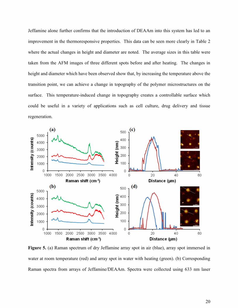
20
Jeffamine alone further confirms that the introduction of DEAAm into this system has led to an
improvement in the thermoresponsive properties. This data can be seen more clearly in Table 2
where the actual changes in height and diameter are noted. The average sizes in this table were
taken from the AFM images of three different spots before and after heating. The changes in
height and diameter which have been observed show that, by increasing the temperature above the
transition point, we can achieve a change in topography of the polymer microstructures on the
surface. This temperature-induced change in topography creates a controllable surface which
could be useful in a variety of applications such as cell culture, drug delivery and tissue
regeneration.
Figure 5. (a) Raman spectrum of dry Jeffamine array spot in air (blue), array spot immersed in
water at room temperature (red) and array spot in water with heating (green). (b) Corresponding
Raman spectra from arrays of Jeffamine/DEAAm. Spectra were collected using 633 nm laser
21
excitation with 5 x 10 second accumulations. (c) Height profile of Jeffamine array spot before (red)
and after (blue) heating to 37°C. Inset: AFM images from which the height profiles were obtained
with white lines highlighting the exact location of the corresponding profile. (d) Corresponding
AFM data for Jeffamine/DEAAm array spots.
Table 1. Area under peaks corresponding to CH and OH stretching modes and comparison of peak
ratios between dry arrays, arrays in water at room temperature and arrays in water at 37°C.
Peak Ratio
(CH Stretch (2835 – 2900 cm-1):CH Stretch
(2907 – 2963 cm-1))
Peak Ratio
(OH Stretch (3046 cm-1 – 3552 cm-1):CH
Stretch (2835 cm-1 – 2963 cm-1))
Dry
Arrays
Jeffamine: 2.08 ± 0.26
Jeffamine/DEAAm: 2.28 ± 0.09
-
Before
Heating
Jeffamine: 0.51 ± 0.12
Jeffamine/DEAAm: 0.74 ± 0.14
Jeffamine: 7.38 ± 0.17
Jeffamine/DEAAm: 16.24 ± 1.90
After
Heating
Jeffamine: 0.84 ± 0.06
Jeffamine/DEAAm: 0.85 ± 0.08
Jeffamine: 2.74 ± 0.48
Jeffamine/DEAAm: 5.29 ± 0.33
Table 2. Changes in height and diameter of Jeffamine and Jeffamine/DEAAm array spots in water
after heating to 37°C.
Average Height Change (nm) Average Diameter Change (たm)
Jeffamine - 33.0 ± 6.1 + 0.2 ± 0.11
Jeffamine/DEAAm - 68.0 ± 12.6 + 0.6 ± 0.05
22
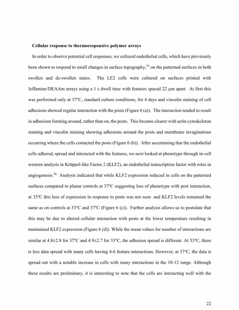
Cellular response to thermoresponsive polymer arrays
In order to observe potential cell responses, we cultured endothelial cells, which have previously
been shown to respond to small changes in surface topography,55 on the patterned surfaces in both
swollen and de-swollen states. The LE2 cells were cultured on surfaces printed with
Jeffamine/DEAAm arrays using a 1 s dwell time with features spaced 22 µm apart. At first this
was performed only at 37oC, standard culture conditions, for 4 days and vinculin staining of cell
adhesions showed regular interaction with the posts (Figure 6 (a)). The interaction tended to result
in adhesions forming around, rather than on, the posts. This became clearer with actin cytoskeleton
staining and vinculin staining showing adhesions around the posts and membrane invaginations
occurring where the cells contacted the posts (Figure 6 (b)). After ascertaining that the endothelial
cells adhered, spread and interacted with the features, we next looked at phenotype through in-cell
western analysis in Krüppel-like Factor 2 (KLF2), an endothelial transcription factor with roles in
angiogenesis.56 Analysis indicated that while KLF2 expression reduced in cells on the patterned
surfaces compared to planar controls at 37oC suggesting loss of phenotype with post interaction,
at 33oC this loss of expression in response to posts was not seen and KLF2 levels remained the
same as on controls at 33oC and 37oC (Figure 6 (c)). Further analysis allows us to postulate that
this may be due to altered cellular interaction with posts at the lower temperature resulting in
maintained KLF2 expression (Figure 6 (d)). While the mean values for number of interactions are
similar at 4.82.8 for 37oC and 4.92.7 for 33oC, the adhesion spread is different. At 33oC, there
is less data spread with many cells having 4-6 feature interactions. However, at 37oC, the data is
spread out with a notable increase in cells with many interactions in the 10-12 range. Although
these results are preliminary, it is interesting to note that the cells are interacting well with the
23
polymer features and that a difference in the cell behaviour can be observed across the transition
temperature, indicating the potential use of the surfaces in biological applications.
Figure 6. (a) Vinculin staining (adhesions seen as white lines within the cell boundaries) showing
that adhesions form around rather than on the printed features (arrows point to adhesions close to
posts). (b) Actin (red) and vinculin (green) staining showing that this adhesion pattern causes
membrane invaginations to form around the posts (arrows point to invaginations around posts)
(nuclei are blue). (c) KLF2 expression showing that the features prevent loss of KLF2 expression
with reduced temperature (n=1). (d) Quantification of number of features individual cells interact
with, showing that cells interact more strongly with the posts at lower temperature (arrows show
vinculin positive adhesions close to features) (50 cells counted per substrate).
24
CONCLUSIONS
Dip-pen nanolithography has been used for the consistent patterning of thermoresponsive
polymer arrays onto a thiol-silanised silicon dioxide substrate with significant control over feature
size. A novel ink formulation based on Jeffamine ED-600 has been developed and we have shown
that this can act as a carrier matrix to improve the printing of DEAAm whilst also increasing the
swelling properties of the hydrogel when compared to the Jeffamine ink alone. Characterisation
of the arrays by Raman and AFM has shown that, as well as a change in hydration state of the
polymer, a temperature-induced change in topography of the arrays on the surface can be observed.
We envisage that this thermally controlled switchable surface could be useful in a variety of
applications ranging from cell culture to micro-actuators for microfluidic devices. Preliminary
experiments show that cells adhere to and interact with the polymer features and that a change in
cell behaviour is observed with changing temperature. This indicates that the controllable surfaces
are biocompatible and have potential use for cellular applications.
ASSOCIATED CONTENT
Supporting Information. Figure S1. Comparison of Raman spectra of the DEAAm printed spot,
the PEG-DMA crosslinker and the whole ink solution. Table S1. Table of Raman vibration
assignments for the printed array spots and bolk material of Jeffamine and Jeff/DEAAm ink
systems. This material is available free of charge via the Internet at http://pubs.acs.org.
AUTHOR INFORMATION
Corresponding Author
25
*Prof. Duncan Graham, Centre of Molecular Nanometrology, Technology and Innovation
Centre, University of Strathclyde, 99 George Street, Glasgow G1 1RD, United Kingdom.
Author Contributions
The manuscript was written through contributions of all authors. All authors have given approval
to the final version of the manuscript. #These authors contributed equally.
Funding Sources
Stacey Laing, Karen Faulds and Duncan Graham received funding from The Leverhulme Trust
(Research Project Grant RPG-2012-758) and EPSRC Doctoral Training Grant (grant no.).
Raffaella Suriano obtained support from Progetto Bandiera “La Fabbrica del Futuro” in the
framework of the funded project “POLYPHAB, POLYmer nanostructuring by two-PHoton
ABsorption.
Notes
The research data associated with this paper will become available at the following link:
http:// 10.15129/b442c62a-8ff2-4854-972e-8d861e9b2aed
ACKNOWLEDGMENT
The authors acknowledge Dr. Laura McNamara for her help in carrying out the cell experiments.
K.F. and S.L. thank the Leverhulme Trust through Research Project Grant RPG-2012-758 for
funding. D.G. acknowledges the Analytical Chemistry Trust Fund, EPSRC for the award of an
analytical studentship which supported and funded this work. R.S. would also like to acknowledge
26
support by Progetto Bandiera “La Fabbrica del Futuro” in the framework of the funded project
“POLYPHAB, POLYmer nanostructuring by two-PHoton ABsorption.
REFERENCES
1. Seliktar, D., Designing Cell-Compatible Hydrogels for Biomedical Applications. Science
2012, 336 (6085), 1124-1128.
2. Kopecek, J., Hydrogel Biomaterials: A Smart Future? Biomaterials 2007, 28 (34), 5185-
5192.
3. Tanaka, T.; Fillmore, D.; Sun, S. T.; Nishio, I.; Swislow, G.; Shah, A., Phase Transitions
in Ionic Gels. Phys. Rev. Lett. 1980, 45 (20), 1636-1639.
4. Tanaka, T., Collapse of Gels and Critical Endpoint. Phys. Rev. Lett. 1978, 40 (12), 820-
823.
5. Vancoillie, G.; Frank, D.; Hoogenboom, R., Thermoresponsive Poly(Oligo Ethylene
Glycol Acrylates). Prog. Polym. Sci. 2014, 39 (6), 1074-1095.
6. Suzuki, A.; Tanaka, T., Phase Transition in Polymer Gels Induced by Visible Light. Nature
1990, 346 (6282), 345-347.
7. Lee, H. I.; Wu, W.; Oh, J. K.; Mueller, L.; Sherwood, G.; Peteanu, L.; Kowalewski, T.;
Matyjaszewski, K., Light-induced Reversible Formation of Polymeric Micelles. Angew. Chem.
Int. Ed. 2007, 46 (14), 2453-2457.
8. Tanaka, T.; Nishio, I.; Sun, S. T.; Uenonishio, S., Collapse of Gels in an Electric Field.
Science 1982, 218 (4571), 467-469.
9. Brun-Graeppi, A. K. A. S.; Richard, C.; Bessodes, M.; Scherman, D.; Merten, O.-W.,
Thermoresponsive Surfaces for Cell Culture and Enzyme-free Cell Detachment. Prog. Polym. Sci.
2010, 35 (11), 1311-1324.
27
10. Rodriguez-Hernandez, J.; Checot, F.; Gnanou, Y.; Lecommandoux, S., Toward 'Smart'
Nano-objects by Self-assembly of Block Copolymers in Solution. Prog. Polym. Sci. 2005, 30 (7),
691-724.
11. Roy, D.; Brooks, W. L. A.; Sumerlin, B. S., New Directions in Thermoresponsive
Polymers. Chem. Soc. Rev. 2013, 42 (17), 7214-7243.
12. Zhang, Q.; Hoogenboom, R., Polymers with Upper Critical Solution Temperature
Behavior in Alcohol/Water Solvent Mixtures. Prog. Polym. Sci. 2015, 48, 122-142.
13. Schmaljohann, D., Thermo- and pH-Responsive Polymers in Drug Delivery. Adv. Drug
Delivery Rev. 2006, 58 (15), 1655-1670.
14. Klouda, L.; Mikos, A. G., Thermoresponsive Hydrogels in Biomedical Applications. Eur.
J. Pharm. Biopharm. 2008, 68 (1), 34-45.
15. Otake, K.; Inomata, H.; Konno, M.; Saito, S., Thermal Analysis of the Volume Phase-
transition with N-isopropylacrylamide Gels. Macromolecules 1990, 23 (1), 283-289.
16. Lin, S. Y.; Chen, K. S.; Liang, R. C., Thermal Micro ATR/FT-IR Spectroscopic System
for Quantitative Study of the Molecular Structure of poly(N-isopropylacrylamide) in Water.
Polymer 1999, 40 (10), 2619-2624.
17. Flory, P. J., Principles of Polymer Chemistry. First ed.; Cornell University Press: Ithaca,
United States, 1953.
18. Okano, T.; Yamada, N.; Okuhara, M.; Sakai, H.; Sakurai, Y., Mechanism of Cell
Detachment from Temperature-modulated, Hydrophilic-hydrophobic Polymer Surfaces
Biomaterials 1995, 16 (4), 297-303.
19. Kushida, A.; Yamato, M.; Konno, C.; Kikuchi, A.; Sakurai, Y.; Okano, T., Decrease in
Culture Temperature Releases Monolayer Endothelial Cell Sheets Together with Deposited
28
Fibronectin Matrix from Temperature-responsive Culture Surfaces. J. Biomed. Mater. Res. 1999,
45 (4), 355-362.
20. Nagase, K.; Kobayashi, J.; Kikuchi, A.; Akiyama, Y.; Kanazawa, H.; Okano, T., Interfacial
Property Modulation of Thermoresponsive Polymer Brush Surfaces and Their Interaction with
Biomolecules. Langmuir 2007, 23 (18), 9409-9415.
21. Canavan, H. E.; Cheng, X. H.; Graham, D. J.; Ratner, B. D.; Castner, D. G., Surface
Characterization of the Extracellular Matrix Remaining After Cell Detachment from a
Thermoresponsive Polymer. Langmuir 2005, 21 (5), 1949-1955.
22. Zhang, R.; Mjoseng, H. K.; Hoeve, M. A.; Bauer, N. G.; Pells, S.; Besseling, R.; Velugotla,
S.; Tourniaire, G.; Kishen, R. E. B.; Tsenkina, Y.; Armit, C.; Duffy, C. R. E.; Helfen, M.;
Edenhofer, F.; de Sousa, P. A.; Bradley, M., A Thermoresponsive and Chemically Defined
Hydrogel for Long-term Culture of Human Embryonic Stem Cells. Nat. Commun. 2013, 4, 1335-
1335.
23. Kavanagh, C. A.; Rochev, Y. A.; Gallagher, W. A.; Dawson, K. A.; Keenan, A. K., Local
Drug Delivery in Restenosis Injury: Thermoresponsive Co-polymers as Potential Drug Delivery
Systems. Pharmacol. Ther. 2004, 102 (1), 1-15.
24. Ma, D.; Chen, H.; Shi, D.; Li, Z.; Wang, J., Preparation and Characterization of Thermo-
responsive PDMS Surfaces Grafted with poly(N-isopropylacrylamide) by Benzophenone-initiated
Photopolymerization. J. Colloid Interface Sci. 2009, 332 (1), 85-90.
25. Okano, T.; Bae, Y. H.; Jacobs, H.; Kim, S. W., Thermally On Off Switching Polymers for
Drug Permeation and Release. J. Controlled Release 1990, 11 (1-3), 255-265.
29
26. Malmstadt, N.; Yager, P.; Hoffman, A. S.; Stayton, P. S., A Smart Microfluidic Affinity
Chromatography Matrix Composed of poly(N-isopropylacrylamide)-coated Beads. Anal. Chem.
2003, 75 (13), 2943-2949.
27. Luo, L.; Zhang, H.-S.; Liu, Y.; Ha, W.; Li, L.-H.; Gong, X.-L.; Li, B.-J.; Zhang, S.,
Preparation of Thermosensitive Polymer Magnetic Particles and Their Application in Protein
Separations. J. Colloid Interface Sci. 2014, 435, 99-104.
28. Shekhar, S.; Mukherjee, M.; Sen, A. K., Synthesis and Characterization of
Thermoresponsive Terpolymer for Protein Separation. Int. J. Polym. Mater. 2014, 63 (8), 389-397.
29. Suzuki, H., Stimulus-responsive Gels: Promising Materials for the Construction of Micro
Actuators and Sensors. J. Intell. Mater. Syst. Struct. 2006, 17 (12), 1091-1097.
30. Kim, Y. S.; Liu, M.; Ishida, Y.; Ebina, Y.; Osada, M.; Sasaki, T.; Hikima, T.; Takata, M.;
Aida, T., Thermoresponsive Actuation Enabled by Permittivity Switching in an Electrostatically
Anisotropic Hydrogel. Nat Mater 2015, 14 (10), 1002-1007.
31. Liu, F.; Jiang, S.; Ionov, L.; Agarwal, S., Thermophilic Films and Fibers from Photo Cross-
linkable UCST-type Polymers. Polym. Chem. 2015, 6 (14), 2769-2776.
32. Sershen, S. R.; Mensing, G. A.; Ng, M.; Halas, N. J.; Beebe, D. J.; West, J. L., Independent
Optical Control of Microfluidic Valves Formed from Optomechanically Responsive
Nanocomposite Hydrogels. Adv. Mater. 2005, 17 (11), 1366-1368.
33. Ryu, S.; Yoo, I.; Song, S.; Yoon, B.; Kim, J.-M., A Thermoresponsive Fluorogenic
Conjugated Polymer for a Temperature Sensor in Microfluidic Devices. J. Am. Chem. Soc. 2009,
131 (11), 3800-3801.
30
34. Ahn, S. J.; Kaholek, M.; Lee, W. K.; LaMattina, B.; LaBean, T. H.; Zauscher, S., Surface-
initiated Polymerization on Nanopatterns Fabricated by Electron-beam Lithography. Adv. Mater.
2004, 16 (23-24), 2141-2145.
35. Jones, D. M.; Huck, W. T. S., Controlled Surface-initiated Polymerizations in Aqueous
Media. Adv. Mater. 2001, 13 (16), 1256-1259.
36. Kaholek, M.; Lee, W. K.; LaMattina, B.; Caster, K. C.; Zauscher, S., Fabrication of
Stimulus-responsive Nanopatterned Polymer Brushes by Scanning-probe Lithography. Nano Lett.
2004, 4 (2), 373-376.
37. Lee, W.-K.; Whitman, L. J.; Lee, J.; King, W. P.; Sheehan, P. E., The Nanopatterning of a
Stimulus-responsive Polymer by Thermal Dip-pen Nanolithography. Soft Matter 2008, 4 (9),
1844-1847.
38. Salaita, K.; Wang, Y.; Mirkin, C. A., Applications of Dip-pen Nanolithography. Nat.
Nanotechnol. 2007, 2 (3), 145-155.
39. Su, M.; Aslam, M.; Fu, L.; Wu, N. Q.; Dravid, V. P., Dip-pen Nanopatterning of
Photosensitive Conducting Polymer Using a Monomer Ink. Appl. Phys. Lett. 2004, 84 (21), 4200-
4202.
40. Hernandez-Santana, A.; Irvine, E.; Faulds, K.; Graham, D., Rapid Prototyping of
poly(dimethoxysiloxane) Dot Arrays by Dip-pen Nanolithography. Chem. Sci. 2011, 2 (2), 211-
215.
41. Stiles, P. L., Direct Deposition of Micro- and Nanoscale Hydrogels using Dip Pen
Nanolithography (DPN). Nat. Methods 2010, 7 (8), I-II.
31
42. Rakickas, T.; Ericsson, E. M.; Ruzele, Z.; Liedberg, B.; Valiokas, R., Functional Hydrogel
Density Patterns Fabricated by Dip-Pen Nanolithography and Photografting. Small 2011, 7 (15),
2153-2157.
43. Garoff, H.; Ansorge, W., Improvements of DNA Sequencing Gels. Anal. Biochem. 1981,
115 (2), 450-457.
44. Kitamura, H.; Okita, K.; Fujikura, D.; Mori, K.; Iwanaga, T.; Saito, M., Induction of Src-
suppressed C Kinase Substrate (SSeCKS) in Vascular Endothelial Cells by Bacterial
Lipopolysaccharide. J. Histochem. Cytochem. 2002, 50 (2), 245-255.
45. Feil, H.; Bae, Y. H.; Feijen, J.; Kim, S. W., Effect of Comonomer Hydrophilicity and
Ionization on the Lower Critical Solution Temperature of N-isopropylacrylamide Copolymers.
Macromolecules 1993, 26 (10), 2496-2500.
46. Fettaka, M.; Issaadi, R.; Moulai-Mostefa, N.; Dez, I.; Le Cerf, D.; Picton, L., Thermo
Sensitive Behavior of Cellulose Derivatives in Dilute Aqueous Solutions: From Macroscopic to
Mesoscopic Scale. J. Colloid Interface Sci. 2011, 357 (2), 372-378.
47. Deguchi, S.; Akiyoshi, K.; Sunamoto, J., Solution Property of Hydrophobized Pullulan
Conjugated with poly(ethylene oxide)-poly(propylene oxide)-poly(ethylene oxide) Block-
Copolymer - Formation of Nanoparticles and Their Thermosensitivity. Macromol. Rapid
Commun. 1994, 15 (9), 705-711.
48. Mocanu, G.; Mihai, D.; Dulong, V.; Picton, L.; Lecerf, D., New Anionic Amphiphilic
Thermosensitive Pullulan Derivatives. Carbohydr. Polym. 2011, 84 (1), 276-281.
49. Hatakeyama, H.; Kikuchi, A.; Yamato, M.; Okano, T., Bio-functionalized
Thermoresponsive Interfaces Facilitating Cell Adhesion and Proliferation. Biomaterials 2006, 27
(29), 5069-5078.
32
50. Agut, W.; Brulet, A.; Taton, D.; Lecommandoux, S., Thermoresponsive Micelles from
Jeffamine-b-poly(L-glutamic acid) Double Hydrophilic Block Copolymers. Langmuir 2007, 23
(23), 11526-11533.
51. Mocanu, G.; Mihai, D.; Dulong, V.; Picton, L.; Le Cerf, D., New Anionic Crosslinked
Multi-responsive Pullulan Hydrogels. Carbohydr. Polym. 2012, 87 (2), 1440-1446.
52. Schmidt, P.; Dybal, J.; Rodriguez-Cabello, J. C.; Reboto, V., Role of Water in Structural
Changes of poly(AVGVP) and poly(GVGVP) Studied by FTIR and Raman Spectroscopy and ab
initio Calculations. Biomacromolecules 2005, 6 (2), 697-706.
53. Dybal, J.; Trchova, M.; Schmidt, P., The Role of Water in Structural Changes of poly(N-
isopropylacrylamide) and poly(N-isopropylmethacrylamide) Studied by FTIR, Raman
Spectroscopy and Quantum Chemical Calculations. Vib. Spectrosc 2009, 51 (1), 44-51.
54. Praprotnik, M.; Janezic, D.; Mavri, J., Temperature Dependence of Water Vibrational
Spectrum: A Molecular Dynamics Simulation Study. J. Phys. Chem. A 2004, 108 (50), 11056-
11062.
55. Dalby, M. J.; Riehle, M. O.; Johnstone, H.; Affrossman, S.; Curtis, A. S. G., In vitro
Reaction of Endothelial Cells to Polymer Demixed Nanotopography. Biomaterials 2002, 23 (14),
2945-2954.
56. Renz, M.; Otten, C.; Faurobert, E.; Rudolph, F.; Zhu, Y.; Boulday, G.; Duchene, J.;
Mickoleit, M.; Dietrich, A.-C.; Ramspacher, C.; Steed, E.; Manet-Dupé, S.; Benz, A.; Hassel, D.;
Vermot, J.; Huisken, J.; Tournier-Lasserve, E.; Felbor, U.; Sure, U.; Albiges-Rizo, C.; Abdelilah-
Seyfried, S., Regulation of く1 integrin-Klf2-mediated Angiogenesis by CCM Proteins. Dev. Cell
2015, 32 (2), 181-190.
33
For Table of Contents Only


![[Suriano & Perry] [Library Design: Community Transformation] IFLA LBES 2016](https://img.pdfslide.us/doc/110x75/58e776eb1a28abac7d8b6379/suriano-perry-library-design-community-transformation-ifla-lbes-2016.jpg)