Embed Size (px)
Citation preview

© Horizon Scientific Press. Offprints from www.ciim.net
*For correspondence: [email protected]
Curr. Issues Intestinal Microbiol. 7: 73–90. Online journal at www.ciim.net
Lactic Acid Bacteria as Probiotics
Åsa Ljungh* and Torkel Wadström
Department of Medical Microbiology, Dermatology and Infection, Lund University, Sölvegatan 23, SE-223 62 Lund, Sweden
AbstractA number of Lactobacillus species, Bifidobacterium sp, Saccharomyces boulardii, and some other microbes have been proposed as and are used as probiotic strains, i.e. live microorganisms as food supplement in order to benefit health. The health claims range from rather vague as regulation of bowel activity and increasing of well-being to more specific, such as exerting antagonistic effect on the gastroenteric pathogens Clostridium
difficile, Campylobacter jejuni, Helicobacter pylori and rotavirus, neutralising food mutagens produced in colon, shifting the immune response towards a Th
2 response,
and thereby alleviating allergic reactions, and lowering serum cholesterol (Tannock, 2002). Unfortunately, most publications are case reports, uncontrolled studies in humans, or reports of animal or in vitro studies.
Whether or not the probiotic strains employed shall be of human origin is a matter of debate but this is not a matter of concern, as long as the strains can be shown to survive the transport in the human gastrointestinal (GI) tract and to colonise the human large intestine. This includes survival in the stressful environment of the stomach – acidic pH and bile – with induction of new genes encoding a number of stress proteins. Since the availability of antioxidants decreases rostrally in the GI tract production of antioxidants by colonic bacteria provides a beneficial effect in scavenging free radicals. LAB strains commonly produce antimicrobial substance(s) with activity against the homologous strain, but LAB strains also often produce microbicidal substances with effect against gastric and intestinal pathogens and other microbes, or compete for cell surface and mucin binding sites. This could be the mechanism behind reports that some probiotic strains inhibit or decrease translocation of bacteria from the gut to the liver. A protective effect against cancer development can be ascribed to binding of mutagens by intestinal bacteria, reduction of the enzymes -glucuronidase and -glucosidase, and deconjugation of
bile acids, or merely by enhancing the immune system of the host. The latter has attracted considerable interest, and LAB have been tested in several clinical trials in allergic diseases. Characteristics ascribed to a probiotic strain are in general strain specific, and individual strains have to be tested for each property. Survival of strains during production, packing and storage of a viable cell mass has to be tested and declared.
IntroductionProbiotics are defined as “living micro-organisms, which upon ingestion in certain numbers, exert health benefits beyond inherent basic nutrition” (Guarner and Schaafsma, 1998; Tannock, 2002) but interest in this area was initiated by Metschnikov 100 years ago (Metschnikoff,1907). Most probiotic microorganisms belong to Lactic Acid Bacteria (LAB), such as Lactobacillus sp, Bifidobacterium
sp and Enterococcus sp (Klein et al., 1998). The yeast Saccharomyces boulardii has been studied extensively (Elmer et al., 1999) and also other bacterial species, like Bacillus sp (Senesi et al., 2001) and Clostridium butyricum
(Takahashi et al., 2004). In some countries the use of Enterococcus sp as a probiotic has been questioned because of safety aspects with regard to transfer of genes conferring antibiotic resistance (Lund and Edlund, 2001). Most scientists agree that probiotic strains shall be able to survive transit through the gastric acid environment as well as exposure to bile and pancreatic juice in the upper small intestine to be able to exert beneficial effects in the lower small intestine and the colon, although there are convincing data on beneficial immunological effects also from dead cells (Mottet and Michetti, 2005). Best effect is achieved if the microorganisms colonise the intestinal surface mucus layer since they then can affect the intestinal immune system, displace enteric pathogens, provide antioxidants and antimutagens, and possibly other effects by cell signalling. That intake of LAB influences multiple systems was elegantly shown for Lactobacillus GG using microarray analysis (Di Caro et
al., 2005). One month treatment resulted in up-regulation of 334 genes and down-regulation of 92 genes involved in inflammation, apoptosis, cell-cell signalling, cell adhesion and differentiation and signal transcription and transduction.
In recent years, multiple reports have described beneficial effects from various aspects on important diseases, like intestinal infections, inflammatory bowel disease (IBD), and allergy by addition of selected strains to food products, often together with fiber or a prebiotic substance. In many countries, there are now several probiotic products on the market but the documentation is often based upon case reports, animal studies or uncontrolled small clinical trials. Furthermore, there is no general acceptance on how to characterize probiotic microorganisms, and few products declare the actual content of live microorganisms.
Cell surface properties mediating adhesionMechanisms of adherence to an epithelial surface involve both receptor-specific binding and charge and hydrophobic interaction. LAB commonly express cell surface hydrophobicity (CSH) as measured by the Salt Aggregation Test (SAT), contact angle and adhesion to xylene (Wadström et al., 1987; Strus et al., 2001). Some

• MALDI-TOF Mass Spectrometry in Microbiology
Edited by: M Kostrzewa, S Schubert (2016) www.caister.com/malditof
• Aspergillus and Penicillium in the Post-genomic Era
Edited by: RP Vries, IB Gelber, MR Andersen (2016) www.caister.com/aspergillus2
• The Bacteriocins: Current Knowledge and Future Prospects
Edited by: RL Dorit, SM Roy, MA Riley (2016) www.caister.com/bacteriocins
• Omics in Plant Disease Resistance
Edited by: V Bhadauria (2016) www.caister.com/opdr
• Acidophiles: Life in Extremely Acidic Environments
Edited by: R Quatrini, DB Johnson (2016) www.caister.com/acidophiles
• Climate Change and Microbial Ecology: Current Research and Future Trends
Edited by: J Marxsen (2016) www.caister.com/climate
• Biofilms in Bioremediation: Current Research and Emerging Technologies
Edited by: G Lear (2016) www.caister.com/biorem
• Microalgae: Current Research and Applications
Edited by: MN Tsaloglou (2016) www.caister.com/microalgae
• Gas Plasma Sterilization in Microbiology: Theory, Applications, Pitfalls and New Perspectives
Edited by: H Shintani, A Sakudo (2016) www.caister.com/gasplasma
• Virus Evolution: Current Research and Future Directions
Edited by: SC Weaver, M Denison, M Roossinck, et al. (2016) www.caister.com/virusevol
• Arboviruses: Molecular Biology, Evolution and Control
Edited by: N Vasilakis, DJ Gubler (2016) www.caister.com/arbo
• Shigella: Molecular and Cellular Biology
Edited by: WD Picking, WL Picking (2016) www.caister.com/shigella
• Aquatic Biofilms: Ecology, Water Quality and Wastewater Treatment
Edited by: AM Romaní, H Guasch, MD Balaguer (2016) www.caister.com/aquaticbiofilms
• Alphaviruses: Current Biology
Edited by: S Mahalingam, L Herrero, B Herring (2016) www.caister.com/alpha
• Thermophilic Microorganisms
Edited by: F Li (2015) www.caister.com/thermophile
• Flow Cytometry in Microbiology: Technology and Applications
Edited by: MG Wilkinson (2015) www.caister.com/flow
• Probiotics and Prebiotics: Current Research and Future Trends
Edited by: K Venema, AP Carmo (2015) www.caister.com/probiotics
• Epigenetics: Current Research and Emerging Trends
Edited by: BP Chadwick (2015) www.caister.com/epigenetics2015
• Corynebacterium glutamicum: From Systems Biology to Biotechnological Applications
Edited by: A Burkovski (2015) www.caister.com/cory2
• Advanced Vaccine Research Methods for the Decade of Vaccines
Edited by: F Bagnoli, R Rappuoli (2015) www.caister.com/vaccines
• Antifungals: From Genomics to Resistance and the Development of Novel Agents
Edited by: AT Coste, P Vandeputte (2015) www.caister.com/antifungals
• Bacteria-Plant Interactions: Advanced Research and Future Trends
Edited by: J Murillo, BA Vinatzer, RW Jackson, et al. (2015) www.caister.com/bacteria-plant
• Aeromonas
Edited by: J Graf (2015) www.caister.com/aeromonas
• Antibiotics: Current Innovations and Future Trends
Edited by: S Sánchez, AL Demain (2015) www.caister.com/antibiotics
• Leishmania: Current Biology and Control
Edited by: S Adak, R Datta (2015) www.caister.com/leish2
• Acanthamoeba: Biology and Pathogenesis (2nd edition)
Author: NA Khan (2015) www.caister.com/acanthamoeba2
• Microarrays: Current Technology, Innovations and Applications
Edited by: Z He (2014) www.caister.com/microarrays2
• Metagenomics of the Microbial Nitrogen Cycle: Theory, Methods and Applications
Edited by: D Marco (2014) www.caister.com/n2
Caister Academic Press is a leading academic publisher of advanced texts in microbiology, molecular biology and medical research. Full details of all our publications at caister.com
Further Reading
Order from caister.com/order

74 Ljungh and Wadström
LAB coaggregate with cells of the same strain or with cells from other species (Kmet et al., 1995; Roos et al., 1999; Kolenbrander, 2000). This mechanism may facilitate adhesion, e.g. to mucus. LAB can also express binding of extracellular matrix molecules (ECM), like collagens, fibronectin and vitronectin which may be shed from the epithelium to the mucus layer, and to mucus components (Aleljung et al., 1994; Sillanpää et al., 1995; Howard et
al., 2000; Lorca et al., 2002A). Strains of L. acidophilus,
L gasseri, L. johnsonii, L. crispatus and others form a Surface (S-) layer which covers the cell surface during growth and may contain substances which mediate adhesion to the intestinal surface (Sillanpää et al., 1995; Smit et al., 2001; Ventura et al., 2002), or convey cell surface hydrophobicity (Van der Mei et al., 2003; Vadillo-Rodríguez et al., 2005). S-layers show a high similarity of their amino acid composition but show small general sequence similarity (Åvall-Jääskeläinen and Palva, 2005). Lactobacillus S-layers have smaller subunits than other S-layers and have a high-predicted pI value. In L. johnsonii
NCC533 (La1), elongation factor Tu was identified as a mediator of adhesion to intestinal epithelium as well as to mucoproteins (Granato et al., 2004), and in L. brevis
an epithelial cell- and fibronectin-binding function was identified in the S1pA of the S-layer (Hyönen et al., 2002).S-layer proteins have been identified in several species of Lactobacillus and shown to mediate adhesion as well as being used as antigen delivery vehicles (Åvall-Jääskeläinen and Palva, 2005). S-layers may also protect the cells from host defense mechanisms.
Cell-surface proteins have been shown to mediate adhesion to mucus by various LAB, and one high-molecular protein from L. reuteri has been purified (Kirjavainen et
al., 1998; Roos and Jonsson, 2002). Maximal binding was achieved at pH 4–5, and could be partially inhibited by fetuin, indicating a lectin-like interaction, as earlier described for collagen binding (Aleljung et al., 1994) Glycolipid-binding and haemagglutinating avtivity by L. reuteri and by L. plantarum possibly also involves one or more adhesins (Mukai et al., 1998; Adlerberth et al., 1996). Strains of Bifidobacterium sp with acquired resistance to bile generally expressed increased CSH and adhered to human mucus to a higher extent than the original variants (Gueimonde et al., 2005). Several bile-resistant variants expressed higher CSH than the original variants but there was no direct correlation between adhesion and CSH. Since the intestinal epithelial cells are covered by mucus screening of adhesion to tissue culture cells, such as Caco-2 or HT29 has limited value in predicting in vivo adhesion (Blum et al., 1999). It is also important to study adhesion to different parts of the gastrointestinal (GI) tract since adhesion is likely to differ between the different compartments (Ouwehand and Salminen, 2003). Interestingly, LAB strains showed no host specificity in adhesion to intestinal mucus from various hosts (Rinkinen et al., 2003).
Survival within the gastrointestinal tractLactobacillus sp and Bifidobacterium sp show a moderate tolerance to acid pH during 90 min incubation which is decreased after 2 h but individual strains vary considerably (Table 1) (Charteris et al., 1998). Acid tolerance can be
mediated by membrane ATPases as described for L. acidophilus (Lorca and Font de Valdez, 2001), B. lactis and B. animalis (Matsumoto et al., 2004). In the presence of milk or other food products the resistance was significantly higher (Saxelin et al., 1999). Most LAB were susceptible to bovine and porcine bile in vitro. However, they were resistant to human bile which correlated with the survival in the human GIT (Dunne et al., 2001). In L. reuteri, bile resistance appeared to be mediated by bile salt hydrolysis (De Boever et al., 2000). This also resulted in precipitation of cholesterol. Similar effects were seen after deconjugation of bile salts by L. acidophilus strains (Ahn et al., 2003; Ashar and Prajapathi, 1998). These reactions could possibly be the mechanism behind a reported decrease of serum cholesterol in patients treated with probiotics (Agerbaek et al., 1995; Ashar and Prajapathi, 2000) as well as an antisclerotic effect of L. bulgaricus (Doncheva et al., 2002). In L. acidophilus
NCFM, two genes encoding bile salt hydrolysis were identified (McAuliffe et al., 2005). One of these, bshA, showed significant similarity to bile salt hydrolases (BSH) of L. johnsonii, B. longum and Listeria monocytogenes
(McAuliffe et al.; 2005; Pridmore et al., 2004; Tanaka et
al., 2000).Pancreatic juice inhibits growth of multiresistant
bacterial strains and for some probiotic bacteria. However, individual strains tolerate growth in media supplemented with pancreatic juice independent of proteolytic activity (Kruszewska et al., 2004). S. boulardii survived transit in the GI tract well but better in the presence of dietary fibers (Elmer et al., 1999). Bacillus sp as probiotics survive the transit very well since they are in the form of spores (Duc et al., 2004).
Influence of stress on LAB
Cold shock-induced proteins in Lactococcus lactis have been extensively studied but also to some extent in L. acidophilus and L. plantarum because of their use as dairy starter cultures (Lorca and Font de Valdez, 1999; Wouters et al., 2000; Derzelle et al., 2000). Several cold shock proteins belong to a family of RNA and DNA chaperones of about 7 kDa which stabilize single stranded regions of RNA and DNA. However, in the GI tract bacteria are exposed to stress in the form of acid pH, bile and pancreatic juice. Exposure to pH 4.5 during 1 h induced de novo synthesis of 9 proteins, 4 of which cross-reacted immunologically with heat shock proteins (HSP) in L. acidophilus, L. paracasei, L. plantarum, Leukonostoc
mesenteroides and Pentococcus pentosaceus (Lorca et
al., 2002B; Kruszewska et al., 2002). At the gene level, 72 genes were induced during passage through the GI tract, 4 of which were involved in stress-related functions
Table 1 Survival of Lactic Acid Bacteria (LAB) in the gastrointestinal tract.
Resistance to pH 4 for 1 hourResistance to 20% human bile for 1 hourAdhesion to mucinBinding of Extracellular Matrix (ECM) proteins (fibronectin, collagens, vitronectin, laminin)Expression of cell surface hydrophobicityProduction of antioxidantsProduction of antimicrobial susbtances (bacteriocins and others)

Lactic Acid Bacteria as Probiotics 75
(Bron et al., 2004). In an interesting study on effects of acid, bile-salt and freezing stresses, log-phase cultures of Lc lactis ssp lactis adapted better to all three stress forms than Lc lactis ssp cremoris. Stationary-phase cultures of both subspecies were quite resistant to all three forms (Kim et al., 1999).
Production of antioxidantsReactive oxygen species are produced during passage of nutrients through the GI tract. The natural production of host antioxidants decreases rostrally. It is well known that oxidative damage forms part in the pathogenesis of cancer, cirrhosis, atherosclerosis and other chronic diseases. B. longum ATCC 15708 and to a lesser extent L. acidophilus ATCC 4356 inhibited linoleic acid peroxidation and scavenged free radicals (Lin and Chang, 2000). We measured total antioxidant activity with a colorimetric assay. P. pentosaceus 16:1 and L. plantarum 2592 produced antioxidants after 18 h growth corresponding to 100 g vitamin C, L. paracasei F19 slightly less but another L. paracasei did not exert antioxidative activity, again emphasizing that these characteristics are strain dependent (Fig. 1) (Kruszewska et al., 2002). In a recent study, obligately homofermentative lactobacilli produced high antioxidant activity whereas this was highly strain dependent among facultatively and obligately heterofermentative lactobacilli (Annuk et al., 2003).
Antimicrobial effectsLAB commonly produce bacteriocins which are peptides with bactericidal activity usually against strains of closely related species. Although bacteriocins may enhance survival of LAB in complex ecological systems interest has focused on prevention of growth of harmful bacteria in the fermentation and preservation of dairy products. It is therefore more interesting with respect to probiotics that individual strains may inhibit growth of or adhesion of pathogenic microorganisms by secreted products, and not merely an effect of acidic pH. The gastric pathogen Helicobacter pylori was inhibited by a protein secreted from L. acidophilus, and Escherichia coli O157:H7 (EHEC) was eradicated in rumen fluid by feed supplement containing S. cerevisiae ssp boulardii (Lorca et al., 2001; Bach et al., 2003). Strains of B. infantis and L. salivarius
isolated from healthy Korean infants exerted bactericidal activity against both Clostridum difficile and EHEC (Lee et al., 2003A). Out of 24 Bifidobacterium strains, six inhibited H. pylori through production of a heat-stable compound which was sensitive to protease treatment (Collado et al., 2005). L. acidophilus LB produced an extracellular substance which inhibited several enteropathogens in vivo and in vitro, including H. pylori
and Salmonella enterica var. Typhimurium (Coconnier et
al., 1998; Coconnier et al., 2000). Lactobacillus treatment also inhibited an adhesion-dependent IL-8 production by S. enterica. Three Pediococcus sp were shown to produce bacteriocins, sensitive to protease treatment but resistant to amylase and pepsin, with activity against several gram-negative and gram-positive bacteria, such as Pseudomonas aeruginosa, Bac. cereus and S. aureus
(Jamuna amd Jeevaratnam, 2004). Faecal isolates from healthy Brazilian volunteers were screened for in
vitro activity against Vibrio cholerae (Silva et al., 2001). One Lactobacillus sp and one Peptostreptococcus sp showed high in vitro actvity, and eliminated vibrios from germ-free mice after bi-association. The broad spectrum antibacterial effect of B. subtilis 3 is, however, due to secretion of antibiotic substances (Pinchuk et al., 2001).
Out of 9 tested L. reuteri strains, two bound to asialo-GM1 and sulfatide, and bound L. reuteri strains inhibited binding of H. pylori to asialo-GM1(Mukai et al., 2002). Likewise, adhesion of enterotoxigenic E. coli K88 to porcine intestinal mucus was inhibited by simultaneous incubation with E. faecium 18C23 (Zin et al., 2000). Enterotoxigenic E. coli expressing CFA/II, a human gut colonization factor, recognize gangliotetraosylceramide. Binding to this glycolipid was inhibited by a soluble substance from B. longum SBT2928 (Fujiwara et al.,1999). L. johnsonii La1 expresses two carbohydrate-binding specificities, one for O-linked oligomannosides and a second for asialo-GM1, indicating that La1 competes with several enteropathogens for carbohydrate receptors in the intestine (Nesser et al., 2000). Competition for mucosal surface binding sites could also be the mechanism behind the finding that L. casei Shirota and L. rhamnosus GG could displace bound enterovirulent E. coli and S. enterica var Typhimurium from Caco-2 cells and from human intestinal mucus (Lee et al., 2003B). Three Lactobacillus strains inhibited adhesion of and displaced S. typhimurium, L. monocytogenes
and C. difficile (Gueimonde et al., 2006). L. crispatus
expressing an S-layer inhibited adhesion of E. coli to the cell matrix basement membrane (Horie et al., 2002), and L. acidophilus R0052 and L. rhamnosus R0011 reduced adhesion by EHEC O157:H7 and EPEC O127:H6, as well as cytoskeletal rearrangements (Sherman et al., 2005).
Genetically engineered probiotics represent a novel promising probiotic therapy. Paton and colleagues introduced a lipopolysaccharide with a terminal trisaccharide sequence which binds Shiga toxin with high affinity into a non-pathogenic E. coli strain (Paton et al., 2000). Mice colonized with this strain were protected from challenge with a Shiga toxin-producing E. coli (EHEC) (Paton et al., 2001). Using a similar approach the same group inserted genes from Neisseria meningitides whereby 3 monosaccharides were added to a cell wall polysaccharide. This novel ganglioside resembles the
Fig. 1 Production of antioxidants by Lactic Acid Bacteria (107 cells) measured by a colorimetric assay (Randox, San Diego, USA). For comparison, vitamin C, 100 g, was included.

76 Ljungh and Wadström
native GM-1 receptor for E. coli LT-1 so that it binds the toxin tightly (Paton et al., 2005). In vitro, the probiotic strain neutralized 95% of LT, and when mice were injected with LT and the probiotic strain in ileum the fluid secretion was significantly reduced. Heat shock protein 60 (Hsp60 or GroEL) of L. johnsonii La1 was expressed in E. coli, shown to be cell surface associated, to bind to mucin and epithelial cells, and to mediate aggregation of H. pylori
but not of other gastrointestinal pathogens (Bergonzelli et al., 2006).
Using a different set-up, it was shown that live probiotic strains L. acidophilus ATCC4356 and Streptococcus
thermophilus ATCC19258 protected human epithelial cell lines (HT-29 and Caco-2) from adhesion, invasion and other deleterious effects by enteroinvasive E. coli (Resta-Lenert and Barrett, 2003).
Another mechanism to inhibit pathogens in the gut is by increasing production of intestinal mucins which may provide protection by functioning as a physicochemical barrier and provide receptors for microbes. Small intestinal mucin fragments have been reported to inhibit viral replication (Yolken et al., 1994). MUC2 and MUC3 gene products dominate in intestinal mucins whereas only MUC2 is present in colonic mucin. L. plantarum 299v was shown to increase expression of MUC2 and MUC3 mRNA in HT29 intestinal cells, and this lead to inhibition of adhesion of enterovirulent E. coli (Mack et al., 1999). Similar results were obtained with other Lactobacillus strains which adhered to different cell lines, Hep-2 and Caco-cells (Forstner and Forstner, 1994; Smith et al., 1995; Mack et al., 2003).
Out of 1200 LAB isolates screened for antifungal activity against Aspergillus fumigatus, 4% showed strong inhibition, and this included inhibition of Rhodotorula
mucilaginosa and Penicillium roqueforti, but not Pichia anomala and Kluyveromyces marxianus. Most of the inhibitory strains were L. salivarius but also L. plantarum
and P. pentosaceus strains inhibited some fungi (Magnusson et al., 2003).
Studies on infections in the gastrointestinal tractLa1 was earlier reported to have a long-time suppressive effect on H. pylori gut colonization when administered as a whey-based culture supernatant (Michetti et al., 1999). Similar results were obtained when administered in milk (Felley et al., 2001). In a study conducted over 16 weeks, gastric mucosa inflammation, particularly of the antrum was reduced although the effects were fairly week (Pantoflickova et al., 2003). Live and heat-killed L. johnsonii La1 and L. paracasei ST11 were given to H. pylori-infected children for 4 weeks in a double blind, randomized, controlled study. Administration of live La1 resulted in a moderate decrease of infection as measured by 13urea breath test (Cruchet et al., 2003). Likewise, administration of live L. casei in milk for 3 weeks gave a slight suppression of H. pylori infection (Cats et al., 2003). This was not related to the concentration of lactic acid. L. gasseri (LG21) suppressed, but did not eradicate, H.
pylori infection in humans, concomitant with a decrease of the pepsinogen, PGI/PGII, ratio (Sakamoto et al., 2001). When given to persons with an asymptomatic H. pylori infection, yoghurt containing 107 CFU of both
B. lactis Bb12 and L. acidophilus La5 twice daily for 6 weeks H. pylori infection was suppressed as measured by 13urea breath test (Wang et al., 2004). In a mouse study, L. casei Shirota administered for 9 months reduced H. pylori colonisation of antrum as well as corpus, and this was concomitant with a reduced intensity of mucosal inflammation (Sgouras et al., 2004).
Symptomatic H. pylori infections are commonly treated with 2 or 3 antibiotics for one week. Side effects are common. In a triple blind, placebo-controlled study patients were given L. rhamnosus GG, S. boulardii, a combination of Lactobacillus sp and Bifidobacterum sp, or placebo (Cremonini et al., 2002). All three probiotic treatments reduced side effects in the form of incidence of diarrhoea and taste disturbance, significantly.
S. boulardii inhibited not only growth of S. typhimurium
and Yersinia enterocolitica but also cell invasion of HeLa cells which correlates with reports on clinical efficiency (Zbinden et al., 1999). Oral feeding of BALB/c mice with B. lactis conferred protection against subsequent challenge with S. typhimurium (Shu et al., 2000). Apart from a ten-fold increase in survival rate, pathogen translocation to viscera was reduced, and this was concomitant with enhanced immune reactions.
Antibiotic-associated colitis is usually caused by proliferation of C. difficile as a result of treatment with broad-spectrum antibiotics. Nosocomial spread within hospitals is common. The clinical presentation varies from mild to severe, with a recurrent form as more serious, often requiring a prolonged antibiotic treatment. There have been a number of case reports and open studies showing beneficial effect of Lactobacillus GG and S. boulardii. In one double-blind, placebo-controlled study with L. plantarum
299v with metronidazol or placebo recurrence of colitis was prevented in 7/11 patients compared to 3/9 receiving placebo (Wullt et al., 2003). Lactobacillus GG (20 109
cfu/d for 14 days) did not reduce the rate of occurrence of antibiotic-associated diarrhoea in a randomized, placebo-controlled trial of 302 hospitalized patients (Thomas et
al., 2001) whereas in a similar study by Vanderhoof and colleagues the incidence of diarrhoea in the Lactobacillus GG group decreased from 26% to 8% and the duration decreased with more than one day (Vanderhoof et al., 1999). A meta-analysis of nine randomised, double blind, placebo-controlled studies on prevention of antibiotic-associated colitis showed significant benefit with the use of S. boulardii, Lactobacillus GG and a combination of B. longum and L. acidophilus but concluded that the efficacy of probiotics in treating antibiotic-associated diarrhoea remains to be proven, and that larger trials are needed (D’Souza et al., 2002).
Interestingly, S. boulardii produces a protease which degrades both C. difficile toxin A and B, the main virulence factors in antibiotic-associated colitis (Castagliuolo et al., 1999).
Viral enteric infections, such as rotavirus, have been the subject of several studies. Whereas breast-feeding effectively prevented nosocomial rotavirus infection, L. rhamnosus GG was ineffective (p=0.003) (Mastretta et
al., 2002). However, L. sporogenes (108 cells/day during one year) had a preventive effect on the incidence as well as duration of acute rotavirus in a placebo-controlled

Lactic Acid Bacteria as Probiotics 77
double-blind study in India (Chandra, 2002). In a double-blind placebo-controlled trial with a mixture of three L. rhamnosus strains (Lakcid , Biomed, Lublin, Poland) the duration of rotavirus diarrhoea was shortened but not diarrhoea of other etiology (Szymanski et al., 2006).
Probiotics have been administered to patients with acute diarrhoea of various aetiologies in several studies. Particularly in studies in children one can assume that rotavirus and enterotoxigenic E.coli (ETEC) dominate as enteric pathogens. L. acidophilus LB added to the oral rehydration therapy during acute diarrhoea reduced the duration of diarrhoea in treated children compared to the placebo group (Simakachorn et al., 2000). A fermented formula with B. breve c50 and St. thermophilus 065,
given daily during 5 months reduced the severity of acute diarrhoea in a large study in France whereas the incidence and duration of diarrhoea did not differ significantly between the groups (Thibault et al., 2004). In New Delhi, India, children with acute diarrhoea were allocated to three groups, one receiving fermented milk (Actimel®, Danone, Paris, France) with 108/g of L. casei
DN 114001, L. bulgaricus and St. thermophilus, the second Indian Dahi containing the same amounts of Lc. lactis, Lc. lactis cremoris and Ln. mesenteroides, and the control group heated yoghurt with no live bacteria (Agarwal and Bhasin, 2002). The preparations were given three times daily along with rehydration therapy. Both regimes containing live bacteria shortened the duration of diarrhoea significantly. A combination of L. rhamnosus
19070–2 and L. reuteri DSM 12246 reduced the duration of diarrhoea in hospitalized children as well as in children attending day-care centers (Rosenfeldt et al., 2002A; Rosenfeldt et al., 2002B).
Infants (no 201) were given humanized cow’s milk formula with B. lactis (BB-12, 107 CFU/g) or L. reuteri (SD 2112, 107 CFU/g) for 12 weeks including follow-up. Formula without supplementation was the control. Breastfeeding was not practised from 2 weeks before the study. Children fed formula supplemented with probiotic strains had fewer and shorter episodes of diarrhoea but there was no difference in the incidence of respiratory illnesses (Weizman et al., 2005). Probiotic supplementation of infant formulas is still a matter of debate (Ghisolfi et al., 2002; Selimoglu, 2006)
Recently, for the first time a probiotic was shown to prevent infection with a protozoan, Giardia intestinalis, including protection against mucosal damage in gerbils (Humen et al., 2005). This confirms an earlier study, showing that a heat-labile low-molecular extracellular product of L. johnsonii La1 inhibited proliferation of G. intestinalis trophozoites in vitro (Pérez et al., 2001).
Influence on the immune system
During the 1990s much interest focused on the effect of LAB on specific as well as non-specific immune functions. Certain strains were found to enhance phagocytosis, secrete lysosomal enzymes and reactivate oxygen species (Gill, 1998), confirmed in humans consuming L. acidophilus La1 or B. bifidum Bb 12 for 3 weeks (Schiffrin et al., 1997). Feeding humans Lactobacillus GG increased the immunogenicity of an oral rotavirus vaccine (Isolauri et al., 1995). Several strains of mainly
lactobacilli were found to induce IL-12 or IL-10, i.e. either a pro- or anti-inflammatory response (Th1 or Th2). L. casei Shirota induced production of proinflammatory cytokines IL-12 with subsequent production of IFN- in murine splenocytes (Kato et al., 1999). Heat-killed L. casei and L. fermentum cells, lipoteichoic acid (LTA) and crude cell extracts, likewise induced a proinflammatory response in the form of TNF- in a macrophage cell line (Table 2). This was preceeded by transcription of NF- B, and shown to be mediated through Toll-like receptor (TLR) 2 (Matsuguchi et al., 2003). Unidentified bacterial components of L. paracasei Ncc2461 induced CD4+ T cells in murine splenocytes, producing IL-10 and TGF- (Von der Weid et al., 2001). In a study on a macrophage cell line, J774.1, eleven strains of lactobacilli induced production of both pro- and anti-inflammatory cytokines in macrophages (Morita et al., 2002). In another macrophage cell line, RAW 264.7, coincubation of culture medium from L. rhamnosus GG with LPS or LTA resulted in inhibition of TNF- production but not of IL-10 synthesis (Pena and Versalovic, 2003). Coincubation of the same growth medium also decreased TNF- production by macrophages activated by growth medium from H. pylori, showing that adhesion not is necessary for immune stimulation. In HT-29 cells, released anti-inflammatory products from B. breve and St. thermophilus were shown to retain their inhibitory effect after transepithelial transport and also to resist digestive enzymes, indicating capability of functions in vivo (Ménard et al., 2004). Oral administration of L. reuteri and L. brevis strains to BALB/c mice induced a proinflammatory response in the form of TNF- , IL-2 and/or IL-1 in the gut (Maassen et al., 2000). Oral administration of L. rhamnosus, L. acidophilus
or B. lactis (109 cells/day) enhanced immunoreactivity of spleen cells and phagocytes, and enhanced serum antibody response to orally and systemically administered antigens (Gill et al., 2000). A mixture of 3 strains, L. acidophilus, L delbrueckii ssp bulgaricus and B. bifidum, designed as Trilac (Krotex, Warszawa, Poland) induced a predominant anti-inflammatory response in monocytes (Michalkiewicz et al., 2003). L. casei Shirota improved murine colitis by inhibiting translocation of NF- B and production of IL-6 (Matsumoto et al., 2005).
L. rhamnosus GG was given orally (2 1010 cfu) daily for 4 weeks to atopic children which generally present with a Th2 picture. This resulted in a significant elevation of the serum concentration of IL-10 (Pessi et al., 2000). This finding was later confirmed in a placebo-controlled trial in atopic children (Kalliomäki et al., 2001). In a randomized,
Table 2. Effects on the immune system by selected strains of Lactic Acid Bacteria.
Transcription of NF- BInduction of pro- and/or anti-inflammatory cytokinesBacterial DNA induce TLR9 signalling anti-inflammatory effect in murine colitisElevation of serum level of IL-10Induction of maturation of dendritic cells (DC)Enhancement of serum antibody response to orally and systemically administered antigensEnhanced immunoreactivity of spleen cells and phagocytesActivation of the gene for human beta defensin 2 in intestinal mucosaInduction of oral tolerance to -lactoglobulinProduction of -galactosidase improvement of lactose intolerance

78 Ljungh and Wadström
controlled trial, administration of L. fermentum PCC decreased the severity of eczema (Weston et al., 2005).
Probiotics given to pregnant women reduced the incidence of atopic disease in the breast-fed infants (Rautava et al. 2002). When L. reuteri DSM 12246 and L. rhamnosus 19070–2 were administered for 6 weeks to children with eczema there was a significant decrease in the frequency of gastrointestinal symptoms, and the lactulose/mannitol ratio was decreased, indicating a reduced permeability of the intestinal epithelium (Rosenfeldt et al., 2004). Interestingly, the severity of eczema was also reduced which may support earlier proposals that impairment of the intestinal mucosal barrier is a factor in the pathogenesis of atopic dermatitis.The effects were more pronounced in children with IgE mediated allergy.
Dendritic cells (DC) are present throughout the GI tract, and play a central role in the regulation of the Th1, Th2 and Th3 balance. In DC, Christensen and coworkers showed that all lactobacilli tested induced maturation of the cells. L. reuteri DSM12246 inhibited the proinflammatory cytokine response by L. casei
CHCC3139, while the level of IL-10 production remained stable (Christensen et al., 2002). DC treated with VSL#3, a probiotic cocktail containing 8 different strains of Bifidobacterium and Lactobacillus sp and St salivarius ssp thermophilus (Sigma-Tau Pharmaceuticals Inc., Italy) for 3 days resulted in substantial production of IL-10 (Drakes et al., 2004). Likewise, VSL#3 inhibited the effects of various proinflammatory stimuli in HT-29 cells and in mice (Jijon et al., 2004). A new in vitro screening method for immunmodulatory properties of LAB strains using chicken spleen cells has been developed and validated (Koenen et al., 2004).
An important part of the mucosal defense in the intestine is production of defensins, small peptides with broad spectrum of antimicrobial activity (Ganz, 2003). Whereas human beta defensin (hBD)-1 is expressed in normal colonic mucosa hBD-2 and hBD-3 are expressed in an inflamed gut mucosa, especially in inflammatory bowel disease (Wehkamp et al., 2003). Heat-killed as well as live strains of E. coli Nissle 1917 and several LAB of different species activated expression of the hBD-2 gene in Caco-2 cells but EPEC and E. coli K12 did not (Wehkamp et al., 2004). Culture supernatant or purified LPS failed to induce hBD-2 expression. Activation of hBD-2 was regulated by NF- B. This may be a hitherto over-looked protective machnism by probiotic bacteria.
Production of -galactosidase by supplied probiotic strains has been proposed to alleviate symptoms of lactose intolerance (Savaiano and Levitt, 1987). This property is declared in some available probiotic strains (Kruszewska et al., 2002). In a study in gnotobiotic and conventional mice on oral tolerance to -lactoglobulin, administration of L. paracasei NCC 2461 to both groups of mice provided better suppression of humoral and cellular responses than L. johnsonii NCC 533 or B. lactis Bb12 (NCC362), and significantly better than gnotobiotic mice challenged with -lactoglobulin (Prioult et al., 2003).
Based on findings from clinical and animal studies on a stimulatory effects on gut antigen-presenting cells to promote protection and switch regulatory mechanism,
the term “immunobiotics” was suggested instead of “probiotics” (Clancy, 2003).
Studies in inflammatory bowel disease
Inflammatory bowel disease (IBD), mainly Mb Crohn and Ulcerative colitis, UC, are chronic inflammations of the terminal ileum and colon with unknown aetiology but three pathogenic factors interact – genetic susceptibility, immune dysregulation and environmental triggering events (Shanahan, 2004). Attempts to link a certain microbe as an aetioloical agent have been in vain. However, several findings link the microbial intestinal flora to the pathogenesis of IBD, such as (i) a faecal stream diversion has a beneficial effect on the clinical course, and relapse coincides with restoration of faecal stream, (ii) patients with Crohn’s disease have serum antibody response to their bacterial intestinal flora, (iii) inflammation occurs in areas with highest bacterial counts, and (iv) experimental colitis cannot be induced in gnotobiotic animals.
A few experimental studies support further clinical trials with probiotics in human patients with IBD. First, IBD patients have a changed ileal microbial flora with increased numbers of entervirulent E.coli, Bacteroides sp and enterococci, and decreased numbers of Lactobacillus and Bifidobacterium sp (Swidsinski et al., 2002). Madsen and coworker studied IL-10 mice which spontaneously develop patchy colitis similar to human Crohn’s disease. Administration of the probiotic mixture VSL#3 restored the epithelial barrier function, which is impaired in UC, and reduced TNF and IFN- secretion (Madsen et al., 2001; Schmitz et al., 1999). In mouse colonic epithelial cells, VSL#3 was shown to inhibit the proteasome and the activation of NF- B (Petrof et al., 2004). Instead, hsp:s with cytoprotective effect on the epithelium, were induced. The attenuation of experimental murine colitis by probiotics was shown to be mediated by their own DNA which induced anti-inflammatory effect by signalling via TLR9 (Rachmilewitz et al., 2004). Thus, live microorganisms were not required. Also L. plantarum 299v and L. salivarius have been shown to attenuate colitis in placebo-controlled trials (Schultz et al., 2002; McCarthy et al., 2003). These effects could, in part, have been mediated by effects reported from an in vitro study on intestinal cells, showing that L. rhamnosus GG activated the anti-apoptotic Akt/protein kinase B and inhibited activation of the pro-apoptotic p38/mitogen-activated protein kinase (Yan and Polk, 2002). This resulted in inhibition of cytokine-induced apoptosis and increased survival of intestinal cells in an environment usually dominated by pro-apoptotic cytokines. In DSS-induced (dextran sodium sulphate) UC in mice L. casei Shirota did not prevent colitis but improved clinical parameters, particularly when given simultaneously with the DSS (Herías et al., 2005).
In small, but well controlled studies on patients with chronic pouchitis administration of VSL#3 prevented relapses better than placebo (p<0.001), and, given as prophylaxis, prevented onset of acute pouchitis (p<0.05) (Gionchetti et al., 2000; Gionchetti et al., 2003). In a study on maintaining remission of pouchitis, patients were randomized to receive VSL#3 or placebo once daily for one year. VSL#3 was effective in maintaining remission

Lactic Acid Bacteria as Probiotics 79
(p<0.0001)(Mimura et al., 2004). Children with Crohn’s disease, treated with L. rhamnosus GG had a significantly reduced score of activity 4 weeks after initation of therapy (Gupta et al., 2000).
In another study on UC patients, one month administration of B. longum together with a prebiotic, Synergy 1 (Orafti,Ticuen,Belgium) an inulin-oligofructose growth substrate, resulted in clinical improvement in the treatment group, significant compared to placebo (Furrie et al., 2005). This shows a beneficial effect of co-administration of the probiotic strain(s) with a growth-promoting substrate, a prebiotic, something which has received much attention during the latest years (Roberfroid, 2001). The most commonly used are non-digestible oligosaccharides which are not absorbed in the upper gut, like fructo-oligosaccharides, but also the polysaccharide inulin which mainly is fermented by bifidobacteria (Losada and Olleros, 2002; Schulz et al., 2004).
Recently, IBD has been reproducibly induced in IL-10-deficient mice by introduction of an entero-hepatic Helicobacter, H. hepaticus. Pretreatment of the mice with L. reuteri and L. paracasei resulted in reduced intestinal inflammation and lower levels of proinflammatory colonic cytokines (Pena et al., 2005).
Studies in other diseases of colon
Irritable bowel syndrome (IBS) is a common functional disorder of the lower intestine, affecting both adults and children, associated with abdominal distention, diarrhoea, constipation, bloating and urgency to defaecate. The aetiology is unknown. However, the microbial intestinal ecosystem is changed compared to normal individuals (Balsari et al., 1982; King et al., 1998). Some studies in adult patients show reduction of symptoms (Brigidi at al., 2001; Sen et al., 2002; Kim et al., 2003) whereas others failed to show efficacy (Barbara et al., 2000). In a recent study on children with IBS Lactobacillus GG with inulin (1010 CFU twice daily for 6 weeks) was not superior to inulin alone in relieving abdominal pain but reduced abdominal distention (Bausserman and Michail, 2005).
Very low birth weight neonates receiving B. infantis,
B. bifidus and S. thermophilus 109CFU/day (Solgar , Israel) had a significantly reduced incidence and severity of necrotizing enterocolitis (NEC) compared to babies not receiving probiotic treatment (Bin Nun, 2005). Similar results were obtained in a larger study with L. acidophilus and B. infantis (Infloran , Swiss Serum and Vaccine Institute, Bern, Switzerland) (Lin et al., 2005). The beneficial effect of probiotics in NEC might be due to mitigation of barrier injury. L. plantarum has been shown to inhibit transepithelial migration of neutrophils induced by enteropathogenic E. coli (Michail and Abernathy, 2003), and L. plantarum MF1298 and L. salivarius
DC5 to increase transepithelial resistance of polarized monolayers of Caco-2 cells (Klingberg et al., 2005). Furthermore, L. casei DN-114 administered to rats with TNBS-induced (trinitrobenzen sulphonic acid) transmural inflammation showed smaller areas of mucosal injury, and lower and less frequent translocation than control rats (Llopis et al., 2005). This could be one of the mechanisms behind reduction of bacterial infection rates after liver
transplantation in patients treated with Synbiotic 2000 (Medipharm, Kågeröd, Sweden), a composition of four LAB and fibers (Rayes et al., 2005).
Effects on plasma lipid levels
Based on observations in patients treated with fermented dairy products and one report on assimilation of cholesterol by L. acidophilus strains (Gilliland et al., 1985) it has been proposed that probiotics could influence the serum lipid metabolism beneficially. Probiotic bacteria can ferment indigestible carbohydrates and produce short-chain fatty acids in the intestine. These inhibit cholesterol synthesis in the liver and/or redistribute cholesterol from plasma to the liver, thereby decreasing levels of lipids in the blood. Individual strains can deconjugate bile salts and may hamper absorption of cholesterol from the gut (De Boever et al., 2000; Doncheva et al., 2002; Ahn et al., 2003). Rats fed on a cholesterol-enriched diet were given youghurt and soy-yoghurt with B. lactis Bb-12 or B. longum Bb-46 for 6 weeks (Abd El-Gawad et al., 2005). The groups fed on the supplemented diet had significantly lower plasma and liver cholesterol levels and higher faecal excretion of bile acids than the rats fed cholesterol-enriched diet alone. The supplement containing Bb-46 was more effective than Bb-12, which emphasizes important differences between individual strains. The strains have so far not been characterized for in vitro bile hydrolase production.
Syrian hamsters were fed skim milk fermented with L. casei Shirota for 14 days. The fermented milk lowered the levels of plasma triglyceride both in animals fed cholesterol-free and -enriched diets compared to control animals (Kikuchi-Hayakawa et al., 2000). Probiotics as well as prebiotics have a potential to decrease serum lipid levels but the mechanisms have to be elucidated (Pereira and Gibson, 2002). One mechanism could be through supply of -glucans which have a cholesterol-lowering effect per se, and stimulate growth of Bifidobacterium spp. These species commonly produce exopolysaccharides which gives a ropy fermented product as recently published in a volunteer study (Nakajima et al., 1992; Biörklund et al., 2005). There are now several clinical studies on-going to study plasma lipid lowering effects of probiotics administered with or without a dairy product. Results of these are awaited with interest.
Beneficial influence on malignancies
An antitumour effect has been reported by oral intake of LAB in in vitro studies but, since colon carcinogenesis is a multistage process the mechanisms, if validated, remain to be elucidated (Table 3) (Hirayama and Rafter, 1999). A preventive effect on malignant development could be mediated by production of antimutagens and LAB binding of mutagens, and this has been reviewed (Lankaputhra and Shah, 1998). In general, live cells of probiotic bacteria showed higher anti-mutagenic activity, and this was permanent, in contrast to killed cells. In this in vitro study, butyric acid, and to a lesser extent, acetic acid inhibited mutagens. Strains of B. lactis were shown to express antimutagenic properties, probably linked to cell wall constituents. The antimutagenic effect was active also after acid and bile treatment, mimicking the GI transport, and interestingly, enhanced in the presence of
80 Ljungh and Wadström
whole milk (Lo et al., 2004). One mechanism for this effect can be binding of mutagens, and heterocyclic aromatic amines were shown to be bound to the cell wall of certain bacteria, such as B. longum and other LAB, and thereby be detoxified (Orrhage et al., 1994; Knasmüller et al., 2001). In L. plantarum KLAB21, however, the antimutagenic effect was mediated by three glycoproteins which are secreted extracellularly (Rhee and Park, 2001).
Antigenotoxic activity against 4-nitroquinoline-1-oxide was shown in in vitro tests for strains of several Lactobacillus species whereas only one L. acidophilus
strain inhibited Nmethyl
N´nitro
Nnitrosoguanidine
. All strains also showed antimutagenic properties and were viable after the tests (Caldini et al., 2005).
Intake of LAB in fermented milk or other products influence gut flora enzymes, like -glucuronidase, -glucosidase and nitroreductase (Wollowski et al.,
2001). LAB and Bifidobacteria have lower activities of these enzymes than Enterobacteriaceae, Clostridia and Bacteroides. A daily intake of L. acidophilus and B. bifidum
for 3 weeks decreased the activity of nitroreductase but increased the activity of -glucosidase (Marteau et al., 1990). This could be an advantage since -glucosidase may release flavonoids which have antimutagenic, antioxidative and immune stimulatory effects (Stoner and Mukhtar, 1995). Daily intake of fermented vegetables for 3 weeks decreased the faecal levels of -glucuronidase and nitroreductase. Most animal and human studies do indicate that feeding certain LAB decrease faecal enzyme levels which may be involved in formation of carcinogens.
Also, beneficial effects can be attributed to immunpotentiating effects by LAB strains. One specific effect was shown by heat-killed L. plantarum L-137 which restored the inhibited IL-12 production in DBA/2 mice with tumors (Murosaki et al., 2000).
Conjugated linoleic acid has been highlighted recently because of its effect to reduce carcinogenesis, atherosclerosis and body fats (Chin et al., 1992; Nicolosi et al., 1997). One LAB strain, L. plantarum JCM 1551 was shown to efficiently produce conjugated linoleic acid from castor oil in the presence of lipase which is an interesting “side effect” of anti-tumor activities of LAB (Ando et al., 2004).
Mycotoxins are well known contaminants of grains and other food raw materials, and e.g. ochratoxin A may accumulate in domestic animals and remain in food products of animal origin (Petzinger and Weidenbach, 2002). Ochratoxin A is carcinogenic, genotoxic, immunosuppressive and nephrotoxic (Petzinger and Ziegler, 2000). In a recent study, strains of Lactobacillus
and Lactococcus were shown to be resistant to Ochratoxin A (5–10 g/disc), and just one strain of L. helveticus was sensitive (0.1 g/disc) (Piotrowska and Zakowska, 2005). Furthermore, all strains tested reduced the amount of Ochratoxin A in the growth medium as measured after 120 h. L. acidophilus CH-5, L. rhamnosus GG and a couple of other strains reduced the initial level of Ochratoxin A by more than 50% (Piotrowska and Zakowska, 2005). Intake of such LAB may potentially reduce carcinogenic/genotoxic effects of consumption of food contaminated by Ochratoxin A.
Safety
LAB are GRAS=Generally Regarded As Safe organisms. However, there are several case reports in the literature on systemic infections caused by LAB. The vast majority deals with severely immunocompromised patients. We have also to bear in mind that the taxonomy of several LAB has been reconstructed during the last decade, and the use of modern polyphasic taxonomy has reclassified several probiotic strains (Klein et al., 1998; Hoa et al., 2000; Temmermann et al., 2004).
Nevertheless, there are a number of case reports of systemic spread of LAB, including strains of species used as probiotics. Using molecular biology techniques, however, there is probably only one case where the causative agent, in this patient of a liver abscess, was indistinguishable from a strain used as probiotics, L. rhamnosus GG (Rautio et al., 1999). Most patients report ingestion of raw milk or other dairy products, but not all (Pellizzer et al., 1996). There is an overrepresentation of patients with endocarditis (Husni et al., 1997). One patient, subjected to dental extraction because of caries and who reported chewing capsules of lyophilized probiotic strains (L. rhamnosus, L. acidophilus and St faecalis) presented with endocarditis with growth of L. rhamnosus in blood cultures, indistinguishable from that of the probiotic capsule (Mackay et al., 1998). Another patient, with endocarditis after colonoscopy, also reported large daily intake of yoghurt. The isolate from blood was identified as L. rhamnosus but no further comparison with probiotic L. rhamnosus were made (Sipsas et al., 2002). A pathogenetic potential of Lactobacillus strains causing infective endocarditis was expression of high cell surface hydrophobocity which could convey adhesive properties (Harty et al., 1993). However, adhesive properties and platelet aggregation properties did not correlate to induction of systemic disease (Apostolou et al., 2001). In a mouse study, induction of cardioangitis by L. casei
could be attributed to induction of inflammatory cytokines (Okitsu-Negishi et al., 1996). Safety and efficacy of perorally administered Lactobacillus sp were assessed in a mouse assay, and proposed as a screening assay (Bernardeau et al., 2002). We have increased the sensitivity of a screening assay by using immuncompromised mice (Kruszewska et al., 2002). Another approach is to assess resistance to killing activity of macrophages and to nitrogen intermediates, two important features of the host innate defense (Asahara et al., 2003). Translocation of bacteria from the intestine, via mesenteric lymph nodes to the portal vein is a prerequisite for systemic spread of intestinal microorganisms. Whereas some probiotic
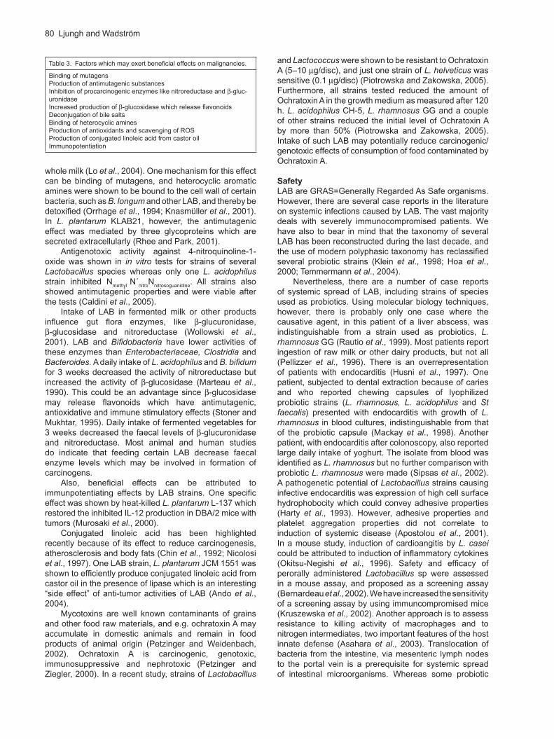
Table 3. Factors which may exert beneficial effects on malignancies.
Binding of mutagensProduction of antimutagenic substancesInhibition of procarcinogenic enzymes like nitroreductase and -gluc-uronidaseIncreased production of -glucosidase which release flavonoidsDeconjugation of bile saltsBinding of heterocyclic aminesProduction of antioxidants and scavenging of ROSProduction of conjugated linoleic acid from castor oilImmunopotentiation

Lactic Acid Bacteria as Probiotics 81
strains have been suggested to inhibit translocation some are themselves translocated which causes concern regarding safety (Ishibashi and Yamazaki, 2001).
Probiotic strains should carry few if any mechanisms for antibiotic resistance, and preferrably no plasmids with antibiotic resistance. Several of the availble probiotic Bacillus products expressed high levels of antibiotic resistance (Hoa et al., 2000).
There are few case reports of invasive disease caused by Bifidobacterium, like one with B. longum in a previously healthy 19 year old man (Ha et al.,1999). There are multiple reports of septicaemia caused by Pediococcus sp, most of them, however, by P. acidilacti
which is not used as a probiotic (Barros et al., 2001).Generally, probiotic strains carry a very low risk of
causing infection. Many probiotic products have been used traditionally over generations, and proven to be safe. Since different characteristics of strains listed above are strain specific, reports of systemic infections caused by various species should not make us exclude these species as probiotics. Each strain should be evaluated in tests for safety, but so far, there is no standard test(s) recommended.
Designing the final probiotic product
Probiotic strains can be administered to the customer in lyophilized form in sealed bags, tablets or capsules, or in yoghurt, other dairy products or in fruit juices. The storage time for lyophilised products is significantly longer. For each final product, the number of live cells has to be declared and the storage time at different temperatures clearly stated. Today, this is the case for but few products. Since probiotic microbes have different beneficial properties an optimal product may need the combination of several strains (Timmerman et al., 2004). It is then essential to show that the strains do not inhibit each other in in vivo conditions. With modern technology there are possibilities to target preparations for delivery at a specific site – stomach, small intestine, large intestine and other organs as elegantly shown for E. coli Nissle 1917 (Westendorf et al., 2005). This strain colonizes the intestinal mucosa and can be used as a carrier for targeted delivery of recombinant proteins and antigens to the intestinal mucosa.
Available data from traditional medicine and clinical use clearly state that probiotics have a great potential, particularly today with the increasing threat of antibiotic over-usage and prevalence of antibiotic resistant microorganisms. Molecular biological techniques will help us elucidate effects on the intestinal microflora, translocation, in vivo genetic transfer of markers for immune mediators as well as on safety issues (Temmermann et
al., 2004; Vaughan et al., 1999). However, randomized, double-blind, placebo-controlled case-control studies are needed.
AcknowledgmentsThe authors’ own studies were supported by the Science Research Council of Sweden (16x-04723) and by Arla Foods.
ReferencesAbd El-Gawad, I. A., El-Sayed, E. M., Hafez, S. A., El-Zeini,
H.M., and Saleh, F.A. (2005). The hypocholesterolaemic effect of milk yoghurt and soy-yoghurt containing bifidobacteria in rats fed on a cholesterol-enriched diet. Int. Dairy J. 15, 37–44.
Adlerberth, I., Ahrné, S., Johansson, M.-L., Molin, G., Hanson, L.-Å. and Wold, A. (1996). A mannose-specific adherence mechanism in Lactobacillus plantarum
conferring binding to the human colonic cell line HT-29. Appl. Environm. Microbiol. 62, 2244–2251.
Agarwal, K.N. and Bhasin, S.K. (2002). Feasibility studies to control acute diarrhoea in children by feeding fermented milk preparations Actimel and Indian Dahi. Eur. J. Clin. Nutr. 56 suppl 4, S56-S59.
Agerbaek, M., Gerdes, L.U. and Richelsen, B. (1995). Hypocholesterolemic effect of a new fermented milk product in healthy middle-aged men. Eur. J. Clin. Nutr. 49, 346–352.
Ahn, Y.T., Kim, G.B., Lim, K.S., Baek, Y.J. and Kim, H.U. (2003). Deconjugation of bile salts by Lactobacillus acidophilus isolates. Int. Dairy J. 13, 303–311.
Aleljung, P., Shen, W., Rozalska, B., Hellman, U., Ljungh, Å. and Wadström, T. (1994). Purification of collagen binding proteins of Lactobacillus reuteri NCIB 11951. Curr. Microbiol. 28, 231–236.
Ando, A., Ogawa, J., Kishino, S. and Shimizu, S. (2004). Conjugated linoleic acid production from castor oil by Lactobacillus plantarum JCM 1551. Enzyme Microb.Technol. 35, 40–45.
Annuk, H., Shchepetova, J., Kullisaar, T., Songisepp, E., Zilmer, M. and Mikelsaar, M. (2003). Characterization of intestinal lactobacilli as putative probiotic candidates. J. Appl. Microbiol. 94, 403–412.
Apostolou, E., Kirjavainen, P., Saxelin, M., Rautelin, H., Valtonen, V., Salminen, S.J. and Ouwehand, A.C. (2001). Good adhesion properties of probiotics: a potential risk for bacteremia? FEMS Immunol. Med. Microbiol. 31, 35–39.
Asahara, T., Takahasi, M., Nomoto, K., Takayama, H., Onoue, M., Morotomi, M., Tanaka, R., Yokohura, T. and Yamashita, N. (2003). Assessment of safety of Lactobacillus strains based on resistance to host innate defense mechanisms.Clin. Diagn. Labor. Immunol. 10, 169–173.
Ashar, M.A. and Prajapathi, J.B. (1998). Bile tolerance, bile deconjugation and cholesterol reducing properties of dietary Lactobacilli. Ind. J. Microbiol. 38, 145–148.
Ashar, M.N. and Prajapathi, J.B. (2000). Verification of hypocholesterolemic effect of fermented milk on human subjects with different cholesterol levels. Folia Microbiol. 45, 263–268.
Åvall-Jääskeläinen, S. and Palva, A. (2005). Lactobacillus surface layers and their applications. FEMS Microbiol. Rev. 29,511–529.
Bach, S.J., McAllister, T.A., Veira, D.M., Gannon, V.P.J. and Holley, R.A. (2003) Effects of a Saccharomyces
cerevisiae feed supplement on Escherichia coli O157:H7 in ruminal fluid in vitro. Anim. Feed Sci. Technol. 104, 179–189.

82 Ljungh and Wadström
Balsari, A., Ceccarelli, A., Dubini, F., Fesce, E., and Poli, G. (1982). The fecal microbial population in the irritable bowel syndrome. Microbiology. 5,185–194.
Barbara, G. and Corinaldesi, R. (2000). Probiotics: could they turn out to be ineffective in irritable bowel syndrome? Dig. Liver Dis. 32, 302–304.
Barros, R.R, Carvalho, M.D.G.S., Peralta, J.M., Facklam, R.R. and Teixeira, L.M. (2001). Phenotypic and genotypic characterization of Pediococcus strains isolated from human sources. J. Clin. Microbiol. 39–40, 1241–1246.
Bausserman, M. and Michail, S. (2005). The use of Lactobacillus GG in irritable bowel syndrome in children: A double blind randomized control trial. J. Ped.147,197–201.
Bergonzelli, G.E., Granato, D., Pridmore, R.D., Marvin-Guy, L.F., Donnicola, D., and Corthésy-Theulaz, I. (2006) GroEL of Lactobacillus johnsonii La1 (NCC533) is cell surface associated: Potential role in interactions with the host and the gastric pathogen Helicobacter
pylori. Infect. Immun. 74, 425–434.Bernardeau, M., Vernoux, J.P. and Gueguen, M. (2002).
Safety and efficacy of probiotic lactobacilli in promoting growth in post-weaning Swiss mice. Int. J. Food Microbiol. 77, 19–27.
Bin-Nun, A., Bromiker, R., Wilschanski, M., Kaplan, M., Rudensky, B., Caplan, M. and Hammerman, C. (2005) Oral probiotics prevent necrotizg enterocolitis in very low birth weight neonates. J. Pediatr. 147, 192–196.
Biörklund, M., Van Rees, A, Mensink, R.P., and Önning, G. (2005). Changes in serum lipids and postprandial glucose and insulin concentrations after consumption of beverages with -glucans from oats or barley: a randomized dose-controlled trial. Eur. J. Clin. Nutr. 59,
1272–1281.Blum, S., Reniero, R., Schiffrin, E.J., Crittenden, R.,
Mattila-Sandholm, T., Ouwehand, A. C., Salminen, S., von Wright, A., Saarela, M., Saxelin, M., Collins, K. and Morelli, L. (1999). Adhesion studies for probiotics: need for validation and refinement. Trends Food Sci. Technol. 10, 405–410.
Boddy, A.V., Elmer G.W., McFarland, L.V. and Levy, R.H. (1991) Influence of antibiotics on the recovery and kinetics of SaccharomyceS. boulardii in rats. Pharm. Res. 8,796–800.
Brigidi, P., Vitali, B., Swennen, E., Bazzocchi, G. and Matteuzzi, D. (2001). Effects of probiotic administration upon the composition and enzymatic activity of human fecal microbiota in patients with irritable bowel syndrome or functional diarrhea. Res. Microbiol. 152, 735–741.
Bron, P.A., Grangette, C., Mercennier, A., de Vos, W.M. and Kleerebezem, M. (2004). Identification of Lactobacillus plantarum genes that are induced in the gastrointestinal tract of mice. J. Bacteriol. 186, 5721–5729.
Calcinaro, F., Dionisi, S., Marinaro, M., Candeloro, P., Bonato, V., Marzotti, S., Corneli, R.B., Ferretti, E., Gulino, A., Grasso, F., De Simone, C., Di Mario, U., Falorni, A., Birivant, M. and Dorra, F. (2005). Oral probiotic administration induces interleukin-10 production and prevents spontaneous autoimmune diabetes in the non-obese diabetic mouse. Diabetologia 48, 1565–1572.
Caldini, G., Trotta, F., Villarini, M., Moretti, M., Pasquini, R., Scassellati-Sforzolini,G., Cenci, G. (2005). Screening of potential lactonacilli antigenotoxicity by microbial and mammalian cell-based tests. Int. J. Food Microbiol. 102, 37–47.
Castagliuolo, I., Riegler, M.F.,Valenick, L., LaMont, J.T., and Pothoulakis, C. (1999). SaccharomyceS. boulardii
protease inhibits the effects of Clostridium difficile toxin A and B in human colonic mucosa. Infect. Immun. 67,302–307.
Castagliuolo, I., Galeazzi, F., Ferrari, S., Elli, M., Brun, P., Cavaggioni, A., Tormen, D., Sturniolo, G.C., Morelli, L. and Palù, G. (2005) Beneficial effect of auto-aggregating Lactobacillus crispatus on experimentally induced colitis in mice. FEMS Immunol. Med. Microbiol. 43, 197–204.
Cats, A., Kuijpers, E.J., Bosschaert, M.A.R., Pot, R.G.J., Vandenbroucke-Grauls, C.M.J.E. and Kusters, J.G. (2003). Effect of fermented consumption of a Lactobacillus casei-containing milk drink in Helicobacter
pylori-colonized subjects. Aliment. Pharmacol. Ther. 17, 429–435.
Chandra, R.K. (2002) Effect of Lactobacillus on the incidence and severity of acute rotavirus diarrhoea in infants. A prospective placebo-controlled double-blind study. Nutr. Res. 22, 65–69.
Charteris, W.P., Kelly, P.M., Morelli, L. and Collins, J.K. (1998). Development and application of an in vitro
methodology to determine transit tolerance of potentially probiotic Lactobacillus and Bifidobacterium species in the upper gastrointestinal tract. J. Appl. Microbiol. 84, 759–768.
Chin, S.E., Liu, W., Storkson, J.M., Ha, Y.L. and Pariza, M.W. (1992). Dietary sources of conjugated dienoic isomers of linoleic acid, a newly recognized class of anticarcinogens. J. Food Comp. Anal. 5, 185–197.
Christensen, H.R, Frøkier, H. and Pestka, J.J. (2002). Lactobacilli differentially modulate expression of cytokines and maturation surface markers in murine dendritic cells. J. Immunol. 168, 171–178.
Clancy, R. (2003). Immunobiotics and the probiotic evolution. FEMS Immunol. Med. Microbiol. 38, 9–12.
Coconnier, M.-H., Liévin, V., Hemery, E. and Servin, A.L. (1998). Antagonistic activity against Helicobacter
infection in vitro and in vivo by the human Lactobacillus acidophilus strain LB. Appl. Environm. Microbiol. 64, 4573–4580.
Coconnier, M.-H., Liévin, V., Lorrot, M. and Servin, A.L. (2000). Antagonistic activity of Lactobacillus acidophilus
LB against intracellular Salmonella enterica serovar Typhimurium infecting human enterocyte-like Caco-2/TC-7 cells. Appl. Environm. Microbiol. 66, 1152–1157.
Collado, M.C., González, A., González, R., Hernández, M., Ferrús, M.A., and Sanz, Y. (2005). Antimicrobial peptides are among the antagonistic metabolites produced by Bifidobacterium against Helicobacter
pylori. Int. J. Antimicrob. Ag. 25, 385–391.Cremonini, F.,Di Caro, S., Covino, M., Armuzzi, A.,
Gabrielli, M., Santarelli,.L, Nista, E.C., Cammarota, G., Gasbarrini, G. and Gasbarrini, A. (2002). Effect of different probiotic preparations on anti-Helicobacter

Lactic Acid Bacteria as Probiotics 83
pylori therapy-related side effects: A parallel group, triple blind, placebo-controlled study. Am. J. Gastroenterol. 97, 2744–2749.
Cruchet, S., Obregon, M.C., Salazar, G., Diaz, E. and Gotteland, M. (2003). Effect of ingestion of a dietary product containing Lactobacillus johnsonii La1 on Helicobacter pylori colonization in children. Nutrition 19, 716–721.
De Boever, P., Wouters, R., Verschaeve, L., Berckmans, P., Schoeters, G. and Verstraete W. (2000). Protective effect of the bile salt hydrolase-active Lactobacillus reuteri against bile salt cytotoxicity. Appl. Microbiol. Biotechnol. 53, 709–714.
Derzelle, S., Hallet, B., Francis, K.P., Ferrain, T., Delcous, J. and Hols, P. (2000). Changes in cspL, cspP and cspC mRNA abundance as a function of cold shock and growth phase in Lactobacillus plantarum. J. Bacteriol. 182, 5105–5113.
Di Caro, S., Tao, H., Grillo, A., Elia, C., Gasbarrini, G., Sepulveda, A.R., Gasbarrini, A. (2005). Effects of Lactobacillus GG on genes expression pattern in small bowel mucosa. Dig. Liver Dis. 37, 320–329.
Doncheva, N.I., Antov, G.P., Softova, E.B. and Nyagolov, Y.P. (2002). Experimental and clinical study on the hypolipidemic and antisclerotic effect of Lactobacillus bulgaricus strain GB N 1 (48). Nutr. Res. 22, 393–403.
Drakes, M., Blanchard, T. and Czinn, S. (2004). Bacterial probiotic modulation of dendritic cells. Infect. Immun. 72, 3299–3309.
D’Souza, A.L., Rajkumar, C., Cooke, J. and Bulpitt, C.J. (2002). Probiotics in prevention of antibiotic-associated diarrhoea: meta-analysis. Brit. Med. J. 324, 1361–1367.
Duc, L.H., Hong, H.A., Barbosa, T.M., Henriques, A.O. and Cutting, S.M. (2004). Characterization of Bacillus probiotics available for human use. Appl. Environm. Microbiol. 70, 2161–2171.
Dunne, C., O’Mahony, L., Murphy, L., Thornton, G., Morrissey, D., O’Halloran, S., Feeney, M., Flynn, S., Fitzgerald, G., Daly, C., Kiely, B., O’Sullivan, G.C., Shanahan, F. and Collins, K.C. (2001).In vitro selection criteria for probiotic bacteria of human origin: correlation with in vivo findings. Am. J. Clin. Nutr. 73, 386S-392S.
Elmer, G.W., Martin, A.W., Horner, K.L., McFarland, L.V. and Levy, R.H. (1999). Survival of SaccharomyceS.
boulardii in the rat gastrointestinal tract and effects of dietary fiber. Microb. Ecol. Health Dis. 11, 29–34.
Felley, C.P. Corthésy-Theulaz, I., Blanco Rivero, J.-L., Sipponen, P., Kaufmann, M., Bauerfeind, P., Wiesel, P.H., Brassart, D., Pfeifer, A., Blum, A.L. and Michetti, P. (2001). Favourable effect of an acidified milk (LC-1) on Helicobacter pylori gastritis in man. Eur. J. Gastroenterol. Hepatol. 13, 25–29.
Forstner, J.F. and Forstner, G.G. (1994). Gastrointestinal mucus. In: Physiology of the gastrointestinal tract (3rd ed) Johnson L R (Ed) (New York, USA, Raven) pp1255–1283.
Fujiwara, S., Hashiba, H., Hirota, T. and Forstner, J.F. (1999). Purification and characterization of a novel protein produced by Bifidobacterium longum SBT2928 that inhibits the binding of enterotoxigenic Escherichia
coli Pb176 (CFA/II) to gangliotetraosylceramide. J. Appl. Microbiol. 86, 615–621.
Furrie, E., Macfarlane, S., Kennedy, A., Cummings, J.H., Walsh, S.V., O’Neill, D.A. and Macfarlane, G.T. (2005). Synbiotic therapy (Bifidobacterium longum/Synergy 1) initiates resolution of inflammation in patients with active ulcerative colitis: a randomised controlled pilot trial. Gut 54, 242–249.
Ganz, T. (2003). Defensins: antimicrobial peptides of innate immunity. Nat. Rev. Immunol. 3, 710–720.
Ghisolfi,J., Roberfroid, M., Rigo, J., Moro, G. and Polanco I. (2002). Infant formula supplemented with probiotics or prebiotics: never, now, or someday? J. Pediatr. Gastroenterol. Nutr. 35, 467–468.
Gill, H.S. (1998). Stimulation of the immune system by lactic cultures. Int. Dairy J. 8, 535–544.
Gill, H.S., Rutherfurd, K.J., Prasad, J. and Gopal, P.K. (2000). Enhancement of natural and acquired immunity by Lactobacillus rhamnosus (HN001), Lactobacillus acidophilus (HN017) and Bifidobacterium lactis (Hn019). Br. J. Nutr. 83, 167–176.
Gilliland, S.E., Nelson, C.R. and Maxwell, C. (1985). Assimilation of cholesterol by Lactobacillus acidophilus.
Appl. Environm. Microbiol. 49, 377–381.Gionchetti, P., Rizzello, F., Venturi, A., Brigidi, P., Mateuzzi,
D., Bazzocchi, G., Poggioli, G., Miglioli, M. and Campieri, M. (2000). Oral bacteriotherapy as maintenance treatment in patients with chronic pouchitis: A double-blind, placebo-controlled trial. Gastroenterology. 119, 305–309.
Gionchetti, P., Rizzello, F., Helwig, U., Venturi, A., Manon Lammers, K., Brigidi, P., Vitali, B., Poggioli, G., Miglioli, M. and Campieri, M. (2003). Prophylaxis of pouchitis onset with probiotic therapy: A double-blind, placebo-controlled trial. Gastroenterology 124,1202–1209.
Granato, D., Bergonzelli, G.E., Pridmore, R.D., Marvin, L., Rouvet, M. and Corthésy-Theulaz, I.F. (2004). Cell surface-associated elongation factor Tu mediates the attachment of Lactobacillus johnsonii NCC533 (La1) to human intestinal cells and mucins. Infect. Immun. 72, 2160–2169.
Guarner, F. and Schaafsma, G.J. (1998). Probiotics. Int.J. Food Microbiol. 39, 237–238.
Gueimonde,M., Noriega, L., Margolles, A., de los Reyes-Gavilan, C,G,. Salminen, S. (2005). Ability of Bifidobacterium strains with acquired resistance to bile to adhere to human intestinal mucus. Int. J. Food Microbiol. 101,341–346.
Gueimonde, M., Jalonen, L., He, F., Hiramtasu, M., and Salminen, S. (2006). Adhesion and competitive inhibition and displacement of human enteropathogens by selected lactobacilli. Food Res. Int. 39, 467–471.
Gupta, P., Andrew, H., Kirschner, B.S. and Guandalini, S. (2000). Is Lactobacillus GG helpful in children with Crohns’disease? Results of a preliminary, open-label study. J. Pediatr. Gastroenterol. Nutr. 31, 453–457.
Ha, G.Y., Yang, C.H., Kim, H. and Chong, Y. (1999). Case of sepsis caused by Bifidobacterium longum. J. Clin. Microbiol. 37, 1227–1228.
Harty, D.W.S., Patrikakis, M. and Knox, K.W. (1993). Identification of Lactobacillus strains isolated from patients with infective endocarditis and comparison of

84 Ljungh and Wadström
their surface-associated properties with those of other strains of the same species. Microb. Ecol. Health Dis. 6, 191–201.
Herías, M.V., Koninkx, J.F.J.G., Vos, J.G., Huis in’t Veld, J.H.J., and van Dijk, J.E. (2005). Probiotic effects of Lactobacillus casei on DSS-induced ulcerative colitis in mice. Int. J. Food Microbiol. 103, 143–155.
Hirayama, K. and Rafter, J. (1999). The role of lactic acid bacteria in colon cancer prevention: mechanistic considerations. Ant. v. Loewenhoek 76, 391–394.
Hoa, N.T., Baccigalupi, L., Huxham, A., Smertenko, A., Van, P.H., Ammendola, S., Ricca, E. and Cutting, S.M. (2000). Characterization of Bacillus species used for oral bacteriotherapy and bacteriprophylaxis of gastrointestinal disorders. Appl. Environm. Microbiol. 66, 5241–5247.
Horie, M., Ishiyama, Y., Fujihira-Ueki, Y., Sillanpää, J., Korhonen, T.K., Toba, T. (2002). Inhibition of the adherence of Escherichia coli strains to the basement membrane by Lactobacillus crispatus expressing an S-layer. J. Appl. Microbiol. 92, 393–403.
Howard J.C., Heinemann, C., Thatcher, B.J., Martin, B., Siang Gan, B. and Reid, G. (2000). Identification of collagen-binding proteins in Lactobacillus spp. with surface-enhanced laser desorption/ionization-time of flight proteinchip technology. Appl. Environm. Microbiol. 68, 4396–4400.
Humen, M.A., De Antoni, G.L., Benyacoub, J., Costas, M.E., Cardozo, M.I., Kozubsky, L., Saudan, K.-Y., Boenzli-Bruand, A., Blum, S., Schiffrin, E.J. and Pérez, P.F. (2005). Lactobacillus johnsonii La1 antagonizes Giardia intestinalis in vivo. Infect. Immun. 75, 1265–1269.
Husni, R.N., Gordon, S.M., Washington, J.A., Longworth, D.L. (1997). Lactobacillus bacteremia and endocarditis: Review of 45 cases. Clin. Infect. Dis. 25:1048–1055.
Hyönen, U., Westerlund-Wikström, B., Palva, A. and Korhonen, T.K. (2002) Identification by flagellum display of an epithelial cell- and fibronectin-binding function in the SipA surface protein of Lactobacillus brevis. Infect. Immun. 184, 3360–3367.
Ishibashi, N. and Yamazaki, S. (2001). Probiotics and safety. Am. J. Clin. Nutr. 73 (suppl), 465S-470S.
Jamuna, M. and Jeevaratnam, K. (2004). Isolation and partial characterization of bacteriocins from Pediococcus
species. Appl. Microbiol. Biotechnol. 65, 433–439.Jijon, H., Backer, J., Diaz, H., Yeung, H., Thiel, D.,
McKaigney, C., de Simone, C. and Madsen, K. (2004). DNA from probiotic bacteria modulates murine and human epithelial and immune function. Gastroenterology 126, 1358–1373.
Kalliomäki, M., Salminen, S., Arvilommi, H., Kero, P., Koskinen, P. and Isolauri, E. (2001). Probiotics in primary prevention of atopic disease: a randomised placebo-controlled trial. Lancet 357, 1076–1079.
Kato, I., Tanaka, K. and Yokokura, T. (1999). Lactic acid bacterium potently induces the production of interleukin-12 and interferon- by mouse splenocytes. Int. J. Immuno-pharmacol. 21, 121–131.
Kikuchi-Hayakawa, H., Shibahara-Sone, H., Osada, K., Onodera-Masuoka, N., Ishikawa, F. and Watanuki, M. (2000). Lower plasma triglyceride level in Syrian
hamsters fed on skim milk fermented with Lactobacillus casei strain Shirota. Biosci. Biotechnol. Biochem. 64, 466–475.
Kim, H.J., Camilleri, M., McKenzie, S., Lempke, M.B. and Burton, D.D. (2003). A randomized controlled trial of a probiotic, VSL#3, on gut transit and symptoms in diarrhea predominant irritable bowel syndrome. Alim. Pharmacol. Ther. 17,895–904.
Kim, W.S., Ren, J. and Dunn, N.W. (1999). Differentiation of Lactococcus lactis subspecies lactis and subspecies cremoris by their adaptive response to stresses. FEMS Microbiol. Lett. 171, 57–65.
King, T.S., Elia, M., and Hunter J.O. (1998). Abnormal colonic fermentation in irritable bowel syndrome. Lancet 352,1187–1189.
Kirjavainen, P.V., Ouwehand, A.C., Isolauri, E. and Salminen, S.J. (1998). The ability of probiotic bacteria to bind to human intestinal mucus. FEMS Microbiol. Lett. 167, 185–189.
Klein, G., Pack, A., Bonaparte, C. and Reuter, G. (1998). Taxonomy and physiology of probiotic lactic acid bacteria. Int. J. Food Microbiol. 41, 103–125.
Klingberg,T.D., Herold Oedersen, M., Cencic, A. and Bjorn Budde, B. (2005). Application of measurements of transepithelial electrical resistance of intestinal epithelial cell monolayers to evaluate probviotic activity. (2005). Appl. Environm. Microbiol. 71,7528–7530.
Kmet, V., Callegari, M.L., Bottazzi, V. and Morelli, L. (1995). Aggregation-promoting factor in pig intestinal Lactobacillus strains. Lett. Appl. Microbiol. 21, 351–353.
Knasmüller, S., Steinknéllner, H., Hirschl, A.M., Rabot, S., Nobis, E.C. and Kassle, F. (2001). Impact of bacteria in dairy products and of intestinal microflora on the genotoxic and carcinogenic effects of heterocyclic aromatic amines. Mut. Res. 480–481, 129–138.
Koenen, M.E., van der Hulst, R., Leering, M., Jeurissen, S.H.M. and Boersma, W.J.A. (2004). Development and validation of a new in vitro assay for selection of probiotic bacteria that express immune-stimulating properties in chicken in vivo. FEMS Immunol. Med. Microbiol. 40, 119–127.
Kolenbrander, P.E. (2000). Oral microbial communities: biofilms, interactions, and genetic systems. Ann. Rev. Microbiol. 54: 413–439.
Kruszewska, D., Lan, J., Lorca, G., Yanagisawa, N., Marklinder, I. and Ljungh, Å. (2002). Selection of Lactic Acid Bacteria as probiotic strains by in vitro tests. Microb. Ecol. Health Dis. 29;37–49.
Kruszewska, D., Ljungh, Å., Hynes, S.O., Pierzynowski, S.G. (2004). Effect of the antibacterial activity of pig pancreatic juice on human multi-resistant bacteria. Pancreas 128, 191–199.
Lankaputhra, W.E.V. and Shah, N.P. (1998). Antimutagenic properties of probiotic bacteria and of organic acids. Mut. Res. 397, 169–182.
Lee, Y.-J., Yu, W.-K. and Heo, T.-R. (2003A). Identification and screening for antimicrobial activity against Clostridium difficile of Bifidobacterium and Lactobacillus species isolated from healthy infant faeces. Int. J. Antimicrob. Ag. 21, 340–346.

Lactic Acid Bacteria as Probiotics 85
Lee, Y.-K., Phuong, K.-Y., Ouwehand, A.C. and Salminen, S. (2003B). Displacement of bacterial pathogens from mucus and Caco-2 cell surface by lactobacilli. J. Med. Microbiol. 52, 925–930.
Lin, M.-Y., and Chang, F.-J. (2000). Antioxidative effect of intestinal bacteria Bifidobacterium longum ATCC 15708 and Lactobacillus acidophilus ATCC 4356. Dig. Dis. Sci. 45, 1617–1622.
Lin, H.-C., Su, B.-H., Chen, A.-C., Lin, T.-W., Tsai, C.-H., Yeh, T.-F. and Oh, W. (2005). Oral probiotics reduce the incidence and severity of necrotizing enterocolitis in very low birth weight infants. Pediatrics 115,1–4.
Llopis, M., Antolin, M., er, F., Salas, A. and Malagelada, J.-R. (2005). Mucosal colonization with Lactobacillus casei mitigates barrier injury induced by exposure to trinitrobenzene sulphonic acid. Gut 54, 855–959.
Lo, P.-R., Yu, R.-C., Chou, C.-C. and Huang, E.-C. (2004). Determinations of the antimutagenic activities of several probiotic bifidobacteria under acidic and bile conditions against benzo[a]pyrene by a modified Ames test. Int. J. Food Microbiol. 93, 249–257.
Lorca, G.L. and Font de Valdez, G. (1999). The effect of suboptimal growth temperature and growth phase on resistance of Lactobacillus acidophilus to environmental stress. Cryobiology 39, 144–149.
Lorca, G.L. and Font de Valdez, G. (2001). Acid tolerance mediated by membrane ATPases in Lactobacillus acidophilus. Biotechnol. Lett. 23, 777–780.
Lorca, G.L., Wadström, T., Font de Valdez, G. and Ljungh, Å. (2001). Lactobacillus acidophilus autolysins inhibit Helicobacter pylori in vitro. Curr. Microbiol. 42, 39–44.
Lorca, G., Torino, M.I., Font de Valdez, G. and Ljungh, Å. (2002A). Lactobacilli express cell surface proteins which mediate binding of immobilized collagen and fibronectin. FEMS Microbiol. Lett. 206, 31–37.
Lorca, G., Font de Valdez, G. and Ljungh, Å. (2002B). Characterizarion of the protein-synthesis dependent acid tolerance response in Lactobacillus acidophilus. J. Molec. Microbiol. Biotechnol. 4:59–65.
Losada, M.A. and Olleros, T. (2002). Towards a healthier diet for the colon: the influence of fructooligosaccharides and lactobacilli on intestinal health. Nutr. Res. 22, 71–84.
Lund, B. and Edlund, C. (2001). Probiotic Enterococcus
faecium strains is a possible recipient of the vanA gene cluster. Clin. Infect. Dis. 32, 1384–1385.
Maassen,C.B.M., van Holten-Neelen, C., Balk, F., Heijne den Bak-Glashouwer, M.-J., Leer, R.J., Laman, J.D., Boersma, W.J.A, and Claassen, E. (2000). Strain-dependent induction of cytokine profiles in the gut by orally administered Lactobacillus strains. Vaccine 18, 2613–1623.
Mack, D.R., Michail, S., Wei, S., McDougall, L. and Hollingsworth, M.A. (1999). Probiotics inhibit enteropathogenic E. coli adherence in vitro by inducing intestinal mucin expression. Am. J. Physiol. (Gastrointest. liver physiol.) 276, G941-G950.
Mack, D.R., Ahrné, S., Hyde, L., Wei, S. and Hollingworth, M.A. (2003). Extracellular MUC3 mucin secretion follows adherence of Lactobacillus strains to intestinal epithelial cells in vitro. Gut 52, 827–833.
Mackay, A.D., Taylor, M.B., Kibbler, C.C. and Hamilton-Miller, J.M.T. (1998). Lactobacillus endocarditis caused by a probiotic organism. Clin. Microbiol. Infect. 5, 290–292.
Madsen, K., Cornish, A., Soper, P., McKaigney, C., Jijon, H., Yachimec, C., Doyle, J., Jewell, L. and de Simone, C. (2001). Probiotic bacteria enhance murine and human intestinal epithelial barrier function. Gastroenterology 121, 580–591.
Magnusson, J., Ström, K., Roos, S., Sjögren, J. and Schnürer, J. (2003). Broad and complex antifungal activity among environmental isolates of lactic acid bacteria. FEMS Microbiol. Lett. 219, 129–135.
Marteau, P., Rochart, P., Flourie, B., Pellier, P., Santos, L., Desjeux, J.F., and Rambaud, J.C. (1990). Effect of chronic ingestion of a fermented dairy product containing Lactobacillus acidophilus and Bifidobacterium bifidum on metabolic activities of the colonic flora in humans. Am. J. Clin. Nutr. 52, 685–688.
Mastretta, E., Longo, P., Laccisaglia, A., Balbo, L., Russo, R., Mazzaccara, A. and Gianino, P. (2002). Effect of Lactobacillus GG and breast-feeding in the prevention of rotavirus nosocomial infection. J. Ped. Gastroenterol. Nutr. 35, 527–531.
Matsuguchi, T., Takagi, A., Matsuzaki, T., Nagaoka, M., Ischikawa, K., Yokohura, T. and Yoshikai, Y. (2003). Lipoteichoic acids from Lactobacillus strains elicit strong tumor necrosis factor alpha-inducing activities in macrophages through Toll-like receptor 2. Clin. Diagn. Labor. Immunol. 10, 259–266.
Matsumoto, M., Ohishi, H. and Benno, Y. (2004). H+-ATPase activity in Bifidobacterium with special reference to acid tolerance. Int. J. Food Microbiol. 93, 109–113.
Matsumoto, S., Hara, T., Hori, T., Mitsuyama, K., Nagaoka, M., Tomiyasu, N., Suzuki, a., and Sata, M. (2005).Probiotic Lactobacillus-induced improvement in murine chronic inflammatory bowel disease is associated with the down-regulation of pro-inflammatory cytokines in lamina propria mononuclear cells. Clin. Exp. Immunol. 140, 417–426.
Mazza, P. (1994). The use of Bacillus subtilis as an antidiarrhoeal microorganism. Boll. Chim. Farm. 133, 3–18.
McAuliffe, O., Cano, R.J., Klaenhammer, T.R. (2005). Genetic analysis of two bile salt hydrolase activities in Lactobacillus acidophilus NCFM. Appl. Environm. Microbiol. 71, 4925–4929.
McCarthy, J., O’Mahony, L., O’Callaghan, L., Sheil, B., Vaughan, E.E., Fitzimons, N., Fitzgibbon, J., O’Sullivan, G.C., Kiely, B., Collins, J.K. and Shanahan, F. (2003). Double blind, placebo controlled trial of two probiotic strains in interleukin 10 knockout mice and mechanistic link with cytokine balance. Gut 52, 975–980.
Ménard, S., Candalh, C., Bambou, J.C., Terpend, K., Cerf-Bensussan, N. and Heyman, M. (2004). Lactic acid bacteria secrete metabolites retaining anti-inflammatory properties after intestinal transport. Gut 53, 821–828.
Metschnikoff, E. (1907). The prolongation of life. Optimistic studies (London, UK, William Heinemann).
Michail, S. and Abernathy, F. (2003). Lactobacillus plantarum inhibits the intestinal transepithelial migration

86 Ljungh and Wadström
of neutrophils induced by enteropathogenic Escherichia
coli. J. Pediatr. Gastroenterol. Nutr. 36, 385–391.Michalkiewicz, J., Krotkiewski, M., Gackowska,
L., Wyszomirska-Golda, M., Helmin-Basa, A., Dzierzanowska, D. and Madalinski, K. (2003). Immunomodulatory effects of Lactic Acid bacteria on human peripheral blood mononuclear cells. Microb. Ecol. Health Dis. 15, 185–192.
Michetti, P., Dorta, G., Wiesel, P.H., Brassart, D., Verdu, E., Herranz, M., Felley, C., Porta, N., Rouvet, M., Blum, A.L. and Corthésy-Theulaz, I. (1999). Effect of whey-based culture supernatant of Lactobacillus acidophilus
(johnsonii) La1 on Helicobacter pylori infection in humans. Digestion 60, 203–209.
Mimura, T., Rizzello, F., Helwig, U., Poggioli, G., Schreiber, S., Talbot, I.C., Nicholls, R.J., Gionchetti, P., Campieri, M. and Kamm, M.A. (2004). Once daily high dose probiotic therapy (VSL#3) for maintaining remission in recurrent or refractory pouchitis. Gut 53, 108–114.
Morita, H., He, F., Fuse, T., Ouwerhand, A.C., Hashimoto, H., Hosoda, M., Mizumachi, K. and Kurisaki, J.-I. (2002). Cytokine production by the murine macrophage cell line J774.1 after exposure to Lactobacilli. Biosci. Biotechnol. Biochem. 66, 1963–1966.
Mottet, C. and Michetti, P. (2005). Probiotics: wanted dead or alive. Dig. Liver Dis. 37, 3–6.
Mukai, T., Kaneko, S. and Ohori, H. (1998). Haemagglutination and glycolipid-binding activities of Lactobacillus reuteri. Lett. Appl. Microbiol. 27, 130–134.
Mukai, T., Asasaka, T., Sato, E., Mori, K., Matsumoto, M. and Ohori, H. (2002). Inhibition of binding of Helicobacter pylori to the glycolipid receptors by probiotic Lactobacillus reuteri. FEMS Immunol. Med. Microbiol. 32, 105–110.
Murosaki, S., Muroyama, K., Yamamoto, Y. and Yoshikai, Y. (2000). Antitumor effect of heat-killed Lactobacillus plantarum L-137 through restoration of impaired interleukin-12 production in tumor-bearing mice. Cancer Immunol. Immunother. 49, 157–164.
Nakajima, H., Suzuki, Y., Kaiza, H., and Hirota, T. (1992). Cholesterol lowering effect of ropy fermented milk. J. Food Sci. 57, 1327–1329.
Nesser, J.-R., Granato, D., Rouvet, M., Servin, A., Teneberg, S. and Karlsson, K.-A. (2000). Lactobacillus johnsonii La1 shares carbohydrate-binding specificities with several enteropathogenic bacteria. Glycobiology 10, 1193–1199.
Nicolosi, R J., Rogers, E.J., Kritschevsky, D., Scimeca, J.A. and Huth, P.J. (1997). Dietary conjugated linoleic acid reduces plasma lipoproteins and early aortic atherosclerosis in hypercholesterolemic hamsters. Artery 22, 266–277.
Okitsu-Negishi, S., Nakano, I., Suzuki, K., Hashira, S., Abe, T. and Yoshino, K. (1996). The induction of cardioangitis by Lactobacillus casei cell wall in mice. Clin. Immunol. Immunopathol. 78, 30–40.
Orrhage, K., Sillerström, E., Gustafsson, J.-Å., Nord, C.-E. and Rafter, J. (1994). Binding of mutagenic heterocyclic amines by intestinal and lactic acid bacteria. Mut. Res. 311, 239–248.
Ouwehand, A.C. and Salminen, S. (2003). In vitro adhesion assays for probiotics and their in vivo relevance: a review. Microb. Ecol. Health Dis. 15, 175–184.
Pantoflickova, D., Corthésy-Theulaz, I., Dorta, G., Stolte, M., Isler, P., Rocjat, F., Enslen, M. and Blum, A.L. (2003). Favourable effect of regular intake of fermented milk containing Lactobacillus johnsonii on Helicobacter
pylori associated gastritis. Aliment. Pharmacol. Ther. 18, 805–813.
Paton, A.W., Morona, R. and Paton, J.C. (2000). A new biological agent for treatment of Shiga toxigenic Escherichia coli infections and dysentery in humans. Nature Med. 6, 265–270.
Paton, J.V., Rogers, T.J., Morona, R. and Paton, A.W. (2001). Oral administration of formalin-killed recombinant bacteria expressing a mimic of the Shiga toxin receptor protects mice from fatal challenge with Shiga toxigenic Escherichia coli. Infect. Immun. 69,
1389–1393.Paton, A.W., Jennings, M.P., Morona, R., Wang, H.,
Focareta, A., Roddam, L.F. and Paton, J.C. (2005). Recombinant probiotics for treatment and prevention of enterotoxigenic Escherichia coli diarrhea. Gastroenterology. 128, 1219–1228.
Pellizzer, G., Benedetti, P., Biavasco, F., Manfrin, V., Franzetti, M., Scagnelli, M., Scarparo, C., and de Lalla, F. (1996). Bacterial endocarditis due to Lactococcus lactis subsp cremoris: case report. Clin. Microbiol. Infect. 2,230–232.
Pena, J.A. and Versalovic, J. (2003). Lactobacillus rhamnosus GG decreases TNF- production in lipopolysacharide-activated murine macrophages by a contact-independent mechanism. Cell. Microbiol. 5, 277–285.
Pena, J.A., Rogers, A.B., Ge, Z., Ng, V., Li, S.Y., Fox, J.G. and Versalovic, J. (2005). Probiotic Lactobacillus spp diminish Helicobacter hepaticus-induced inflammatory bowel disease in Interleukin-10-deficient mice. Infect. Immun. 73, 912–920.
Pereira, D.I.A. and Gibson, G.R. (2002). Effects of consumption of probiotics and prebiotics on serum lipid levels in humans. Crit. Rev. Biochem. Molec. Biol. 37, 259–281.
Pérez, P.F., Minnard, J., Bouvet, M., Knabenhans, C., Brassart, D., De Antoni, G. and Schiffrin, E.J. (2001). Inhibition of Giardia intestinalis by extracellular factors from Lactobacilli: an in vitro study. Appl. Environm. Microbiol. 47, 5087–5092.
Pessi, T., Sütas, Y., Hurme, M. and Isolauri, E. (2000). Interleukin-10 generation in atopic children following oral Lactobacillus rhamnosus GG. Clin. Exp. Allergy
30, 1804–1808.Petrof, E.O., Kojima, K., Ropeleski, M.J., Musch, M.W.,
Tao, Y., de Simone, C. and Chang, E.B. (2004). Probiotics inhibit nuclear factor - B and induce heat shock proteins in colonic epithelial cells through proteasome inhibition. Gastroenterol. 127, 1474–1487.
Petzinger, E. and Ziegler, K. (2000).Ochratoxin A from a toxicological perspective. J. Vet. Pharmacol. Therap. 23, 91–98.

Lactic Acid Bacteria as Probiotics 87
Petzinger, E. and Weidenbach, A. (2002). Mycotoxins in the food chain: the role of ochratoxins. Livestock Prod. Sci. 76, 245–250.
Pinchuk, I.V., Bressollier, P., Verneuil, B., Fenet, B., Sorokulova, I.B., Mégraud, F. and Urdaci, M.C. (2001). In vitro anti-Helicobacter pylori activity of the probiotic strain Bacillus subtilis 3 is due to secretion of antibiotics. Antimicrob. Ag. Chemother. 45, 3156–3161.
Piotrowska, M. and Zakowska, Z. (2005). The elimination of Ochratoxin A by Lactic Acid Bacteria strains. Polish J. Microbiol. 54, 279–286.
Pridmore, R.D., Berger, B., Desiere, F., Vilanova, D., Barretto, C., Pittet, A.C., Zwahlen, M.C., Rouvet, M., Attermann, E., Barrangou, R., Mollet, B., Mercenier, A., Klaenhammer, T.R., Arigoni, F., and Schell, M.A. (2004). The genome sequence of the probiotic intestinal bacterium Lactobacillus johnsonii NCC 533. Proc. Natl. Acad. Sci. USA 101, 2512–2517.
Prioult, G., Fliss, I. and Pecquet, S. (2003). Effect of probiotic bacteria on induction and maintenance of oral tolerance to -lactoglobulin in gnotobiotic mice. Clin. Diagn. Lab. Immunol. 10, 787–792.
Rachmilewitz, D., Katakura, K., Karmeli, F., Hayashi, T., Reinus, C., Rudensky, B., Akira, S., Takeda, K., Lee, J., Takabayashi, K. and Raz, E. (2004). Toll-like receptor 9 signaling mediates the anti-inflammatory effects of probiotics in murine experimental colitis. Gastroenterology 126, 520–528.
Rautava, S., Kalliomäki, M. d Isolauri, E. (2002). Probiotics during pregmancy and breast-feeding might confer immunomodulatory protection against atopic disease in the infant. J. Allergy Clin. Immunol. 109, 119–121.
Rautio, M., Jousimies-Somer, H., Kauma, H., Pietarinen, I., Saxelin, M., Tynkynen, S., Koskela, M. (1999). Liver abscess due to a Lactobacillus rhamnosus
indistinguishable from L. rhamnosus strain GG. Clin. Infect. Dis. 28,1159–1160.
Rayes, N., Seehofer, D., Theruvath, T., Schiller, R.A., Langrehr, J.M., Jonas, S. Bengmark, S. and Neuhaus, P. (2005). Supply of pre- and probiotics reduces bacterial infection rates after liver transplantation – a randomized, double-blind trial. Am J. Transplant. 5,125–130.
Resta-Lenert, S. and Barrett, K.E. (2003). Live probiotics protect intestinal epithelial cells from the effects of infection with enteroinvasive Escherichia coli (EIEC). Gut 52, 988–997.
Rhee, C.-H. and Park, H.-D. (2001). Three glycoproteins with antimutagenic activity identified in Lactobacillus plantarum KLAB21. Appl. Environm. Microbiol. 67, 3445–3449.
Rinkinen, M., Westermarck, E., Salminen, S. and Ouwehand, A.C. (2003). Absence of host specificity for in vitro adhesion of probiotic lactic acid bacteria to intestinal mucus. Vet. Microbiol. 97, 55–61.
Roberfroid, M.B. (2001). Prebiotics. Preferential substrates for specific germs. J. Clin. Nutr. 73, 406S-409S.
Roos, S. and Jonsson, H. (2002). A high-molecular-mass cell-surface protein from Lactobacillus reuteri 1063 adheres to mucus components. Microbiol. 148, 433–442.
Roos, S., Lindgren, S., and Jonsson, H. (1999). Autoaggregation of Lactobacillus reuteri is mediated
by a putative DEAD-box helicase. Molec. Microbiol. 32, 427–436.
Rosenfeldt, V., Michaelsen, K.F., Jakobsen, M., Larsen, C.N., Moller, P.L., Tvede, M., Weyrehter, H., Valerius, N.H. and Paerregaard, A. (2002A). Effect of probiotic Lactobacillus strains on acute diarrhea in a cohort of nonhospitalized children attending day-care centers. Pediatr. Infect. Dis. J. 21, 417–419.
Rosenfeldt, V., Michaelsen, K.M., Jakobsen, M., Larsen, C.N., Moller, P.L., Pedersen, P., Tvede, M., Weyrehter, H., Valerias, N.H. and Paerregaard, A. (2002B). Effect of probiotic Lactobacillus strains in young children hospitalized with acute diarrhea. Pediatr. Infect. Dis. J. 21, 411–416.
Rosenfeldt, V., Benfeldt, E., Valerius, N.H., Paerregaard, A., Michaelsen, K.F. (2004). Effect of probiotics on gastrointestinal symptoms and small intestinal permeability in children with atopic dermatitis. J. Pediatr. 145, 612–616.
Sakamoto, I., Igarashi, M., Kimura, K., Takagi, A., Miwa, T. and Koga,Y. (2001). Suppressive effect of Lactobacillus gasseri OLL 2716 (LG21) on Helicobacter pylori infection in humans. J. Antimicrob. Chemother. 47, 709–710.
Savaiano, D.A. and Levitt, M.D. (1987). Milk intolerance and microbe-containing dairy foods. J. Dairy Sci. 70, 397–406.
Saxelin, M., Grenov, B., Svensson, U., Fondén, R., Reniero, R. and Mattila-Sandholm, T. (1999) The technology of probiotics. Trends Food Sci. Technol. 10, 387–392.
Schiffrin, E.J., Brassart, D., Servin, A., Rochat, F. and Donnet-Hughes, A. (1997). Immune modulation of blood leukocytes in humans by lactic acid bacteria: criteria for strain selection. Am. J. Clin. Nutr. 66, 515S-520S.
Schmitz, H., Barmeyer, C., Fromm, M., Runkel, N., Foss, H.D., Bentzel, C.J., Riecken, E.O.D., Schulzke, J.D. (1999). Altered tight junction structure contributes to the impaired epithelial barrier function in ulcerative colitis. Gastroenterology 116, 301–309.
Schultz, M., Veltkamp, C., Dieleman, L.A., Grenther, W.B., Wyrick, P.B., Tonkonogy, S.L. and Sartor, R.B. (2002). Lactobacillus plantarum 299v in the treatment and prevention of spontaneous colitis in interleukin-10-deficient mice. Inflamm. Bowel Dis. 8, 71–80.
Schulz, M., Munro, K., Tannock, G.W., Melchner, I., Göttl, C., Schwietz, H., Schölmerich, J. and Rath, H.C. (2004). Effects of feeding a probiotic preparation (SIM) containing inulin on the severity of colitis and on the composition of the intestinal microflora in HLA-B27 transgenic rats. Clin. Diagn. Lab. Immunol. 11,581–587.
Selimoglu, M.A. (2006). Supplemented infant formulas: Which is the best? Curr. Nutr. Food Sci. 2, 59–67.
Sen, S., Mullan, M.M., Parker, T.J., Woolner, J.T., Tarry, S.A. and Hunter, J.O. (2002). Effects of Lactobacillus plantarum 299v on colonic fermentation and symptoms of irritable bowel syndrome. Dig. Dis. Sci. 47, 2615–2620.
Senesi, S., Celandroni, F., Tavanti, A. and Ghelardi, E. (2001). Molecular characterization and identification of Bacillus clausii strains marketed for use in oral

88 Ljungh and Wadström
bacteriotherapy. Appl. Environm. Microbiol. 67, 834–839.
Sgouras, D., Maragkoudakis, P., Petraki, K., Martinez-Gonzalez, B., Eriotou, E., Michopoulos, S., Kalatzopoulos, G., Tsakalidou, E. and Mentis, A. (2004). In vitro and in vivo inhibition of Helicobacter pylori by Lactobacillus casei strain Shirota. Appl. Environm. Microbiol. 70, 518–526.
Shanahan, F. (2004). Probiotics in inflammatory bowel disease – therapeutic rationale and role. Adv. Drug Deliv. Rev. 56, 809–818.
Sherman, P.M., Johnson.-Henry, K.C., Yeong, H.P., Ngo, P.S.C., Goulet, J., and Tompkins, T.A. (2005). Probiotics reduce enterohemnorrhagic Escherichia coli O157:H7- and enteropathogenic E. coli O127:H6-induced changes in polarized T84 epithelial cell monolayers by reducing bacterial adhesion and cytoskeletal rearrangements. Infect. Immun. 73,5183–5188.
Shu, Q., Lin, H., Rutherford, K.J., Fenwick, S.G., Prasad, J., Gopal, P.K. and Gill, H.S. (2000). Dietary Bifidobacterium lactis (HN019) enhances resistance to oral Salmonella typhimurium infection in mice. Microbiol. Immunol. 44, 213–222.
Sillanpää, J., Martinez, B., Antikainen, J., Toba, T., Kalkkinen, N., Tankka, S., Lounatmaa, K., Keränen, J., Höök, M., Westerlund-Wiksten, B.,Virkola, R., Westerlund, B., Pouwels, P.H. and Korhonen, T.K. (1995). A collagen-binding S-layer protein in Lactobacillus crispatus. Appl. Environm. Microbiol. 61, 2467–2471.
Silva, S.H., Vieira, E.C., Dias, R.S. and Nicoli, J.R. (2001). Antagonism against Vibrio cholerae by diffusible substances produced by bacterial components of the human faecal microbiota. J. Med. Microbiol. 50, 161–164.
Simakachorn, N., Pichaipat, V., Rithipornpaisarn, P., Kongkaew, C., Tongpradit, P. and Varavithya, W. (2000). Clinical evaluation of the addition of lyophilized, heat-killed Lactobacillus acidophilus LB to oral rehydration therapy in the treatment of acute diarrhea in children. J. Ped. Gastroenterol. Nutr. 30, 68–72.
Sipsas, N.V., Zonios, D.I. and Kordossis, T. (2002). Safety of Lactobacillus strains used as probiotic agents. Clin. Infect. Dis. 34, 1283–1284.
Smit, E., Oling, F., Demel, R., Martinez, B. and Pouwles, P.H. (2001). The S-layer protein of L. acidophilus ATCC 4356: identification and characterisation of domains responsible for S-protein assembly and cell wall binding. J. Molec. Biol. 305, 245–257.
Smith, C.J., Kaper, J.B. and Mack, D.R. (1995). Intestinal mucus inhibits adhesion of human enteropathogenic Escherichia coli to Hep-2 cells. J. Pediatr. Gastroenterol. Nutr. 21, 269–276.
Stoner, G.D. and Mukhtar, H. (1995). Polyphenols as cancer chemopreventive agents. J. Cell. Biochem. 22, 169–180.
Strus, M., Marewicz, E., Kukla, G., Ruranska-Smutnicka, D., Przondo-Mordarska, A. and Heczko, P.B. (2001). Surface properties of Lactobacillus strains of human origin. Microb. Ecol. Health Dis. 13, 240–245.
Swidsinski, A., Ladhoff, A., Pernthaler, A., Swidsinski, S., Loenig-Baucke, V., Ortner, M., Weber, J., Hoffman, U.,
Schreiber, S., Dietel, M. and Lochs, H. (2002). Mucosal flora in inflammatory bowel disease. Gastroenterology 122, 44–54.
Szymanski, H., Pejcz, J., Jaw, M., Chmielarcyks, A., Strus, M. and Heczko, P.B. (2006). Treatment of acute infectious diarrhoea in infants and children with a mixture of three Lactobacillus rhamnosus strains – a randomized, double-blind, placebo-controlled trial. Aliment. Pharmacol. Ther. 23,247–253.
Takahashi, M., Taguchi, H., Yamaguchi, H., Osaki, T., Komatsu, A. and Kamiya, S. (2004). The effect of probiotic treatment with Clostridium butyricum on enterhemorrhagic Escherichia coli O157:H7 infection in mice. FEMS Immunol. Med. Microbiol 41, 219–226.
Tanaka, H., Hashiba, H., Kok, J. and Mierau, I. (2000). Bile salt hydrolase of Bifidobacterium longum. Appl. Environm. Microbiol. 66, 2502–2512.
Tannock, G.W. (2002). Probiotics and prebiotics. Where are we going? (Norfolk, UK: Caister Acad Press).
Temmermann, R., Huys, G. and Swings, J. (2004). Identification of lactic acid bacteria: culture-dependent and culture-independent methods. Trends Food Science Technol. 15, 348–359.
Thibault, H., Aubert-Jacquin, C. and Gouket, O. (2004). Effects of long-term consumption of a fermented infant formula (with Bifidobacterium breve c50 and StreptococcuS. thermophilus 065) on acute diarrhea in healthy infants. J. Ped. Gastroenterol. Nutr. 39, 147–152.
Thomas, M.R., Litin, S.C., Osmon, D.R., Corr, A. P., Weaver, A.L. and Lohse, C.M. (2001). Lack of effect of Lactobacillus GG on antibiotic-associated diarrhea: A randomized, placebo-controlled trial. Mayo Clin. Proc. 76, 883–889.
Timmerman H M, Koning C J M, Mulder L, Rombouts F M and Beynen A C (2004). Monostrain, multistrain and multispecies probiotics – A comparison of functionality and efficacy. Int. J. Food Microbiol. 96, 219–233.
Vadillo-Rodriguez, Busscher, H.J., van der Mei, H.C., de Vries, J., Norde, W. (2005). Role of lactobacillus cell surface hydrophobicity as probed by AFM in adhesion to surfaces at low and high ionic strength. Coll. Surf. B: Biointerfaces 41,33–41.
Van der Mei H. C, van de Belt-Gritter B, Pouwels P. H, Martinez B and Busscher H. J (2003). Cell surface hydrophobicity is conveyed by S-layer proteins – a study in recombinant lactobacilli. Colloids Surf B: Biointerfaces 28, 127–134.
Vanderhoof, J.A., Whitney, D.B., Antonson, D.L., Hanner, T.L., Lupo, J.V. and Young, R.J. (1999). Lactobacillus GG in the prevention of antibiotic-associated diarrhea in children. J. Pediatr. 135,564–568.
Vaughan, E.E., Heilig, H.G.H.J., Zoetendal, E.G., Satokari, R, Collins J K, Akkermans A D L and de Vos, W.M. (1999). Molecular approaches to study probiotic bacteria. Trends Food Sci. Technol. 10, 400–404.
Ventura, M., Jankovic, I., Walker, D.C., Pridmore, R.D. and Zink, R. (2002). Identification and characterization of novel surface proteins in Lactobacillus johnsonii and Lactobacillus gasseri. Appl. Environm. Microbiol. 68, 6172–6181.

Lactic Acid Bacteria as Probiotics 89
Von der Weid,T,. Bulliard, C. and Schiffrin, E.J. (2001). Induction by a lactic acid bacterium of a population of CD4+ T cells with low proliferative capacity that produce transforming growth factor and interleukin-10. Clin. Diagn. Labor. Immunol. 8, 695–701.
Wadström, T., Andersson, K., Sydow, M., Axelsson, L., Lindgren, S. and Gullmar, B. (1987). Surface properties of lactobacilli isolated from the small intestine of pigs. J. Appl. Microbiol. 62, 513–520.
Wang, K.-Y., Li, S.-N., Liu, C.-S., Perng, D.-S., Su, Y.-C., Wu, D.-C., Jan, C.-M., Lai C.H., Wehkamp, J., Harder, J., Weichental, M., Mueller, O., Herrlinger, K.R., Fellermann, K., Schroeder, J.M. and Stange, E.F. (2003). Inducible and constitutive beta-defensins are differentially expressed in Crohn’s disease and ulcerative colitis. Inflamm. Bowel Dis. 9, 215–223.
Wang, K.-Y., Li, S.-N., Liu, C.-S., Perng, D.-S., Su, Y.-C., Wu, D.-C., Jan, C.-M., Lai, C.H., Wang, T.-N. and Wang, W.-M. (2004). Effects of ingesting Lactobacillus-
and Bifidobacterum-containing yougurt in subjects with colonized Helicobacter pylori. Am. J. Clin. Nutr. 80, 737–741.
Wehkamp, J., Harder, J., Weichtal, M., Mueller, O., Herrlinger, K.R., Fellermann, K., Schroeder, J.M. and Stange, E.F. (2003). Inducible and constitutive beta-defensins are differentially expressed in Crohn’s disease and ulcerative colitis. Inflamm. Bowel Dis. 9,215–223.
Wehkamp, J., Harder, J., Wehkamp, K., Wehkamp-Meissner, B., Schlee, M., Enders, C., Sonnenborn, U., Nuding, S., Bengmark, S., Fellermann, K., Schröder, J.M. and Stange, E.F. (2004). NF- B and AP-1-mediated induction of human beta defensin-2 in intestinal epithelial cells by Escherichia coli Nissle 1917: A novel effect of a probiotic bacterium. Infect. Immun. 72, 5750–5758.
Weizman, Z., Asli, G. and Alsheikh, A. (2005). Effect of a probiotic infant formula on infections in child care
centers: Comparison of two probiotic agents. Pediatrics 115, 5–9.
Weston, s., Halbert, A., Richmond, P. and Prescott, S.L. (2005). Effects of probiotics on atopic dermatitis: a randomised controlled trial. Arch. Dis. Child. 90, 892–897.
Wollowski, I., Rechkemmer, G. and Pool-Zobel, B.L. (2001). Protective role of probiotics and prebiotics in colon cancer. Am. J. Clin. Nutr. 73(suppl), 451S–455S.
Wouters, J.A, Mailhes, M., Rombouts, F.M., de Vos, W.M., Kuipers, O.P. and Abee, T. (2000). Physiological and regulatory effects of controlled overproduction of five cold shock proteins of Lactococcus lactis MG1363. Appl. Environm. Microbiol. 66,3756–3763.
Wullt, M., Johansson-Hagslätt, M.-L. and Odenholt, I. (2003). Lactobacillus plantarum 299v for the treatment of recurrent Clostridium difficile-associated diarrhoea: A double-blind, placebo-controlled trial. Scand. J. Infect. Dis. 35, 365–367.
Yan, F. and Polk, D.B. (2002). Probiotic bacterium prevents cytokine-induced apoptosis in intestinal epithelial cells. J. Biol. Chem. 277, 50959–50965.
Yolken, R.H., Ojeh, C., Khatri, I.A., Sajjan, U. and Forstner, J.F. (1994). Intestinal mucins inhibit rotavirus replication in an oligosaccharide-dependent manner. J. Infect. Dis. 169, 1002–1006.
Zbinden, R., Gönczi, E.-E. and Altwegg, M. (1999). Inhibition of SaccharomyceS. boulardii (nom. inval.) on cell invasion of Salmonella typhimurium and Yersinia
enterocolitica. Microb. Ecol. Health Dis. 11, 158–162.Zin, L.Z., Marquardt, R.R. and Zhao, X. (2000). A strain
of Enterococcus faecium (18C23) inhibits adhesion of enterotoxigenic Escherichia coli K88 to porcine small intestine mucus. Appl. Environm. Microbiol. 66, 4200–4204.