Embed Size (px)
Citation preview
Lack of Effect of Peritubular Protein on Passive
NaCi Transport in the Rabbit Proximal Tubule
CHRISTINE A. BERRY, Department of Physiology and the Cardiovascular ResearchInstitute, University of California, San Francisco, California 94143
A B ST R A CT The effect of peritubular protein re-moval on passive NaCi transport was examined in theisolated rabbit proximal convoluted tubule (PCT).Three modes of passive NaCl transport were tested:(a) paracellular backflux of NaCl, (b) convective flowof NaCl through junctional complexes, and (c) aniongradient-dependent NaCl transport. The effect of per-itubular protein removal on the paracellular perme-ability to NaCl was examined using transepithelial spe-cific resistance. Eight PCT were perfused with ul-trafiltrate (UF) and bathed in either serum or UF.Transepithelial specific resistance averaged 14.5±1.9in the presence and 13.7±1.7 f cm2 in the absence ofperitubular protein. The effect of peritubular proteinremoval on the convective flow of a NaCl solutionacross junctional complexes was examined in the ab-sence of active transport by using colloid osmotic pres-sure (COP) gradients. 12 PCTwere perfused with sim-ple salt solutions in Donnan equilibrium with andwithout protein at 20'C. A COPgradient of 60.1 and-60.1 mmHgdrove only 0.06 and -0.23 nl/min, re-spectively. These values are -10% of the value pre-dicted for an effect of peritubular protein on NaClsolution flow (1.98 nl/min) and are approximatelyequal to the value predicted for pure water equilibra-tion for the small osmotic pressure difference betweensolutions in Donnan equilibrium (0.17-0.18 nl/min).The effect of peritubular protein removal on the pas-sive absorption of NaCl driven by anion concentrationgradients was examined in seven PCT perfused witha high chloride solution simulating late proximal tu-bular fluid and bathed in either serum or UF at 20'C.Volume absorption averaged 0.34±0.20 in the pres-ence and 0.39±0.20 nl/mm min in the absence of per-itubular protein. In conclusion, peritubular proteinremoval did not significantly affect any of the threedistinct modes of passive NaCl transport tested. The
Received for publication 23 November 1981 and in re-vised form 6 October 1982.
lack of effect of peritubular protein removal on passiveparacellular NaCl transport suggests that protein mod-ulates an active transcellular NaCl transport process.
INTRODUCTION
We recently reported that in isolated perfused prox-imal convoluted tubules (PCT), removal of peritubularprotein specifically inhibits NaCl absorption withoutaffecting glucose and bicarbonate absorption (1). It wasnot possible from these studies to determine whetherprotein removal reduced volume absorption by inhib-iting active transcellular or passive paracellular NaCltransport. The present studies investigate the effect ofperitubular protein on passive NaCl transport in thein vitro perfused rabbit PCT.
Three distinct models have been proposed to ac-count for an effect of peritubular protein on passiveNaCl transport. First, and most widely accepted, is theparacellular backflux hypothesis. Changes in peritu-bular protein concentration are suggested to alter thepassive backleak of sodium through the paracellularpathway without affecting active transcellular trans-port (2, 3, 4). This view has been supported by theobservation that transepithelial specific resistance (Rm),a property of the paracellular pathway (3, 4), decreasesafter a reduction in peritubular protein in the in vivoPCT (3, 4, 5). However, we have argued indirectlythat resistance and the relative contribution of junc-tional complexes and lateral intercellular spaces toparacellular resistance are not affected by alterationsin peritubular protein in vitro (6). Second, is the sol-vent drag hypothesis. According to this concept, thecolloid osmotic pressure (COP) of peritubular proteinprovides a driving force for NaCl and water movementacross junctional complexes in the same manner as itdoes across capillaries. Data relating to this hypothesishave been conflicting. Somestudies report that proteinadded to the luminal perfusate inhibits spontaneousvolume absorption (7, 8), whereas other studies havefailed to show an effect of luminal protein (9, 10).
268 J. Clin. Invest. © The American Society for Clinical Investigation, Inc. - 0021-9738/83/02/0268/14 $1.00Volume 71 February 1983 268-281
Third, is the hypothesis that in some manner peritu-bular protein is regulating the passive reabsorption ofNaCI that is driven by anion gradients. This view hasnot been tested. However, the passive component ofNaCi transport accounts for 30-60% of the NaCl ab-sorption observed when PCT are perfused with solu-tions simulating late proximal tubular fluid and bathedin serum (11). This hypothesis would require that peri-tubular protein influence the diffusive and/or the con-vective movement of NaCl.
The purpose of the present in vitro microperfusionstudy was to examine directly each of the three pro-posed models for the effect of peritubular protein onpassive NaCl transport. Three groups of experimentswere performed. In the first group, the effect of peritu-bular protein removal on Rm was examined in PCTperfused with protein-free ultrafiltrates (UF) of rabbitserum. In the second group, the direct effect of COPgradients from bath to lumen and from lumen to bathon volume absorption was measured in PCT perfusedwith simple NaCl solutions in the absence of activetransport. In the third group, the direct effect of peri-tubular protein removal on the passive component ofanion gradient-dependent NaCl transport was mea-sured in PCT perfused at 20'C with a solution stim-ulating late proximal tubular fluid. The results showthat peritubular protein removal does not affect Rmorthe passive component of anion gradient-dependentNaCl transport. Furthermore, COPgradients actingacross junctional complexes cannot account for theeffect of protein on volume reabsorption. In conclu-sion, the lack of effect of peritubular protein removalon passive NaCl transport suggests that protein mod-ulates active transcellular NaCl transport.
GLOSSARY
AAAR change in absolute absorptive rateb bathC concentrationCi mean ion concentrationCOP colloid osmotic pressureDe electrical diameterDo optical diameterI. injected currentJv net volume absorptive rateA length constantI lumenL tubular lengthMPD mean paired differenceAosm osmotic gradientosmB bath osmolalityosmp perfusate osmolalityPNa sodium permeability
PiPCTPDPDL
PD.RCRiRmRTpUFVVHZOVLVO
ion permeabilityproximal convoluted tubulepotential differencePD at collection endPD at perfusion endcore resistanceinput resistancetransepithelial specific resistancetransepithelial resistancespecific resistivityultrafiltratepotential differenceabsolute pure water absorptive ratecollection rateperfusion rate
METHODS
General. Isolated segments of rabbit PCT were dissectedand perfused as previously described (6, 12, 13), except thatthe collection end of the tubule was cannulated as describedby Stoner and Roch-Ramel (14). Briefly, kidneys from femaleNew Zealand white rabbits were cut into slices in a planeperpendicular to the surface. Individual PCTwere dissectedin a UF-like solution that contained 104 mMNaCl, 25 mMNaHCO3, 4 mMNa2HPO4, 7.5 mMNaAcetate, 1 mMCaC12,1 mMMgSO4, 5 mMKCI, 5 mMglucose, 5 mMalanine, and5 mMurea. PCT were identified as juxtamedullary if ob-tained from immediately above the corticomedullary junc-tion, and as superficial if obtained from the remaining cor-tex. Early PCT attached to glomeruli were avoided. All tu-bules were perfused at 39 and/or 20°C in a 1.2-mltemperature-controlled bath.
Three protocols were performed: (a) determination of theeffect of peritubular protein removal on resistance, (b) ex-amination of the direct effect of COPon NaCl solution flow,and (c) investigation of the effect of peritubular protein re-moval on the passive component of anion gradient-depen-dent NaCl absorption. Each protocol consisted of three orfour experimental periods. The first period began after anequilibration time of 30 min. Subsequent periods were sep-arated by an equilibration time of 20 min.
The effect of protein on passive NaCl absorption was ex-amined by using rabbit serum (Irvine Scientific, Santa Ana,CA) and a protein-free UF of rabbit serum. The UF wasprepared using low pressure with an ultrafiltration mem-brane (UM-10 Amicon Corp., Scientific Sys. Div., Lexington,MA) at 5°C. The composition of UF and rabbit serum aregiven in Table I.
Net volume absorption (Jv, nanoliters per millimeter min-ute) was measured as the difference between the perfusionand collection rates (nanoliter per minute), normalized permillimeter of tubule length. Exhaustively dialyzed [meth-oxy-3H]inulin (New England Nuclear, Boston, MA) wasadded to the perfusate at a concentration of 50 ACi/ml sothat perfusion rate could be calculated. The collection ratewas measured with a 50-nl constant-volume pipette. Thelength (L, in millimeters) and the diameter of the tubule(optical diameter [D.], in micrometers) were measured withan eyepiece micrometer.
The transepithelial potential difference (PD in millivolts)due to active ion transport was measured by using the per-fusion pipette as the bridge into the tubular lumen. The
Protein and Passive NaCI Transport 269
TABLE IComposition of UF and Serum'
Na K+ C1 Osmolality Protein
megqliter mosmol/kg g/dl
UF (10) 138.3±0.8 4.83±0.03 108.9±0.65 293.8±1.5 -Serum (10) 137.3±0.5 4.81±0.1 100.2±1.2 293.5±1.4 5.5±0.07
e The bicarbonate concentration was adjusted to 25 meq/liter. Solutions were bubbled with a C02-02gas mixture to give an averge pCO2 of 40 mmHg.
perfusion and bath solutions were connected to the recordingand reference calomel half-cells, respectively, via a bridgecontaining perfusion or bath solution in series with a 3.6 MKCI/0.9 MKNO3agarose bridge. This arrangement avoidsdirect contact of the KCl/KNO3 agarose bridges with solu-tions bathing PCT. When the bath was a protein-containingsolution, a protein-free UF replaced the protein-containingsolution in the bridge. This arrangement eliminates the mea-surement of the Donnan potential when the bath solutioncontains protein (1). The recording and reference calomelhalf-cells were connected to the high and low impedenceside, respectively, of a WPI Dual Electrometer (model F-223, W-P Instruments, Inc., New Haven, CT). The voltageoutput was displayed on an Electronik 194 Lab Recorder(Honeywell, Inc., Fort Washington, PA).
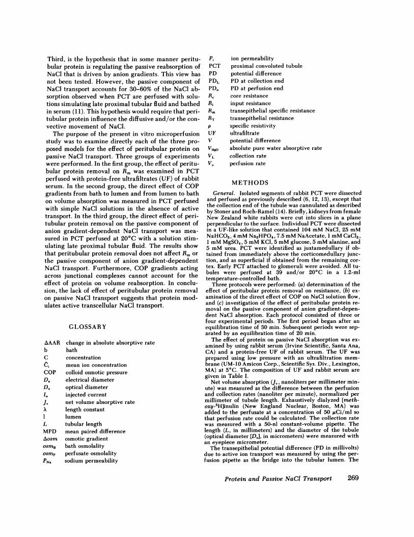
I. Rm. In physiologic experiments designed to measureRm, the perfusion solution was UF and the bath solution waseither UF or rabbit serum (Table I). In other experimentsRmwas measured by using either a simple sodium chlorideor choline chloride solution as the perfusate and bath. Thesimple salt solutions contained 160 mMNaCl or Choline Cl,4 mMNa2HPO4, 1.0 mMCaCl2, and 1.0 mMMgSO4. ThePD was measured at both the perfusion and collection endsof the tubule. The measurement of PD at the collection end(PDL) was identical to that described for the perfusion end(PD.) except that the KC1/KNO3 agarose bridge made directcontact with the collected fluid. As the perfusion rates were>20 nl/min and the PCT were short, there should be nosolute concentration-gradients along the length of the tubule,and the liquid junctions at the perfusion and collection endKCl/KNO3 bridges should be symmetrical.
The Rm was determined by using cable analysis of thevoltage attenuation after point source current injection (15)as shown in Fig. 1. A constant-current generator (CVRIElectronics, San Francisco, CA) injected a pair of 1-sec pulsesof 50-100 nA of alternating current into the tubular lumenthrough a current-passing pipette that was concentricallyarranged inside the tubule perfusion pipette and filled with0.16 MNaCl. The constant-current generator was connectedto the current-passing pipette and to the bath solution viaAg-AgCl electrodes. The resistance of the current-passingpipette was between 15 and 25 X 106 Q. During an exper-iment, the relative position of the current-passing pipetteand the perfusion pipette was kept constant, with the cur-rent-passing pipette extending beyond the perfusion pipetteby at least 10 ,m. The coupling resistance between the cur-rent-passing pipette and the voltage-recording pipette at theperfusion end was 41.8±4.5 103 Q (n = 13). This means thatinjection of 100 nA through the current-passing pipette inthe absence of a tubule resulted in a voltage deflection of4.2 mVat the perfusion end. This voltage was subtractedfrom the total voltage deflection observed in the presenceof a tubule.
Cable analysis for a truncated cable (15) was used to cal-culate the length constant (X, centimeters), the input resis-tance (R,, ohms), the transepithelial resistance (RT, Q cen-timeter), the core resistance (RC, ohms per centimeter), theelectrical diameter (D., micrometers), and the Rm (ohms-centimeter squared) according to the equation:
A = L/cosh-' (APD0/APDL), (1)
FIGURE 1 Schematic arrangement for measuring Rm. Square wave current pulses of 50-100nA and 1-s duration are injected into the tubule lumen through a current-passing pipetteinserted through the center of the perfusion pipette. Specific resistance is calculated from theinduced voltage deflections at both the perfusion and the collection ends.
270 C. A. Berry
where APDOand APDL are the voltage deflections inducedby the injected current (I.) after correction for the voltagedeflection due to coupling resistance.
R = APDO/IO
(APD0)(X)(tanh L/X)RT=
(APD.)(tanh L/X)
De = (4p/rR0)1/2,
(2)
(3)
(4)
(5)where p is the specific resistivity of the perfusion solution,55 Q cm (15) for UF, 63.2 ( cm for sodium chloride, and69.3 (2 cm for choline chloride. The latter were measuredwith a conductivity meter (Western, model 90).
Rm= 2(rpX3R,)'12(tanh L/X)/2. (6)Both transepithelial PD and specific resistance were mea-
sured by using tightly fitting tubule holding and cannulatingpipettes as electrical seals at the perfusion and the collectionends. Recently, Sakin and Boulpaep (16) have shown thatthe use of an outermost pipette containing liquid Sylgard184 did not enhance electrical insulation during measure-ments of Rmwhen both ends of the tubule are cannulated.In order to examine this issue transepithelial PD and R, (Eq.2) were measured in the presence and in the absence of anoutermost pipette containing liquid Sylgard 184 as describedby Lutz et al. (15). No significant difference in either par-ameter was observed. Transepithelial PD averaged -2.49mVwith Sylgard 184 and -2.52 mVwithout Sylgard 184with a mean paired difference (MPD) of -0.03±0.09 mV(n = 5). R, averaged 158 X 10' Q with Sylgard 184 and 146X 103 Q without Sylgard 184 with an MPDof 0.12±0.06X 10³ ( (n = 5). The increase in R, observed with Sylgard184, although not significant, is probably due to terminationof the cable at a point closer to the site of current injection.These observations agree with the conclusions of Sakin andBoulpaep (16) and suggest that valid measurements of PDand resistance can be obtained in the absence of an outermostpipette containing Sylgard 184.
II. COP. In experiments designed to examine the effectof COP (mmHg) on volume absorption in the absence ofactive transport, simple salt solutions were designed. Thesesolutions did not contain preferentially reabsorbed solutes,such as glucose and sodium bicarbonate, or high molecularweight solutes that might be sieved, such as raffinose. Thesimple salt solution without protein (Salt) contained 160 mMNaCl, 4 mMNa2HPO4, 5 mMKCI, 1 mMCaC12, and 1 mMMgSO4. The simple salt solution with protein (Protein) wasprepared by addition of -12 g/dl dialyzed albumin to theSalt solution and subsequent overnight dialysis against theSalt solution at 50C. The composition of the Salt and theProtein solution are given in Table II. These solutions werenot osmotically adjusted, in order to maintain Donnan equi-librium.
The COPof rabbit serum and of the protein solution werecalculated from the Landis-Pappenheimer equations forserum and albumin, Eqs. 7 and 8, respectively (17).
COP=2.1C + 0.16C2 + 0.009C3,
TABLE IIComposition of Salt and Protein Solution*
Na' K+ Osmolality Protein
meq/liter mosmol/Ikg g/dI
Salt (14) 160.2±1.2 5.01±0.02 306.3±2.2Protein (14) 163.8±1.5 5.11±0.06 311.7±2.6 10.3±0.3
e Solutions were bubbled with 100% 02 gas; pH was 7.4.
sion and bathing solutions can be calculated from the fol-lowing equation:'
VH£O = V0(AOSm/OSmB), (9)
where V. is the perfusion rate in nanoliters per minute, osmBis the osmolality of the bath in milliosmoles per kilogram,and Aosm is the osmotic gradient in milliosmoles per kilo-gram.
III. Anion gradient-dependent NaCi absorption. In ex-periments designed to examine the effect of protein on thepassive component of anion gradient-dependent NaCl ab-sorption, tubules were perfused at 39 and 20'C with a highchloride solution simulating late proximal tubular fluid. Thehigh chloride solution (High Cl-) contained 146.5 mMNaCl,5 mMNaHCO3, 4 mMNa2HPO4, 1 mMCaC12, 1 mMMgSO4, 5 mMKCI, and 5 mMurea. This solution was bub-bled with a CO2-O2 gas mixture to give an average Pco2 of40 mmHgand was osmotically adjusted to the serum andUF bathing solutions.
Statistics. There were two to three measurements of eachparameter in a given period for a given tubule. The meanvalues for individual periods in an individual tubule wereused to calculate the mean value for that period. Data areexpressed as mean±SEM. P values for the MPDwere com-puted by using the paired two-tailed t test. For comparisonof Rmduring sodium and choline chloride perfusions, a one-tailed Spearman's Rank Correlation Test (18) was used. Cho-line is an impermeant cation (19). Substitution of choline forsodium must therefore increase Rm and justifies the use ofa one-tailed test. Significance was accepted at the 0.05 level.
RESULTS
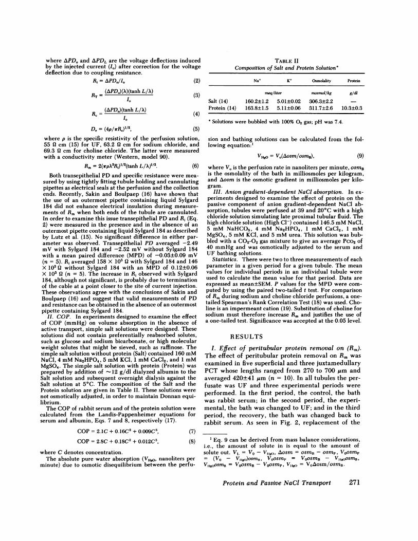
I. Effect of peritubular protein removal on (Rm).The effect of peritubular protein removal on Rmwasexamined in five superficial and three juxtamedullaryPCT whose lengths ranged from 270 to 700 ,m andaveraged 420±41 um (n = 10). In all tubules the per-fusate was UF and three experimental periods wereperformed. In the first period, the control, the bathwas rabbit serum; in the second period, the experi-mental, the bath was changed to UF; and in the thirdperiod, the recovery, the bath was changed back torabbit serum. As seen in Fig. 2, replacement of the
(7)COP= 2.8C + 0.18C2 + 0.012C3, (8)
where C denotes concentration.The absolute pure water absorption (VHO, nanoliters per
minute) due to osmotic disequilibrium between the perfu-
' Eq. 9 can be derived from mass balance considerations,i.e., the amount of solute in is equal to the amount ofsolute out. VI, = V0 - V,120, Aosm = osmB - osmp, Voosmp= (VO - VI,,)osmB, VOosmP = VOosmB - VI$)osmB,VHIOpsmB = VOosmB- VOosmP, V,,,) = VOAosm/osmB.
Protein and Passive NaCI Transport 271
PERFUSATE UF UF UF
BATH SERUM UF SERUM
26-24-222018-
(i1cm2) 14-
12 -
864-2
MEAN(8) 15.13* 2.16 13.66 1.65 13.79 t1.64
M PD - 1. 5t2.8 0.13*t1.0
p NS NS
FIGURE 2 Effect of protein on Rm. The effect of peritubularprotein removal on Rmwas examined in five superficial (opencircles) and three juxtamedullary (closed circles) PCT. Dataand statistics are given below.
serum bath solution with protein-free UF did not sig-nificantly affect Rm. Rmwas 15.1±2.2, 13.7±1.7, and13.8±1.6 Q cm2 in the control, experimental, and re-covery periods, respectively. There is a tendency forRmto decrease from the control to the recovery period;however, the MPDwas not significant, 1.3±0.9 (2 cm2(n = 8). Nonetheless, to control for this tendency forRm to decline, two additional PCT were bathed withUF in the control and recovery periods and with serumin the experimental period: R. was 13.7±3.4, 13.0±3.4,and 12.4±3.4 ( cm2, respectively. Table III shows thatthis tendency to decrease with time was also observedwith the PDOand the PDL of the tubule. In previousexperiments, PD. tended to increase between the con-trol and the recovery periods (1). Thus, the tendencytoward a time-dependent decrease in Rm, PDo, andPDL is probably related to the injection of current.
In a separate group of five tubules, Rmwas measuredduring perfusion at 20'C with simple sodium and cho-
line chloride solutions in the perfusate and bath. Thepurpose of these experiments was to show that thepresent measurements can detect changes in Rm. Dur-ing perfusion with simple sodium chloride, Rm was12.06±1.6 Qcm2 (n = 5). During perfusion with simplecholine chloride solutions, Ri increased in every tu-bule, averaging 19.8±5.0 Q cm2 (n = 5). These dataare significantly different at the 0.05 level using a one-tailed Spearman's Rank Correlation test (18).
A test of the adequacy of cable analysis is a com-parison between the De and D. of the core of the cable.The diameter of the core calculated from electricalmeasurements (Eq. 5) was 28.1±2.8 lsm (n = 10), withUF perfusate, 27.7±0.5 Jim (n = 5) with sodium chlo-ride perfusate, and 22.3±2.0 jim (n = 5) with cholinechloride perfusate. The diameter of the core measuredoptically was 20.0±1.6 jAm (n = 10) with UF and was21.2±0.8 Mm(n = 5) with simple salt solutions. Themean De was usually greater than the mean DV. Acomparable discrepancy has been observed by mostinvestigators who have used cable analysis to measureR. (Table IV). As pointed out by Lutz et al. (15), itscause probably is due to the fact that optical mea-surements include the microvilli of the brush border,-'3 1Am in perfused PCT (20). Accordingly, the barrierto current passage would approximate the level of thejunctional complex.
The average Rm observed in the presence and ab-sence of peritubular protein was 14.04±0.94 ( cm2(n = 30), at least twice the value reported by otherinvestigators (Table IV). Intranephron heterogeneity,in which Rm is higher in early than in late PCT (21),is not the cause of this discrepancy, because PCT at-tached to their glomerulus were excluded at dissection.The value calculated for Rm is most sensitive to vari-ation in A PDL. Therefore, it is likely that the higherRm reported in Table III and Fig. 1 is due to cannu-lation of the PCT at the collection end.
II. Effect of COPgradients on volume absorptionin the absence of active transport. Numerous inves-tigators have shown that in the isolated PCT perfusedwith UF at 390C, removal of peritubular protein re-duced spontaneous volume absorption coupled to ac-tive transport (1, 10, 22-24). The present experiments
TABLE IIIEffect of Peritubular Protein Removal on PD and Cable Analysis Parameters
PD. PDL APDI APDL A R, RT R. R.
mV ',m 1O'3 1O3fl cm 1O0,/cm a cms
Protein (8) -3.2±0.3 -3.3±0.6 13.9±1.3 1.3±0.3 139.3±12 127±12 1.65±0.2 9.7±1.4 15.1±2.2UF (8) -3.3±0.3 -3.9±0.6 13.4±2.8 1.3±0.3 135.9±8.7 132±15 1.49±0.1 9.6±1.4 13.7±1.7Protein (8) -3.0±0.3 -3.1±0.6 13.5±1.3 1.1±0.2 132.3±14 119±13 1.47±0.2 9.9±1.3 13.8±1.6
272 C. A. Berry
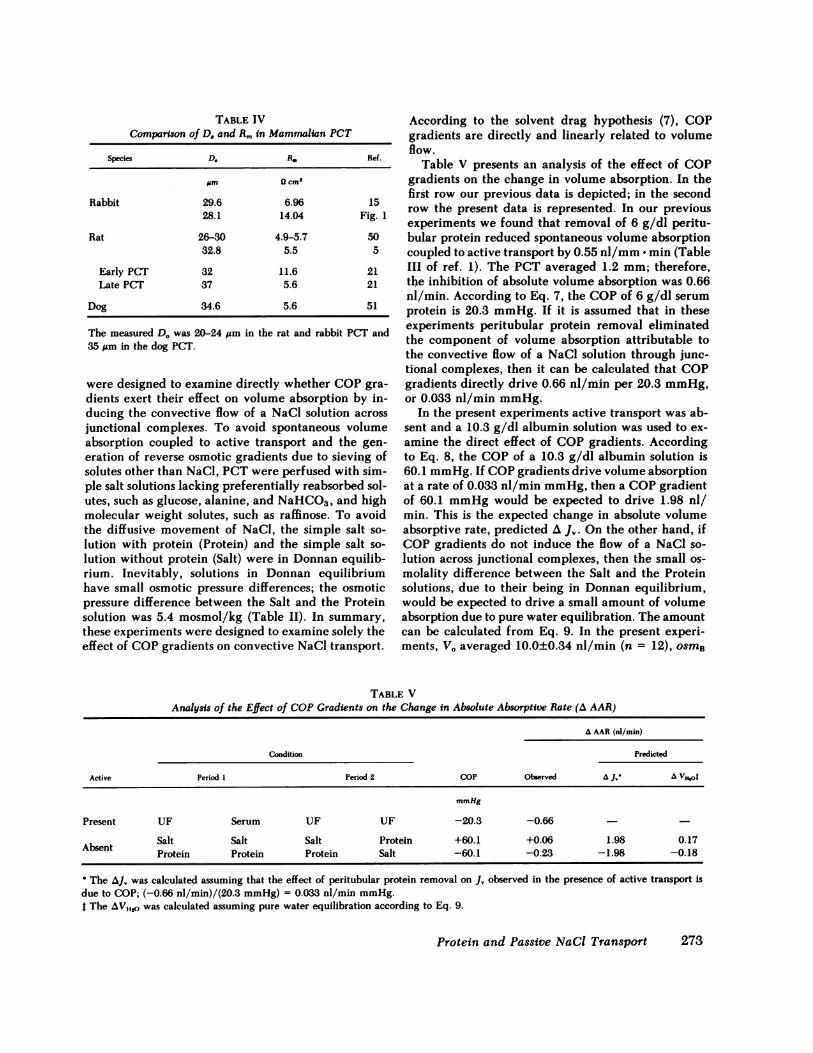
TABLE IVComparison of De and Rm in Mammalian PCT
Species D. R. Ref.
,Wm a cms
Rabbit 29.6 6.96 1528.1 14.04 Fig. 1
Rat 26-430 4.9-5.7 5032.8 5.5 5
Early PCT 32 11.6 21Late PCT 37 5.6 21
Dog 34.6 5.6 51
The measured D. was 20-24 jm in the rat and rabbit PCT and35 ;&m in the dog PCT.
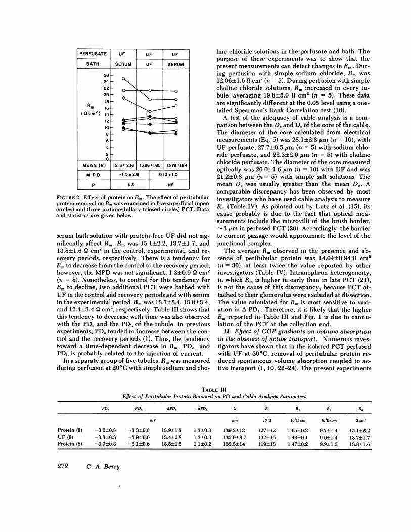
were designed to examine directly whether COPgra-dients exert their effect on volume absorption by in-ducing the convective flow of a NaCl solution acrossjunctional complexes. To avoid spontaneous volumeabsorption coupled to active transport and the gen-eration of reverse osmotic gradients due to sieving ofsolutes other than NaCl, PCTwere perfused with sim-ple salt solutions lacking preferentially reabsorbed sol-utes, such as glucose, alanine, and NaHCO3, and highmolecular weight solutes, such as raffinose. To avoidthe diffusive movement of NaCl, the simple salt so-lution with protein (Protein) and the simple salt so-lution without protein (Salt) were in Donnan equilib-rium. Inevitably, solutions in Donnan equilibriumhave small osmotic pressure differences; the osmoticpressure difference between the Salt and the Proteinsolution was 5.4 mosmol/kg (Table II). In summary,these experiments were designed to examine solely theeffect of COPgradients on convective NaCl transport.
According to the solvent drag hypothesis (7), COPgradients are directly and linearly related to volumeflow.
Table V presents an analysis of the effect of COPgradients on the change in volume absorption. In thefirst row our previous data is depicted; in the secondrow the present data is represented. In our previousexperiments we found that removal of 6 g/dl peritu-bular protein reduced spontaneous volume absorptioncoupled to active transport by 0.55 nl/mm * min (TableIII of ref. 1). The PCT averaged 1.2 mm; therefore,the inhibition of absolute volume absorption was 0.66nl/min. According to Eq. 7, the COPof 6 g/dl serumprotein is 20.3 mmHg. If it is assumed that in theseexperiments peritubular protein removal eliminatedthe component of volume absorption attributable tothe convective flow of a NaCl solution through junc-tional complexes, then it can be calculated that COPgradients directly drive 0.66 nl/min per 20.3 mmHg,or 0.033 nl/min mmHg.
In the present experiments active transport was ab-sent and a 10.3 g/dl albumin solution was used to ex-amine the direct effect of COPgradients. Accordingto Eq. 8, the COPof a 10.3 g/dl albumin solution is60.1 mmHg. If COPgradients drive volume absorptionat a rate of 0.033 nl/min mmHg, then a COPgradientof 60.1 mmHgwould be expected to drive 1.98 nl/min. This is the expected change in absolute volumeabsorptive rate, predicted A Jv. On the other hand, ifCOPgradients do not induce the flow of a NaCl so-lution across junctional complexes, then the small os-molality difference between the Salt and the Proteinsolutions, due to their being in Donnan equilibrium,would be expected to drive a small amount of volumeabsorption due to pure water equilibration. The amountcan be calculated from Eq. 9. In the present experi-ments, V. averaged 10.0±0.34 nl/min (n = 12), OsmB
TABLE VAnalysis of the Effect of COPGradients on the Change in Absolute Absorptive Rate (A AAR)
A AAR (ni/min)
Condition Predicted
Active Period 1 Period 2 COP Observed a Ji A Vot
mmHg
Present UF Serum UF UF -20.3 -0.66 -
Salt Salt Salt Protein +60.1 +0.06 1.98 0.17Absent Protein Protein Protein Salt -60.1 -0.23 -1.98 -0.18
The AJ, was calculated assuming that the effect of peritubular protein removal on J, observed in the presence of active transport isdue to COP; (-0.66 nl/min)/(20.3 mmHg) = 0.033 nl/min mmHg.I The AVHOwas calculated assuming pure water equilibration according to Eq. 9.
Protein and Passive NaCi Transport 273
averaged 311.7 and 306.3 mosmol/kg (Table II), andA osm averaged 5.4 mosmol/kg (Table II). Accordingto Eq. 9, the predicted VH±O was 0.17 nl/min when thebath solution was Protein and -0.18 nl/min when thebath solution was Salt. These are the expected changesin VH±0, Predicted A VH,0-
Fig. 3 shows that the MPDin volume absorptioninduced by COPgradients of +60.1 mmHgand -60.1mmHgwere 0.05±0.07 and -0.19±0.06 nl/mm -minin the absorptive and secretory directions, respec-tively. Because the tubular length averaged 1.2 mm,the observed A AAR are 0.06 and -0.23 nl/min. Theobserved AAAR and those predicted for A J, andA VH_. are in the last three columns of the second rowin Table V. Comparison shows that the observed AAARare consistent with pure water equilibration due to thesmall osmotic gradient (Predicted A VH20), but not withthe induction of a convective flow of a NaCl solutionby COPgradients (Predicted A J).
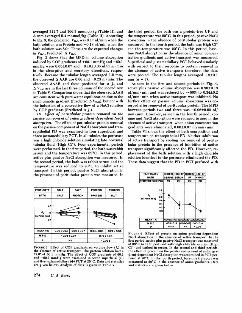
III. Effect of peritubular protein removal on thepassive component of anion gradient-dependent NaClabsorption. The effect of peritubular protein removalon the passive component of NaCl absorption and tran-sepithelial PD was examined in four superficial andthree juxtamedullary PCT. In all tubules the perfusatewas a high chloride solution simulating late proximaltubular fluid (High Cl-). Four experimental periodswere performed. In the first period, the bath was rabbitserum and the temperature was 390C. In this period,active plus passive NaCl absorption was measured. Inthe second period, the bath was rabbit serum and thetemperature was reduced to 20'C to inhibit activetransport. In this period, passive NaCl absorption inthe presence of peritubular protein was measured. In
PERFUSATE SALT SALT PROTEIN PROTEINBATH SALT PROTEIN PROTEIN SALT
0.4-
(nI/mm min)-OA-
MEAN(9) j.00t Q0.03 |+.8t0.07 -0.04*0.03 0.06M P D + 0.05 ± 0.07 -0.19t0.06
P NS e0.025
FIGURE 8 Effect of COPgradients on volume flow (J,) inthe absence of active transport. The protein solution had aCOPof 60.1 mmHg. The effect of COPgradients of 60.1and -60.1 mmHgwere examined in seven superficial (0)and five juxtamedullary (@) PCTat 200C. Data and statisticsare given below. Analysis of data is given in Table V.
the third period, the bath was a protein-free UF andthe temperature was 200C. In this period, passive NaClabsorption in the absence of peritubular protein wasmeasured. In the fourth period, the bath was High Cl-and the temperature was 20'C. In this period, base-line NaCl absorption in the absence of anion concen-tration gradients and active transport was measured.Superficial and juxtamedullary PCTbehaved similarlywith respect to their response to protein removal inthe absence of active transport; therefore, the datawere pooled. The tubular lengths averaged 1.3±0.1mm(n = 7).
As seen in the first and second periods in Fig. 4,active plus passive volume absorption was 0.90±0.13nl/mm -min and was reduced by -60% to 0.34±0.2nl/mm * min when active transport was inhibited. Nofurther effect on passive volume absorption was ob-served after removal of peritubular protein. The MPDbetween periods two and three was -0.06±0.08 nl/mm* min. However, as seen in the fourth period, vol-ume and NaCl absorption were reduced to zero in theabsence of active transport, when anion concentrationgradients were eliminated, 0.00±0.07 nl/mm * min.
Table VI shows the effect of bath composition andtemperature on transepithelial PD. Neither inhibitionof active transport by cooling nor removal of peritu-bular protein in the presence of inhibition of activetransport significantly affected the PD. However, re-placement of the bath solution with a high chloridesolution identical to the perfusate eliminated the PD.These data suggest that the PD in PCT perfused with
PERFUSATE HIGH Cl HIGH Cr HIGH C- HIGH Cl-BATH SERUM SERUM UF HIGH CW
TEMPERATURE(IC) 39 20 20 20
1.2
J 1.0
Am_0.8 ;(n/mi)0.60.4-0.20.0
MEAN(7) 090:kQI1 I34 ±0.2 39Q2 *O007M P D LQ56SC.18I [6C O±Q4*0.16
< 0.01 NS I < 0.
FIGURE 4 Effect of protein on anion gradient-dependentNaCl absorption in the absence of active transport. In thefirst period, active plus passive NaCl transport was measuredat 390C in PCT perfused with high chloride solution (HighCl-) and bathed in serum. In the second and third periods,the effect of protein on the passive component of anion gra-dient-dependent NaCl absorption was examined in PCTper-fused at 20'C. In the fourth period, base-line transport wasmeasured at 20'C in the absence of anion gradients. Dataand statistics are given below.
274 C. A. Berry
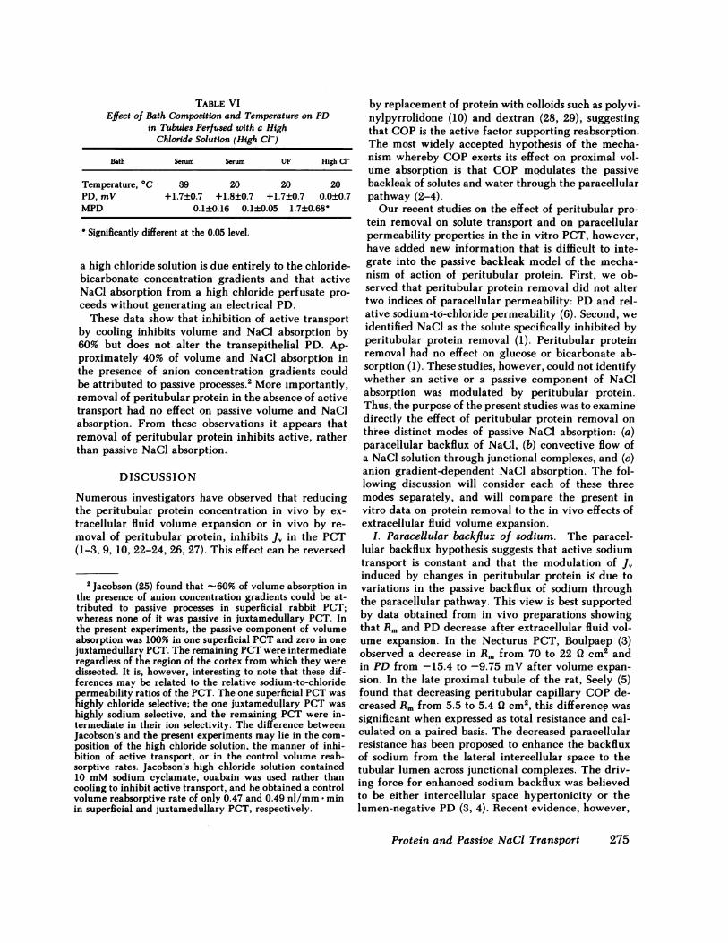
TABLE VIEffect of Bath Composition and Temperature on PD
in Tubules Perfused with a HighChloride Solution (High Cl-)
Bath Serum Serum UF High Cl1
Temperature, 0C 39 20 20 20PD, mV +1.7±0.7 +1.8±0.7 +1.7±0.7 0.0±0.7MPD 0.1±0.16 0.1±0.05 1.7±0.68°
* Significantly different at the 0.05 level.
a high chloride solution is due entirely to the chloride-bicarbonate concentration gradients and that activeNaCl absorption from a high chloride perfusate pro-ceeds without generating an electrical PD.
These data show that inhibition of active transportby cooling inhibits volume and NaCl absorption by60% but does not alter the transepithelial PD. Ap-proximately 40% of volume and NaCl absorption inthe presence of anion concentration gradients couldbe attributed to passive processes.2 More importantly,removal of peritubular protein in the absence of activetransport had no effect on passive volume and NaClabsorption. From these observations it appears thatremoval of peritubular protein inhibits active, ratherthan passive NaCl absorption.
DISCUSSION
Numerous investigators have observed that reducingthe peritubular protein concentration in vivo by ex-tracellular fluid volume expansion or in vivo by re-moval of peritubular protein, inhibits J, in the PCT(1-3, 9, 10, 22-24, 26, 27). This effect can be reversed
2 Jacobson (25) found that '60% of volume absorption inthe presence of anion concentration gradients could be at-tributed to passive processes in superficial rabbit PCT;whereas none of it was passive in juxtamedullary PCT. Inthe present experiments, the passive component of volumeabsorption was 100% in one superficial PCTand zero in onejuxtamedullary PCT. The remaining PCTwere intermediateregardless of the region of the cortex from which they weredissected. It is, however, interesting to note that these dif-ferences may be related to the relative sodium-to-chloridepermeability ratios of the PCT. The one superficial PCTwashighly chloride selective; the one juxtamedullary PCT washighly sodium selective, and the remaining PCT were in-termediate in their ion selectivity. The difference betweenJacobson's and the present experiments may lie in the com-position of the high chloride solution, the manner of inhi-bition of active transport, or in the control volume reab-sorptive rates. Jacobson's high chloride solution contained10 mMsodium cyclamate, ouabain was used rather thancooling to inhibit active transport, and he obtained a controlvolume reabsorptive rate of only 0.47 and 0.49 nl/mm * minin superficial and juxtamedullary PCT, respectively.
by replacement of protein with colloids such as polyvi-nylpyrrolidone (10) and dextran (28, 29), suggestingthat COPis the active factor supporting reabsorption.The most widely accepted hypothesis of the mecha-nism whereby COPexerts its effect on proximal vol-ume absorption is that COP modulates the passivebackleak of solutes and water through the paracellularpathway (2-4).
Our recent studies on the effect of peritubular pro-tein removal on solute transport and on paracellularpermeability properties in the in vitro PCT, however,have added new information that is difficult to inte-grate into the passive backleak model of the mecha-nism of action of peritubular protein. First, we ob-served that peritubular protein removal did not altertwo indices of paracellular permeability: PD and rel-ative sodium-to-chloride permeability (6). Second, weidentified NaCl as the solute specifically inhibited byperitubular protein removal (1). Peritubular proteinremoval had no effect on glucose or bicarbonate ab-sorption (1). These studies, however, could not identifywhether an active or a passive component of NaClabsorption was modulated by peritubular protein.Thus, the purpose of the present studies was to examinedirectly the effect of peritubular protein removal onthree distinct modes of passive NaCl absorption: (a)paracellular backflux of NaCl, (b) convective flow ofa NaCl solution through junctional complexes, and (c)anion gradient-dependent NaCl absorption. The fol-lowing discussion will consider each of these threemodes separately, and will compare the present invitro data on protein removal to the in vivo effects ofextracellular fluid volume expansion.
I. Paracellular backflux of sodium. The paracel-lular backflux hypothesis suggests that active sodiumtransport is constant and that the modulation of J,induced by changes in peritubular protein is due tovariations in the passive backflux of sodium throughthe paracellular pathway. This view is best supportedby data obtained from in vivo preparations showingthat Rmand PD decrease after extracellular fluid vol-ume expansion. In the Necturus PCT, Boulpaep (3)observed a decrease in Rm from 70 to 22 ( cm2 andin PD from -15.4 to -9.75 mVafter volume expan-sion. In the late proximal tubule of the rat, Seely (5)found that decreasing peritubular capillary COPde-creased Rm from 5.5 to 5.4 Q2 cm2, this difference wassignificant when expressed as total resistance and cal-culated on a paired basis. The decreased paracellularresistance has been proposed to enhance the backfluxof sodium from the lateral intercellular space to thetubular lumen across junctional complexes. The driv-ing force for enhanced sodium backflux was believedto be either intercellular space hypertonicity or thelumen-negative PD (3, 4). Recent evidence, however,
Protein and Passive NaCI Transport 275
indicates that lateral intercellular spaces are not hy-pertonic (6, 30) and that the PDalong all but the initialmammalian superficial proximal tubule is lumen-pos-itive (30, 31). In the absence of lateral intercellularspace hypertonicity and in the presence of a lumen-positive PD, a decrease in paracellular resistancewould enhance the passive reabsorption of sodium,rather than the passive backflux of sodium. These theo-retical considerations question the paracellular back-flux hypothesis.
Recently, three observations in the in vitro PCThave suggested indirectly that paracellular resistanceis not altered by protein removal. First, in rabbit PCTperfused with UF, volume absorption is reduced buttransepithelial PD is not affected by removal of peritu-bular protein (1, 6, 10, 22, 23). Second, in rabbit PCTthe relative sodium-to-chloride permeability ratio isnot altered by removal of peritubular protein (6). Thisobservation suggests that removal of peritubular pro-tein does not alter either the ion selectivity of the junc-tional complexes or the relative contribution of thejunctional complexes and lateral intercellular spacesto the paracellular resistance (6). Third, in amphibianPCT, volume absorption is inhibited, but neither tran-sepithelial nor basolateral membrane PD are reducedby removal of bath colloid (16).
In the present study, Rmwas measured directly inthe rabbit PCTand found to be unaltered by removalof peritubular protein (Fig. 2). At this point it is worth-while to estimate the decrease in Rmthat would havebeen required to account for our previously observed40% decrease in J, (1) according to the paracellularbackflux hypothesis. The paracellular backflux hy-pothesis proposes that the rate of active sodium ab-sorption at the basolateral membrane is constant andthat J, is decreased after peritubular protein removalbecause of an increase in the net passive sodium back-flux. The net passive sodium backflux is increased be-cause of an increase in paracellular sodium perme-ability. The increase in sodium permeability requiredto account for a 40% decrease in net sodium absorptioncan be calculated with the data shown in Table VIIand the equations for unidirectional passive sodiumfluxes (32).
Passive influx:VF [NalievF(RT
Na PaRT 1 - eVF/RT (10)
Passive efflux:
JP(I-b) =VF [Nalb (11)
where b is the bath, 1 is the lumen, V is potential
TABLE VIIData Used for Calculation of Unidirectional
Passive Sodium Fluxes'
Bath Protein Present Absent
Jv.nl/mm min 1.4 0.84JiNet,pmol/mm- mint 210 126PD, mV -3 -3PNaw cm/s 1O-4[Nall = [Nalb, mM 150 150Tubule diameter, Um 20 20
All data are approximated from Ref. 1 except for PN. in thepresence of bath protein which was obtained from Ref. 35.t Calculated assuming J, is isonatric.
difference, PNa is the sodium permeability and R, T,and F have their usual meanings.
Fig. 5 shows the calculated unidirectional passivesodium fluxes in the presence and absence of bath pro-tein. In the presence of bath protein, the net sodiumflux of 210 peq/mm min is -20% of the unidirec-tional passive sodium fluxes. But because the PD islumen-negative, passive sodium influx exceeds passivesodium efflux by 140 peq/mm * min and the calculatedrate of active sodium absorption is 350 peq/mm * min.In the absence of bath protein, the net sodium flux is126 peq/mm * min, and according to the paracellularbackflux hypothesis the rate of active sodium absorp-tion is constant at 350 peq/mm - min. Thus, in the ab-sence of bath protein, the net passive backflux of so-dium must be 224 peq/mm -min. The PNa requiredto give a net passive sodium efflux of 224 peq/mm-min can be calculated from solution of Eq. 12.
Eq. 10 + Eq. 11 = -224 peq/mm -min. (12)
As shown at the bottom right of Fig. 5, PNa in theabsence of bath protein must be increased to1.6 X 10' cm/s. This calculation shows that a 60%increase in PNa is required to account for a 40% de-crease in net sodium transport. The increase in PNa willbe inversely related to the decrease in Rm accordingto Eq. 13 (19).
2: PiCi = RT/F2(1/Rm), (13)where Pi is the ion permeability, Ci is the mean ionconcentration, and R, T, and F have their usual mean-ings.
The increase in Pi will be equivalent for Na and Clbecause the relative solution-to-chloride permeabilityratio is not altered by peritubular protein removal (6).Accordingly, a 60% increase in Pi will be associatedwith a 40% decrease in Rm. This would have meantthat Rmwould have had to decrease from an average
276 C. A. Berry
Bath ProteinPresent
1200
1060 350
-I -Fpeq mm mmn
P =1 -4 cms-No
Bath ProteinAbsent
X mVm
1.
EI Passive
E-IiZD Active
1920...2
224 12E.: I-
1696 350
- I -Ipeqrmm min
P = 1.6 * 104 cmSNNo
FIGURE 5 Calculated unidirectional sodium fluxes in the presence and in the absence of bathprotein. Active and passive sodium fluxes are calculated from equations 10 and 11 and the datain Table VII. In the absence of bath protein, the PNa required to account for a 40% decreasein net sodium absorption according to the paracellular backflux hypothesis is 1.6 X lo-4 cm/s, a value 60% greater than observed in the presence of bath protein.
of 14 ( cm2 in the presence of bath protein to 8.4 Qcm2 in the absence of bath protein.3 The present data,therefore, indicate that the effect of removal of peritu-bular protein on proximal solute and water absorptioncannot be accounted for by a change in Rmas suggestedby the paracellular backflux hypothesis.
II. NaCi solution flow through junctional com-plexes. Carl Ludwig (34) first proposed that the COPof serum proteins could be responsible for tubular saltand water reabsorption in 1843. He envisioned thatthe tubular wall was freely permeable to small mo-lecular weight solutes, but impermeable to serum pro-teins. As a result, COPprovided the driving force forsolute and water absorption, obviating the need foractive solute transport. Because this mechanism cannotaccount for urine whose osmolality is greater than thatof plasma, it was discarded. More recently, Schner-mann (7) has revived Ludwig's theory to explain theeffect of peritubular protein on isosmotic proximal tu-bular solute and water absorption, while maintainingthe view that active solute transport is the principledriving force for water absorption. This modern Lud-wig-like theory proposed that the junctional complexesare highly permeable to small molecular weight solutesand water, but impermeable to peritubular protein.The COPprovided the driving force for the protein-sensitive component of proximal volume absorption.
3 This calculation provides a minimum estimate of therequired change in Rmbecause it assumes that the PD alongthe tubule is constant. PD becomes less negative and maybecome positive with length (31, 33), thus decreasing thedriving force for sodium backflux.
This view is best supported by data showing that ad-dition of luminal protein dramatically inhibited vol-ume absorption in the rat PCT (7, 8). Other studies(9, 10) in both the rat and the rabbit PCT, however,find that luminal protein does not influence volumeabsorption.
In order to examine the effect of COPgradients ona component of proximal volume absorption not as-sociated with active solute transport, the present stud-ies measured volume absorption at 20'C. Fig. 3 andTable V show that COPgradients do not drive a com-ponent of volume absorption larger than can be at-tributed to pure water equilibration between perfusionand bathing solutions. These data indicate that thereis not a highly NaCl- and water-permeable pathwayin the PCT across which COPgradients operate. Thisconclusion is in accord with measured NaCl reflectioncoefficients in the PCT. The reflection coefficient forNaCl in the PCT has been measured to be between0.7 and 0.9 (35, 36). In either case, COPgradientswould cause significant sieving of luminal NaCl andeventual osmotic equilibration.
It might be argued that the permeability propertiesof the pathway across which protein exerts its directcolloid osmotic effect, presumably the junctional com-plexes, have been altered by inhibition of active trans-port. For instance, the lateral intercellular spaces havebeen observed to be collapsed in the absence of activetransport (37). Their width, however, is still substan-tial, -200 A(38). Although such a constrained envi-ronment might restrict the diffusion of albumin by asmuch as one to two orders of magnitude, the albuminconcentration at the contraluminal surface of the junc-
Protein and Passive NaCl Transport 277
tional complexes would be equal to that in the bathafter the 20-min equilibration period and in the pres-ence of negligible fluid velocities within the lateralintercellular spaces (39). Accordingly, a convectiveflow of a NaCl solution across junctional complexesshould have occurred if the junctional complexes hada high water permeability and a low reflection coef-ficient for NaCl, irrespective of changes in paracellulargeometry.
III. Passive component of anion gradient-depen-dent NaCi absorption. It is generally agreed thatpassive NaCl transport processes account for 30-60%of the NaCl transport in the late superficial PCTwhereanion gradients are present (11). The passive compo-nent of anion gradient-dependent NaCl transport hasbeen attributed to both diffusive and convective trans-port mechanisms. A comparison of periods two andthree in Fig. 4 shows that removal of peritubular pro-tein did not influence the passive component of aniongradient-dependent NaCl transport; the MPD was-0.06±0.08 nl/mm - min. Although it is possible thatthe diffusive and convective components are changingreciprocally, it is more likely that neither the diffusivenor the convective component of NaCl transport areinfluenced by peritubular protein. The conclusion thatthe diffusive component of NaCl transport is constantis supported by Imai and Kokko's (23) observation thatPNa was not altered by protein and with our data thatshow that neither the relative sodium-to-chloride per-meability (6) nor the Rm(Fig. 2) were altered by peri-tubular protein removal. If the diffusive componentis constant, then it follows that the convective com-ponent is also constant, indicating that the reflectioncoefficients for sodium chloride, sodium bicarbonate,and organic solutes are unchanged by peritubular pro-tein removal.
IV. Effect of peritubular protein removal in vitro.The present observations show that peritubular proteindoes not act on three distinct modes of passive NaCltransport. First, directly determined Rm is not alteredby protein removal. Consequently, the inhibitory ef-fect of protein removal on proximal volume absorptioncannot be attributed to an increase in the backflux ofsodium across junctional complexes. Second, COPgra-dients in the absence of active transport do not gen-erate volume absorption over that that can be attrib-uted to pure water equilibration between the luminaland peritubular solutions. Consequently, the inhibi-tory effect of protein removal on proximal volumeabsorption cannot be attributed to removal of a directeffect of protein on the flow of a NaCl solution acrossjunctional complexes. Third, the directly determinedpassive component of anion gradient-dependent NaCltransport is not changed by peritubular protein re-moval. Consequently, the inhibitory effect of protein
cannot be attributed to alterations in diffusive or con-vective NaCl transport.
The failure of peritubular protein removal to inhibitany of the three modes of passive NaCl transport ex-amined suggests that protein modulates active NaCltransport. The magnitude of the effect of protein re-moval on active NaCl transport can be estimated froma comparison of the effect of protein removal in thepresence of active transport and of inhibition of activetransport on volume absorption from a high chloridesolution. These comparisons are shown in Table VIII.The first column gives volume absorption in the pres-ence of active transport and peritubular protein, 0.90nl/mm * min; the second column gives volume absorp-tion in the presence of active transport but in the ab-sence of peritubular protein, 0.37 nl/mm - min; andthe third column gives volume absorption in the ab-sence of active transport and in the presence of peritu-bular protein, 0.34 nl/mm min. Comparison of thefirst and second columns shows that removal of peri-tubular protein in the presence of active transport,reduced volume absorption by -60%. Comparison ofthe first and third columns shows that inhibition ofactive transport in the presence of peritubular proteinalso reduced volume absorption by -60%. The iden-tity of the percentages of volume absorption inhibitedby removal of peritubular protein in the presence ofactive transport and by inhibition of active transportsuggests that peritubular protein removal inhibits vir-tually all active volume and NaCl absorption from ahigh chloride solution.
The actual mode of active NaCl transport inhibitedby peritubular protein removal is unknown. Becauseperitubular protein addition or removal occurs adja-cent to the basolateral cell membrane, it is likely thatprotein exerts its effect on the basolateral, rather thanon the luminal cell membrane. In support of the viewthat protein is acting on the basolateral cell membraneare the recent observations in proximal straight tubulesthat protein addition depolarizes the basolateral PD
TABLE VIIIComparison of the Effect of Protein Removal and Inhibition of
Active Transport on Volume Absorption (Jo) from a HighChloride Solution (High Cl-)
Perfusate High Cl- High Cl1 High Cl1
Bath Serum UF SerumTemperature, 0C 39 39 20J,, nl/mm min 0.90 I 0.370 0.34t
o Data are from Table V of Ref. 1. The J, in the presence of proteinand active transport was 0.90 nl/mm - min.t These are data from Fig. 4.
278 C. A. Berry
(40). Wehave shown previously that peritubular pro-tein removal does not inhibit substantially either glu-cose or sodium bicarbonate reabsorption in PCT per-fused with UF (1). Failure to inhibit these sodium co-transport processes was interpreted as evidence againstan effect of protein removal on the Na-K ATPase pumpsystem located in the basolateral cell membrane. TableV of ref. 1 and Table VI of this paper show that re-moval of peritubular protein does not alter transepi-thelial PD at either 39 or 20'C, respectively. Thus, weconclude that peritubular protein removal specificallyinhibits neutral NaCl transport4 across the basolateralcell membrane without inhibiting the Na-K ATPasepump system.
V. Comparison of the effects of peritubular proteinremoval in vitro to extracellular fluid volume expan-sion in vivo. The relevance of these in vitro studiesto the in vivo circumstance where peritubular proteinconcentration is lowered by expansion of extracellularvolume with noncolloidal salt solutions should be con-sidered. Removal of peritubular protein in vitro andextracellular fluid volume expansion in vivo have sim-ilar effects on proximal transport processes and dissim-ilar effects on proximal paracellular permeabilityproperties. On the one hand, both experimental modelsshow a reduction in volume absorption with an inhi-bition of NaCl transport (1, 41, 42), but no effect onNaHCO3(1, 41, 42). On the other hand, the availablein vivo data strongly support an increase in paracel-lular permeability properties (3, 4, 5); whereas in vitrodata show no change in paracellular permeabilityproperties (Fig. 2, ref. 1, 6, 23). Wehave previouslysuggested that the changes in paracellular permeabil-ity properties observed in vivo may be secondary toincreases in interstitial hydrostatic pressure and arenot the primary cause for the reduction in volume andNaCl transport (1). An argument in favor of this viewis that an increase in paracellular permeability in vivowould stimulate, rather than inhibit, NaCl absorption:The chloride concentration is greater in the lumenthan in the blood, favoring chloride absorption and thetransepithelial PD, in the late proximal tubule wherethe bulk of NaCl is absorbed, is lumen-positive, fa-voring sodium absorption. Thus, two lines of evidencesuggest that the present in vitro results on peritubularprotein removal bear importantly on extracellular
4 Protein removal inhibits NaCl transport without chang-ing the transepithelial PD (Table IV, ref. 1), suggesting thattranscellular NaCl transport is neutral. The fact that tran-scellular NaCl transport is neutral does not define the mech-anism of NaCl transport across the luminal or basolateralmembranes. It is consistent with several modes of neutraltransport and does imply direct one for one coupling via aneutral NaCl symporter.
fluid volume expansion in vivo. First, both modelsshow NaCl specificity. Second, in both models a de-crease in tubular resistance would stimulate, not in-hibit, NaCl absorption. Ultimately, however, the rel-evance of the in vitro protein removal data to in vivoextracellular fluid volume expansion resides in themechanism by which changes in peritubular capillaryprotein concentrations in vivo are transduced to thebasolateral membrane of the proximal tubule.
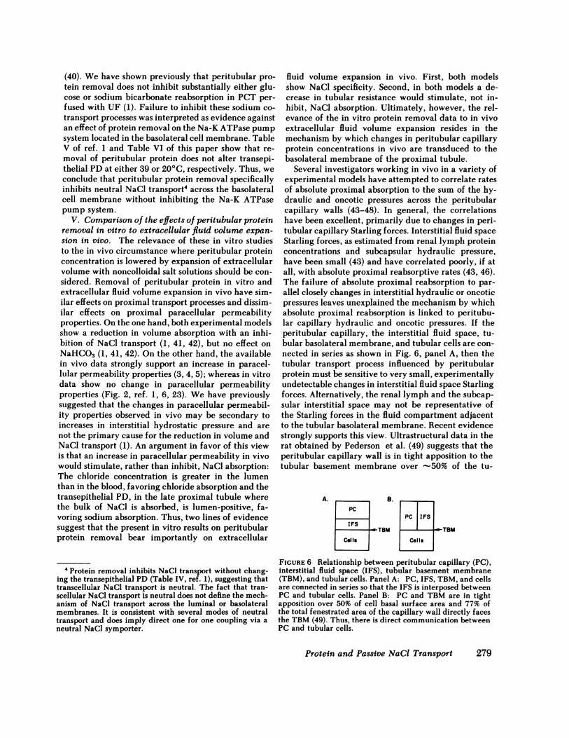
Several investigators working in vivo in a variety ofexperimental models have attempted to correlate ratesof absolute proximal absorption to the sum of the hy-draulic and oncotic pressures across the peritubularcapillary walls (43-48). In general, the correlationshave been excellent, primarily due to changes in peri-tubular capillary Starling forces. Interstitial fluid spaceStarling forces, as estimated from renal lymph proteinconcentrations and subcapsular hydraulic pressure,have been small (43) and have correlated poorly, if atall, with absolute proximal reabsorptive rates (43, 46).The failure of absolute proximal reabsorption to par-allel closely changes in interstitial hydraulic or oncoticpressures leaves unexplained the mechanism by whichabsolute proximal reabsorption is linked to peritubu-lar capillary hydraulic and oncotic pressures. If theperitubular capillary, the interstitial fluid space, tu-bular basolateral membrane, and tubular cells are con-nected in series as shown in Fig. 6, panel A, then thetubular transport process influenced by peritubularprotein must be sensitive to very small, experimentallyundetectable changes in interstitial fluid space Starlingforces. Alternatively, the renal lymph and the subcap-sular interstitial space may not be representative ofthe Starling forces in the fluid compartment adjacentto the tubular basolateral membrane. Recent evidencestrongly supports this view. Ultrastructural data in therat obtained by Pederson et al. (49) suggests that theperitubular capillary wall is in tight apposition to thetubular basement membrane over -50% of the tu-
A. B
PC I FSI IFS
TBM TBM
CellIs Cells
FIGURE 6 Relationship between peritubular capillary (PC),interstitial fluid space (IFS), tubular basement membrane(TBM), and tubular cells. Panel A: PC, IFS, TBM, and cellsare connected in series so that the IFS is interposed betweenPC and tubular cells. Panel B: PC and TBMare in tightapposition over 50% of cell basal surface area and 77% ofthe total fenestrated area of the capillary wall directly facesthe TBM(49). Thus, there is direct communication betweenPC and tubular cells.
Protein and Passive NaCl Transport 279
bular basal surface. There appears to be little, if any,fluid in the intervening space (49). Furthermore, atleast 77% of the fenestrations in the peritubular cap-illary are in the portion of peritubular capillary thatabuts the tubular basement membrane (49). Accord-ingly, the relationship between the peritubular cap-illaries, the interstitial fluid space, the tubular baso-lateral membrane, and the tubular cells is probably asdepicted in Fig. 6, panel B. In Fig. 6, panel B twodistinctly different pathways for reabsorbate to passfrom proximal tubule cells to peritubular capillary lu-men are represented. First, there is a long and complexpathway that passes through the interstitial fluid spaceand enters the peritubular capillary where fenestra-tions or endothelial pores are sparse. Second, there isthe short and direct pathway from tubule cell acrossthe peritubular capillary surface with a large fractionof the endothelial pores. This second pathway mightwell represent the route by which changes in peritu-bular Starling forces are transduced to the proximaltubule cell. The mechanism for the transduction wouldbe direct, without involving the interstitial fluid space,and as such, would be similar for in vitro protein re-moval and in vivo extracellular fluid volume expan-sion.
In summary, we have shown that peritubular proteinremoval in vitro specifically inhibits active neutralNaCl transport4 from proximal convoluted tubules.The relevance of this in vitro observation to the mech-anism of extracellular fluid volume expansion in vivoremains to be explored. In view of the present in vitrodata, however, it is possible that the effects of extra-cellular fluid volume expansion in vivo are twofold.First, there would be a specific effect of capillary pro-tein concentration on volume and NaCl absorption.This effect would occur in vivo and in vitro and wouldbe primary. Second, there would be an effect of in-terstitial hydrostatic pressure on paracellular perme-ability properties. This effect would be small and sec-ondary.
ACKNOWLEDGMENTS
I would like to thank Mr. Kenneth Wong for expert anddedicated technical assistance and Dr. Floyd C. Rector, Jr.for his continuing support and valuable suggestions.
This investigation was supported by U. S. Public HealthService Research grants HL-06285 and RO1-AM07219 fromthe National Institutes of Arthritis, Metabolism, and Diges-tive Diseases.
REFERENCES
1. Berry, C. A., and M. G. Cogan. 1981. Influence of peritu-bular protein on solute absorption in the rabbit proximaltubule. A specific effect on NaCl transport. J. Clin. In-vest. 68: 506-516.
2. Lewy, J. E., and E. E. Windhager. 1968. Peritubularcontrol of proximal tubular fluid reabsorption in the ratkidney. Am. J. Physiol. 214: 943-954.
3. Boulpaep, E. L. 1972. Permeability changes of the prox-imal tubule of Necturus during saline loading. Am. J.Physiol. 222: 517-529.
4. Grandchamp, A., and E. L. Boulpaep. 1974. Pressurecontrol of sodium reabsorption and intercellular back-flux across proximal kidney tubule. J. Clin. Invest. 54:69-82.
5. Seely, J. F. 1973. Effects of peritubular oncotic pressureon rat proximal tubule electrical resistance. Kidney Int.4: 28-35.
6. Berry, C. A., and F. C. Rector, Jr. 1978. Relative sodiumto chloride permeability in proximal convoluted tubule.Am. J. Physiol. 235(6): F592-F604.
7. Schnermann, J. 1973. Physical forces and transtubularmovement of solutes and water. In Kidney and UrinaryTract Physiology. K. Thurau, editor. Butterworths Univ.Park Press, Baltimore, 157-198.
8. Persson, A. E. G., J. Schnermann, B. Agerup, and N. E.Ericksson. 1975. The hydraulic conductivity of the ratproximal tubular wall determined with colloidal solu-tions. Pfiuegers Arch. Eur. J. Physiol. 360: 25-44.
9. Green, R., E. E. Windhager, and G. Giebisch. 1974.Protein oncotic pressure effects on proximal tubular fluidmovement in the rat. Am. J. Physiol. 226: 265-276.
10. Imai, M., and J. P. Kokko. 1974. Transtubular oncoticpressure gradients and net fluid transport in isolatedproximal tubules. Kidney Int. 6: 138-145.
11. Berry, C. A., and F. C. Rector, Jr. 1980. Active andpassive sodium transport in the proximal tubule. Miner.Electrolyte Metab. 4: 149-160.
12. Berry, C. A. 1981. Electrical effects of acidification inrabbit proximal convoluted tubule. Am. J. Physiol. 240:F459-F470.
13. Berry, C. A., D. G. Warnock, and F. C. Rector, Jr. 1978.Ion selectivity and proximal salt reabsorption. Am. J.Physiol. 235: F234-F245.
14. Stoner, L. C., and F. Roch-Ramel. 1979. The effects ofpressure on the water permeability of the descendinglimb of Henle's loops of rabbits. Pfluegers Arch. Eur. J.Physiol. 382: 7-15.
15. Lutz, M., J. Cardinal, and M. G. Burg. 1973. Electricalresistance of renal proximal tubule perfused in vitro.Am. J. Physiol. 225: 729-734.
16. Sakin, H., and E. L. Boulpaep. 1981. Isolated perfusedsalamander proximal tubule: methods electrophysiologyand transport. Am. J. Physiol. 241: F39-F52.
17. Landis, E. M., and J. R. Pappenheimer. 1963. Exchangeof substances through the capillary walls. Handb. Phy-siol. Circulation. II: 961-1034.
18. Zar, J. H. 1974. Biostatistical Analysis. Prentice-Hall,Inc., Englewood Cliffs, NJ.
19. Fromter, E. 1973. Electrophysiology and isotonic ab-sorption of proximal tubules of mammalian kidney. InKidney and Urinary Tract Physiology. K. Thurau, editor.Butterworths Univ. Park Press, Baltimore, 1-38.
20. Burg, M. G., and J. J. Grantham. 1971. Ion movementsin renal tubules. In Membranes and Ion Transport.E. E. Bittar, editor. Wiley, Sussex, England. 3: 49.
21. Seely, J. 1973. Variation in electrical resistance alongthe length of rat proximal convoluted tubule. Am. J.Physiol. 255: 48-57.
22. Burg, M., C. Patlak, N. Green, and D. Villey. 1976.Organic solutes in fluid absorption by renal proximalconvoluted tubules. Am. J. Physiol. 231: 627-637.
280 C. A. Berry
23. Imai, M., and J. P. Kokko. 1972. Effect of peritubularprotein concentration on reabsorption of sodium andwater in isolated perfused proximal tubules. J. Clin. In-vest. 51: 314-325.
24. Grantham, J. J., P. B. Qualizza, and L. W. Welling. 1972.Influence of serum proteins on net fluid reabsorption ofisolated proximal tubules. Kidney Int. 2: 66-75.
25. Jacobson, H. R. 1979. Characteristics of volume reab-sorption in rabbit superficial and juxtamedullary prox-imal convoluted tubules. J. Clin. Invest. 63: 410-418.
26. Brenner, B. M., K. H. Falchuk, R. I. Keimowitz, andR. W. Berliner. 1969. The relationship between peri-tubular capillary protein concentration and fluid reab-sorption by the renal proximal tubule. J. Clin. Invest.48: 1519-1531.
27. Weinman, E. J., M. Kashgarian, and J. P. Hayslett. 1971.Role of peritubular protein concentration in sodiumreabsorption. Am. J. Physiol. 221: 1521-1528.
28. Brazy, P. C., R. G. Balaban, S. R. Gullans, L. J. Mandel,and V. W. Dennis. 1980. Inhibition of renal metabolism.Relative effects of arsenate on sodium, phosphate, andglucose transport by the rabbit proximal tubule. J. Clin.Invest. 66: 1211-1221.
29. Windhager, E. E., J. E. Lewy, and A. Spitzer. 1969.Intrarenal control of proximal tubular reabsorption ofsodium and water. Nephron. 6: 247-259.
30. Schafer, J. A., C. S. Patlak, and T. E. Andreoli. 1977.A mechanism for isotonic fluid absorption linked to ac-tive and passive ion flows in the mammalian superficialpars recta. Am. J. Physiol. 233: F154-F167.
31. Barratt, L. J., F. C. Rector, J. P. Kokko, and D. W.Seldin. 1974. Factors governing the transepithelial po-tential difference across the proximal tubule of the ratkidney. J. Clin. Invest. 53: 454-464.
32. Curran, P. F., and S. G. Schultz. 1974. Some thermo-dynamic and kinetic principles governing solvent andsolute transport across membranes. In The Kidney.B. M. Brenner and F. C. Rector, Jr., editors. W. B. Saun-ders Company, Philadelphia. 104-125.
33. Fromter, E., and K. Gessner. 1974. Active transport po-tentials, membrane diffusion potentials and streamingpotentials across rat kidney proximal tubule. PfluegersArch. Eur. J. Physiol. 351: 85-98.
34. Ludwig, E. 1843. Beitrage zur Lehre vom Mechanismusder Harnsecretion. N. G. Elwert Verlags, Marburg, WestGermany.
35. Kokko, J. P., M. B. Burg, and J. Orloff. 1971. Charac-teristics of NaCl and water transport in the renal prox-imal tubule. J. Clin. Invest. 50: 69-76.
36. Hierholtzer, K., S. Kawamura, D. W. Seldin, J. P. Kokko,and H. R. Jacobson. 1980. Reflection coefficients of var-ious substrates across superficial and juxtamedullaryproximal convoluted segments of rabbit nephrons. Miner.Electrolyte Metab. 3: 172-180.
37. Bentzel, C. J., B. Parsa, and D. K. Hare. 1969. Osmotic
flow across proximal tubule of Necturus: correlation ofphysiologic and anatomic studies. Am. J. Physiol. 217:570-580.
38. Gorzalez, E., P. Cappi-Medina, and G. Whittembury.1982. Cell osmotic water permeability of isolated rabbitproximal straight tubules. Am. J. Physiol. 242: F321-F330.
39. Welling, P. J., L. W. Welling, and J. J. Hill. 1978. Phe-nomenological model relating cell shape to water reab-sorption in proximal nephron. Am. J. Physiol. 234(4):F308-F317.
40. Biagi, B., T. Kubota, M. Sohtell, and G. Giebisch, 1981.Intracellular potentials in rabbit proximal tubules per-fused in vitro. Am. J. Physiol. 240: F200-F210.
41. Cogan, M. G., D. A. Maddox, M. S. Lucci, and F. C.Rector, Jr. 1979. Control of proximal bicarbonate reab-sorption in normal and acidotic rats. J. Clin. Invest. 64:1168-1180.
42. Alpern, R. J., M. G. Cogan, and F. C. Rector, Jr. Effectsof extracellular fluid volume and plasma bicarbonatereabsorption in proximal acidification in the rat. J. Clin.Invest. In press.
43. Ichikawa, I., J. R. Hoyer, M. W. Seiter, and B. M. Bren-ner. 1982. Mechanism of glomerulotubular balance inthe setting of heterogeneous glomerular injury. J. Clin.Invest. 69: 185-198.
44. Blantz, R. C., and B. J. Tucker. 1975. Determinants ofperitubular capillary fluid uptake in hydropenia and sa-line and plasma expansion. Am. J. Physiol. 288: 1927-1935.
45. Tucker, B. J., and R. C. Blantz. 1978. Determinants ofproximal tubular reabsorption as mechanisms of glomer-ulotubular balance. Am. J. Physiol. 235: F142-F150.
46. Agerup, B. 1975. Influence of peritubular hydrostaticand oncotic pressures on fluid reabsorption in proximaltubules of the rat kidney. Acta Physiol. Scand. 93: 184-194.
47. Quinn, M. D., and D. J. Marsh. 1979. Peritubular cap-illary control of proximal tubular reabsorption in the rat.Am. J. Physiol. 236: F478-487.
48. Ott, C. E. 1981. Effect of saline expansion on peritubularcapillary pressures and reabsorption. Am. J. Physiol.240: F106-F11O.
49. Pedersen, J. C., A. E. G. Persson, and A. B. Maumbach.1980. Ultrastructure and quantitative characterizationof the cortical interstitium in the rat kidney. In Func-tional Ultrastructure of the Kidney. A. B. Maunsbach,T. S. Olsen, and E. I. Christensen, editors. AcademicPress, Inc., London.
50. Hegel, U., E. Fromter, and T. Wick. 1967. Der elek-trische Wandwiderstand des proximalen Kenvolutes derRattenniere. Pfleugers Arch. Ges. Physiol. 294: 274-290.
51. Boulpaep, E. L., and J. F. Seely. 1971. Electrophysiologyof proximal and distal tubules in the autoperfused dogkidney. Am. J. Physiol. 221: 1084-1096.
Protein and Passive NaCI Transport 281


![NANOINDENTATION TESTING OF HUMAN ENAMEL AND DENTIN€¦ · dentin-enamel junction towards the pulp [7, 10]. The tubules are surrounded by highly mineralized cylinders of peritubular](https://img.pdfslide.us/doc/110x75/5f3dc5968fe42175d60d313e/nanoindentation-testing-of-human-enamel-and-dentin-dentin-enamel-junction-towards.jpg)


![New RESEARCHARTICLE Evaluationofthe2013SoutheastAsianHaze … · 2017. 4. 13. · generation, suchas theeffect ofdust,duststorms, windspeed,angle, and shedding [9].In 1942,Hotteland](https://img.pdfslide.us/doc/110x75/60106f03680181237e1a940d/new-researcharticle-evaluationofthe2013southeastasianhaze-2017-4-13-generation.jpg)