Embed Size (px)
Citation preview

Labrador Retriever, m, castrated, 7y.
Case of the month
June 2006
Acute head tilt and circling to the right. Slightly reduced right sided pupillary reflex, other cranial nerves without abnormalities. Ears unremarkable.
Treatment with 1 mg/kg Cortisone; no improvement; after 5 days acute worsening:Unable to walk, reduced menace response on the right.
Skull radiographs and examination of the ears under anesthesia were unremarkable.
History
Referral to Neurology Division of the Vetsuisse Faculty University Berne
Results of neurologic exam• Head tilt to the right• Tetraparesis• Vestibular strabism• Bilaterally reduced proprioception of hindlimbs
MRI of the head

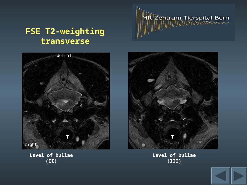
FSE T2-weighting transverse
T: trachea
dorsal
right
Level of optic chiasm Level of bullae (b)
b b
TT T
dorsal
right
Level of bullae (III)Level of bullae (II)
T T
FSE T2-weighting transverse

FE 3D T1 MPR dorsal1mm slice thickness
plain + C plain + Crostral
right

Contrast enhanced, fat suppressed SE T1-weightingtransverse
Level of optic chiasmLevel of orbita
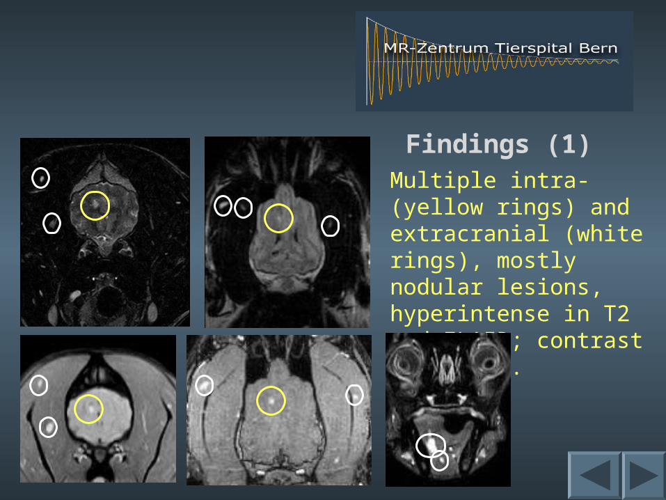
Findings (1)Multiple intra- (yellow rings) and extracranial (white rings), mostly nodular lesions, hyperintense in T2 and FLAIR; contrast enhancing.
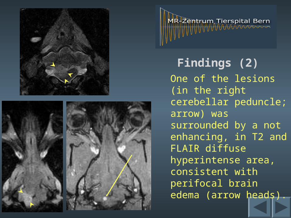
Findings (2)One of the lesions (in the right cerebellar peduncle; arrow) was surrounded by a not enhancing, in T2 and FLAIR diffuse hyperintense area, consistent with perifocal brain edema (arrow heads).
Interpretation
There are multiple lesions within the brain and the musculature of the head including the tongue. The contrast uptake indicates break down of the blood/brain (blood/tissue resp.) barrier. Most lesions are rounded, some could be confluent. One of them is surrounded by diffuse brain edema.These findings are typical of metastases.
Final diagnosis
The muscle lesions were biopsied under ultrasonographic guidance and turned out to be metastases from an hemangiosarcoma.
Comment
Hemangiosarcomas occur in the spleen, liver, heart base and lungs of older dogs. They usually metastasize into the lungs, but spine and brain-metastases have also been described.
Due to its excellent soft tissue contrast, MRI allows the detection of even small metastases. The application of contrast agent increases the sensitivity of the examination. Metastases can be diagnosed by their disseminated, rounded appearance with strong contrast enhancement. Hemangiosarcoma metastases appear bright in all fluid sensitive sequences.