Embed Size (px)
Citation preview
J. Anat. (1983), 137, 2, pp. 371-385 371With 7 figuresPrinted in Great Britain
Postnatal maturation of the vascularisation of thesuprasylvian gyrus of the cat
C. BEN HAMIDA*, J. C. BISCONTEt ANDS. MARGULESt
*Laboratoire de Neuropathologie, Centre de Neurologie-Hopital La Rabta,Tunis, Tunisie and t Laboratoire de Microscopie Quantitative, UER Biomedicale,
74 rue Marcel Cachin, 93 012 Bobigny, France
(Accepted 30 December 1982)
INTRODUCTION
In the course of a general study of the postnatal development of the cat supra-sylvian gyrus, special attention has been paid to changes in its vascularisation duringmaturation. Among numerous earlier studies devoted to the maturation of thecerebral cortex, those concerned with its vascularisation (Dunning & Wolff, 1937;Caley & Maxwell, 1970; Bar & Wolff, 1973; Conradi, Eins & Wolff, 1979) havesought to establish whether or not this feature might be considered as an indicatorof maturation.The present study attempts to determine whether vascularisation could be a
functional indicator, that is, one related closely to cell formation, to synaptogenesisand to myelination, as well as an indicator of structure. For example, it might berelated to cytoarchitectonics, or be related to differences or similarities in the super-ficial and deep parts of the cortex.The study of different vascular parameters, particularly the orientation of the
vessels, is made possible by automatic image analysis, and may provide answers tosome of the questions raised. Therefore, in this investigation, an evaluation has beenmade of mitoses, vascular densities and diameters, the ratio of vascular surface areato area of cerebral tissue (vascularisation coefficient), and the preferential orientationsof vessels in the developing cat cerebral cortex.
MATERIAL AND METHODS
Two experimental series of cats were anaesthetised by an intraperitoneal injectionof Nembutal (0 04 ml/10 g body weight) before tissue fixation.
Mitosis, vascular density and diameterSeventeen kittens with postnatal ages of 2 hours to 42 days, and three adult cats,
were given aortic perfusions of 2.5 % glutaraldehyde in a phosphate buffer(Na2HPO4, KH2PO4) to which paraformaldehyde was added in various concentra-tions to give osmolarities of from 300 mosM (younger animals) to 600 mosM (olderanimals). The fixative was placed in a flask 1 2-1 5 metres above the animal, hence thepressure used, during perfusion, was approximately 150cm water. This made possiblefixation of cytological structures, and of vessels in particular, without distension orshrinkage artefacts. The same material was used in another study using the electron
C. BEN HAMIDA, J. C. BISCONTE AND S. MARGULES
A
100Cerebral cortex AWhite matter A
50 7 7 7 7 7
a , - --A. _ __
10 20 30 40 50 60 70 adu ItAge (days)
B
100 -A ---~~~~~~~~~~~~~
.. *-. .A-.-̂ Cerebral cortex A
% , . 4White matter
A~.,,
50 ,
10 20 30 40 50 60 70Age (days)
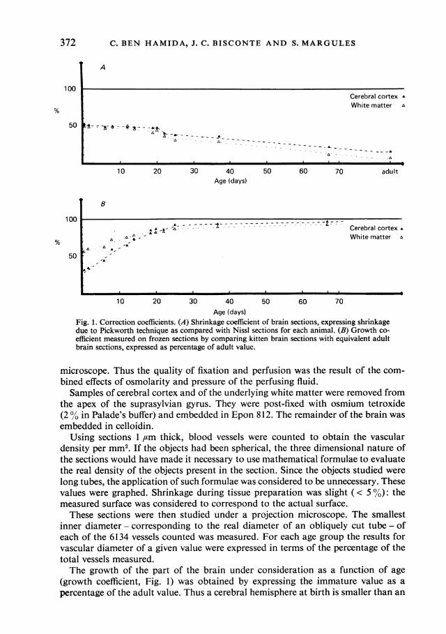
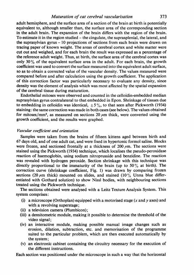
Fig. 1. Correction coefficients. (A) Shrinkage coefficient of brain sections, expressing shrinkagedue to Pickworth technique as compared with Nissl sections for each animal. (B) Growth co-efficient measured on frozen sections by comparing kitten brain sections with equivalent adultbrain sections, expressed as percentage of adult value.
microscope. Thus the quality of fixation and perfusion was the result of the com-bined effects of osmolarity and pressure of the perfusing fluid.
Samples of cerebral cortex and of the underlying white matter were removed fromthe apex of the suprasylvian gyrus. They were post-fixed with osmium tetroxide(2 0 in Palade's buffer) and embedded in Epon 812. The remainder of the brain wasembedded in celloidin.
Using sections 1 ,um thick, blood vessels were counted to obtain the vasculardensity per mm2. If the objects had been spherical, the three dimensional nature ofthe sections would have made it necessary to use mathematical formulae to evaluatethe real density of the objects present in the section. Since the objects studied werelong tubes, the application of such formulae was considered to be unnecessary. Thesevalues were graphed. Shrinkage during tissue preparation was slight ( < 5 0): themeasured surface was considered to correspond to the actual surface.These sections were then studied under a projection microscope. The smallest
inner diameter - corresponding to the real diameter of an obliquely cut tube - ofeach of the 6134 vessels counted was measured. For each age group the results forvascular diameter of a given value were expressed in terms of the percentage of thetotal vessels measured.The growth of the part of the brain under consideration as a function of age
(growth coefficient, Fig. 1) was obtained by expressing the immature value as apercentage of the adult value. Thus a cerebral hemisphere at birth is smaller than an
372
Maturation of cat cerebral vascularisationadult hemisphere, and the surface area of a section of the brain at birth is potentiallyequivalent to, although smaller than, the surface area of the corresponding sectionin the adult brain. The expansion of the brain differs with the region of the brain.To estimate it in the region studied - the cingulate, the suprasplenial, the lateral, andthe suprasylvian gyrus - 10 projections of sections from each brain were drawn ontracing paper of known weight. The areas of cerebral cortex and white matter werecut out and weighed, and for each brain the result was expressed as a percentage ofthe reference adult weight. Thus, at birth, the surface area of the cerebral cortex wasonly 30% of the equivalent surface area in the adult. For each brain, the growthcoefficient was used to convert the surface measured into the equivalent adult surface,so as to obtain a corrected value of the vascular density. The values measured werecompared before and after calculation using the growth coefficient. The applicationof this correction factor was particularly necessary to evaluate any density, sincedensity was the element of analysis which was most affected by the spatial expansionof the cerebral tissue during maturation.
Endothelial mitoses were observed and counted in the celloidin-embedded mediansuprasylvian gyrus contralateral to that embedded in Epon. Shrinkage of tissues dueto embedding in celloidin was identical, ± 5 %, to that seen after Pickworth (1934)staining: the same correction was made in both cases (see below). The values obtainedfor mitoses/mm2, as measured on sections 20 ,tm thick, were converted using thegrowth coefficient, and the results were graphed.
Vascular coefficient and orientationSamples were taken from the brains of fifteen kittens aged between birth and
67 days old, and of one adult cat, and were fixed in hypertonic formol saline. Blockswere frozen, and sectioned frontally at a thickness of 200 prm. The sections werestained using the Pickworth (1934) technique, which localises the pseudo-peroxidasereaction of haemoglobin, using sodium nitroprusside and benzidine. The reactionwas revealed with hydrogen peroxide. Section shrinkage with this technique wasdirectly proportional to the immaturity of the brain (up to 50% at birth). Thecorrection curve (shrinkage coefficient, Fig. 1) was drawn by comparing frozensections (20,tm thick) mounted on slides, and stained (10% Unna blue differ-entiated with Gothard solution) to show Nissl bodies, with neighbouring sectionstreated using the Pickworth technique.The sections obtained were analysed with a Leitz Texture Analysis System. This
system comprises:(i) a microscope (Orthoplan) equipped with a motorised stage (x and y axes) and
with a revolving superstage;(ii) a television camera (Plumbicon);(iii) a densitometric module, making it possible to determine the threshold of the
video signal;(iv) an interactive module, making possible manual image changes such as
erosion, dilation, subtraction, etc. and memorisation of the programmesuited to the particular problem, which are then executed automatically bythe system;
(v) an electronic cabinet containing the circuitry necessary for the execution ofthe different instructions.
Each section was positioned under the microscope in such a way that the horizontal
373
C. BEN HAMIDA, J. C. BISCONTE AND S. MARGULES
y
x
Y+120 Y+240
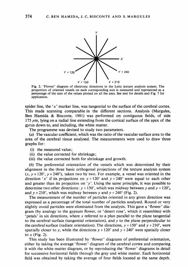
Y + 150 Y + 210Fig. 2. 'Flower' diagram of electronic directions in the Leitz texture analysis system. Theproportion of oriented vessels on each corresponding axis is measured and represented as apercentage of the sum of the values plotted on all the axes. See text for details and Fig. 7 forapplication.
spider line, the 'x' marker line, was tangential to the surface of the cerebral cortex.This made scanning comparable in the different sections. Analysis (Margules,Ben Hamida & Bisconte, 1981) was performed on contiguous fields, of side175 ,um, lying on a radial line extending from the cortical surface of the apex of thegyrus down to, and including, the white matter.The programme was devised to study two parameters.(a) The vascular coefficient, which was the ratio of the vascular surface area to the
area of the cerebral tissue analysed. The measurements were used to draw threegraphs for:
(i) the measured value;(ii) the value corrected for shrinkage;(iii) the value corrected both for shrinkage and growth.(b) The preferential orientation of the vessels which was determined by their
alignment in the three basic orthogonal projections of the texture analysis system(x, y + 120°, y + 240°), taken two by two. For example, a vessel was oriented in thedirection 'x' if its projections on y + 1200 and y + 240° were equal to each otherand greater than its projection on 'y'. Using the same principle, it was possible todetermine two other directions: y + 1500, which was midway between y and y + 120°,and y + 210°, which was midway between y and y + 2400 (Fig. 2).The measurement of the number of particles oriented in any given direction was
expressed as a percentage of the total number of particles analysed. Round or veryslightly ovoid particles were eliminated from the analysis. This gave a 'flower' dia-gram (by analogy to the gypsum flower, or 'desert rose', which it resembles) with'petals' in six directions, where x referred to a plan parallel to the plane tangentialto the cerebral surface (tangential orientation), and y to the plane perpendicular tothe cerebral surface (radiate orientation). The directions, y + 1500 and y + 210°, werespatially closer to y, while the directions y + 120° and y + 240° were spatially closerto x (Fig. 2).
This study has been illustrated by 'flower' diagrams of preferential orientation,either by taking the average 'flower' diagram of the cerebral cortex and comparingit with the white matter diagram, or by reproducing the 'flower' diagrams in detailfor sutccessive horizontal fields through the grey and white matter. Each horizontalfield was obtained by taking the average of four fields located at the same depth.
374
Maturation of cat cerebral vascularisation 375
Vascular mitosis
6 A Cerebral cortex
* White matterEEE
4
A
2 A
AA
0 5 10 1 5 20 25 30 35 40 45Age (days)
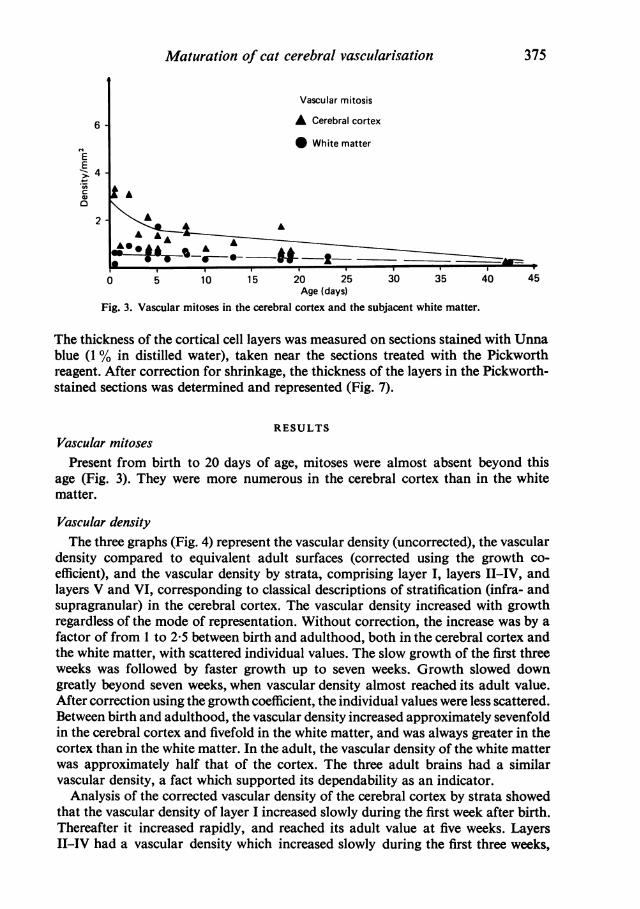
Fig. 3. Vascular mitoses in the cerebral cortex and the subjacent white matter.
The thickness of the cortical cell layers was measured on sections stained with Unnablue (1 % in distilled water), taken near the sections treated with the Pickworthreagent. After correction for shrinkage, the thickness of the layers in the Pickworth-stained sections was determined and represented (Fig. 7).
RESULTSVascular mitosesPresent from birth to 20 days of age, mitoses were almost absent beyond this
age (Fig. 3). They were more numerous in the cerebral cortex than in the whitematter.
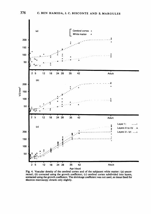
Vascular densityThe three graphs (Fig. 4) represent the vascular density (uncorrected), the vascular
density compared to equivalent adult surfaces (corrected using the growth co-efficient), and the vascular density by strata, comprising layer I, layers II-IV, andlayers V and VI, corresponding to classical descriptions of stratification (infra- andsupragranular) in the cerebral cortex. The vascular density increased with growthregardless of the mode of representation. Without correction, the increase was by afactor of from 1 to 2-5 between birth and adulthood, both in the cerebral cortex andthe white matter, with scattered individual values. The slow growth of the first threeweeks was followed by faster growth up to seven weeks. Growth slowed downgreatly beyond seven weeks, when vascular density almost reached its adult value.After correction using the growth coefficient, the individual values were less scattered.Between birth and adulthood, the vascular density increased approximately sevenfoldin the cerebral cortex and fivefold in the white matter, and was always greater in thecortex than in the white matter. In the adult, the vascular density of the white matterwas approximately half that of the cortex. The three adult brains had a similarvascular density, a fact which supported its dependability as an indicator.
Analysis of the corrected vascular density of the cerebral cortex by strata showedthat the vascular density of layer I increased slowly during the first week after birth.Thereafter it increased rapidly, and reached its adult value at five weeks. LayersII-IV had a vascular density which increased slowly during the first three weeks,
C. BEN HAMIDA, J. C. BISCONTE AND S. MARGULES
(a) E Cerebral cortex AWhite matter a
la
A -,
A I- A A
-- A~~~~~ A.. -A
*A ,,A0, AA A
A
6 a
Y Y V
2 5 12 18 24 28 35 42 Adult
(b)
- ---4- ^ ^ ,\ . ., -- --- -A
m-
.A*.
-. ..A
2 5 12 18 24 28 35 42 'Adult
Layer 1: --(c) : LayerslIItoIV.*
.. Layers V--VI -
.- --.'.,',~.Z.,,..',-,.'*V
2 5 12 18 24 28 35 42 AdultAge (days)
Fig. 4. Vascular density of the cerebral cortex and of the subjacent white matter: (a) uncor-rected; (b) corrected using the growth coefficient; (c) cerebral cortex subdivided into layers,corrected using the growth coefficient. The shrinkage coefficient was not used, as tissue fixed forelectron microscopy shrank only slightly.
376
200-
150 -
100
50 -
200
EE 150-a
100 '
50 -
200 -
150
100 ^
50 -
A& -
Maturation of cat cerebral vascularisation
12 hours to 17 days
O> lo Am C.C. = 49%-L W.M.=172%
nb- S ~~~NX' cn)CN CD 84 C~ObCM (D ~~N X' C')
31-42 days
>>10Om [C.C. =62%-iW.M. = 12%---
.li . . . -- . . . . . '... . . . . . . . . . . . . . . . ..
50
VL)0
II-
0
0-0
50'A
4-0
0e
18-30 days¢,,O,lm:C.C = 7.7%I>10ml [ X
C' CO= i- C°Nd- Cn 00
_ ~_N cn
Adults
¢> 10Um W.M.- 13-1 %=
l Lcs r - m -r co1 a) Is I I I;_ , cs cs:,,
0)) ~L O I- C ') 0M(C6 7 4 3 ot-4N c) (a0 e (6 6 4 6 C ~ C_- _ CN (c) c() _- %_ N cn E
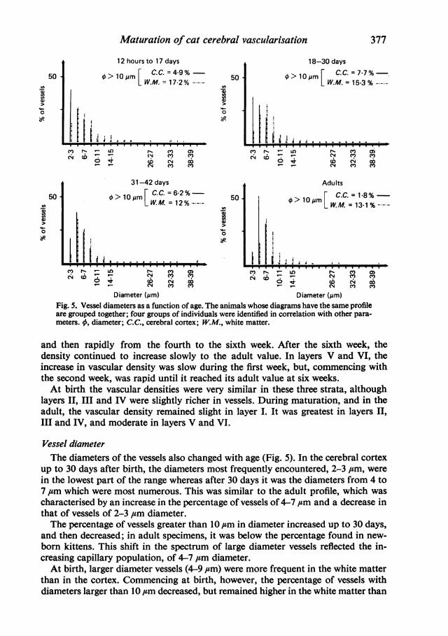
Diameter (,im) Diameter (,um)Fig. 5. Vessel diameters as a function of age. The animals whose diagrams have the same profileare grouped together; four groups of individuals were identified in correlation with other para-meters. 0, diameter; C.C., cerebral cortex; W.M., white matter.
and then rapidly from the fourth to the sixth week. After the sixth week, thedensity continued to increase slowly to the adult value. In layers V and VI, theincrease in vascular density was slow during the first week, but, commencing withthe second week, was rapid until it reached its adult value at six weeks.At birth the vascular densities were very similar in these three strata, although
layers II, III and IV were slightly richer in vessels. During maturation, and in theadult, the vascular density remained slight in layer I. It was greatest in layers II,
III and IV, and moderate in layers V and VI.
Vessel diameterThe diameters of the vessels also changed with age (Fig. 5). In the cerebral cortex
up to 30 days after birth, the diameters most frequently encountered, 2-3 #m, were
in the lowest part of the range whereas after 30 days it was the diameters from 4 to7 ,um which were most numerous. This was similar to the adult profile, which was
characterised by an increase in the percentage of vessels of 4-7 ,um and a decrease inthat of vessels of 2-3 ,um diameter.The percentage of vessels greater than 10 ,um in diameter increased up to 30 days,
and then decreased; in adult specimens, it was below the percentage found in new-
born kittens. This shift in the spectrum of large diameter vessels reflected the in-creasing capillary population, of 4-7 ,um diameter.At birth, larger diameter vessels (4-9 ,csm) were more frequent in the white matter
than in the cortex. Commencing at birth, however, the percentage of vessels withdiameters larger than 10 ,um decreased, but remained higher in the white matter than
377
50
00
50
-
o0o
. v v v v v v v v v v T-T - v v v v T T-
n
378
300
200
100
EEE
300
200
100
300
200
100
C. BEN HAMIDA, J. C. BISCONTE AND S. MARGULES
(a) Cerebral cortex £White matter a
.,
A . A - ,..
AA . A, A. .. -...
2 5 10 20 30 40 50 60 70 Adult
(b)
£~~~~~~~~~~~~~~~~~~~~~~~~~~ - . .
*~~~~~~~~
2 5 10 20 30 40 50 60 70 Adult
(c)
- - - - - - - - - - - - - - - A
2 -
M *...A...2 5 10 20 30 40 50 60 70 Adult
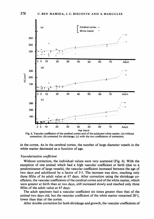
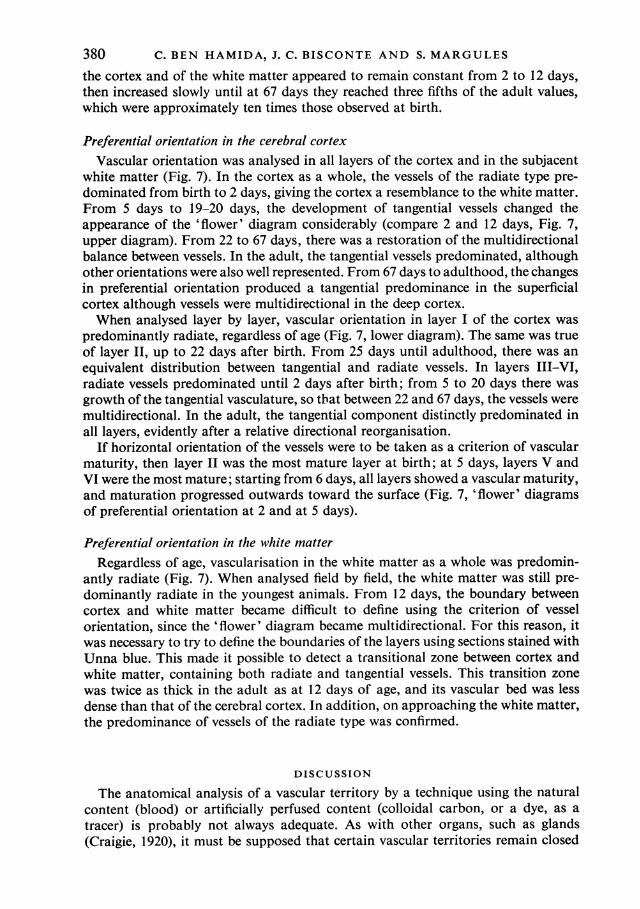
Age (days)Fig. 6. Vascular coefficient of the cerebral cortex and of the subjacent white matter: (a) without
correction; (b) corrected for shrinkage; (c) with the two coefficients of correction.
in the cortex. As in the cerebral cortex, the number of large diameter vessels in thewhite matter decreased as a function of age.
Vascularisation coefficientWithout correction, the individual values were very scattered (Fig. 6). With the
exception of one animal which had a high vascular coefficient at birth (due to apredominance of large vessels), the vascular coefficient increased between the age oftwo days and adulthood by a factor of 3 5. The increase was slow, reaching onlythree fifths of its adult value at 67 days. After correction using the shrinkage co-efficient, the vascular coefficients of the cerebral cortex and of the white matter, whichwere greater at birth than at two days, still increased slowly and reached only threefifths of the adult value at 67 days.The adult specimen had a vascular coefficient six times greater than that of the
animal two days old, but the vascular coefficient of the white matter remained 20%lower than that of the cortex.
After double correction for both shrinkage and growth, the vascular coefficients of
Maturation of cat cerebral vascularisation 379
Age(days)
0 1 5 2 5 6 8 12 12 14 19 20 22 25 37 67 Adult
UJj* t $**4%*-**f I + ,* * * + -Superficial--cortex
1f
1t
1 1 i j 14t f e | + * % | $ Deep corte>
Age.-'-days'
LayerWz - 0 1.5 2 5 6 8 12 12 14 19 29 22 25 37 67 Adult
LDa + %J I * t r' s % I * * 4 I
ol tt+++++ t+ -F+*±± -4 ±+ * Lx . + + + + + + + + + + +;;
- + t+ -+ t+ + + + + + + + >-uv * t.+ t 4 4+ 4+ + + 4 It -
V {*sB+ + + + + ++0 + + + + J - TZ
Newborn -4- + + + + + _ + + t, 4<, j
4 . t L -
E~ ~ ~ ~ ~ 7 1msqae 7- .r
a)~~~~~~~~~~~~~~~~i
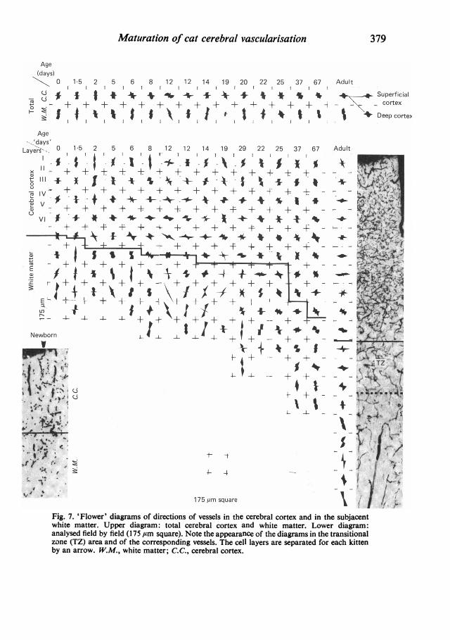
( hit mater $pe iga:ttlcrba otxadwiemte.Lwrdarm
Newborn4--4Wt~~~~~~~~~
analysed field by field (175 gum square). Note the appearance of the diagrams in the transitionalzone (TZ) area and of the corresponding vessels. The cell layers are separated for each kittenby an arrow. W.M., white matter; C.C., cerebral cortex.
C. BEN HAMIDA, J. C. BISCONTE AND S. MARGULES
the cortex and of the white matter appeared to remain constant from 2 to 12 days,then increased slowly until at 67 days they reached three fifths of the adult values,which were approximately ten times those observed at birth.
Preferential orientation in the cerebral cortexVascular orientation was analysed in all layers of the cortex and in the subjacent
white matter (Fig. 7). In the cortex as a whole, the vessels of the radiate type pre-dominated from birth to 2 days, giving the cortex a resemblance to the white matter.From 5 days to 19-20 days, the development of tangential vessels changed theappearance of the 'flower' diagram considerably (compare 2 and 12 days, Fig. 7,upper diagram). From 22 to 67 days, there was a restoration of the multidirectionalbalance between vessels. In the adult, the tangential vessels predominated, althoughother orientations were also well represented. From 67 days to adulthood, the changesin preferential orientation produced a tangential predominance in the superficialcortex although vessels were multidirectional in the deep cortex.When analysed layer by layer, vascular orientation in layer I of the cortex was
predominantly radiate, regardless of age (Fig. 7, lower diagram). The same was trueof layer II, up to 22 days after birth. From 25 days until adulthood, there was anequivalent distribution between tangential and radiate vessels. In layers III-VI,radiate vessels predominated until 2 days after birth; from 5 to 20 days there wasgrowth of the tangential vasculature, so that between 22 and 67 days, the vessels weremultidirectional. In the adult, the tangential component distinctly predominated inall layers, evidently after a relative directional reorganisation.
If horizontal orientation of the vessels were to be taken as a criterion of vascularmaturity, then layer II was the most mature layer at birth; at 5 days, layers V andVI were the most mature; starting from 6 days, all layers showed a vascular maturity,and maturation progressed outwards toward the surface (Fig. 7, 'flower' diagramsof preferential orientation at 2 and at 5 days).
Preferential orientation in the white matterRegardless of age, vascularisation in the white matter as a whole was predomin-
antly radiate (Fig. 7). When analysed field by field, the white matter was still pre-dominantly radiate in the youngest animals. From 12 days, the boundary betweencortex and white matter became difficult to define using the criterion of vesselorientation, since the 'flower' diagram became multidirectional. For this reason, itwas necessary to try to define the boundaries of the layers using sections stained withUnna blue. This made it possible to detect a transitional zone between cortex andwhite matter, containing both radiate and tangential vessels. This transition zonewas twice as thick in the adult as at 12 days of age, and its vascular bed was lessdense than that of the cerebral cortex. In addition, on approaching the white matter,the predominance of vessels of the radiate type was confirmed.
DISCUSSION
The anatomical analysis of a vascular territory by a technique using the naturalcontent (blood) or artificially perfused content (colloidal carbon, or a dye, as atracer) is probably not always adequate. As with other organs, such as glands(Craigie, 1920), it must be supposed that certain vascular territories remain closed
380
Maturation of cat cerebral vascularisationtemporarily. Thus, it is possible that the results obtained with the Pickworth tech-nique on non-perfused brains, i.e. the measurements of vascular coefficient and of-preferential vascular orientation, may correspond more to the physiological realitythan to the strictly anatomical reality. The results obtained on perfused brains, forevaluation of vascular diameter and density, may correspond more to the anatomicalthan to the physiological reality.The shrinkage coefficient was used in this study to correct the experimental data
and give them greater real precision, and is a method proposed by Eins & Wilhems(1976). The application of a correction using the growth coefficient to the curvesobtained from our results has made it possible to compare potentially and function-ally similar parts of the brain, regardless of the age of the animal.
Stages of developmentThe postnatal cerebral vascular maturation of the cat is slower than that of the
rat and takes twice as long to attain the vascular profile of the adult. Using fiveindicators of vascular maturation (vascular mitosis, density, diameter, vascularcoefficient, and the preferential vascular orientation), different consecutive periodsare observed.The first week of immaturity, exhibiting active mitoses, corresponds to the period
of sprouting of Bar & Wolff (1973), with constant vascular diameter and vascularcoefficient, an immature diameter (predominantly 2-3 ,tm), and a primarily radiatepreferential orientation comparable to that of the white matter. This period is almostidentical to that in the rat, which has a low and constant vascular density (Craigie,1955; Singh & Nathaniel, 1975; Bar, 1978). This is succeeded by the period ofvascular growth, as established by the indicator of vascular density, which reachesalmost its maximum value. This period covers the second to the seventh weeks afterbirth and corresponds to the second and third weeks in the rat. It is characterised bya rapidly increasing vascular density and by an increase in the number of capillaryramifications (Bar & Wolff, 1973). Nevertheless, taking into consideration otherindicators such as vascular diameter, vascular coefficient, and preferential orienta-tion, this period can be subdivided into two stages in the cat.The first stage, from the second week to 30 days after birth (premature period) is
characterised by vascularisation of an immature type (maximum percentage ofdiameters, 2-3 ,um), an increasing percentage of diameters greater than 10 ,um, anda predominantly tangential orientation of vessels.The second stage, from 30 to 49 days (pre-adult period), is characterised by a
maximum but decreasing percentage of vascular diameters greater than 10 ,um, andby both radiate and tangential orientation of vessels. The adult period resembles thepre-adult period, but with preferentially tangential orientation of vessels; some largevessels disappear in both the cortex and the white matter.
Gradients in the cerebral cortexParallel to a gradient of maturation of cortical neurons from the deep strata to-
wards the superficial strata (Berry, Rogers & Eayrs, 1964; Rabinovitch, 1964; Caley& Maxwell, 1968 a, b), it has been noted by Bar (1978) in the newborn rat, that thereis a greater capillary density in the deep cerebral cortex than in the superficialcerebral cortex. Up to 3 weeks of age, he observes a more rapid development in thesuperficial and deep cortex, with a delay of 4 days in layer IV.
In the newborn kitten, the vascular densities in the superficial and the deep layers
381
382 C. BEN HAMIDA, J. C. BISCONTE AND S. MARGULES
of the suprasylvian gyrus are approximately equal. Consideration of the postnatalchanges of vascular density in the cortical layers (Fig. 4) suggests that extrapolationinto the prenatal period would give a vascular density of layers II-IV which is lessthan the vascular density of layers V-VI before birth, in agreement with observa-tions made in the rat. This indicates that deep vascular maturation precedessuperficial vascular maturation. Thereafter, superficial vascularisation develops toa greater extent than deep vascularisation until adulthood. This is in agreementwith the description of a more highly developed vascularisation of the suprasylviangyrus of the cat in layers III and IV (Campbell, 1939) than in other layers.
Vessels greater than 10,um in diameter continue to form until 30 days, completingthe population of transitional vessels existing at birth. It should be noted that at42 days almost all indicators have reached their adult value, apart from the vascularcoefficient, which remains at 37 % of the adult value. Consequently, the vessels in-crease essentially in length, growing neither in number nor in diameter. This is inagreement with Sakla(1965), who attributes the decrease in the number of vesselslarger than10,um diameter to longitudinal vessel growth, a consequence of theincrease in-cerebral volume. The vessels which form after birth are mainly capillaries,i.e. their diameters are less than or equal to 8,um (Bar & Wolff, 1973; Hunziker,Prey & Schulz, 1974; Mato & Ookawara, 1979; Conradi, Engvall & Wolff, 1980).Indeed between birth and adulthood it is these vessels (4-84um diameter) whichincrease in number, while those of other diameters decrease.
MyelinationThe formation of the vascular bed and the commencement of myelination have
been compared chronologically (Kennedy, Grove, Jehle & Sokoloff, 1970, 1972; Bar,1978), but analysis of the present observations does not seem to reveal any directcorrelation between myelination and vascular growth. While vascular densityincreases greatly in the cerebral cortex starting from the seventh day, myelinationdoes not reach the cortex until 19 days, that is, 12 days after the beginning of vas-cular growth. Two studies have been devoted to the vascularisation of the whitematter. Following anatomical studies of vasculogenesis (Craigie, 1955; De Reuck,1972; Conradi & Sourander, 1980), Kennedy et al. (1972), studying the vascularflow, note a transitory increase in flow in relation to myelination.
White matter and transitional zoneIn the kitten, the low vascular density and the high vascular coefficient of the
white matter in comparison with the cerebral cortex indicate that the vessels in thewhite matter are of greater diameter. The vascular bed of the white matter is com-posed preferentially of radiate vessels, i.e. transitional vessels formed at an earlystage (Wolff, 1978; Conradi et al. 1980), and a well represented group of capillaries,which is approximately constant up to 42 days after birth (70 to 73 % of the vascularpopulation), reducing slightly in the adult (66 %), and probably belonging to thetransitional zone. The transitional zone appears at the age of 12 days, and containsa vascular network with larger mesh than that of the cerebral cortex. The richnessof this vascular network is expressed in its relatively high percentage of capillaries andin a preferential tangential orientation of its vessels (Fig. 7), recalling the appearanceof the cortex. The transitional zone, supplied by corticomedullary vessels (De Reuck,1972), seems to correspond to a zone rich in basal dendrites of pyramidal cells, inapical dendrites of abnormally oriented pyramidal cells (Van der Loos, 1965; Globus
382
Maturation of cat cerebral vascularisation& Scheibel, 1967), and in dendrites of other cells. It is not possible, either in the whitematter or in the cerebral cortex, to detect different stages of development, for all theindicators increase slowly and regularly. This is the case even between 13 and 19 days,which is the period of progression of myelination between the white matter of thesuprasylvian gyrus and layer VI of the cortex.The vessels of the white matter are the first to form; proliferation occurs essentially
in the fetal period, and their growth pre- and postnatally.
SynaptogenesisConcerning the relationship with synaptogenesis observed in the cerebral cortex
(Bar, 1978; Conradi et al. 1979, 1980), work in progress suggests the existence of aclose correlation between synaptogenesis and vascular maturation. Synaptic growthand neuropil development commence from the second postnatal week in the supra-sylvian gyrus of the cat, producing maximum synaptic density of the superficialcortex where the zone of maximum vascular density (layers II-IV) is located.
Structural significanceWith respect to capillaries 2-3 ,um in diameter it appears from the work of Mato
& Ookawara (1979) that they may be functional, since rheology experiments demon-strate the extreme ease of distortion of erythrocytes. This provides no information,of course, concerning possible exchange through the vascular wall. The populationof small vessels - potential capillaries, arterioles and venules - is very large at birth(40%) and up to 30 days (36 %). In the immature state, all these vessels may play arole in metabolic exchange. With the maturation of the vascular system, each typeof vessel seems to acquire its adult characteristics, i.e. selectivity and a preponderantrole in metabolic exchange as compared with that of the intercellular space.According to Johanson (1980), the permeability of the cerebral and cerebellar
cortices to certain substances decreases pari passu with increase in vascularisation,and the formation of the blood-brain barrier is related to a decrease in the extra-cellular space. The permeability of small, immature vessels, such as capillaries,arterioles, venules, may therefore be temporary. Selective exchange comes withmaturity; it concerns primarily the capillaries, whose endothelial membranesgradually become enriched in proteins which play a role in selectivity. The imper-meability of the walls of arteriole and venule seems to be directly related to theirthickness.The extracellular spaces seem to regress in parallel with the development of the
vessels (Caley & Maxwell, 1970) and of the neuropil. Until recently, there have beenno definitive arguments making it possible to establish whether extracellular spaceslarger than 20 nm are artifactual (Pappas & Purpura, 1964) or not (Luse, 1960; DeRobertis & Gerschenfeld, 1961; Caley & Maxwell, 1970). However, the detection ofanions in extracellular spaces (Derer & Nakanishi, 1980) seems to indicate that thesespaces are indeed preferential routes of passage of metabolites during the perinatalperiod. As the vascular density is very low in the newborn as compared with theadult (even after correction using the growth coefficient), and the vascular diameteris small (40% of the vessels, 2-3 ,um), the passage of metabolites would naturallybe via the extracellular spaces.The assumption that extracellular space accounts for 17-20% of the adult mam-
malian brain (Cragg, 1979) suggests that it continues to play a role in the passage ofmetabolites in the adult. The capillaries may then selectively complement the extra-
383
13 ANA137
C. BEN HAMIDA, J. C. BISCONTE AND S. MARGULES
cellular space, necessary but not sufficient for the normal functioning of the cerebraltissue.
SUMMARY
Vascular growth in the median suprasylvian gyrus of the cat has been analysedquantitatively with respect to mitoses, vascular density, vascular diameters, vascularcoefficient and preferential vascular orientation. After correction for shrinkage andgrowth, four maturation periods were identified:
(i) Immature period (first postnatal week), when the tissue exhibited numerousvascular mitoses, a low but constant vascular density and vascular coefficient, pre-ferentially radiate vessels and immature (small) vascular diameters.
(ii) Premature period (second to fourth week), with few mitoses, a rapidly in-creasing vascular density, immature (small) vascular diameters, an increase in thenumber of vessels more than 10 /am in diameter, and a preferential orientation oftangential vessels.
(iii) Pre-adult period (fifth to sixth week), without mitoses. The vascular densityincreased greatly, the vascular diameters reached adult profile, the number of vesselsover 10 ,um in diameter decreased, the vascular orientations were both tangentialand radiate and the vascular coefficient remained slight.
(iv) Adult period, when the vascular density and the vascular coefficient weremaximal, the vessel diameters were of adult type, and the orientation was tangential.From six weeks, the vessels increased only in length. Vascular maturation pro-
ceeded from the depth toward the surface of the cerebral cortex. Layers II-IV werethose most highly vascularised, regardless of age. This is discussed in relation tosynaptic growth. There was no direct relation between vasculogenesis and myelina-tion.The white matter had a typically radiate vascularisation. A transitional zone be-
tween cortex and white matter was identified. It had a loose mesh vascular networkand corresponded to the area in which dendrites of inverted pyramidal cells werefound.The role of immature vascularisation in the nutrition of the neuropil is discussed.
REFERENCES
BAR, TH. (1978). Morphometric evaluation of capillaries in different laminae of rat cerebral cortex byautomatic image analysis: changes during development and aging. In Advances in Neurology, vol. 20(ed. J. Cerves-Navarro et al.), pp. 1-9. New York: Raven Press.
BAR, TH. & WOLFF, J. R. (1973). Quantitative Beziehungen zwischen der Verzweigungsdichte undLinge von Capillaren in Neocortex der Ratte wahrend der post-natalen Entwicklung. Zeitschrift farAnatomie und Entwicklungsgeschichte 141, 207-221.
BERRY, M., ROGERS, A. W. & EAYRS, J. T. (1964). The pattern and mechanism of migration of theneuroblasts of the developing cerebral cortex. Journal of Anatomy 98, 291-292.
CALEY, D. W. & MAXWELL, D. S. (1968a). An electron microscopic study of neurons during post-nataldevelopment of rat cerebral cortex. Journal of Comparative Neurology 133, 17-43.
CALEY, D. W. & MAXWELL, D. S. (1968b). An electron microscopic study of the neuroglia during post-natal development of the rat cerebrum. Journal of Comparative Neurology 133, 45-70.
CALEY, D. W. & MAXWELL, D. S. (1970). Development of blood vessels and extracellular spaces duringpostnatal maturation of rat cerebral cortex. Journal of Comparative Neurology 138, 31-48.
CAMPBELL, A. C. F. (1939). Variation in vascularity and oxidase content in different gyrus of the brainof the rat. Archives of Neurology and Psychiatry 41, 223-242.
CONRADI, N. C., EINS, S. & WOLFF, J. R. (1979). Postnatal vascular growth in the neocortex of normaland protein-deprived rats. Acta neuropathologica 47, 123-130.
384
Maturation of cat cerebral vascularisation 385CONRADI, N. C., ENGVALL, J. & WOLFF, J. R. (1980). Angioarchitectonics of rat cerebellar cortex during
pre- and postnatal development. Acta neuropathologica 50, 131-138.CONRADI, N. C. & SOURANDER, P. (1980). The early internal vascularization of the rat brain. Morpho-
logical studies on foetuses of normal and protein deprived mothers. Acta neuropathologica 50, 221-226.CRAGG, B. (1979). Brain extracellular space fixed for electron microscopy. Neuroscience Letters 15,
301-306.CRAIGIE, E. H. (1920). On the relative vascularity of various parts of the central nervous system of the
albino rat. Journal of Comparative Neurology 31, 429-464.CRAIGIE, E. H. (1955). Vascular patterns of the developing nervous system. In Biochemistry of the De-
veloping Nervous System (ed. H. Warlsch), pp. 28-51. New York: Academic Press.DERER, P. & NAKANISHI, S. (1980). Extracellular anionic site localisation in the developing cortex of
'normal' and 'reeler' mutant mice. Abstract, 1st ISDN Congress, Strasbourg.DE REUCK, J. (1972). The cortico-subcortical arterial angio-architecture in the human brain. Acta
neurologica belgica 72, 323-329.DE ROBERTIs, E. & GERSCHENFELD, H. M. (1961). Submicroscopic morphology and function of glial cells.
International Review of Neurobiology 3, 1-65.DUNNING, H. J. & WOLFF, H. G. (1937). The relative vascularity of various parts of the central and
peripheral nervous system of the cat and its relation to function. Journal of Comparative Neurology67, 433-450.
EINS, S. & WILHELMS, E. (1976). Assessment of preparative volume changes in central nervous tissueusing automatic image analysis. Microscope, 24, 29-38.
GLOBUS, A. & SCHEIBEL, A. B. (1967). Pattern and field in cortical structure: the rabbit. Journal ofComparative Neurology 131, 155-172.
HUNZIKER, C., PREY, M. & SCHULZ, U. (1974). Morphometric investigations of the capillaries in the braincortex of the cat. A contribution to the microcirculation of the brain. Acta cardiologica, Suppl. XIX,227-233.
JOHANSON, C. E. (1980). Permeability and vascularity of the developing brain: cerebellum vs cerebralcortex. Brain Research 190, 3-16.
KENNEDY, C., GRAVE, G. D., JEHLE, J. W. & SOKOLOFF, L. (1970). Blood flow to white matter duringmaturation of the brain. Neurology 20, 613-618.
KENNEDY, C., GRAVE, G. D., JEHLE, J. W., & SOKOLOFF, L. (1972). Changes in blood flows in the com-ponent structure of the dog brain during postnatal maturation. Journal of Neurochemistry 19,2423-2435.
LUSE, S. T. (1960). Electron microscopic observations of the central nervous system. In Inhibition in theNervous System and Gamma-amino butyric Acid (ed. E. Roberts), pp. 29-33. New York. PergamonPress.
MARGULES, S., BEN HAMIDA, C. & BISCONTE, J. C. (1981). Determination of the preferential orientationof blood vessels in cat brain. Evaluation of the method, using a T.A.S. (Leitz) image analyser. Congressof Quantitative Histology, Sheffield, July.
MATO, M. & OOKAWARA, S. (1979). A simple method for observations of capillary nets in rat braincortex. Experientia 55, 4, 501-503.
PAPPAS, G. D. & PURPURA, D. P. (1964). Electron microscopy of immature human and feline neocortex.In Progress in Brain Research, vol. 4 (ed. D. P. Purpura & J. P. Schade), pp. 126-186. New York:Elsevier.
PICKWORTH, F. A. (1934). A new method of study of the brain capillaries and its application to theregional localisation of mental disorder. Journal ofAnatomy 69, 62-78.
RABINOVITCH, TH. (1964). The cerebral cortex of the premature infant of the 8th month. In Growth andmaturation of the brain. Progress in Brain Research 4, 39-92.
SAKLA, F. B. (1965). Post-natal growth of neuroglia cells and blood vessels of the cervical spinal cord ofthe albino mouse. Journal of Comparative Neurology 124, 189-202.
SINGH, D. N. P. & NATHANIEL, E. J. H. (1975). Post natal development of blood vessels (capillaries) inthe rat olfactory bulb: a light and ultrastructural study. Neuroscience Letters 1, 203-208.
VAN DER Loos, (1965). The 'improperly' oriented pyramidal cells in the cerebral cortex and its possiblebearing on problems of neuronal growth and cells orientation. Bulletin of the Johns Hopkins Hospital117, 4, 228-250.
WOLFF, J. R. (1978). Cytogenetic aspects of cortical architecture: Lamination. In Architectonics ofthe Cerebral Cortex (ed. M. A. E. Brazier & H. Petsche), pp. 159-173. New York: Raven Press.
13-2