Embed Size (px)
Citation preview
Korean J Radiol 8(5), October 2007 365
Labeling Efficacy of SuperparamagneticIron Oxide Nanoparticles to HumanNeural Stem Cells: Comparison ofFerumoxides, Monocrystalline Iron Oxide,Cross-linked Iron Oxide (CLIO)-NH2 andtat-CLIO
Objective: We wanted to compare the human neural stem cell (hNSC) labelingefficacy of different superparamagnetic iron oxide nanoparticles (SPIONs),namely, ferumoxides, monocrystalline iron oxide (MION), cross-linked iron oxide(CLIO)-NH2 and tat-CLIO.
Materials and Methods: The hNSCs (5 105 HB1F3 cells/ml) were incubatedfor 24 hr in cell culture media that contained 25 g/ml of ferumoxides, MION orCLIO-NH2, and with or without poly-L-lysine (PLL) and tat-CLIO. The cellular ironuptake was analyzed qualitatively with using a light microscope and this wasquantified via atomic absorption spectrophotometry. The visibility of the labeledcells was assessed with MR imaging.
Results: The incorporation of SPIONs into the hNSCs did not affect the cellularproliferations and viabilities. The hNSCs labeled with tat-CLIO showed thelongest retention, up to 72 hr, and they contained 2.15 0.3 pg iron/cell, whichare 59 fold, 430 fold and six fold more incorporated iron than that of the hNSCslabeled with ferumoxides, MION or CLIO-NH2, respectively. However, when PLLwas added, the incorporation of ferumoxides, MION or CLIO-NH2 into the hNSCswas comparable to that of tat-CLIO.
Conclusion: For MR imaging, hNSCs can be efficiently labeled with tat-CLIOalone or with a combination of ferumoxides, MION, CLIO-NH2 and the transfec-tion agent PLL.
uman neural stem cells (hNSCs) can be used to replace dead tissue inpatients suffering with Alzheimer’s disease, amyotrophic lateral sclerosis,Huntington’s disease, stroke and spinal cord injury (1, 2). There is a
growing interest in in vivo visualization of transplanted cells with using noninvasivetechniques such as magnetic resonance (MR) imaging. The development of anappropriate MR imaging technique with optimized cell labeling conditions will beuseful to monitor the effectiveness of cell implantation, homing and differentiation (3).
Superparamagnetic iron oxide nanoparticles (SPIONs) have been used for celllabeling agents in both preclinical and clinical settings. Ferumoxide (AMI-25), astandard SPION agent, is clinically approved and commercially available (4 6). It iscoated with dextran and has a hydrodynamic diameter of approximately 100 nm.Monocrystalline iron oxides (MIONs) are smaller (28 nm) than the standard SPIONs so
Miyeoun Song, PhD1
Woo Kyung Moon, MD2
Yunhee Kim, MS1
Dongyeol Lim, PhD3
In-Chan Song, PhD2
Byung-Woo Yoon, MD1
Index terms:Human neural stem cellIron oxide nanoparticlesMagnetic resonance (MR)
Korean J Radiol 2007;8:365-371Received April 11, 2006; accepted after revision January 23, 2007.
1Department of Neurology, ClinicalResearch Institute, Seoul NationalUniversity Hospital, Seoul NationalUniversity, Seoul 110-744, Korea;2Department of Diagnostic Radiology,Seoul National University Hospital, and theInstitute of Radiation Medicine, SeoulNational University Medical ResearchCenter, Seoul 110-744, Korea;3Department of Applied Chemistry, SejongUniversity, Seoul 143-747, Korea
This research was supported by theKorean Health 21 R & D Project, Ministryof Health & Welfare (Project No.A040004), by the National R&D Programfor Cancer Control, Ministry of Health &Welfare, Republic of Korea (Grant no.0420080-1), and by the National R & DProgram of the Korean Ministry of Scienceand Technology (Project No. SC-3111).
Address reprint requests to:Woo Kyung Moon, MD, PhD, Departmentof Diagnostic Radiology, Seoul NationalUniversity Hospital, and the Institute ofRadiation Medicine, Seoul NationalUniversity Medical Research Center, 28,Yongon-dong, Chongno-gu, Seoul 110-744, KoreaTel. (822) 2072-3928Fax. (822) 743-6385e-mail: [email protected]
H
that they can easily pass through the capillary endothelium.The cross-linked iron oxide (CLIO)-NH2 is a modified formof MIONs, and it had the same biophysical properties asMIONs (7). In terms of the particle size, MIONs or CLIO-NH2 might be more useful for performing cellular andmolecular MR imaging. However, many cell types, includ-ing most stem cells, do not take up appreciable amounts ofunmodified iron oxide preparations (8 11). One solutionhas been to modify the surface of the particles withmonoclonal antibody or tat-peptide (12), which may resultin increased internalization of the particles. Tat-peptide ispurified from the human immunodeficiency virus (HIV) tat-peptide, and it carries both a transmembrane and nuclearlocalization signal within its sequence; it is capable oftranslocating exogeneous molecules into cells (13 15). Theother approach is to use a transfection agent such assuperfect, lipofectamine or poly-L-lysine (PLL) (16 19).Recent studies have demonstrated that ferumoxides orMIONs with using the transfection agent PLL are suitablefor labeling human stem cells (6). To the best of ourknowledge, no study has been performed that’s comparedthe cell labeling efficacy of surface modified SPIONs withusing transfection agents. Thus, the purpose of this studywas to compare the hNSC labeling efficacy of differentSPIONs, namely, ferumoxides, MION, CLIO-NH2 and tat-CLIO.
MATERIALS AND METHODS
Cell Line and Culture ConditionsHB1F3 is a commercially available hNSC line (1), and
this was a gift from a donor (Ajou University, Suwon,Korea). The cells were grown in a humidified 5% CO2
atmosphere at 37 in Dulbecco’s modified Eagle medium(Sigma, St. Louis, MO) that was supplemented with 5%fetal bovine serum, 100 g/ml penicillin, 100 U/ml strepto-mycin (Sigma), 5% horse serum, 50 g/ml recombinanthuman epidermal growth factor (PeproTech Inc., RockyHill, NJ), and 1 l/ml recombinant human fibroblastgrowth factor-basic (PeproTech Inc.).
Iron Oxide LabelingHB1F3 cells (5 105 cell/ml) were separately labeled for
24 hours with using the four different types of SPIONs at25 g/ml, i.e., ferumoxides (Feridex IV; AdvancedMagnetics, Cambridge, MA), MION-47 (The Center forMolecular Imaging Research, Massachusetts GeneralHospital, Charlestown, MA), CLIO-NH2 and tat-CLIO (20).The CLIO-NH2 was synthesized by cross-linking MION in astrong base containing epichlorohydrin, and by reacting theobtained product with ammonia. Tat-CLIO was synthesized
with using CLIO and tat-peptide, and the tat-peptide waspurified from the HIV tat-peptide. The synthesized CLIO-NH2 and tat-CLIO had the same chemical properties aspreviously reported (7). After 24 hr, the agents werewashed out with phosphate-buffered saline (PBS) and thecells were washed three times with PBS. In order to detectthe iron in the cells after labeling, the cells were fixed withparaformaldehyde and stored at 4 .
A transfection agent PLL (Sigma) at 0.25, 0.5, 1 or 2 g/ml was mixed with ferumoxides, MION-47 or CLIO-
NH2 for 60 minutes in a cell culture medium at roomtemperature on a rotating shaker, and these mixed mediawere then used to label the HB1F3 cells.
Retention of Iron Oxides in the CellsIn order to investigate retention of iron oxide in HB1F3
cells after in vitro culture, the iron labeled cells wereincubated for up to 72 hr in cell culture media. Afterincubating for 0, 6, 24, 30, 48 or 72 hr, the cultured cellswere fixed with 4% paraformaldehyde and stored at 4until they were used for Prussian blue staining (PBS).
Prussian Blue StainingThe fixed HB1F3 cells were washed three times with
PBS, incubated for 30 min with 5% potassiumferrocyanide in 5% hydrochloric acid, rewashed and thencounterstained with nuclear fast red. Representativelabeled cells were examined under a light microscope todetermine the intracellular iron oxide distributions.
Cell Proliferation and ViabilityThe proliferative activities and the viabilities of the iron
oxide labeled HB1F3 cells were evaluated by performinglong-term (10 days) cell proliferation assays and trypanblue exclusion testing, respectively. The proliferativeactivities after the magnetic labeling were confirmed bynoting the increased amount of cells during the periods ofculture time compared with the control. The cell viabilityvalue, as compared to a control cell group, was estimatedto be 100%.
Measurement of the Iron ContentsThe iron contents of the labeled HB1F3 cells were
determined by performing atomic absorption spectropho-tometry (SpectrAA 800; Varian, Walnut Creek, CA).Briefly, cell suspensions (4 106 cells/2 ml PBS) werecompletely digested in a mixture (2.4 ml) of 35%hydrochloric acid (1.8 ml) and 65% nitric acid (0.6 ml) byheating them for at least three hours at 60 . They werethen diluted to a volume of 10 ml with destabilized waterand this was next filtered. The iron concentrations in the
Song et al.
366 Korean J Radiol 8(5), October 2007
samples were calculated with employing a standard curveobtained with using ferrous chloride calibration standardsthat contained 0, 250, 500 or 1,000 g/L of iron in theabove-mentioned mixture. The iron contents were alsoconfirmed by performing ferrozine-based spectrophoto-metric assays with using triplicate samples of the acid-digested cell suspensions. The average iron contents percell were calculated as mean values divided by the numberof cells in each sample.
MR ImagingPhantom cell suspensions (312, 625, 1,250, 2,500, 5,000
and 10,000 cells/ l) were prepared in 2% agarose gel. MRimaging of the phantoms was performed under a standardknee coil and with using a 1.5 T MR imager (SignaHorizon; GE Medical Systems, Milwaukee, WI) to obtainthe T2-axial images. The sequence parameters were arepetition time = 5,000 ms, an effective echo time = 90 ms,a field of view = 120 120 mm, a flip angle = 90 , amatrix = 256 160, a slice thickness = 2.0 mm a sliceseparation = 0 mm and the number of excitations = 3.0.
The T2 and T2* values were measured by using theconventional spin-echo (TR/TE = 2000 ms/10, 15, 20, 25,30, 40, 50, 60 and 70 ms) and gradient-echo sequences(TR/TE = 1000 ms/4, 11, 18, 25, 32 and 39 ms) with oneecho for each sequence, while varying the TE. The T2 andT2* values were calculated by fitting the decreased signalintensities with the increasing TEs into a mono-exponentialfunction. The T1 was determined by using the inversion-recovery fast spin-echo sequences (TR/TE/TI =2200/18/50, 100, 200, 500, 800, 1200, 2100 ms) while
varying the TI and keeping the TR and TE constant. Theimage intensities with the various TIs were proportional to|[1 (1 k)exp( TI/T1)]Mo| and T1 was measured using aleast squares fit of the image intensities in this equation(25).
Statistical AnalysisAll the data is presented as means standard deviations.
The data was statistically processed using the Mann-Whitney test. For all the tests, p values of < 0.05 wereconsidered to indicate statistical significance. All thecalculations were performed using commercially availablestatistical software (GraphPad Prism, version 4; GraphPad,San Diego, CA).
RESULTS
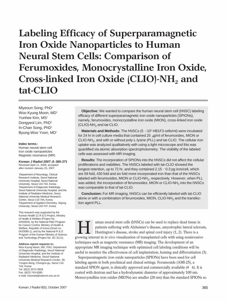
Labeling The HB1F3 cells exposed to the ferumoxides, MION-47,
CLIO-NH2 or tat-CLIO showed intracellular uptake of theiron oxide (Fig. 1). However, no intracellular uptake of theiron oxide was detected in cells incubated with MION-47.
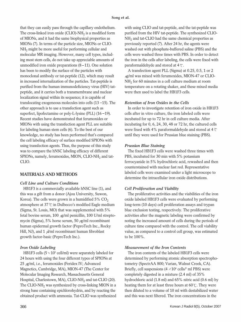
RetentionThe number of iron-containing cells decreased as the
incubation time increased for all the SPION-labeled cells(Fig. 2). The cells loaded with tat-CLIO showed a greaternumbers of blue stained cells at all time points comparedto the ferumoxides and the CLIO-NH2 labeled cells. Theferumoxide-exposed cells and the CLIO-NH2 exposed cellsshowed iron labeling until 24 48 hr, whereas iron-
Superparamagnetic Iron Oxide Nanoparticles to Human Neural Stem Cells
Korean J Radiol 8(5), October 2007 367
Fig.1. Photomicrographs of the hNSCstreated for 24 hr with ferumoxides (A),MION-47 (B), CLIO-NH2 (C) or tat-CLIO(D) at 25 g/ml. The intracellular uptakeof iron oxide nanoparticles (arrows) isseen in cells exposed to ferumoxides (A),CLIO-NH2 (C) or tat-CLIO (D). However,no intracellular uptake of iron oxide wasfound for the cells incubated with MION-47 (B). (Prussian blue stain, objectivemagnification: 40)
C D
A B
containing cells were visible after 72 hr for the tat-CLIOexposed cells. However, more than 90% of the HB1F3cells labeled with tat-CLIO were unlabeled after 48 hr ofincubation.
Viability and ProliferationThe average viability of the control cells, as determined
by trypan blue exclusion assay, was 100 4.5%. Theviable percentage of the ferumoxide-labeled, MION-47
Song et al.
368 Korean J Radiol 8(5), October 2007
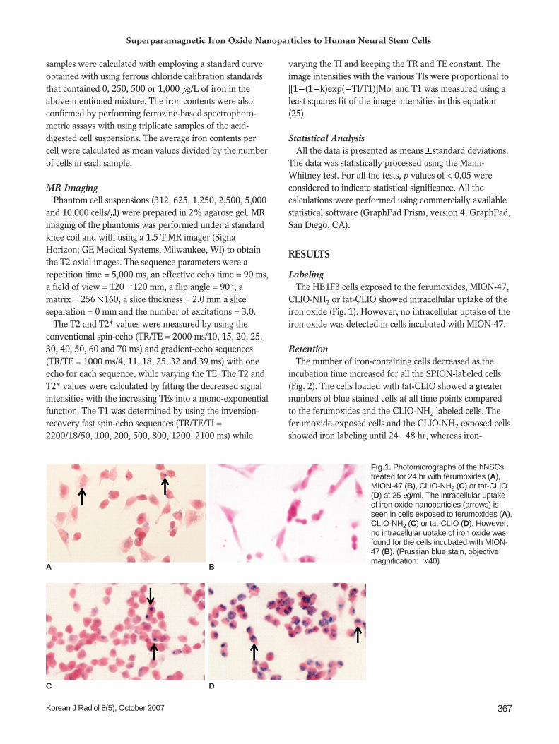
Fig. 3. Photomicrographs of hNSCs treated for 24 hr with three different SPIONs (ferumoxides, MION-47 or CLIO-NH2) at 25 g/ml in thepresence of different doses of PLL. As the PLL concentration increased in the media from 0.25 g/ml to 2 g/ml, more iron oxidenanoparticles are seen inside the labeled cells. (Prussian blue stain, objective magnification: 40)
Fig. 2. Retention of SPIONs in hNSCs. Iron oxide nanoparticles (arrows) are seen within the ferumoxides labeled cells and the CLIO-NH2 labeled cells for up to 24 and 48 hr, respectively and inside the tat-CLIO labeled cells for up to 72 hr. (Prussian blue stain, objectivemagnification: 40)
PLL
+
Ferumoxides
CLIO-NH2
Tat-CLIO
Ferumoxides
MION-47
CLIO-NH2
0.25 g/ml 0.5 g/ml 1 g/ml 2 g/ml
6h 24h 30h 48h 72h
labeled, CLIO-NH2 labeled or tat-CLIO-labeled HB1F3cells versus the unlabeled control cells was 105 3.9, 101
5.1, 101 2.5 and 95 6.5, respectively. The cellsexposed to these SPIONs showed no differences of viabil-ity versus the unlabeled cells, and there was no effect onthe proliferative capability of the cells labeled with all fourSPIONs during 10 days of culture.
Increased Intracellular Iron Uptake Induced by Poly-L-Lysine
Prussian blue staining of the SPION-exposed cells withPLL revealed a dose-dependent increase of the iron oxideuptake into the cells (Fig. 3). Almost 100% of the cellswere labeled with iron oxide when the HB1F3 cells wereincubated in SPION media that contained 2 g/ml PLL. So,the concentration 2 g/ml of PLL was selected to transfectiron oxide into the cells. The viabilities and proliferationsof the SPION exposed cells with using PLL were similar tothose of the unlabeled control cells.
Iron Contents with and without Poly-L-LysineAtomic absorption spectrophotometry revealed the
highest iron incorporation in the tat-CLIO-exposed cells (20.3 pg/cell) and the lowest in the MION-47-exposed cells
Superparamagnetic Iron Oxide Nanoparticles to Human Neural Stem Cells
Korean J Radiol 8(5), October 2007 369
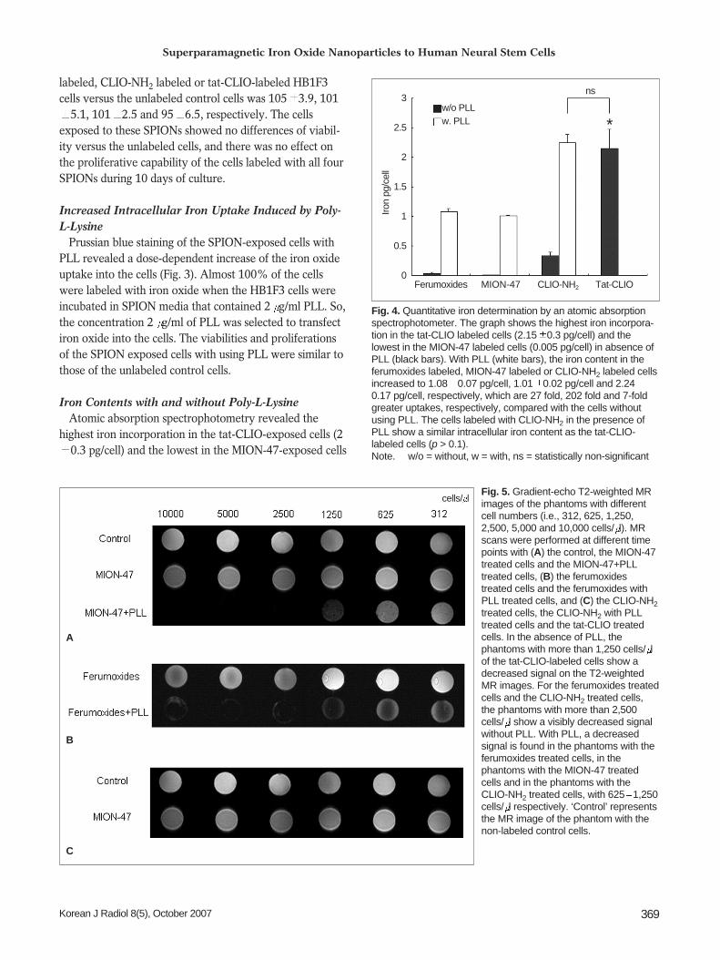
Fig. 4. Quantitative iron determination by an atomic absorptionspectrophotometer. The graph shows the highest iron incorpora-tion in the tat-CLIO labeled cells (2.15 0.3 pg/cell) and thelowest in the MION-47 labeled cells (0.005 pg/cell) in absence ofPLL (black bars). With PLL (white bars), the iron content in theferumoxides labeled, MION-47 labeled or CLIO-NH2 labeled cellsincreased to 1.08 0.07 pg/cell, 1.01 0.02 pg/cell and 2.240.17 pg/cell, respectively, which are 27 fold, 202 fold and 7-foldgreater uptakes, respectively, compared with the cells withoutusing PLL. The cells labeled with CLIO-NH2 in the presence ofPLL show a similar intracellular iron content as the tat-CLIO-labeled cells (p > 0.1). Note. w/o = without, w = with, ns = statistically non-significant
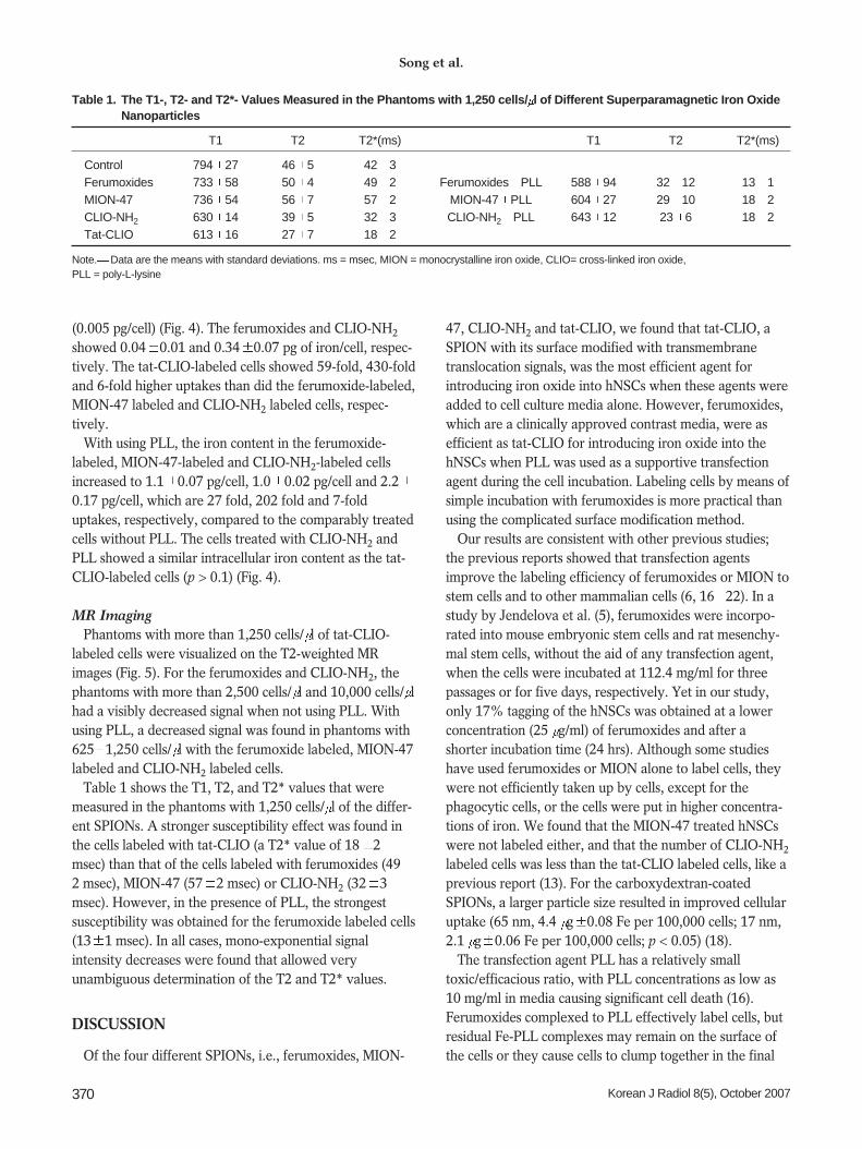
Fig. 5. Gradient-echo T2-weighted MRimages of the phantoms with differentcell numbers (i.e., 312, 625, 1,250,2,500, 5,000 and 10,000 cells/ l). MRscans were performed at different timepoints with (A) the control, the MION-47treated cells and the MION-47+PLLtreated cells, (B) the ferumoxidestreated cells and the ferumoxides withPLL treated cells, and (C) the CLIO-NH2treated cells, the CLIO-NH2 with PLLtreated cells and the tat-CLIO treatedcells. In the absence of PLL, thephantoms with more than 1,250 cells/ lof the tat-CLIO-labeled cells show adecreased signal on the T2-weightedMR images. For the ferumoxides treatedcells and the CLIO-NH2 treated cells,the phantoms with more than 2,500cells/ l show a visibly decreased signalwithout PLL. With PLL, a decreasedsignal is found in the phantoms with theferumoxides treated cells, in thephantoms with the MION-47 treatedcells and in the phantoms with theCLIO-NH2 treated cells, with 625 1,250cells/ l respectively. ‘Control’ representsthe MR image of the phantom with thenon-labeled control cells.
B
C
A
Ferumoxides
cells/ l
MION-47 CLIO-NH2 Tat-CLIO
3
2.5
2
1.5
1
0.5
0
ns
*
Iron
pg/c
ell
w/o PLLw. PLL
(0.005 pg/cell) (Fig. 4). The ferumoxides and CLIO-NH2
showed 0.04 0.01 and 0.34 0.07 pg of iron/cell, respec-tively. The tat-CLIO-labeled cells showed 59-fold, 430-foldand 6-fold higher uptakes than did the ferumoxide-labeled,MION-47 labeled and CLIO-NH2 labeled cells, respec-tively.
With using PLL, the iron content in the ferumoxide-labeled, MION-47-labeled and CLIO-NH2-labeled cellsincreased to 1.1 0.07 pg/cell, 1.0 0.02 pg/cell and 2.20.17 pg/cell, which are 27 fold, 202 fold and 7-folduptakes, respectively, compared to the comparably treatedcells without PLL. The cells treated with CLIO-NH2 andPLL showed a similar intracellular iron content as the tat-CLIO-labeled cells (p > 0.1) (Fig. 4).
MR ImagingPhantoms with more than 1,250 cells/ l of tat-CLIO-
labeled cells were visualized on the T2-weighted MRimages (Fig. 5). For the ferumoxides and CLIO-NH2, thephantoms with more than 2,500 cells/ l and 10,000 cells/ lhad a visibly decreased signal when not using PLL. Withusing PLL, a decreased signal was found in phantoms with625 1,250 cells/ l with the ferumoxide labeled, MION-47labeled and CLIO-NH2 labeled cells.
Table 1 shows the T1, T2, and T2* values that weremeasured in the phantoms with 1,250 cells/ l of the differ-ent SPIONs. A stronger susceptibility effect was found inthe cells labeled with tat-CLIO (a T2* value of 18 2msec) than that of the cells labeled with ferumoxides (492 msec), MION-47 (57 2 msec) or CLIO-NH2 (32 3msec). However, in the presence of PLL, the strongestsusceptibility was obtained for the ferumoxide labeled cells(13 1 msec). In all cases, mono-exponential signalintensity decreases were found that allowed veryunambiguous determination of the T2 and T2* values.
DISCUSSION
Of the four different SPIONs, i.e., ferumoxides, MION-
47, CLIO-NH2 and tat-CLIO, we found that tat-CLIO, aSPION with its surface modified with transmembranetranslocation signals, was the most efficient agent forintroducing iron oxide into hNSCs when these agents wereadded to cell culture media alone. However, ferumoxides,which are a clinically approved contrast media, were asefficient as tat-CLIO for introducing iron oxide into thehNSCs when PLL was used as a supportive transfectionagent during the cell incubation. Labeling cells by means ofsimple incubation with ferumoxides is more practical thanusing the complicated surface modification method.
Our results are consistent with other previous studies;the previous reports showed that transfection agentsimprove the labeling efficiency of ferumoxides or MION tostem cells and to other mammalian cells (6, 16 22). In astudy by Jendelova et al. (5), ferumoxides were incorpo-rated into mouse embryonic stem cells and rat mesenchy-mal stem cells, without the aid of any transfection agent,when the cells were incubated at 112.4 mg/ml for threepassages or for five days, respectively. Yet in our study,only 17% tagging of the hNSCs was obtained at a lowerconcentration (25 g/ml) of ferumoxides and after ashorter incubation time (24 hrs). Although some studieshave used ferumoxides or MION alone to label cells, theywere not efficiently taken up by cells, except for thephagocytic cells, or the cells were put in higher concentra-tions of iron. We found that the MION-47 treated hNSCswere not labeled either, and that the number of CLIO-NH2
labeled cells was less than the tat-CLIO labeled cells, like aprevious report (13). For the carboxydextran-coatedSPIONs, a larger particle size resulted in improved cellularuptake (65 nm, 4.4 g 0.08 Fe per 100,000 cells; 17 nm,2.1 g 0.06 Fe per 100,000 cells; p < 0.05) (18).
The transfection agent PLL has a relatively smalltoxic/efficacious ratio, with PLL concentrations as low as10 mg/ml in media causing significant cell death (16).Ferumoxides complexed to PLL effectively label cells, butresidual Fe-PLL complexes may remain on the surface ofthe cells or they cause cells to clump together in the final
Song et al.
370 Korean J Radiol 8(5), October 2007
Table 1. The T1-, T2- and T2*- Values Measured in the Phantoms with 1,250 cells/ l of Different Superparamagnetic Iron OxideNanoparticles
T1 T2 T2*(ms) T1 T2 T2*(ms)
Control 794 27 46 5 42 3Ferumoxides 733 58 50 4 49 2 Ferumoxides PLL 588 94 32 12 13 1MION-47 736 54 56 7 57 2 MION-47 PLL 604 27 29 10 18 2CLIO-NH2 630 14 39 5 32 3 CLIO-NH2 PLL 643 12 23 6 18 2Tat-CLIO 613 16 27 7 18 2
Note. Data are the means with standard deviations. ms = msec, MION = monocrystalline iron oxide, CLIO= cross-linked iron oxide, PLL = poly-L-lysine
cell preparation prior to infusion. A recent report indicatedthat labeling mesenchymal stem cells with ferumoxides-PLL complexes inhibited the chondrogenic differentiationcapacity of mesenchymal stem cells (23). In contrast,labeling cells with ferumoxides-protamine sulfatecomplexes did not alter the viability and functionalcapacity of a variety of cell types (24). In our study,SPIONS, including tat-CLIO, showed no adverse effect onthe cell proliferations and viabilities. However, we did notcheck the effect on cell differentiation and so furtherstudies on these issues are necessary.
In conclusion, our study indicates that hNSCs can besafely and efficiently labeled for MR imaging with usingeither tat-CLIO alone or a combination of ferumoxides,MION-47 and CLIO-NH2 and the transfection agent PLL.These labeling methods could be used to noninvasivelytrack hNSCs.
References1. Kim SU. Human neural stem cells genetically modified for brain
repair in neurological disorders. Neuropathology 2004;24:159-171
2. Lindvall O, Kokaia Z, Martinez-Serrano A. Stem cell therapyfor human neurodegenerative disorders-how to make it work.Nat Med 2004;10:S42-50
3. Bulte JW, Duncan ID, Frank JA. In vivo magnetic resonancetracking of magnetically labeled cells after transplantation. JCereb Blood Flow Metab 2002;22:899-907
4. Wang YX, Hussain SM, Krestin GP. Superparamagnetic ironoxide contrast agents: physicochemical characteristics andapplications in MR imaging. Eur Radiol 2001;11:2319-2331
5. Jendelova P, Herynek V, Urdzikova L, Glogarova K,Kroupova J, Andersson B, et al. Magnetic resonance tracking oftransplanted bone marrow and embryonic stem cells labeled byiron oxide nanoparticles in rat brain and spinal cord. J NeurosciRes 2004;76:232-243
6. Daldrup-Link HE, Rudelius MR, Piontek G, Metz S, Brauer R,Debus G, et al. Migration of iron oxide-labeled humanhematopoietic progenitor cells in a mouse model: in vivomonitoring with 1.5-T MR imaging equipment. Radiology2005;234:197-205
7. Wunderbaldinger P, Josephson L, Weissleder R. CrosslinkedIron Oxides (CLIO): a new platform for the development oftargeted MR contrast agents. Acad Radiol 2002;9:S304-S306
8. Kukowska-Latallo JF, Bielinska AU, Johnson J, Spindler R,Tomalia DA, Baker JR Jr. Efficient transfer of genetic materialinto mammalian cells using starburst polyamidoaminedendrimers. Proc Natl Acad Sci USA 1996;93:4897-4902
9. Tang MX, Redemann CT, Szoka FC Jr. In vitro gene delivery bydegraded polyamidoamine dendrimers. Bioconjug Chem1996;7:703-714
10. Plank C, Mechtler K, Szoka FC Jr, Wagner E. Activation of thecomplement system by synthetic DNA complexes: a potentialbarrier for intravenous gene delivery. Human Gene Ther
1996;7:1437-144611. DeLong R, Stephenson K, Loftus T, Fisher M, Alahari S, Nolting
A, et al. Characterization of complexes of oligonucleotides withpolyamidoamine starburst dendrimers and effects on intracellu-lar delivery. J Pharm Sci 1997;86:762-764
12. Josephson L, Tung CH, Moore A, Weissleder R. High-efficiencyintracellular magnetic labeling with novel superparamagnetic-tatpeptide conjugate. Bioconjug Chem 1999;10:186-191
13. Lewin M, Carlesso N, Tung CH, Tang XW, Cory D, ScaddenDT, et al. Tat peptide-derivatized magnetic nanoparticles allowin vivo tracking and recovery of progenitor cells. NatureBiotechnol 2000;18:410-414
14. Zhao M, Kircher MF, Josephson L, Weissleder R. Differentialconjugation of tat peptide to superparamagnetic nanoparticlesand its effect on cellular uptake. Bioconjug Chem 2002;13:840-844
15. Frankel AD, Pabo CO. Cellular uptake of the tat protein fromhuman immunodeficiency virus. Cell 1988;55:1189-1193
16. Arbab AS, Yocum GT, Wilson LB, Parwana A, Jordan EK,Kalish H, et al. Comparison of transfection agents in formingcomplexes with ferumoxides, cell labeling efficiency, andcellular viability. Mol Imaging 2004;3:24-32
17. Arbab AS, Bashaw LA, Miller BR, Jordan EK, Lewis BK, KalishH, et al. Characterization of biophysical and metabolic proper-ties of cells labeled with superparamagnetic iron oxide nanopar-ticles and transfection agent for cellular MR imaging. Radiology2003;229:838-846
18. Matuszewski L, Persigehl T, Wall A, Schwindt W, Tombach B,Fobker M, et al. Cell tagging with clinically approved ironoxides: feasibility and effect of lipofection, particle size, andsurface coating on labeling efficiency. Radiology 2005;235:155-161
19. Montet-Abou K, Montet X, Weissleder R, Josephson L.Transfection agent induced nanoparticle cell loading. MolImaging 2005;4:165-171
20. Wunderbaldinger P, Josephson L, Weissleder R. Tat peptidedirects enhanced clearance and hepatic permeability of magneticnanoparticles. Bioconjug Chem 2002;13:264-268
21. Weissleder R, Cheng HC, Bogdanova A, Bogdanova A Jr.Magnetically labeled cells can be detected by MR imaging. JMagn Reson Imaging 1997;7:258-263
22. Frank JA, Miller BR, Arbab AS, Zywicke HA, Jordan EK, LewisBK, et al. Clinically applicable labeling of mammalian and stemcells by combining superparamagnetic iron oxides and transfec-tion agents. Radiology 2003;228:480-487
23. Bulte JW, Kraitchman DL, Mackay AM, Pittenger MF.Chondrogenic differentiation of mesenchymal stem cells isinhibited after magnetic labeling of with ferumoxides. Blood2004;104:3410-3412
24. Arbab AS, Yocum GT, Rad AM, Khakoo AY, Fellowes V, ReadEJ, et al. Labeling of cells with ferumoxides-protamine sulfatecomplexes does not inhibit function or differentiation capacityof hematopoietic or mesenchymal stem cells. NMR Biomed2005;18:553-559
25. Zhu DC, Penn RD. Full-brain T1 mapping through inversionrecovery fast spin echo imaging with time-efficient sliceordering. Magn Reson Med 2005;54:725-731
Superparamagnetic Iron Oxide Nanoparticles to Human Neural Stem Cells
Korean J Radiol 8(5), October 2007 371


![· Web viewSynthesis of iron oxide nanoparticles The superparamagnetic Fe3O4 MNPs were prepared by recrystallization of hematite, as described by Cheng et al [38]. A mixture of bulk](https://img.pdfslide.us/doc/110x75/5e5a6458836da45bd4631290/web-view-synthesis-of-iron-oxide-nanoparticles-the-superparamagnetic-fe3o4-mnps.jpg)


![The Uses of Iron (II) Oxide By: Isabel Rimando. Iron (II) oxide [FeO] Not to be confused with iron (III) oxide (rust)](https://img.pdfslide.us/doc/110x75/5a4d1bbd7f8b9ab0599d1c20/the-uses-of-iron-ii-oxide-by-isabel-rimando-iron-ii-oxide-feo-not-to.jpg)