Embed Size (px)
Citation preview
© 2 0 0 7 A M E R I C A N C H E M I C A L S O C I E T Y
l a b fa b
First DIBs on protocell networksResearchers assemble networks of primitive artifi cial cells that communicate through shared lipid bilayers.
Like nosy neighbors passing along the latest bits of juicy gossip, cells
constantly receive and transmit signals from each other and from the extracel-lular environment. Cellular communica-tion in the form of ion flow through gap junctions or membrane pores underlies physiological processes such as the fir-ing of neurons and the coordination of contractions in the heart. However, the complex milieu of ions, signaling molecules, and membrane proteins in living cells complicates the study of cellular communication. To sim-plify these studies, Matthew Holden, Hagan Bayley, and David Needham at the University of Oxford (U.K.) and Duke University fabricated net-works of tiny, lipid-encased water droplets that mimic rudimentary cells (J. Am. Chem. Soc. 2007, 129, 8650–8655).
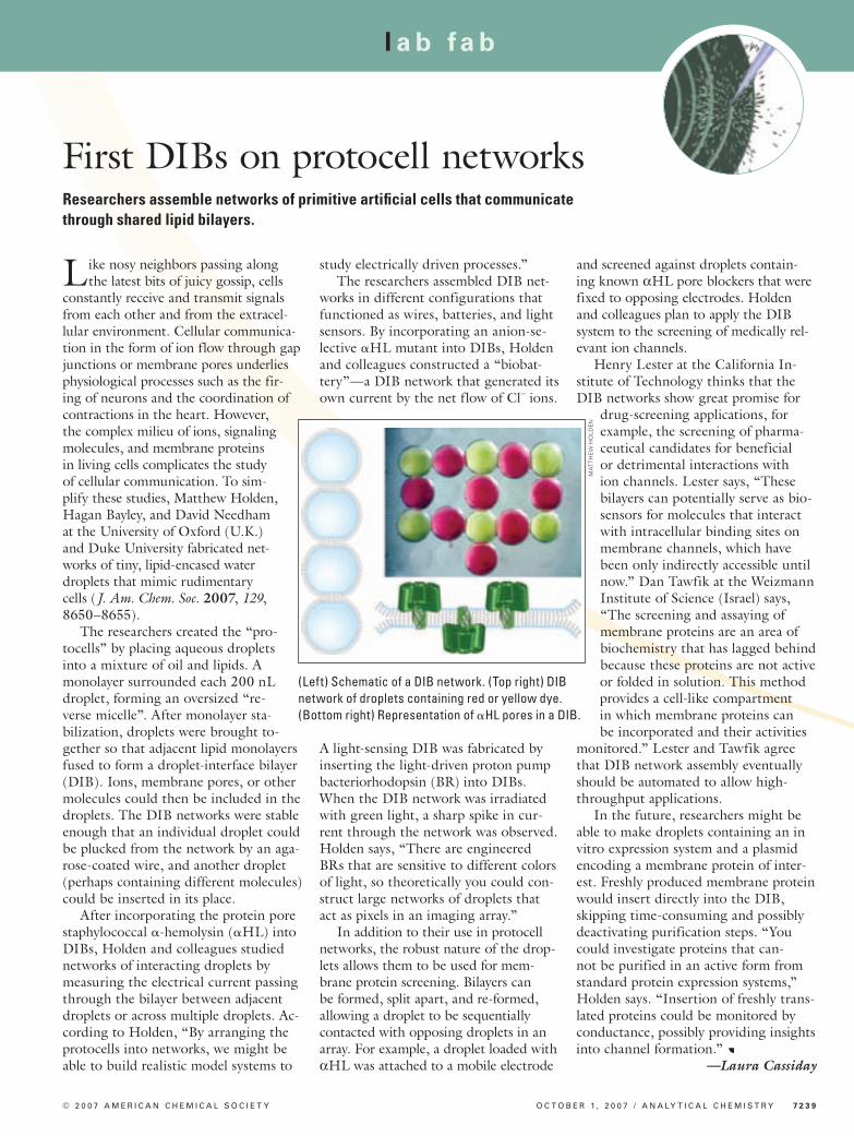
The researchers created the “pro-tocells” by placing aqueous droplets into a mixture of oil and lipids. A monolayer surrounded each 200 nL droplet, forming an oversized “re-verse micelle”. After monolayer sta-bilization, droplets were brought to-gether so that adjacent lipid monolayers fused to form a droplet-interface bilayer (DIB). Ions, membrane pores, or other molecules could then be included in the droplets. The DIB networks were stable enough that an individual droplet could be plucked from the network by an aga-rose-coated wire, and another droplet (perhaps containing different molecules) could be inserted in its place.
After incorporating the protein pore staphylococcal -hemolysin (HL) into DIBs, Holden and colleagues studied networks of interacting droplets by measuring the electrical current passing through the bilayer between adjacent droplets or across multiple droplets. Ac-cording to Holden, “By arranging the protocells into networks, we might be able to build realistic model systems to
study electrically driven processes.”The researchers assembled DIB net-
works in different configurations that functioned as wires, batteries, and light sensors. By incorporating an anion-se-lective HL mutant into DIBs, Holden and colleagues constructed a “biobat-tery”—a DIB network that generated its own current by the net flow of Cl– ions.
A light-sensing DIB was fabricated by inserting the light-driven proton pump bacteriorhodopsin (BR) into DIBs. When the DIB network was irradiated with green light, a sharp spike in cur-rent through the network was observed. Holden says, “There are engineered BRs that are sensitive to different colors of light, so theoretically you could con-struct large networks of droplets that act as pixels in an imaging array.”
In addition to their use in protocell networks, the robust nature of the drop-lets allows them to be used for mem-brane protein screening. Bilayers can be formed, split apart, and re-formed, allowing a droplet to be sequentially contacted with opposing droplets in an array. For example, a droplet loaded with αHL was attached to a mobile electrode
and screened against droplets contain-ing known αHL pore blockers that were fixed to opposing electrodes. Holden and colleagues plan to apply the DIB system to the screening of medically rel-evant ion channels.
Henry Lester at the California In-stitute of Technology thinks that the DIB networks show great promise for
drug-screening applications, for example, the screening of pharma-ceutical candidates for beneficial or detrimental interactions with ion channels. Lester says, “These bilayers can potentially serve as bio-sensors for molecules that interact with intracellular binding sites on membrane channels, which have been only indirectly accessible until now.” Dan Tawfik at the Weizmann Institute of Science (Israel) says, “The screening and assaying of membrane proteins are an area of biochemistry that has lagged behind because these proteins are not active or folded in solution. This method provides a cell-like compartment in which membrane proteins can be incorporated and their activities
monitored.” Lester and Tawfik agree that DIB network assembly eventually should be automated to allow high-throughput applications.
In the future, researchers might be able to make droplets containing an in vitro expression system and a plasmid encoding a membrane protein of inter-est. Freshly produced membrane protein would insert directly into the DIB, skipping time-consuming and possibly deactivating purification steps. “You could investigate proteins that can-not be purified in an active form from standard protein expression systems,” Holden says. “Insertion of freshly trans-lated proteins could be monitored by conductance, possibly providing insights into channel formation.” a
—Laura Cassiday
O C T O B E R 1 , 2 0 0 7 / A N A LY T I C A L C H E M I S T R Y 7 2 3 9
(Left) Schematic of a DIB network. (Top right) DIB network of droplets containing red or yellow dye. (Bottom right) Representation of HL pores in a DIB.
MA
TTH
EW
HO
LDE
N


![DIBS: Just-in-time Congestion Mitigation for Data Centers · In the rest of the paper, we will describe the DIBS idea in detail, and evaluate DIBS extensively using a NetFPGA [8]](https://img.pdfslide.us/doc/110x75/602679ab4a1cc911d92a1281/dibs-just-in-time-congestion-mitigation-for-data-centers-in-the-rest-of-the-paper.jpg)