Embed Size (px)
Citation preview

Lab Exercise: Diversity of Eukaryotic Microbes
OBJECTIVES
1. To observe representatives of major types of microbes. 2. To cultivate select representatives of major types of microbes. 3. Understand key characteristics of the different eukaryotic microbes and groups found
within these Kingdoms. INTRODUCTION Eukaryotic organisms have a nucleus in a membrane. They are typically more complex than prokaryotic organisms. They make up the Domain Eukarya and include the major kingdoms of Protista, Fungi, Plantae and Animalia. Protista is a diverse group that includes many different types of organisms, divided into the animal-like protists, or protozoa, and the plant-like protists, or algae. Protozoa are examples of Protistans that we will survey in this lab. Fungi are a kingdom of organisms that may be unicellular or multicellar, yeasts or molds. All fungi absorb their nutrients from their environment. Animals are multicelluar organisms that ingest their nutrients. This lab will be presented in three parts, one focusing on Protozoa, one on Fungi and one on Helminths (parasitic worms of the animal kingdom). Protozoa These are unicelluar eukaryotes. They do not have cell walls, but do have a membrane called a pellicle surrounding the cell. They have a nucleus and membrane bound organelles. They typically form cysts, a hardy dormant life-form that allows survival of harsh environments. Protozoa are classified into four phyla or groups based on their means of locomotion:
• Flagellates (or phylum Mastigophora) • Amoebae (or phylum Sarcodina) • Sporozoans (or phylum Apicomplexa) and • Ciliates (or phylum Ciliophora).
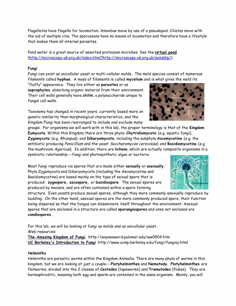
Flagellates have flagella for locomotion. Amoebae move by use of a pseudopod. Ciliates move with the aid of multiple cilia. The sporozoans have no means of locomotion and therefore have a lifestyle that makes them all internal parasites. Pond water is a great source of assorted protozoan microbes. See the virtual pond (http://microscopy-uk.org.uk/index.html?http://microscopy-uk.org.uk/ponddip/). Fungi Fungi can exist as unicellular yeast or multi-cellular molds. The mold species consist of numerous filaments called hyphae. A mass of filaments is called mycelium and is what gives the mold its “fluffy” appearance. They live either as parasites or as saprophytes, absorbing organic material from their environment. Their cell walls generally have chitin, a polysaccharide unique to fungal cell walls. Taxonomy has changed in recent years, currently based more on genetic similarity than morphological characteristics, and the Kingdom Fungi has been rearranged to include and exclude many groups. For organisms we will work with in this lab, the proper terminology is that of the Kingdom Eumycota. Within this Kingdom there are three phyla: Chytridiomycota (e.g. aquatic fungi), Zygomycota (e.g. Rhizopus), and Dikaryomycota, including the subphyla Ascomycotina (e.g. the antibiotic producing Penicillium and the yeast Saccharomyces cerevisiae) and Basidiomycotina (e.g. the mushroom Agaricus). In addition, there are lichens, which are actually composite organisms in a symbiotic relationship---fungi and photosynthetic algae or bacteria.
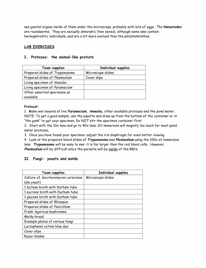
Most fungi reproduce via spores that are made either sexually or asexually. Phyla Zygomycota and Dikaryomycota (including the Ascomycotina and Basidiomycotina) are based mainly on the type of sexual spore that is produced: zygospore, ascospore, or basidiospore. The sexual spores are produced by meiosis, and are often contained within a spore forming structure. Even yeasts produce sexual spores, although they more commonly asexually reproduce by budding. On the other hand, asexual spores are the more commonly produced spore, their function being dispersal so that the fungus can disseminate itself throughout the environment. Asexual spores that are enclosed in a structure are called sporangiospores and ones not enclosed are condiospores.
For this lab, we will be looking at fungi as molds and as unicellular yeast. Web resources: The Amazing Kingdom of Fungi, http://waynesword.palomar.edu/ww0504.htm UC Berkeley's Introduction to Fungi http://www.ucmp.berkeley.edu/fungi/fungisy.html Helminths Helminths are parasitic worms within the Kingdom Animalia. There are many phyla of worms in this kingdom, but we are looking at just a couple---Platyhelminthes and Nematoda. Platyhelminthes are flatworms, divided into the 2 classes of Cestodes (tapeworms) and Trematodes (flukes). They are hermaphroditic, meaning both egg and sperm are contained in the same organism. Mainly, you will
see genital organs inside of them under the microscope, probably with lots of eggs. The Nematodes are roundworms. They are sexually dimorphic (two sexes), although some also contain hermaphroditic individuals, and are a bit more evolved than the platyhelminthes.
LAB EXERCISES I. Protozoa: the animal-like protists
Team supplies Individual supplies Prepared slides of Trypanosoma Microscope slides Prepared slides of Plasmodium Cover slips Living specimen of Amoeba Living specimen of Paramecium Other assorted specimens as available
Protocol: 1. Make wet mounts of live Paramecium, Ameoba, other available protozoa and the pond water. NOTE: To get a good sample, use the pipette and draw up from the bottom of the container or in “the gunk” to get your specimen. Do NOT stir the specimen container first. 2. Start with the 10x lens and go to 40x lens. Oil-immersion will magnify too much for most pond water protozoa. 3. Once you have found your specimen, adjust the iris diaphragm for even better viewing. 4. Look at the prepared blood slides of Trypanosoma and Plasmodium using the 100x oil immersion lens. Trypanosoma will be easy to see: it is far larger than the red blood cells. However, Plasmodium will be difficult since the parasite will be inside of the RBCs. II. Fungi: yeasts and molds
Team supplies Individual supplies Culture of Saccharomyces cerevisiae (ale yeast)
Microscope slides
1 lactose broth with Durham tube 1 sucrose broth with Durham tube 1 glucose broth with Durham tube Prepared slides of Rhizopus Prepared slides of Penicillium Fresh Agaricus mushrooms Moldy bread Example plates of various fungi Lactophenol cotton blue dye Cover slips Razor blades
Protocols: Saccharomyces cerevesiae 1. Make a wet mount of the culture by mixing a small drop of S. cerevisiae into a drop of lactophenol cotton blue. 2. Observe under the microscope. 3. As a team, inoculate S. cerevisiae into 3 sugar broth tubes: lactose, sucrose, and glucose. Next class, you will be observing these broths for evidence of fermentation with production of CO2 Prepared slides of Rhizopus and Penicillium 1. For Rhizopus, identify hyphae, sporangia, and sporangiospores. Also differentiate between the
sexual zygospores and the asexual sporangiospores on the slides. 2. For Penicillium, identify hyphae, conidia and conidiospores. Agaricus mushrooms 1. Using your razor blade, cut a thin gill section from the mushroom cap. Make a wet mount of 1 thin gill section in a drop of lactophenol cotton blue. 2. Observe under the microscope, 10X and 40X, looking for basiospores and basidia. Plates of various fungi 1. Make macroscopic observations of any molds growing on plates, noting color and reproductive
hyphae. Bread mold 1. Make a wet mount of your mold mold by mixing a small sample with a drop of lactophenol cotton
blue. 2. Observe under the microscope. 3. Try to identify fungal class (and perhaps even the genus) based on spore characteristics.
Compare to previous examples of molds and see Alexander Atlas pages 19-27. III. Helminths
Team supplies
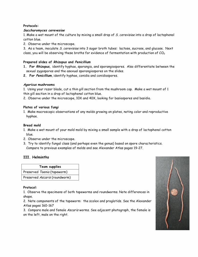
Preserved Taenia (tapeworm) Preserved Ascaris (roundworm) Protocol: 1. Observe the specimens of both tapeworms and roundworms. Note differences in shape. 2. Note components of the tapeworm: the scolex and proglotids. See the Alexander Atlas pages 160-167 3. Compare male and female Ascaris worms. See adjacent photograph, the female is on the left, male on the right.
DATA AND OBSERVATIONS I. Protozoa 1. Draw representatives of the following protozoa: Amoeba Paramecium Total magnification _______ Total magnification _______ Trypanosoma Plasmodium Total magnification _______ Total magnification _______
II. Fungi 1. Draw S. cerevisiae: S. cerevisiae
Total magnification _______
2. Observe S. cerevisiae growth in sugar tubes. Note presence of growth, color of medium, and presence/absence of a gas bubble in the Durham tube: Glucose broth: Lactose broth: Sucrose broth: 3. Draw and label representatives of spores/ spore structures in Rhizopus, Penicillium, and Agaricus: Rhizopus Penicillium Total magnification _______ Total magnification _______
Agaricus Total magnification _______ 4. Make macroscopic observations of molds provided: Name _________________ Name _________________ Name _________________
5. Draw observations of the wet mount of bread mold: Total magnification _______ DISCUSSION I. Protozoa 1. Compare and contrast the three mechanisms of motility displayed by protozoa. 2. Trypanosoma and Plasmodium are both found in blood. What diseases do these microbes cause? II. Fungi 1. Define the term yeast. Why are yeast cells larger than bacterial cells?
2. Was S. cerevisiae capable of fermenting all sugars? What were the indications of fermentation, and which tubes showed positives? Relate your results to what you know about the normal biotechnological use of this yeast. 3. Complete the following table
Organism Sexual spores Phylum Rhizopus
Aspergillus
Penicillium
Agaricus
4. Which phylum and genus did your bread mold belong to? 5. How do fungal spores differ from bacterial endospores? III. Helminths 1. How does a male roundworm differ from a female roundworm? 2. Name a disease caused by a roundworm.


![[TITLE WITH CAPITAL LETTERS]pure.au.dk/portal/files/79630819/AW_Bacteria_and_protozoa_in_soil... · Protozoa ›Protista: unicellular eukaryotic organisms: protozoa, unicellular algae,](https://img.pdfslide.us/doc/110x75/606ca0b8d91e76743244800e/title-with-capital-letterspureaudkportalfiles79630819awbacteriaandprotozoainsoil.jpg)