Embed Size (px)
Citation preview
Lab Activity 13
Spinal Cord
Portland Community CollegeBI 232
2
Definitions
• Tracts: collections of axons in CNS
• Nerves:collections of axons in PNS
• Ganglia: collections of neuron cell bodies in
PNS
• Nucleus (nuclei) : collections of neuron cell
bodies in CNS
3
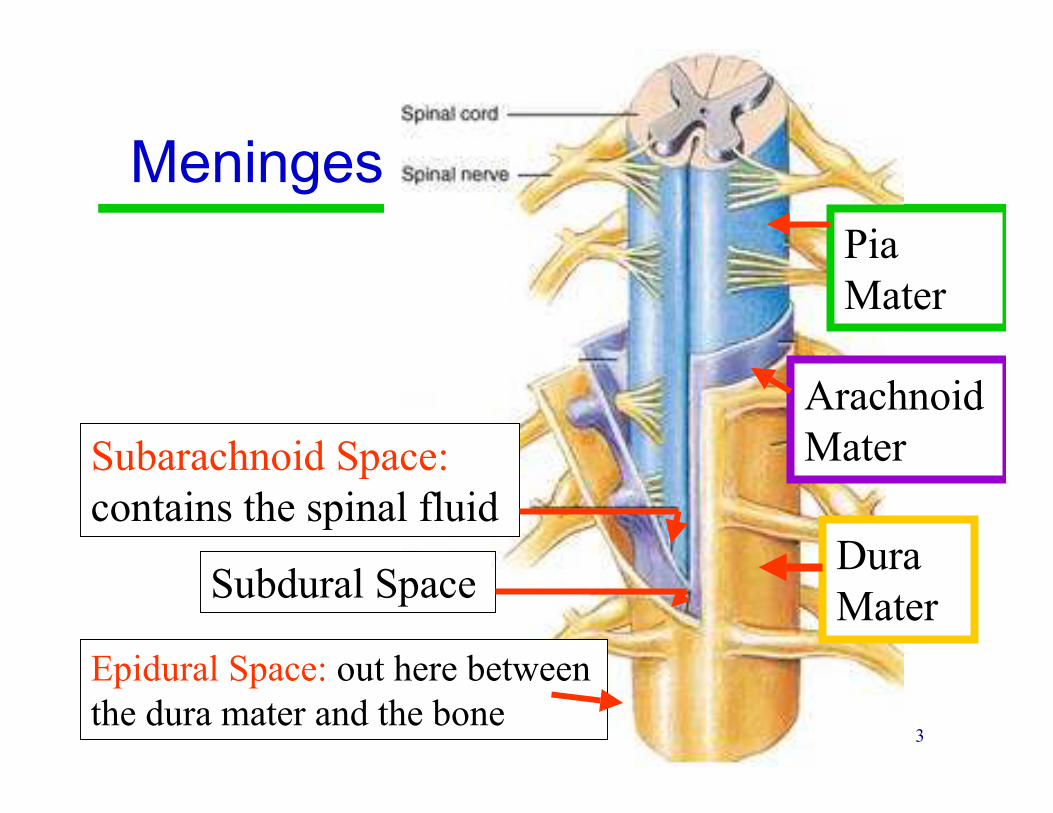
Meninges
Pia
Mater
Arachnoid
Mater
Dura
Mater
Subarachnoid Space:
contains the spinal fluid
Subdural Space
Epidural Space: out here between
the dura mater and the bone
4
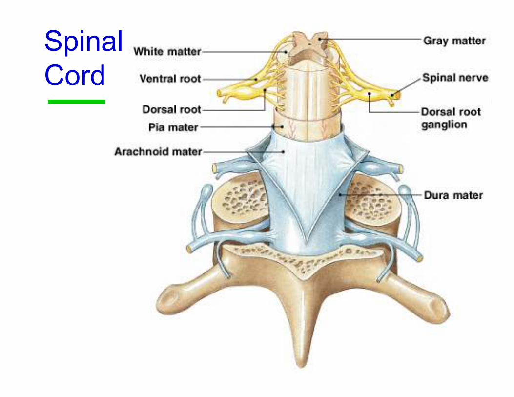
Spinal
Cord

5
Spinal Cord: Conus MedullarisConus Medullaris
Ends at the level of L1 or L2
6
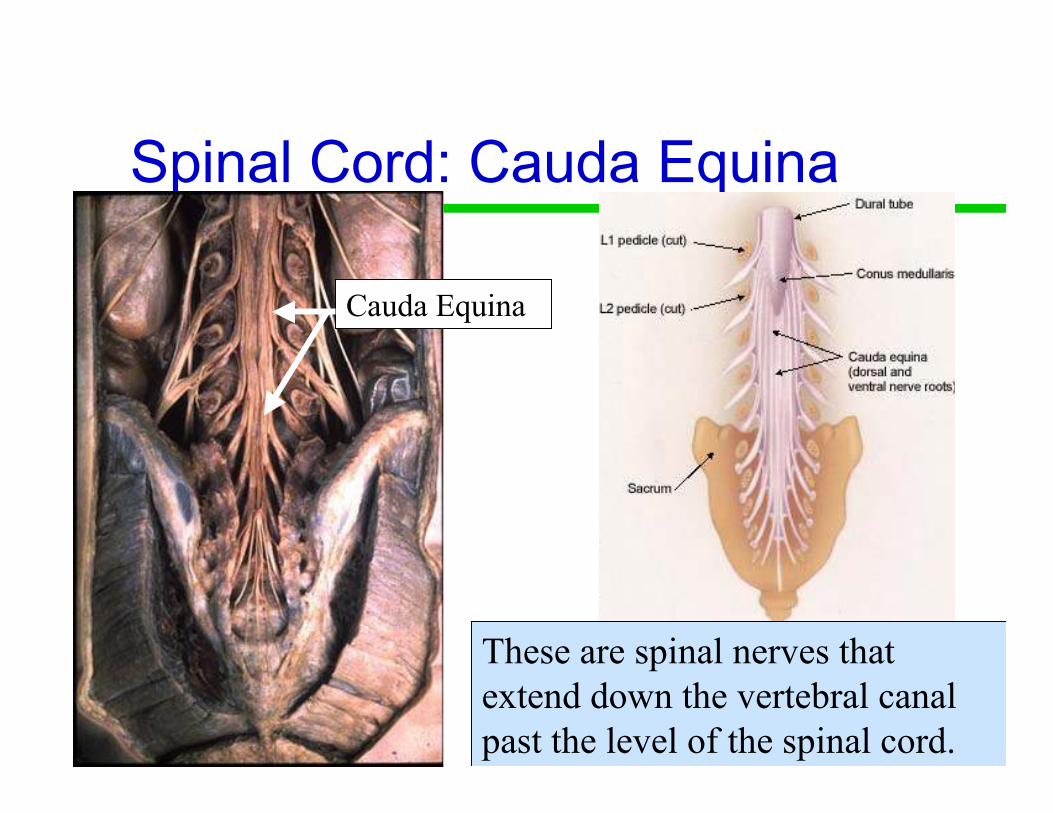
Spinal Cord: Cauda Equina
Cauda Equina
These are spinal nerves that
extend down the vertebral canal
past the level of the spinal cord.
7
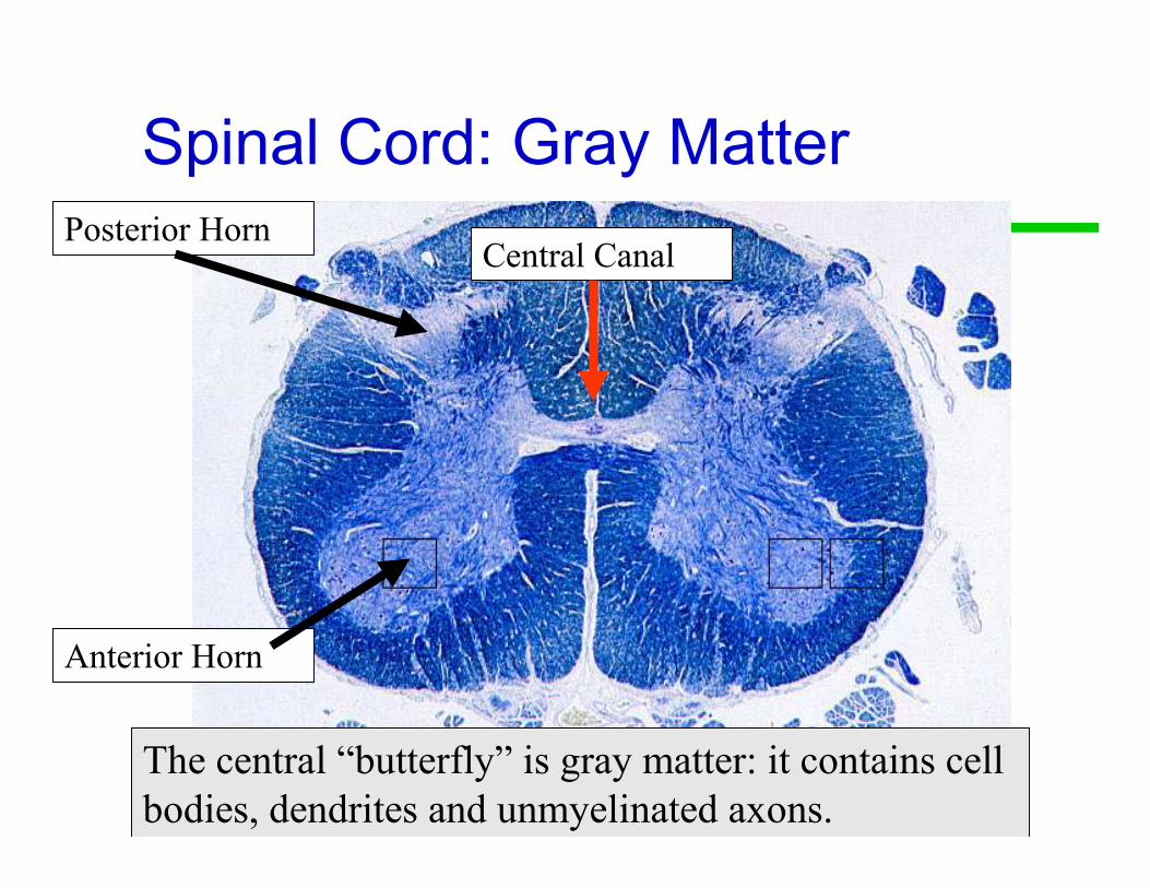
Spinal Cord: Gray Matter
Anterior Horn
Posterior HornCentral Canal
The central “butterfly” is gray matter: it contains cell
bodies, dendrites and unmyelinated axons.

8
Gray Matter Horns
• Posterior gray horns contain somatic and
visceral sensory nuclei
• Anterior gray horns contain somatic motor
nuclei
• Lateral gray horns (only located in the
thoracic and lumbar segments) contain
visceral motor nuclei
9
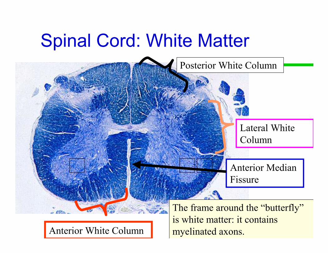
Spinal Cord: White Matter
The frame around the “butterfly”
is white matter: it contains
myelinated axons.
Posterior White Column
Lateral White
Column
Anterior White Column
Anterior Median
Fissure
10
White Matter Columns
• Each column contains tracts (axons)
• Ascending tracts carry sensory information
from the body toward the brain
• Descending tracts carry motor commands to
the spinal cord

11
Spinal Cord Structures

12
Dorsal Root
(Afferent=Sensory)
Dorsal Root
Ganglion
(Cell bodies
of sensory
neurons)
Posterior
(Dorsal)
Anterior
(Ventral)
Ventral Root
(Efferent=Motor)
Spinal Nerve Mixed
motor and sensory.

13
Poliomyelitis
• Polio means gray matter
• The polio virus causes inflammation of the
gray matter in the anterior horn motor
neurons.
• These neurons innervate muscles
• Symptoms: causes muscle paralysis

14
Lou Gehrig’s Disease
Amyotrophic Lateral Sclerosis
• ALS is a genetic disease that causes
progressive destruction of anterior horn
motor neurons.
• Leads to paralysis and death

15
Spinal Nerves:
31 PairCervical: 8
Thoracic: 12
Lumbar: 5
Sacral: 5
8 + 12 + 5 + 5 + 1 = 31
Coccygeal: 1
C1-C7 Emerge above the
vertebra for which they are
named
C8 Emerges between C7 and T1
Thoracic, Lumbar, Sacral and
Coccygeal spinal nerves emerge
below the vertebra for which they
are named
16
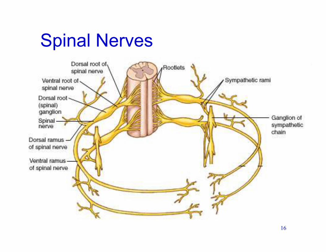
Spinal Nerves
17
Spinal Nerves→Nerve Plexus
• Dorsal and Ventral roots exit the spinal cord
and join together to make a spinal nerve
• The spinal nerve then splits into dorsal and
ventral rami (ramus)
• Some ventral rami give off branches to the
sympathetic ganglion
• The other ventral rami mix and match to make
up nerve plexuses
18
Ventral Rami
• The Dorsal Root only contains sensory
neurons going toward the spinal cord
• The Ventral Root only contains motor
neurons going out of the spinal cord
• Ventral Rami contain BOTH sensory and
motor neurons
• As the spinal nerves, rami and plexus are
crisscrossing, everything gets mixed
around.
19
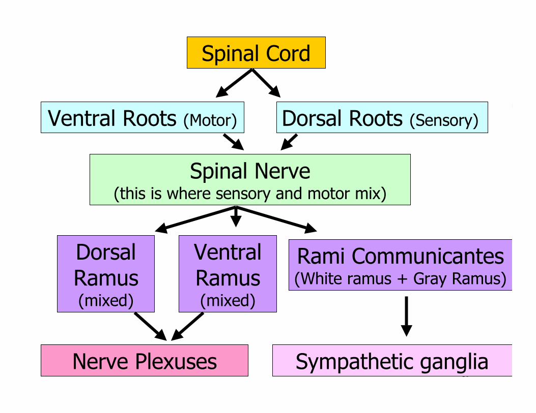
Spinal Cord
Dorsal Ramus(mixed)
Ventral Ramus(mixed)
Rami Communicantes(White ramus + Gray Ramus)
Nerve Plexuses
Ventral Roots (Motor)
Spinal Nerve(this is where sensory and motor mix)
Dorsal Roots (Sensory)
Sympathetic ganglia
20
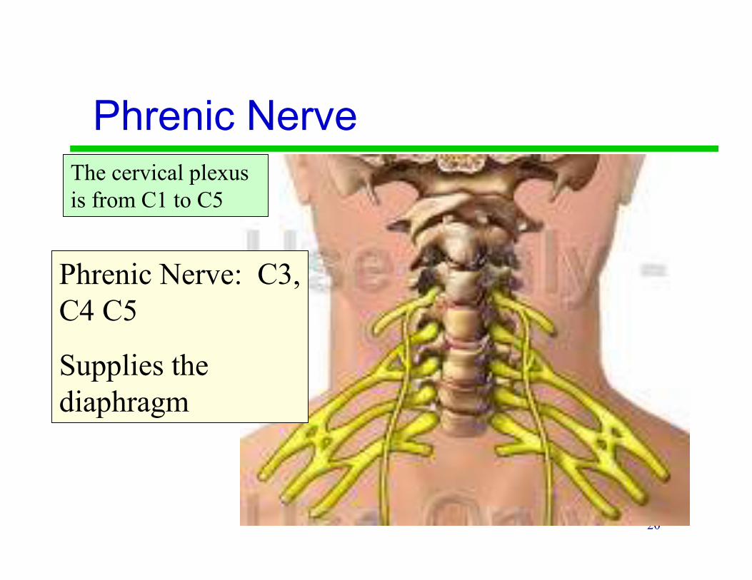
Phrenic Nerve
Phrenic Nerve: C3,
C4 C5
Supplies the
diaphragm
The cervical plexus
is from C1 to C5
21
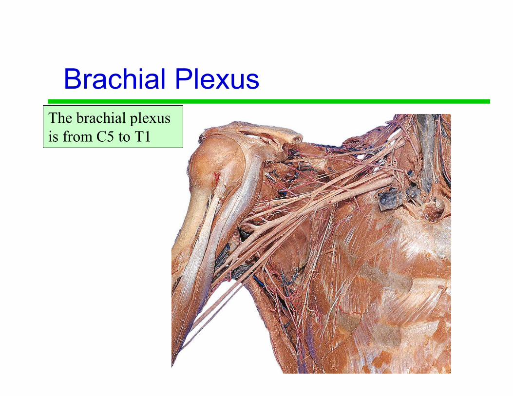
Brachial Plexus
The brachial plexus
is from C5 to T1
22
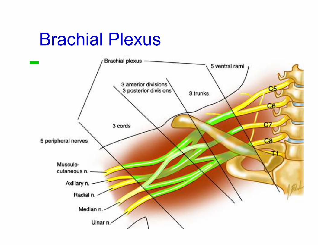
Brachial Plexus
23
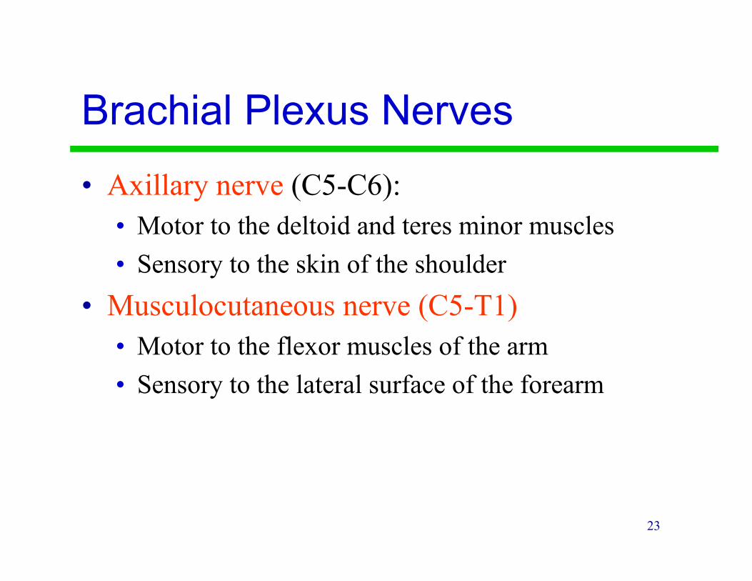
Brachial Plexus Nerves
• Axillary nerve (C5-C6):
• Motor to the deltoid and teres minor muscles
• Sensory to the skin of the shoulder
• Musculocutaneous nerve (C5-T1)
• Motor to the flexor muscles of the arm
• Sensory to the lateral surface of the forearm

24
Brachial
Plexus Nerves
• Radial nerve (C5-T1)
• Motor to muscles of the
posterior arm and forearm
• Sensory to the posterior-
lateral side of the hand,
but not the fingers (purple
in picture)
Radial
Nerve
25
Brachial Plexus
Nerves
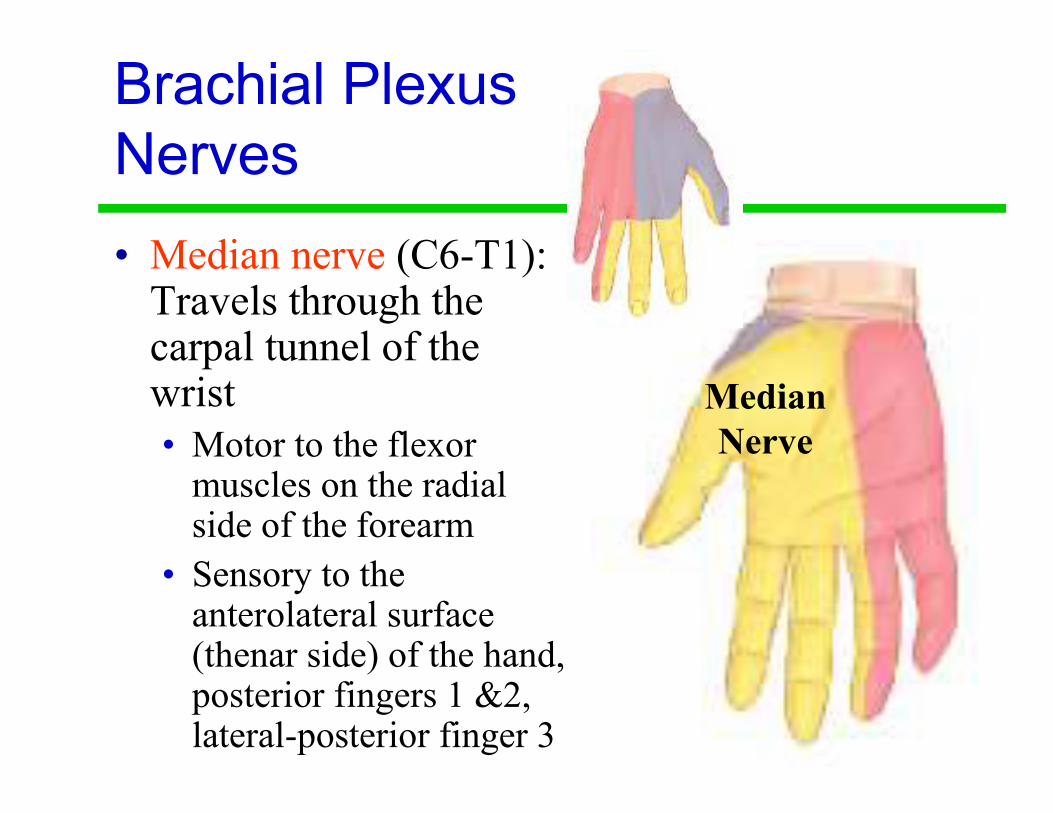
• Median nerve (C6-T1): Travels through the carpal tunnel of the wrist
• Motor to the flexor muscles on the radial side of the forearm
• Sensory to the anterolateral surface (thenar side) of the hand, posterior fingers 1 &2, lateral-posterior finger 3
Median
Nerve

26
Brachial Plexus
Nerves
• Ulnar nerve (C8-T1)
• Motor to many flexor
muscles of forearm and
hand on ulnar side
• Sensory to the medial
surface of the hand.
Ulnar
Nerve
27
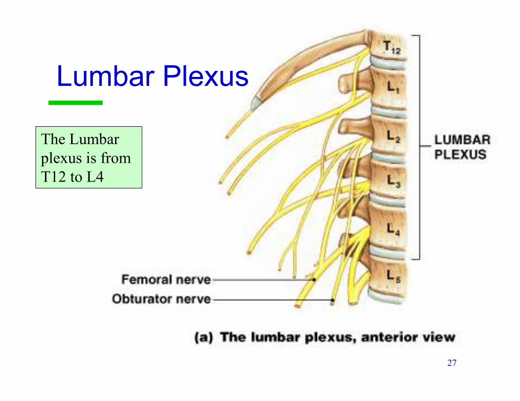
Lumbar Plexus
The Lumbar
plexus is from
T12 to L4

28
Lumbar Plexus
• The major nerves:
• Femoral nerve L2-L4• Motor to Quadriceps group, Pectineus and Iliopsoas muscles, sensory anterior-medial thigh and medial surface of leg and foot.
• Injury to femoral nerve causes inability to extend leg & loss of sensation in thigh
• Obturator nerve L2-L4• Motor to adductors of hip. Sensory to medial surface of thigh.
• Injury to obturator nerve causes paralysis of thigh adductors
29
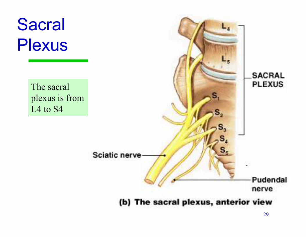
Sacral
Plexus
The sacral
plexus is from
L4 to S4
30
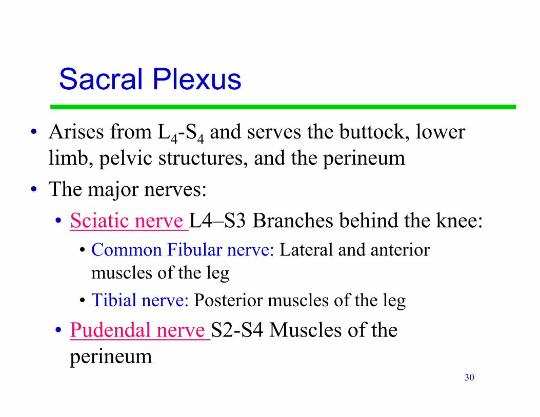
Sacral Plexus
• Arises from L4-S4 and serves the buttock, lower
limb, pelvic structures, and the perineum
• The major nerves:
• Sciatic nerve L4–S3 Branches behind the knee:
• Common Fibular nerve: Lateral and anterior
muscles of the leg
• Tibial nerve: Posterior muscles of the leg
• Pudendal nerve S2-S4 Muscles of the
perineum
31
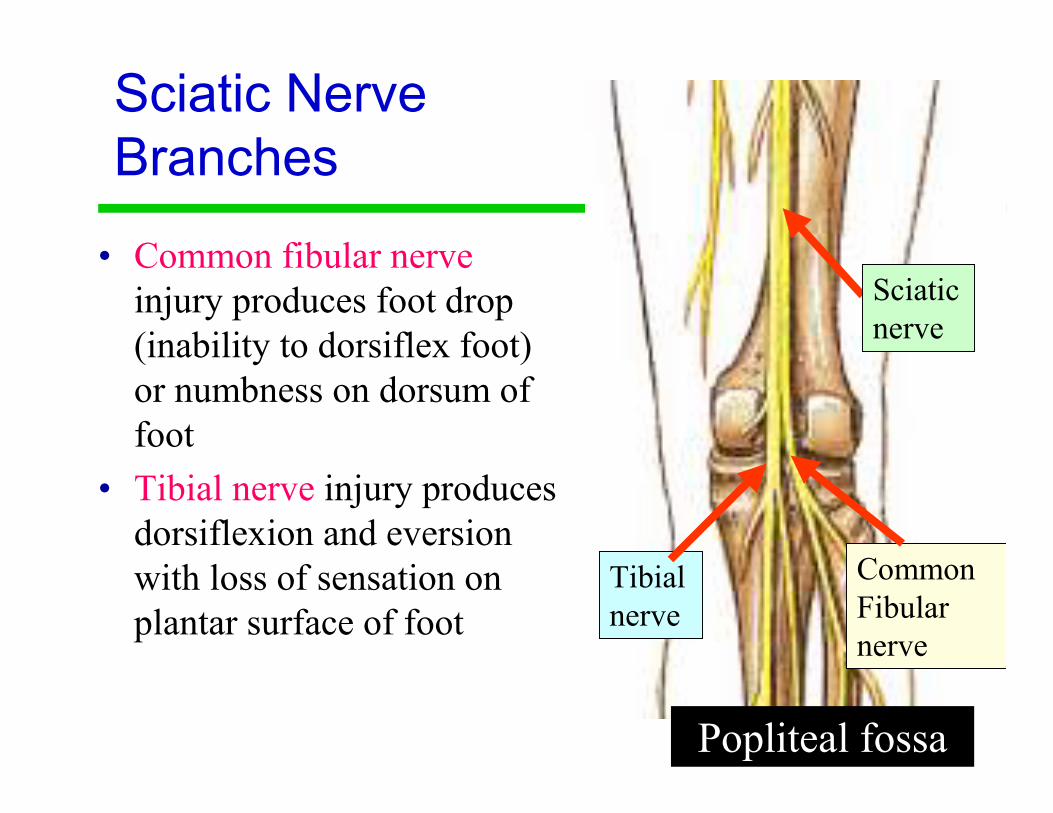
Sciatic Nerve
Branches
• Common fibular nerve
injury produces foot drop
(inability to dorsiflex foot)
or numbness on dorsum of
foot
• Tibial nerve injury produces
dorsiflexion and eversion
with loss of sensation on
plantar surface of foot
Popliteal fossa
Common
Fibular
nerve
Tibial
nerve
Sciatic
nerve
32
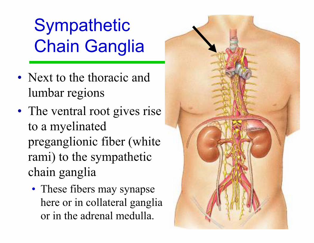
Sympathetic
Chain Ganglia
• Next to the thoracic and
lumbar regions
• The ventral root gives rise
to a myelinated
preganglionic fiber (white
rami) to the sympathetic
chain ganglia
• These fibers may synapse
here or in collateral ganglia
or in the adrenal medulla.

33
Lab Activity 14
Reflexes
35
Reflexes
• A reflex is a rapid, predictable motor
response to a stimulus
• Reflexes may:
• Be inborn (intrinsic) or learned (acquired)
• Involve only peripheral nerves and the spinal
cord (aka: spinal reflexes)
• Involve higher brain centers as well
36
Reflex Arc
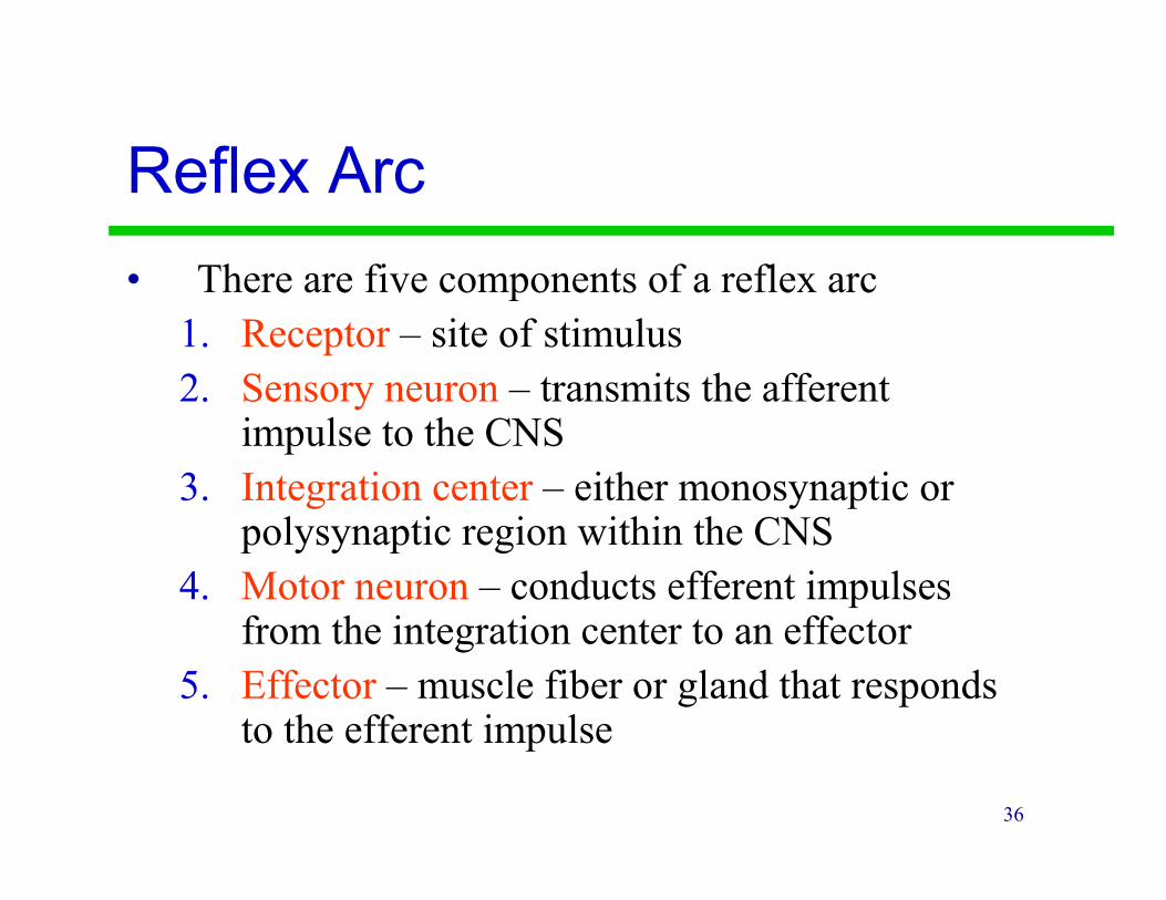
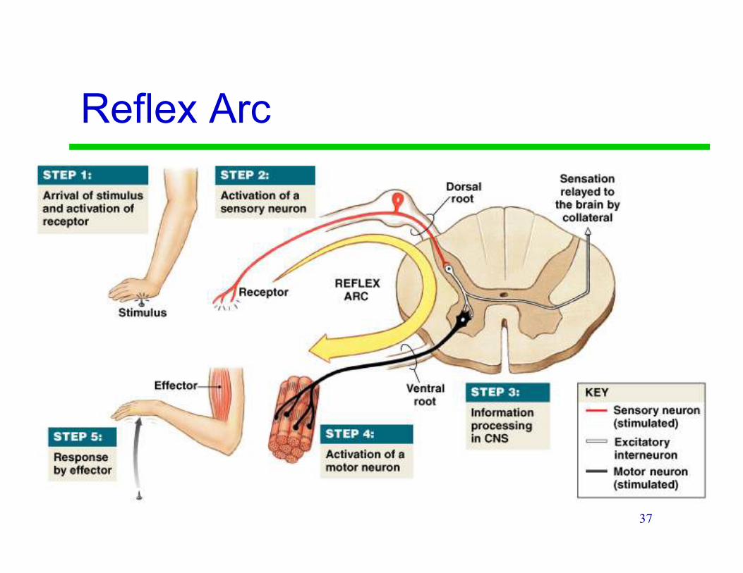
• There are five components of a reflex arc
1. Receptor – site of stimulus
2. Sensory neuron – transmits the afferent impulse to the CNS
3. Integration center – either monosynaptic or polysynaptic region within the CNS
4. Motor neuron – conducts efferent impulses from the integration center to an effector
5. Effector – muscle fiber or gland that responds to the efferent impulse
37
Reflex Arc

38
Innate Reflexes
• Innate reflexes: Reflexes you are born with.
• The are genetically or developmentally
programmed
• Examples:
• Withdrawing from pain
• Suckling
• Chewing
• Tracking objects with the eyes
39
Acquired Reflexes
• Acquired reflexes are learned motor patterns
• Generally more complex than innate reflexes
• Examples:
• Slamming on the break when driving
• Professional skier making quick adjustments in body position
40
Reflexes
• Visceral (Autonomic) reflexes regulate
body functions
• Digestion, blood pressure, sweating ect…
• Somatic reflexes involve skeletal muscles
• Function to maintain posture, balance and
locomotion
41
Reflexes
• Spinal reflexes: The important
interconnections and processing events
occur in the spinal cord.
• Cranial reflexes: The integration center is in
the brain
42
Types of Reflexes
• Monosynaptic reflexes: The sensory neuron
synapse directly on a motor neuron.
• The delay between stimulus and the response is
minimized.
• The synapse is considered the integration center
• Polysynaptic reflexes: There is at least one
interneuron between the sensory and motor
neuron
• More complex responses
43
Upper Motor Neurons
• Upper motor neurons: Starts in the motor
cortex of the brain and terminates within the
medulla (another part of the brain) or within
the spinal cord.
• Damage to upper motor neurons can result in
spasticity and exaggerated reflexes (because of
the loss of inhibition) “Spastic Paralysis”
44
Lower Motor Neurons
• Lower motor neurons go from the spinal cord to a muscle.
• The cell body of a lower motor neuron is in the spinal cord and its termination is in a skeletal muscle.
• The loss of lower motor neurons leads to weakness, twitching of muscle (fasciculation), and loss of muscle mass (muscle atrophy). “Flaccid Paralysis”
45
Reflexes
• Intact reflexes require
• Intact sensory afferent nerves (coming to the
spinal cord)
• Intact synapse within the spinal cord
• Intact efferent motor nerves coming from the
spinal column
• Adequately functioning muscle.
46
Testing Reflexes
• Reflexes can also be modified by conditions higher in the cord than the relevant synapse including the brain itself.
• The purpose of testing reflexes is to check the integrity of the system as a whole.
• An absent reflex indicates a problem somewhere in the reflex arc but it does not tell you where.
47
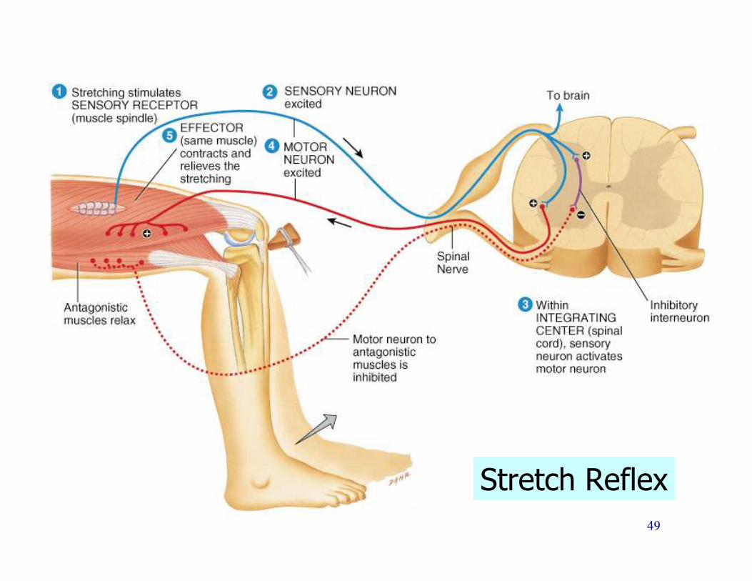
Stretch Reflexes
• 1. Stretching of the muscle activates a muscle spindle
• A muscle spindle is a bundle of specialized skeletal muscle fibers that act as sensory receptors
• 2. An impulse is transmitted by afferent fibers to the spinal cord
• 3. Motor neurons in the spinal cord cause the stretched muscle to contract
• 4. The integration area in the spinal cord causes the antagonist muscle to relax (reciprocal inhibition)
48
Stretch Reflex Example
Patellar Reflex (L2, L3, L4)
• Tap the patellar tendon
• muscle spindle signals stretch of muscle
• motor neuron activated & muscle contracts
• Quadriceps muscle contracts
• Hamstring muscle is inhibited (relaxes)
• Reciprocal innervation (polysynaptic- interneuron)
• antagonistic muscles relax as part of reflex
• Lower leg kicks forward
• Demonstrates sensory and motor connections between muscle and spinal cord are intact.
49
Stretch Reflex
50
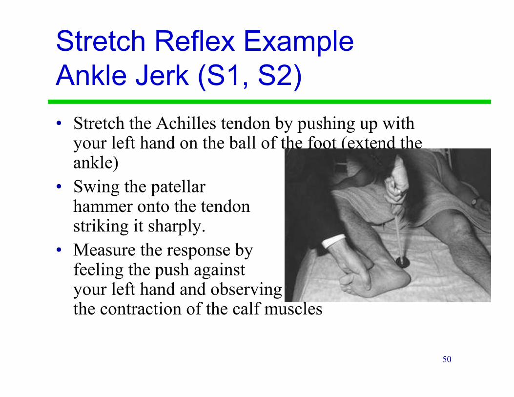
Stretch Reflex Example
Ankle Jerk (S1, S2)
• Stretch the Achilles tendon by pushing up with your left hand on the ball of the foot (extend the ankle)
• Swing the patellar hammer onto the tendon striking it sharply.
• Measure the response by feeling the push against your left hand and observing the contraction of the calf muscles
51
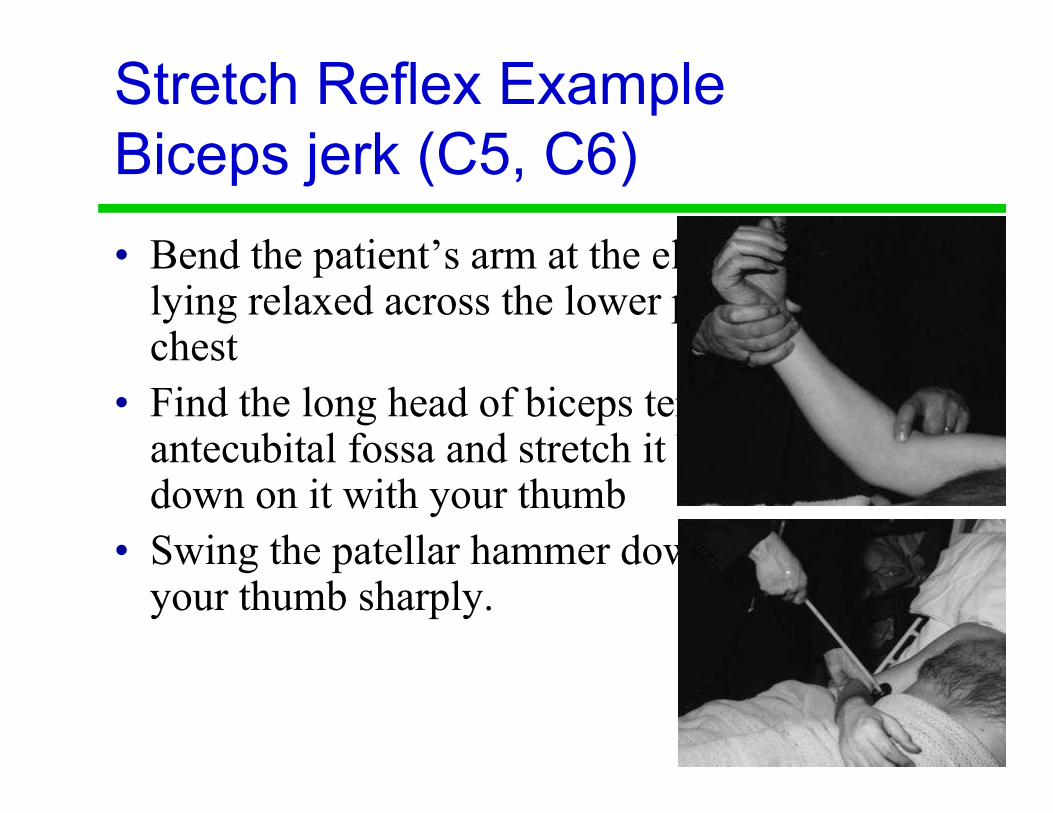
Stretch Reflex Example
Biceps jerk (C5, C6)
• Bend the patient’s arm at the elbow so it is lying relaxed across the lower part of the chest
• Find the long head of biceps tendon in the antecubital fossa and stretch it by pushing down on it with your thumb
• Swing the patellar hammer down and strike your thumb sharply.
52
Grading Reflexes
• Grading of reflexes:
• 0+ = absent
• 1+ = hyporeflexic (reduced reflex)
• 2+ = normal
• 3+ = hyperreflexia (exaggerated reflex)
• 4+= clonus
• Say “one plus”
• Conditions such as hypothyroidism and spinal shock diminish reflexes.
• Stimulant drugs, anxiety, and hyperthyroidism increase reflexes.

53
Tendon Reflexes
• Controls muscle tension by causing muscle
relaxation that prevents tendon damage
• Golgi tendon organs in tendon
• Activated by stretching of tendon
• Inhibitory neuron is stimulated (polysynaptic)
• Motor neuron is hyperpolarized and muscle relaxes
• Both tendon & muscle are protected
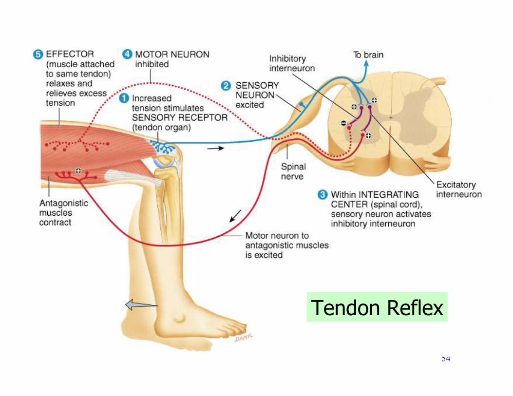
54
Tendon Reflex

55
Flexor Reflex
• Withdrawal reflex
• When pain receptors are activated it causes automatic withdrawal of the threatened body part.
• Reciprocal inhibition: Interneurons in the spinal cord prevent a stretch reflex in the antagonistic muscles
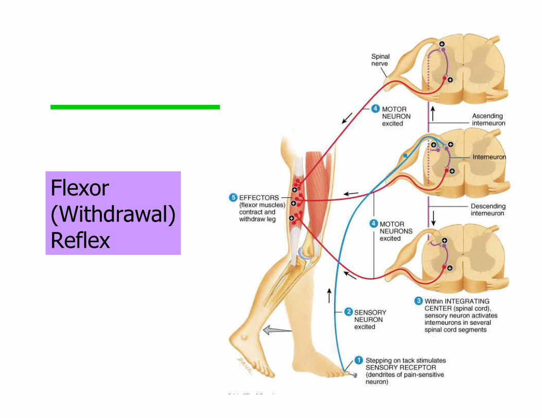
56
Flexor (Withdrawal)Reflex
57
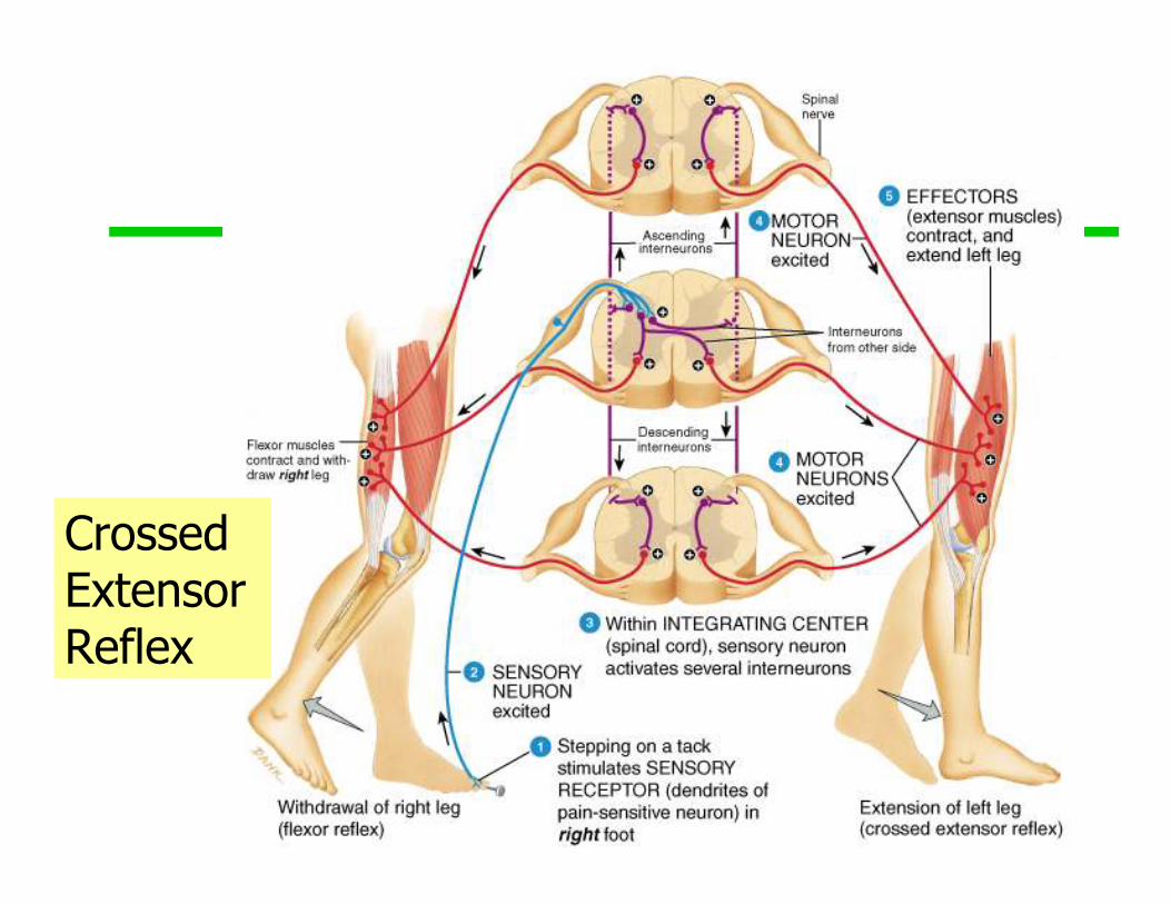
Crossed Extensor Reflex
• Complex reflex that consists of an
ipsilateral withdrawal reflex and a
contralateral extensor reflex
• This keeps you from falling over, for
example if you step on something painful.
When you pull your foot back, the other leg
responds to hold you up.
58
Crossed Extensor Reflex
59
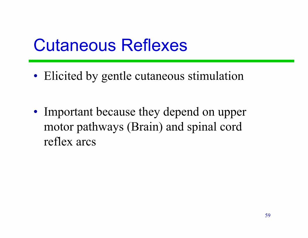
Cutaneous Reflexes
• Elicited by gentle cutaneous stimulation
• Important because they depend on upper
motor pathways (Brain) and spinal cord
reflex arcs
60
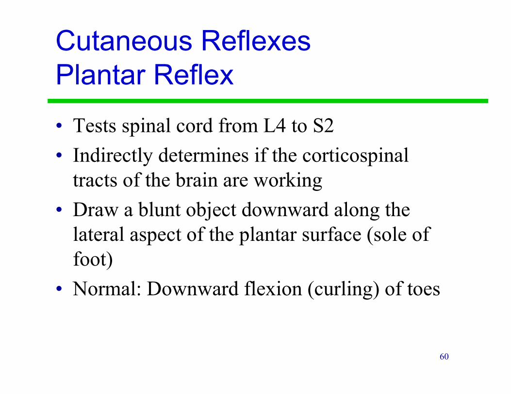
Cutaneous Reflexes
Plantar Reflex
• Tests spinal cord from L4 to S2
• Indirectly determines if the corticospinal
tracts of the brain are working
• Draw a blunt object downward along the
lateral aspect of the plantar surface (sole of
foot)
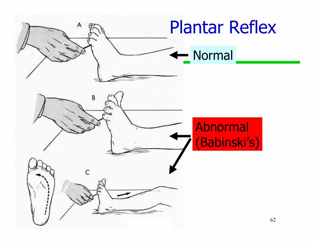
• Normal: Downward flexion (curling) of toes
61
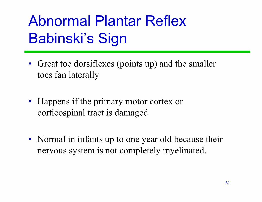
Abnormal Plantar Reflex
Babinski’s Sign
• Great toe dorsiflexes (points up) and the smaller
toes fan laterally
• Happens if the primary motor cortex or
corticospinal tract is damaged
• Normal in infants up to one year old because their
nervous system is not completely myelinated.
62
Normal
Abnormal(Babinski’s)
Plantar Reflex

63
The End


![Identification and Characterization of Pleural Neurons ......dulin, sensory neuron, motor neuron, inhibition, neural cir- cuit, Aplysia] The sensory and motor neurons that mediate](https://img.pdfslide.us/doc/110x75/5fc497a9642d1777a877bb71/identification-and-characterization-of-pleural-neurons-dulin-sensory-neuron.jpg)