Embed Size (px)
Citation preview

1
Lab 12
Nervous System II
Laboratory Objectives
Identify the structural components of the central nervous system
Label the functional areas of human cerebral cortex.
Given a deficit affecting one or more of those functional areas, be able to predict a
patient’s symptoms.
Be able to locate the 12 cranial nerves on a diagram.
Know at least one function associated with each of the 12 cranial nerves.
Compare and contrast the autonomic (involuntary) and somatic (voluntary) nervous
systems.
Divide the autonomic nervous system into sympathetic and parasympathetic
components.

2
Activity 1: Cadaver Video: The Nervous System
Navigation: WileyPlus > Read, Study, and Practice > Chapter 14: The Brain and Cranial
Nerves > See > Cadaver Video: The Nervous System/Brain (25:50)
1. What is the space between the arachnoid mater and pia mater?
2. What groove divides the left and right hemispheres?
3. What are the ridges of the cerebrum called?
4. What separates the frontal and parietal lobes?
5. Where do the lateral corticospinal tracts cross?

3
Activity 2: Cadaver Video: Cadaver Video: The Nervous System/Spinal Cord
Navigation: WileyPlus > Read, Study, & Practice > Chapter 15. The Autonomic Nervous
System > See > Cadaver Video: The Nervous System/Spinal Cord (time: - 6:08)
1. What the outer layer of the meninges?
2. What does the pia mater contain?
3. What are the two spinal enlargements?
4. What is the name of the bundle of nerves extending from the lumbar region of the
spinal cord?
5. What is the name of the ligamentous extensions of the pia mater that support the
spinal cord laterally?

4
Activity 3: Visual Anatomy
Navigation: WileyPlus > Read, Study, and Practice > Lab Exercise 20.Brain Structure and
Function > Do > Visual Anatomy > Brain and Cranial Nerves > Cadaver Practicals
Complete the following sections:
Cerebrum, right lateral view
Brain, midsagittal section
Transverse section, choroid plexuses, superior view
Cerebellum, midsagittal section
1. List the regions of the brainstem from inferior to posterior.
2. What gland is part of the epithalamus?
3. What is the name of the dural extension between the two hemispheres?

5
Activity 4 Functional Areas of the Brain
Navigation: WileyPlus > Read, Study, and Practice > Lab Exercise 20.Brain Structure and
Function > Do > Interactive Exercise: Paint the Functional Areas of the Brain
1. Which area is associated with the production of language?
2. Which lobe of the brain includes the olfactory center?
3. Which lobe of the brain receives somatosensory information?
4. Primary visual centers are located in which lobe?

6
Activity 5: PowerAnatomy
Navigation: WileyPlus > Read, Study, and Practice > Lab Exercise 20.Brain Structure and
Function > PowerAnatomy > Table of Contents > 8 - Structure and Function of the Brain >
Exercises and Review
Complete the following:
Exercise A. Cerebrospinal Fluid
Exercise B . Location of Functional Areas of the Cerebral Cortex
Review 1. Functions of the Brain Regions
Review 2. The Effects of Damage to the Brain
1. What is the name of the tissue that produces cerebrospinal fluid (CSF)?
2. Name several structures through which CSF flows.
3. What is the name of the structure that resorbs CSF?
4. The composition of CSF is very close to ...

7
5. The location of a stroke (a loss of blood supply, which damages brain tissue) is
shown in the left column. In the right column, fill in one or more symptoms you
would expect in the patient. (Some of the difficult or unclear ones are filled out for
you already)
Structure affected by stroke Symptoms which might be caused (include side where appropriate)
Primary motor area
Primary somatosensory area
Broca’s speech area (motor)
Primary visual area
Primary auditory area Because hearing projects to both hemispheres, strokes in this area do not result in
measurable hearing loss
Primary gustatory area As for hearing, taste is generally represented on both sides. Some patients may
report diminished taste sensation, or odd tastes (parageusias)
Wernicke’s area
Medulla
Cerebellum
Pineal gland
Hypothalamus
Thalamus Motor: Damage to the motor part of the thalamus can result in abnormalities of
movement. For example, strokes in the subthalamic nucleus lead to a weird
condition called hemiballismus.
Pons
Thalamus Sensory: Patients with strokes in this area have pain which cannot be relieved.
Corpus callosum Strokes here are rare. Some patients have agenesis of the corpus callosum where
the two hemispheres are disconnected, like Sperry’s split brain patients

8
Activity 5: The Case of the Man with the Tingling Arm
Navigation: WileyPlus > Read, Study, and Practice > Lab Exercise 20.Brain Structure and
Function > Do > Homeostatic Imbalance: The Case of the Man with the Tingling Arm
1. Why does the narrator mention Mr. Jackson’s race, body weight, blood pressure
and diabetes? Are all of these relevant to his case?
2. What area of the brain receives sensations from the right upper extremity and
right face (be sure to include the side of the brain)?
3. What area of the brain controls the right upper extremity (include side)?
4. What area of the brain controls speaking (include side)? Is this always on the side
that you mentioned, or almost always?
5. What area of the brain controls the muscles on the right side of the face (include
side)?
6. Is it important that all of the answers to questions 2-5 describe the same general
area of the brain? Why or why not?

9
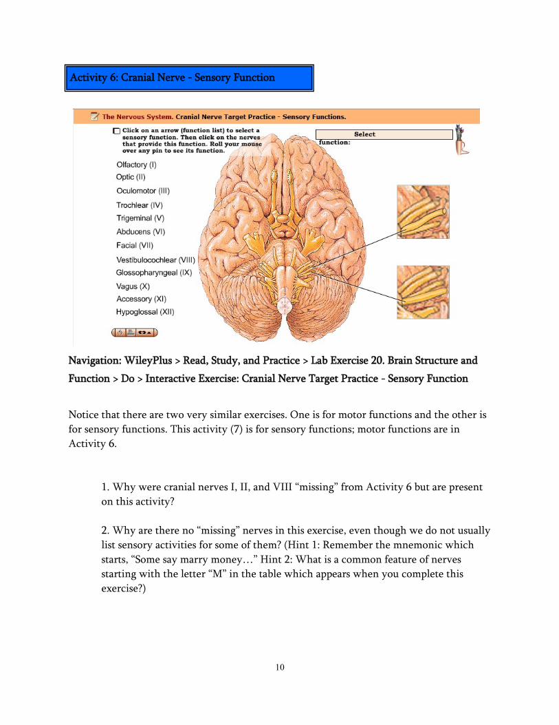
Activity 6: Cranial Nerve - Motor Function
Navigation: WileyPlus > Read, Study, and Practice > Lab Exercise 20. Brain Structure and
Function > Do > Interactive Exercise: Cranial Nerve Target Practice - Motor Functions
Notice that there are two very similar exercises. One is for motor functions and the other is
for sensory functions. This activity (6) is for motor functions; sensory functions are in
Activity 7.
1. What is meant by the term, “mixed nerve”?
2. Damage to the abducens nerve would demonstrate what dysfunction?
3. Damage to the accessory nerve would produce what symptom?
4. Damage to the hypoglossal nerve would produce what symptom?
10
Navigation: WileyPlus > Read, Study, and Practice > Lab Exercise 20. Brain Structure and
Function > Do > Interactive Exercise: Cranial Nerve Target Practice - Sensory Function
Notice that there are two very similar exercises. One is for motor functions and the other is
for sensory functions. This activity (7) is for sensory functions; motor functions are in
Activity 6.
1. Why were cranial nerves I, II, and VIII “missing” from Activity 6 but are present
on this activity?
2. Why are there no “missing” nerves in this exercise, even though we do not usually
list sensory activities for some of them? (Hint 1: Remember the mnemonic which
starts, “Some say marry money…” Hint 2: What is a common feature of nerves
starting with the letter “M” in the table which appears when you complete this
exercise?)
Activity 6: Cranial Nerve - Sensory Function
11
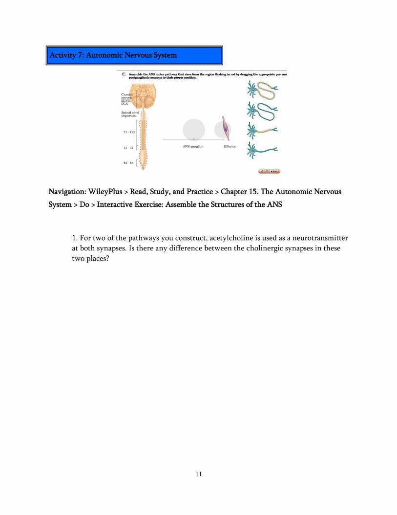
Navigation: WileyPlus > Read, Study, and Practice > Chapter 15. The Autonomic Nervous
System > Do > Interactive Exercise: Assemble the Structures of the ANS
1. For two of the pathways you construct, acetylcholine is used as a neurotransmitter
at both synapses. Is there any difference between the cholinergic synapses in these
two places?
Activity 7: Autonomic Nervous System

12
Navigation: WileyPlus > Read, Study, and Practice > Chapter 15. The Autonomic Nervous
System > Do > Interactive Exercise: Sort ANS Functions
1. In the list of organs in this exercise, one of the organs is not paired (there is only
one icon, not two). What is the name of this organ, which list is it missing from, and
why?
2. The activity instructions say the size of the icons is significant (indicating
increased/decreased activity). What other differences do you note in the icons, if
any?
3. In the parasympathetic nervous system, which organs are controlled by the
brainstem, and which are controlled by the lumbar spinal cord?
Activity 8: Autonomic Nervous System

13
Activity 9: What is your ANS status?
Navigation: WileyPlus > Read, Study, and Practice > Chapter 15. The Autonomic Nervous
System > Do > Interactive Exercise: What is your ANS status
1. What is meant by the term dual innervation?
2. Name some of the factors that go into “deciding” which response to evoke? Why
would the same event provoke a sympathetic response in some people, but not in
others?
3. Which system is activated all at once? Which one is activated organ by organ? Is
this reflected in the innervation pattern? Is this reflected in the changes in blood
chemistry? Is there an abnormal or pathological condition in which one of these
systems is activated all at once?