Embed Size (px)
Citation preview
Master’s thesis
L-rhamnose-1-dehydrogenase gene and L-rhamnosecatabolism in the yeast Pichia stipitis
Outi Koivistoinen
Division of Microbiology
Department of Applied Chemistry and Microbiology
Faculty of Agriculture and Forestry
University of Helsinki
June 2008
1
HELSINGIN YLIOPISTO HELSINGFORS UNIVERSITET UNIVERSITY OF HELSINKI
Tiedekunta/Osasto Fakultet/Sektion FacultyFaculty of Agriculture and Forestry
Laitos Institution DepartmentDepartment of Applied Chemistry andMicrobiology
Tekijä Författare AuthorOuti KoivistoinenTyön nimi Arbetets titel TitleL-rhamnose-1-dehydrogenase gene and L-rhamnose catabolism in the yeast Pichia stipitisOppiaine Läroämne SubjectMicrobiologyTyön laji Arbetets art LevelMaster’s thesis
Aika Datum Monthand yearJune 2008
Sivumäärä Sidoantal Number of pages63
Tiivistelmä Referat AbstractThe purpose of this work was to identify some of the genes of the catabolic route of L-rhamnose in the yeast Pichia stipitis. There are at least two distinctly different pathways for L-rhamnose catabolism. The one described in bacteria has phosphorylated intermediates and theenzymes and the genes of this route have been described. The pathway described in yeast doesnot have phosphorylated intermediates. The intermediates and the enzymes of this pathway areknown but none of the genes have been identified. The work was started by purifying the L-rhamnose dehydrogenase, which oxidates L-rhamnose to rhamnonic acid- -lactone. NAD isused as a cofactor in this reaction. A DEAE ion exchange column was used for purification.The active fraction was further purified using a non-denaturing PAGE and the active proteinidentified by zymogram staining. In the last step the protein was separated in a SDS-PAGE,the protein band trypsinated and analysed by MALDI-TOF MS. This resulted in theidentification of the corresponding gene, RHA1 , which was then, after a codon change,expressed in Saccharomyces cerevisiae. Also C- or N-terminal histidine tags were added but asthe activity of the enzyme was lost or strongly reduced these were not used. The kineticproperties of the protein were analysed in the cell extract. Substrate specifity was tested withdifferent sugars; L-rhamnose, L-lyxose and L-mannose were oxidated by the enzyme. Vmax
values were 180 nkat/mg, 160 nkat/mg and 72 nkat/mg, respectively. The highest affinity wastowards L-rhamnose, the Km value being 0.9 mM. Lower affinities were obtained with L-lyxose, Km 4.3 mM, and L-mannose Km 25 mM. Northern analysis was done to study thetranscription of RHA1 with different carbon sources. Transcription was observed only on L-rhamnose suggesting that RHA1 expression is L-rhamnose induced. A RHA1 deletion cassettefor P. stipitis was constructed but the cassette had integrated randomly and not targeted todelete the RHA1 gene.Enzyme assays for L-lactaldehyde dehydrogenase were done similarly to L-rhamnosedehydrogenase assays. NAD is used as a cofactor also in this reaction where L-lactaldehyde isoxidised to L-lactate. The observed enzyme activities were very low and the activity was lostduring the purification procedures.Avainsanat Nyckelord KeywordsL-Rhamnose, L-Rhamnose dehydrogenase, EC 1.1.1.174, L-rhamnose catabolism, Pichiastipitis, L-rhamnonate, MALDI-TOF MS, L-lactaldehyde, L-lactaldehyde dehydrogenaseSäilytyspaikka Förvaringsställe Where depositedThe reference library of the Division of Microbiology, Department of Applied Chemistry andMicrobiologyMuita tietoja Övriga uppgifter Further informationSupervisor: PhD Peter Richard, Funded by: Academy of Finland
2
HELSINGIN YLIOPISTO HELSINGFORS UNIVERSITET UNIVERSITY OF HELSINKI
Tiedekunta/Osasto Fakultet/Sektion FacultyMaatalous-metsätieteellinen tiedekunta
Laitos Institution Department
Soveltavan kemian ja mikrobiologian laitosTekijä Författare AuthorOuti Koivistoinen
Työn nimi Arbetets titel TitleL-ramnoosi-1-dehydrogenaasigeeni ja L-ramnoosin kataboliareitti Pichia stipitis -hiivassaOppiaine Läroämne SubjectMikrobiologiaTyön laji Arbetets art Level
Pro gradu
Aika Datum Month and yearKesäkuu 2008
Sivumäärä Sidoantal Number of pages63
Tiivistelmä Referat AbstractTyön tarkoitus oli identifioida L-ramnoosin kataboliareitin geenejä Pichia stipitis hiivassa.Toistaiseksi on löydetty kaksi erilaista L-ramnoosin kataboliareittiä. Bakteereilla esiintyvässäreitissä on fosforyloidut välituotteet. Tämän reitin entsyymit ja niitä koodaavat geenit onkuvattu. Sienillä L-ramnoosin katabolireitin tuotteet ovat fosforyloimattomia. Reitinvälituotteet ja entsyymit on kuvattu, mutta yhtään entsyymiä koodaavaa geeniä ei oleidentifioitu. Työ aloitettiin puhdistamalla L-ramnoosidehydrogenaasi, joka katalysoi L-ramnoosin hapettumista ramnonihappo- -laktoniksi. NAD toimii reaktiossa kofaktorina.DEAE ioninvaihtopylvästä käytettiin entsyymin puhdistukseen. Aktiiviset osat ajettiin ei-denaturoivassa PAGE-geelissä ja zymogram-värjättiin. Proteiini ajettiin SDS-PAGE –geelissäja trypsinoitiin. Aminohapposekvenssin analysointi tehtiin MALDI-TOF MS laitteella javastaava DNA-sekvenssi nimettiin RHA1-geeniksi. Kodoninvaihdon jälkeen RHA1ekspressoitiin Saccharomyces cerevisiae –hiivassa. Proteiinin C- ja N-terminaalisiin päihinlisättiin histidiiniketjut, mutta seurauksena entsyymin aktiivisuus joko katosi tai vähenivoimakkaasti. Proteiinin kineettiset ominaisuudet analysoitiin solu-uutteesta.Substraattispesifisyyttä testattiin eri sokereilla. L-Ramnoosin, L-lyksoosin ja L-mannoosintodettiin hapettuvan entsyymin vaikutuksesta. Vmax arvot olivat vastaavassa järjestyksessä 180nkat/mg, 160 nkat/mg ja 72 nkat/mg. Suurin affiniteetti oli L-ramnoosilla, jonka Km arvo oli0.9 mM. L-Lyksoosin Km arvo oli 4.3 mM ja L-mannoosin Km arvo oli 25 mM. Northern-analyysilla tutkittiin eri sokerien vaikutusta RHA1:n transkriptioon. Transkriptiota havaittiinvain L-ramnoosilla, joten RHA1:n ekspressio on L-ramnoosin indusoimaa. P. stipitis –hiivalletehtiin RHA1-deleetiokasetti, mutta sen todettiin integroituneen sattumanvaraisesti eikä sekohdistunut oikein, joten RHA1-geenin deleetio epäonnistui.L -Laktaldehydidehydrogenaasin entsyymianalyysi tehtiin samoin kuin L -ramnoosidehydrogenaasin entsyymianalyysi. NAD toimii kofaktorina myös tässä reaktiossa,jossa L-laktaldehydi hapetetaan L-laktaatiksi. Havaitut entsyymiaktiivisuudet olivat kuitenkinhyvin alhaisia, ja entsyymi inaktivoitui puhdistusprosessissa.
Avainsanat Nyckelord KeywordsL-ramnoosi, L-ramnoosidehydrogenaasi, EC 1.1.1.174, L-ramnoosikatabolia, Pichia stipitis,L-rhamnonaatti, MALDI-TOF MS, L-laktaldehydi, L-laktaldehydidehydrogenaasi
Säilytyspaikka Förvaringsställe Where depositedSoveltavan kemian ja mikrobiologian laitoksen mikrobiologian osaston käsikirjastoMuita tietoja Övriga uppgifter Further informationTyön ohjaus: Peter Richard, Rahoittaja: Suomen Akatemia
3
Table of Contents
ACKNOWLEDGEMENTS.................................................................................. 5
LIST OF TABLES .............................................................................................. 7
LIST OF ABBREVIATIONS............................................................................... 8
1 INTRODUCTION....................................................................................... 10
1.1 Pichia stipitis .............................................................................................. 10
1.2 Production of biofuels by yeast.................................................................... 11
1.3 Oxidation of hexoses ................................................................................... 13
1.3.1 Embden-Meyerhof-Parnas pathway ..................................................... 13
1.3.2 Entner-Doudoroff pathway .................................................................. 13
1.3.3 Non-phosphorylated Entner-Doudoroff pathway.................................. 14
1.4 L-Rhamnose ................................................................................................ 16
1.4.1 Biosynthetic pathway of L-rhamnose ................................................... 16
1.4.2 L-Rhamnose catabolism....................................................................... 17
1.5 Catabolic routes similar to the L-rhamnose route ......................................... 19
1.5.1 Fungal L-fucose catabolism.................................................................. 19
1.5.2 Fungal D-galactose catabolism............................................................. 20
1.5.3 Fungal D-gluconate catabolism ............................................................ 20
1.5.4 Fungal D-galacturonate catabolism....................................................... 21
1.6 Aim of the work .......................................................................................... 23
2 MATERIALS AND METHODS.................................................................. 24
2.1 Yeast and bacterial strains and plasmids ...................................................... 24
2.2 Media and growth conditions ...................................................................... 25
2.3 Recombinant DNA techniques .................................................................... 26
2.4 Transformations .......................................................................................... 28
2.4.1 E. coli transformations......................................................................... 28
2.4.2 S. cerevisiae transformations ............................................................... 29
2.4.3 P. stipitis transformations .................................................................... 29
4
2.5 Sequencing.................................................................................................. 29
2.6 Enzyme purification .................................................................................... 29
2.6.1 Anion exchange and gradient............................................................... 30
2.6.2 Zymogram staining.............................................................................. 30
2.6.3 SDS PAGE.......................................................................................... 31
2.7 In-gel digestion MALDI-TOF MS............................................................... 31
2.8 Site directed mutagenesis kit for changing the CTG codon .......................... 32
2.9 Cloning of the RHAI.................................................................................... 33
2.10 Enzyme assays ............................................................................................ 34
2.11 Deletion cassette for P. stipitis..................................................................... 36
2.12 Synthesis of L-rhamnoate and L-lactaldehyde .............................................. 39
2.13 Northern analysis ........................................................................................ 39
3 RESULTS ................................................................................................. 41
3.1 Purification of L-rhamnose dehydrogenase .................................................. 41
3.2 MALDI-TOF MS........................................................................................ 42
3.3 Enzyme activity........................................................................................... 44
3.4 RHA1 deletion cassette ................................................................................ 48
3.5 Northern blot............................................................................................... 49
3.6 L-Lactaldehyde activity ............................................................................... 50
4 DISCUSSION............................................................................................ 51
4.1 L-Rhamnose dehydrogenase ........................................................................ 51
4.1.1 Kinetic properties ................................................................................ 51
4.1.2 RHA1 deletion cassette ........................................................................ 53
4.2 L-Lactaldehyde dehydrogenase.................................................................... 54
4.3 Transformation of P. stipitis ........................................................................ 54
4.4 Northern blot............................................................................................... 55
4.5 Conclusions................................................................................................. 56
5 REFERENCES.......................................................................................... 57
5
Acknowledgements
The experimental part of this work was done at the Metabolic Engineering group at
Technical Research Centre of Finland (VTT), Espoo.
I thank my supervisor PhD Peter Richard for the guidance and valuable advice for the
experimental and written work. I wish to thank MSc Satu Hilditch for the help and
support during my work. I want to express my sincere gratitude to Peter and Satu for
their patience and encouragement during the work. I would also like to thank Sanni
Voutilainen and Harry Boer for the essential help with the MALDI-TOF MS. Finally a
special thanks to Outi Könönen, Aili Grundström and to the other people at the VTT
who were always ready to help in the lab.
This research was funded by an Academy Research Fellowship for Peter Richard from
the Academy of Finland.
6
List of figures
Figure 1 The Entner-Doudoroff pathway.
Figure 2 Fungal pathway for L-rhamnose catabolism.
Figure 3 (a) Fungal pathway for L-fucose, (b) Fungal pathway for D-galactose, (c)
Fungal pathway for D-gluconate and (d) Fungal pathway for D-
galacturonate.
Figure 4 Map of p3132 plasmid.
Figure 5 The sequence of the Nat1woCUG gene.
Figure 6 The cassette made to delete RHA1 gene of P. stipitis.
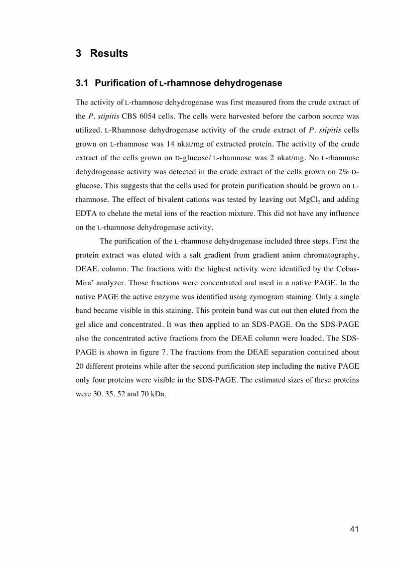
Figure 7 Picture of the Coomassie Blue stained SDS-PAGE.
Figure 8 The nucleotide sequence of the P. stipitis L-rhamnose dehydrogenase and
the corresponding protein sequence.
Figure 9 L-Rhamnose dehydrogenase activities with different sugar substrates
calculated with a Michaelis-Menten kinetic model.
Figure 10 The structure of L-rhamnose, L-lyxose and L-mannose.
Figure 11 Eadie-Hofstee plots of L-rhamnose (A), L-lyxose (B) and L-mannose (C).
Figure 12 pH dependency of the L-rhamnose dehydrogenase expressed in S.
cerevisiae.
Figure 13 The effect of the NAD concentration to the L-rhamnose dehydrogenase
activity calculated with a Michaelis-Menten kinetic model.
Figure 14 RNA gel stained with SYBR Green II RNA gel stain.
Figure 15 Northern blot analysis of RHA1 expression.
7
List of tables
Table 1 Plasmids used in this work.
Table 2 Primers used for PCR and sequencing reactions.
8
List of abbreviations
DDIW Two times distilled ion changed water
DNA Deoxyribonucleic acid
EDTA Ethylenediamine tetraacetic acid
KM The substrate concentration (mol/l) at which
an enzyme reaction proceeds at half its
maximal rate.
LB Luria-Bertani
MALDI-TOF MS Matrix Assisted Laser Desorption Ionisation
Time-of-flight Mass Spectrometry
NAD(H) Nicotinamide adenine dinucleotide
NADP(H) Nicotinamide adenine dinucleotide phosphate
PAGE Polyacrylamide gel electrophoresis
PGK1 promoter/terminator Promoter/terminator of the 3-
phosphoglycerate kinase of S. cerevisiae
RHA1 Gene encoding L-rhamnose dehydrogenase
RNA Ribonucleic acid
SC Synthetic complete
SCD Synthetic complete medium with D-glucose
SDS Sodium dodecyl sulphate
SDS-PAGE Sodium dodecyl sulphate polyacrylamide gel
electrophoresis
SOC recovery broth used after electroporation
TKT promoter Promoter of the transketolase gene of P.
stipitis
URA3 Gene encoding the orotidine-5’-phosphate
decarboxylase of S. cerevisiae
Vmax The substrate concentration where constant
rate of product formation is achieved.
YNB Yeast nitrogen base
YPD Yeast extract peptone with D-glucose
YPX Yeast extract peptone with D-xylose
9
X-gal 5-bromo-4-chloro-3-indolyl- - D-
galactorytanoside
XYL promoter/terminator Promoter/terminator of the xylose reductase
gene of P. stipitis
10
1 Introduction
Yeasts have served mankind for several thousands of years. Bread baking and produc-
tion of alcoholic beverages have exploited the fermentation process by yeasts already a
long time before “yeast” was found. The shared path of human and yeast has been food
related for most of the history. Only for a couple of decades ago it was understood that
yeast are providing also other prospects. First yeast was introduced as an experimental
system for molecular biology. Increasing attention has been received as the develop-
ment of yeast molecular biology has offered new possibilities. Besides being a model
organisms for eukaryotic mode of life, yeast are of commercial interest because of the
possibilities they offer to biotechnology, agriculture and medicine. Now the public
interest is towards biofuels and the enhanced ethanol production.
Yeasts are eukaryotic microbes, a group of fungi. Fungi are divided into differ-
ent phylas, the Ascomycota and Basidiomycota being sister groups which are classified
as a subkingdom of Dikarya (Hibbett et al. 2007). Members of both phylas can grow as
yeast or as hyphae, or both. Most of the yeast species are part of the Hemiascomycetes
of the Ascomycota phyla. The well known yeasts, such as Saccharomyces cerevisiae
and Candida albicans, are part of this group but not all Hemiascomycetes are exclu-
sively yeast and some of the species are even predominately filamentous. Typical to
Hemiascomycetes is that they lack fruiting bodies, structures for spore forming (Oliver
and Schweizer 1999).
1.1 Pichia stipitis
P. stipitis (Pignal 1967) is a haploid, homothallic hemiascomycetous yeast and one of
the 91 accepted species of the Pichia genus (Kurtzman, Fell 1998). P. stipitis was found
from the gut of a wood-inhabiting passalid beetle. P. stipitis has the highest known
native capacity to ferment xylose to ethanol. As xylose is one of the major components
of plant lignocellulose, there has been great intrest to study possibilities to use P. stipitis
for the production of biofuels. P. stipitis induces fermentative activity in response to
oxygen limitation unlike Saccharomyces cerevisiae, which regulates fermentation by
sensing the presence of fermentable sugars (Hertz-Fowler and Pain 2007, Jeffries et al.
2007).
11
The genome of P. stipitis CBS 6054 strain was published in March 2007 by
Jeffries et al. (2007). The size of the genome is 15.4 Mb and it encodes 5 851 genes.
There are eight chromosomes which vary in size from 3.5 to 0.97 Mbp. Comparison
with eight other yeast revealed 25 gene families representing 72 proteins that are spe-
cific to P. stipitis. The closest sequenced relative to P. stipitis is the yeast
Debaryomyces hansenii sharing 151 gene families that are not found in the other ge-
nomes. The origin in beetle gut, where the environment has a limited amount of oxygen
and the energy source is partially digested wood, explains the numerous genes related to
endoglucanase, -glucosidase, xylanase, mannanase and chitinase (Hertz-Fowler and
Pain 2007, Jeffries et al. 2007). P. stipitis has an alternative yeast codon usage where
CUG codes for leucine instead of serine (Laplaza et al. 2006).
1.2 Production of biofuels by yeast
Biomass covers already about 10% of the world’s primary energy demand (Antoni,
Zverlov and Schwarz 2007). Rising oil prices, depletion of fossil resources, and envi-
ronmental challenges are reasons why there has been a growing interest towards biofu-
els. For now bioethanol is the only microbially produced biofuel, which is produced on
an industrial scale. Also biodiesel is produced on industrial scale but microbial proc-
esses are presently not involved in its production although use of enzymes and biologi-
cal systems in transesterification is under development (Antoni, Zverlov and Schwarz
2007). S. cerevisiae is used to ferment sugar cane molasses or enzymatically hydrolysed
starch from grains to produce ethanol. Plant biomass is a renewable source of energy
and the future will hopefully see a technical process using lignocellulosic hydrolysates.
For being cost-effective the biofuel industry should be able to exploit feedstocks, such
as agricultural residues, wood, municipal solid waste and dedicated energy. These
biomasses are composed primarily of cellulose (40–50%), hemicellulose (25–35%) and
lignin (15–20%) unlike grain where the major carbohydrate is starch (Gray, Zhao and
Emptage 2006). Cellulose has a highly crystalline and compact structure where glucose
is linked via -1,4 glycosidic linkage. This makes cellulose very resistant to microbial
degradation. Hemicellulose consists of xylan backbone with various branches of sugars,
such as mannose, arabinose and galactose. Ferulic acid ester linkages can covalently
link lignin to hemicellulose (Gray, Zhao and Emptage 2006). The main concern for
using S. cerevisiae for lignocellulosic fermentation is its inability to ferment pentose
12
sugars, such as xylose and arabinose. Most S. cerevisiae strains do not utilize xylose
and metabolic engineering is needed to produce strains suitable for biofuel production
(Hahn-Hägerdal et al. 2007).
Complete substrate utilization is important in order to make lignocellulosic
ethanol processes economically competitive. New cellulases and hemicellulases are
being developed. All types of sugars in cellulose and hemicellulose should be converted
to ethanol. These sugars are a mixture of hexoses, primarily glucose, and pentoses,
mainly xylose. Xylose reductase (XYL1) and xylitol dehydrogenase (XYL2) genes of P.
stipitis have been introduced to recombinant S. cerevisiae strains (Jeffries 2006). Also a
xylose isomerase gene of the anaerobic fungus Piromyces sp. E2 has been expressed in
S. cerevisiae (Kuyper et al. 2003). Both recombinant strains are able to utilize xylose
but rates for xylose utilization have been very low.
The strains have to tolerate high sugar and ethanol concentrations in order to de-
crease the distillation costs. This leads to increased osmolality in the solutions, and thus
osmotolerant strains are needed. The fermenting organism must also tolerate inhibitors,
which are generated as side products in the hydrolysis processes. These include low
molecular weight organic acids, furans and aromatics. Ethanol yield and ethanol pro-
ductivity are, however, most important factors. Ethanolic fermentation of glucose and
starch already reach 90-95 % of the theoretical yield but the yields from other sugars,
such as xylose or other pentoses are lower (Hahn-Hägerdal et al. 2007).
The use of ethanol as a fuel is not a new development. Already in the 1860s an
ethanol using prototype of a spark ignition engine was designed and ethanol was used as
fuel for the T model Ford in the beginning of 1900s. Between 1925 and 1945 ethanol
was added to gasoline as an anti-knocking additive. Then ethanol production was abol-
ished due to the low price of gasoline until Brazil decided to start using ethanol again as
a fuel in the 1970s (Antoni, Zverlov and Schwarz 2007). However, ethanol is not an
ideal fuel because it has lower energy density than gasoline, and it is hygroscopic,
which causes problems for storage and distribution (Atsumi, Hanai and Liao 2008).
Higher alcohols (C4 or higher) would be better choices as their energy densities are
closer to gasoline and they are not hygroscopic. The problem is that no micro-organism
has been identified to produce industrially relevant quantities of higher alcohols. Atsumi
et al. suggests synthetic approach and metabolic engineering as a solution to produce
these higher alcohols. One solution is the utilization of 2-keto acid degradation of the
13
Ehrlich pathway where amino acids are catabolised to -keto acids and then decar-
boxylated to aldehydes (Hazelwood et al. 2008). 2-Keto-acid decarboxylases are im-
portant for this alcohol production method and they are common in yeasts, fungi and
plants but rare in prokaryotes (Atsumi, Hanai and Liao 2008). Alcohol dehydrogenases
can then be used to produce alcohols from aldehydes (Atsumi, Hanai and Liao 2008,
Hazelwood et al. 2008). The problem also in this case is that expression of non-native
pathways may cause metabolic imbalance and accumulation of heterologous metabo-
lites may cause cytotoxicity (Atsumi, Hanai and Liao 2008).
1.3 Oxidation of hexoses
1.3.1 Embden-Meyerhof-Parnas pathway
Embden-Meyerhof-Parnas pathway, also called glycolytic pathway, is the most com-
monly used oxidation route for hexoses. As a result of oxidation it forms two pyruvate,
two ATP and two NADH molecules from each hexose molecule. Glycolysis can be
considered as one of the classic central metabolism pathways together with pentose
phosphate pathway and citric acid cycle. It has been found that alternative pathways and
abbreviated forms can replace all these pathways.
1.3.2 Entner-Doudoroff pathway
Not all microbes have phosphofructokinase, which is a key enzyme of glycolysis. These
microbes use an alternative route for sugar degradation, which is called Entner-
Doudoroff pathway. In addition to Embden-Meyerhof-Parnas and pentose phosphate
pathways it can be considered as one of three pathways found in nature that provide
pyruvate. It is found to be common especially in gram-negative bacteria but it is present
in all three phylogenic domains: Bacteria, Archaea and Eukaryota (Peekhaus, Conway
1998). Sometimes different pathways are used for different sugars: Halococcus
metabolizes glucose via a modified Entner-Doudoroff pathway but fructose is degraded
via the Embden-Meyerhof-Parnas pathway. In fact, all possible combinations of classic,
alternative and abbreviated metabolic pathways are found together in micro-organisms
(Romano and Conway 1996).
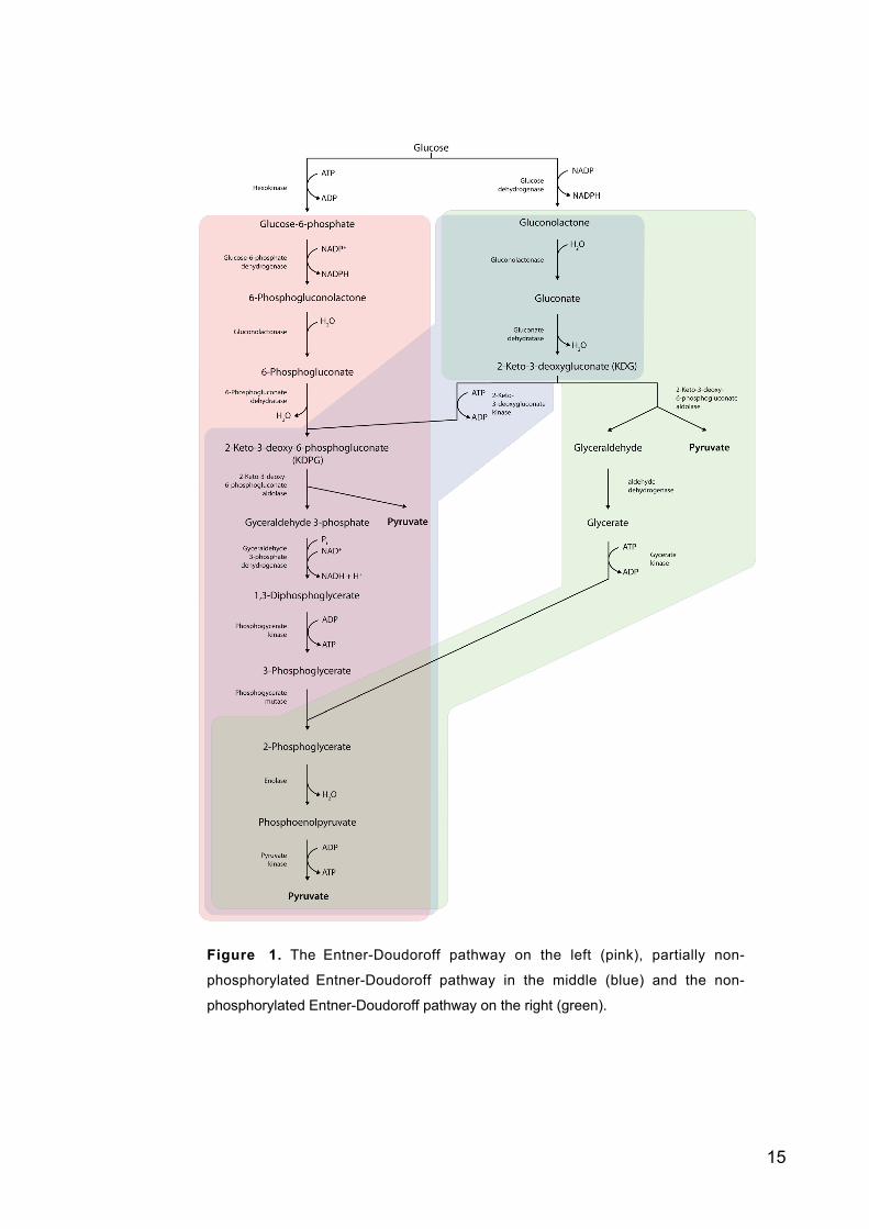
The first step of the Entner-Doudoroff pathway is oxidation of glucose-6-phos-
phate to 6-phosphogluconate as in the pentose phosphate pathway (figure 1). The
Entner-Doudoroff pathway has only two reactions different from the glycolytic and
14
pentose phosphate pathway. 6-phosphogluconate dehydratase forms 2-keto-3-deoxy-6-
phosphogluconate (D-erythro-3-deoxy-hexulosonate) which is then cleaved by an aldo-
lase to form pyruvate and glyceraldehyde-3-phosphate. From this on the pathway is
similar to the glycolytic pathway. The main differences between these pathways are that
in the Entner-Doudoroff pathway one NADPH is formed and the net gain is only one
ATP unlike in glycolysis where two ATP and two NADH are formed.
1.3.3 Non-phosphorylated Entner-Doudoroff pathway
There are two known modifications of the Entner-Doudoroff pathway (figure 1). The
partially or semi non-phosphorylated pathway is found in halophilic archaebacteria and
a few eubacteria. Here glucose is oxidized to gluconate and then phosphorylated to 2-
keto-3-deoxy-6-phosphogluconate. From this step on the pathway continues as the
normal Entner-Doudoroff pathway. The other modification for Entner-Doudoroff is the
non-phosphorylated Entner-Doudoroff pathway where none of the intermediates are
phosphorylated. This pathway is used by archaebacterial genera Thermoplasma,
Thermoproteus and Sulfolobus. The first steps are as in the partially non-phosphorylated
Entner-Doudoroff pathway described above. Then 2-keto-3-deoxygluconate is cleaved
to glyceraldehyde and puryvate. Glyceraldehyde is oxidised further to form glycerate
and then phosphorylated to form 2-phosphoglycerate. From this point on the pathway
continues as the normal Entner-Doudoroff pathway. Enolase and pyruvate kinase are
metabolising 2-phosphoglycerate to pyruvate. There is no net synthesis of ATP from the
metabolism of the hexose to pyruvate by this pathway. (Lengeler, Drews and Schlegel
1999)
15
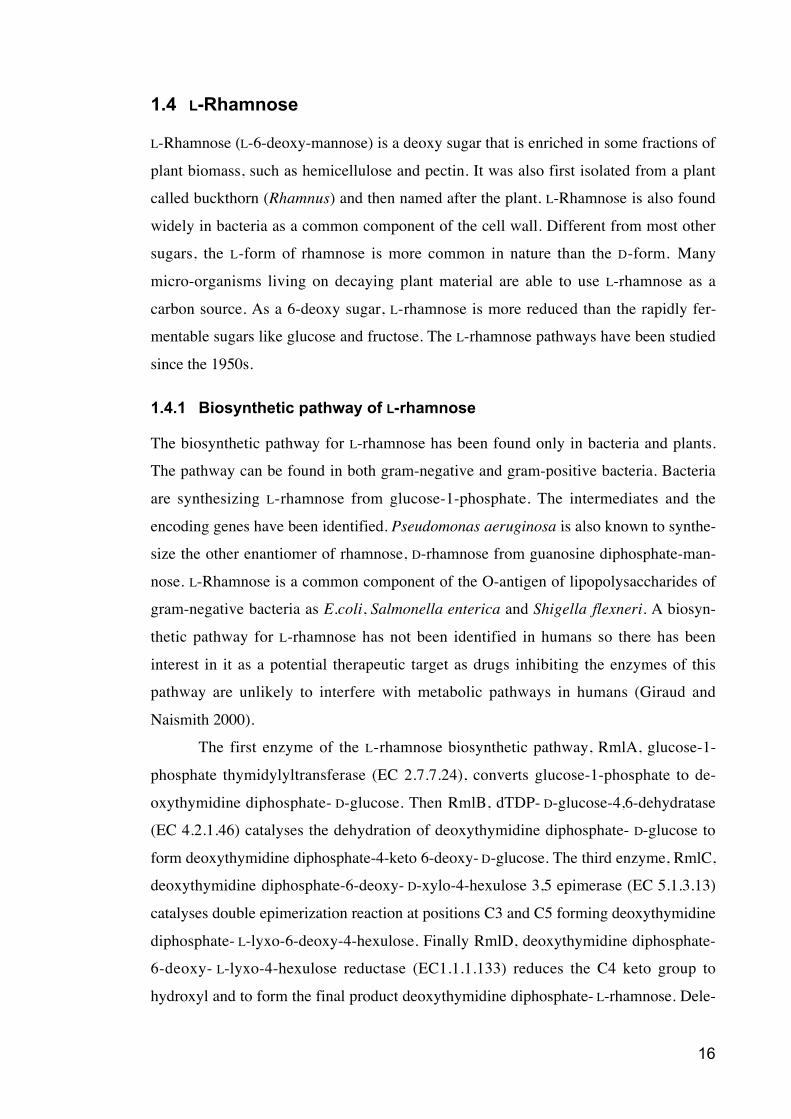
Figure 1. The Entner-Doudoroff pathway on the left (pink), partially non-
phosphorylated Entner-Doudoroff pathway in the middle (blue) and the non-
phosphorylated Entner-Doudoroff pathway on the right (green).
16
1.4 L-Rhamnose
L-Rhamnose (L-6-deoxy-mannose) is a deoxy sugar that is enriched in some fractions of
plant biomass, such as hemicellulose and pectin. It was also first isolated from a plant
called buckthorn (Rhamnus) and then named after the plant. L-Rhamnose is also found
widely in bacteria as a common component of the cell wall. Different from most other
sugars, the L-form of rhamnose is more common in nature than the D-form. Many
micro-organisms living on decaying plant material are able to use L-rhamnose as a
carbon source. As a 6-deoxy sugar, L-rhamnose is more reduced than the rapidly fer-
mentable sugars like glucose and fructose. The L-rhamnose pathways have been studied
since the 1950s.
1.4.1 Biosynthetic pathway of L-rhamnose
The biosynthetic pathway for L-rhamnose has been found only in bacteria and plants.
The pathway can be found in both gram-negative and gram-positive bacteria. Bacteria
are synthesizing L-rhamnose from glucose-1-phosphate. The intermediates and the
encoding genes have been identified. Pseudomonas aeruginosa is also known to synthe-
size the other enantiomer of rhamnose, D-rhamnose from guanosine diphosphate-man-
nose. L-Rhamnose is a common component of the O-antigen of lipopolysaccharides of
gram-negative bacteria as E.coli, Salmonella enterica and Shigella flexneri. A biosyn-
thetic pathway for L-rhamnose has not been identified in humans so there has been
interest in it as a potential therapeutic target as drugs inhibiting the enzymes of this
pathway are unlikely to interfere with metabolic pathways in humans (Giraud and
Naismith 2000).
The first enzyme of the L-rhamnose biosynthetic pathway, RmlA, glucose-1-
phosphate thymidylyltransferase (EC 2.7.7.24), converts glucose-1-phosphate to de-
oxythymidine diphosphate- D-glucose. Then RmlB, dTDP- D-glucose-4,6-dehydratase
(EC 4.2.1.46) catalyses the dehydration of deoxythymidine diphosphate- D-glucose to
form deoxythymidine diphosphate-4-keto 6-deoxy- D-glucose. The third enzyme, RmlC,
deoxythymidine diphosphate-6-deoxy- D-xylo-4-hexulose 3,5 epimerase (EC 5.1.3.13)
catalyses double epimerization reaction at positions C3 and C5 forming deoxythymidine
diphosphate- L-lyxo-6-deoxy-4-hexulose. Finally RmlD, deoxythymidine diphosphate-
6-deoxy- L-lyxo-4-hexulose reductase (EC1.1.1.133) reduces the C4 keto group to
hydroxyl and to form the final product deoxythymidine diphosphate- L-rhamnose. Dele-
17
tion of one or several of Rml genes results for example severe colonization defects for
Vibrio cholerae, inhibits cell-wall polysaccharide synthesis of Streptococcus mutans
and leads E. coli to a loss of serum resistance (Giraud and Naismith 2000).
1.4.2 L-Rhamnose catabolism
There are at least two different pathways for L-rhamnose catabolism. The one described
in bacteria, for example in Escherichia coli, involves the following intermediates: L-
rhamnulose, L-rhamnulose 1-phosphate, dihydroxyacetone phosphate and L-lactalde-
hyde. The corresponding enzymes for this pathway involving the phosphorylated inter-
mediates include L-rhamnose isomerase ( EC 5.3.1.14) (Takagi and Sawada 1964a,
Wilson and Ajl 1957a), rhamnulokinase (EC 2.7.1.5) (Takagi and Sawada 1964b,
Wilson and Ajl 1957b) and rhamnulose-1-phosphate aldolase (EC 4.1.2.19) (Sawada
and Takagi 1964). Depending on the redox conditions, L-lactaldehyde can then be re-
duced to 1,2-propanediol or oxidized to lactate by lactaldehyde reductase (EC 1.1.1.77)
or lactaldehyde dehydrogenase (EC 1.2.1.22). Gene sequences for all these enzymes
have been described (Moralejo et al. 1993).
The pathway without phosphorylated intermediates is distinctly different and it
has been described in three different yeast species. First it was found by Rigo et al. in
the yeast like fungus Aureobasidium pullulans (Rigo et al. 1985). In 1984 the pathway
involving oxidative intermediates was described also in P. stipitis and Debaryomyces
polymorphus by Twerdochlib et al. (1994). The intermediates in this pathway are L-
rhamno-1,4-lactone, L-rhamnoate, L-erythro-3,6-dideoxyhexulosonate, pyruvate and L-
lactaldehyde. The corresponding enzymes are NAD-utilizing L-rhamnose-1-dehydro-
genase (EC 1.1.1.173), L-rhamno-1,4-lactonase (EC 3.1.1.65), L-rhamnoate dehydratase
(EC 4.2.1.90), and L-erythro-3,6-dideoxyhexulosonate aldolase (EC 4.1.2.-). Finally the
L-lactaldehyde is oxidized to L-lactate in an NAD-coupled reaction, as in the pathway
with the phosphorylated intermediates. The pathway is shown in the figure 2. This
pathway has some similarities to the non-phosphorylated Entner-Doudoroff pathway.
Common is that the L-erythro-3,6-dideoxy hexulosonic acid, which is similar to the 2-
keto-3-deoxygluconate of the Entner-Doudoroff pathway, is split by an aldolase to
pyruvate and an aldehyde.
It is known that S. cerevisiae does not utilize rhamnose. Van Maris et al. (2006)
also reported that S. cerevisiae genome does not have genes with a clear homology to
genes encoding rhamnose-catabolizing enzymes. As only the genes of the prokaryotic
18
L-rhamnose route have been known, this would not necessarily mean that there are no
homologies to the eukaryotic route. Also the L-rhamnose uptake rate through the plasma
membrane of S. cerevisiae cells is extremely low. The successful introduction of L-
rhamnose pathway to S. cerevisiae would require the introduction of the transporter and
the enzymes. Engineered strains that can efficiently convert all potentially fermentable
substrates of plant biomass hydrolysates are important for enhanced fuel ethanol
production.
Van Maris et al. (2006) points out that the introduction of the four enzymes and
transporter of the fungal L-rhamnose pathway should enable the conversion of L-rham-
nose to equimolar amounts of ethanol, lactaldehyde and CO2. No ATP would be formed
in this pathway. The prokaryotic pathway with the phosphorylated intermediates results
in the net generation of two ATP molecules per equimolar amounts of ethanol, L-lactal-
dehyde and CO2. This enables the option to select for growth on L-rhamnose but the
resulting biomass formation would go at the expense of the ethanol yield. One of the
end products of both pathways is L-lactaldehyde, which could be converted to 1,2-pro-
panediol. S. cerevisiae already has an enzyme for this reduction, L-lactaldehyde reduc-
tase but also one extra NADH per L-lactaldehyde would be required. As a sink for
excess reduction equivalents, during fermenting, anaerobic growth, S. cerevisiae nor-
mally uses glycerol formation for this at the cost of one ATP. This reduces the ethanol
yield. Production of 1,2-propanediol might function as an alternative path for excess
redox sink and it would actually increase the ethanol yield. Van Maris et al. (2006)
came up with the conclusion that bacterial pathway together with a fungal transporter
for engineering S. cerevisiae would be preferable because this would enable the L-
rhamnose selection for growth during the strain construction and long-term industrial
cultivation. It is still debatable whether the fungal pathway enabling greater ethanol
yield would be better for the end result.
19
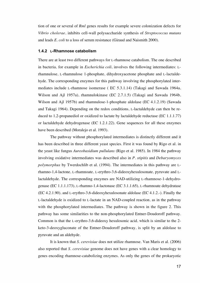
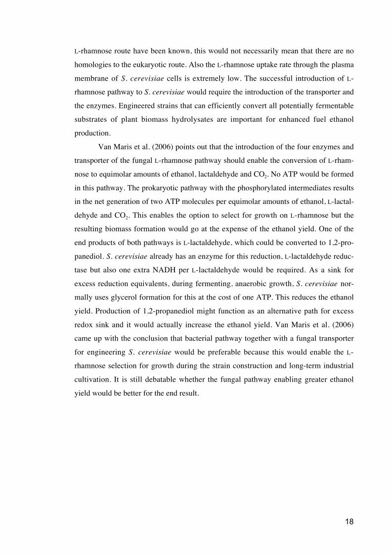
Figure 2. Fungal pathway for L-rhamnose catabolism. The intermediates and en-
zymes but not the corresponding gene sequences of this pathway have been de-
scribed previously.
1.5 Catabolic routes similar to the L-rhamnose route
1.5.1 Fungal L-fucose catabolism
L-Fucose is a 6-deoxyhexose like L-rhamnose. The L-fucose pathway (figure 3) in eu-
karyotic microbes is very similar to the corresponding L-rhamnose pathway. For the
yeast like fungus Aureobasidium pullulans (synonym to Pullula pullulans) this pathway
has been described by Guimarães and Veiga (1990). The intermediates in this pathway
are L-fucono-1,4-lactone, L-fuconate, 2-keto-3-deoxy- L-fuconate, pyruvate and L-lac-
taldehyde. The first step of the pathway is catalyzed by an NAD-utilizing L-fucose-
dehydrogenase. The second step is spontaneous hydrolysis due to the instability of the
L-fuconate-1,4-lactone. Then the L-fucose dehydratase dehydrates L-fuconate to 2-keto-
3-deoxy- L-fuconate which is cleaved to pyruvate and L-lactaldehyde. The enzyme
activities have been described but the corresponding protein and gene sequences are not
known.
20
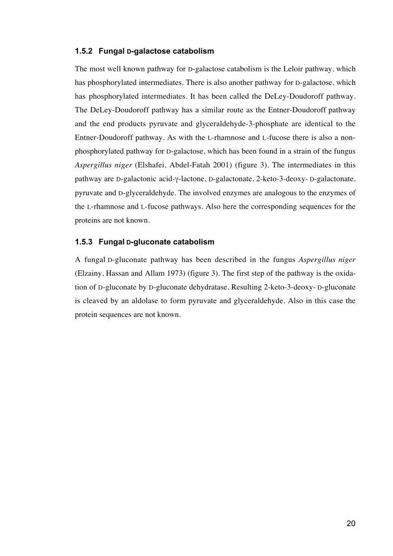
1.5.2 Fungal D-galactose catabolism
The most well known pathway for D-galactose catabolism is the Leloir pathway, which
has phosphorylated intermediates. There is also another pathway for D-galactose, which
has phosphorylated intermediates. It has been called the DeLey-Doudoroff pathway.
The DeLey-Doudoroff pathway has a similar route as the Entner-Doudoroff pathway
and the end products pyruvate and glyceraldehyde-3-phosphate are identical to the
Entner-Doudoroff pathway. As with the L-rhamnose and L-fucose there is also a non-
phosphorylated pathway for D-galactose, which has been found in a strain of the fungus
Aspergillus niger (Elshafei, Abdel-Fatah 2001) (figure 3). The intermediates in this
pathway are D-galactonic acid- -lactone, D-galactonate, 2-keto-3-deoxy- D-galactonate,
pyruvate and D-glyceraldehyde. The involved enzymes are analogous to the enzymes of
the L-rhamnose and L-fucose pathways. Also here the corresponding sequences for the
proteins are not known.
1.5.3 Fungal D-gluconate catabolism
A fungal D-gluconate pathway has been described in the fungus Aspergillus niger
(Elzainy, Hassan and Allam 1973) (figure 3). The first step of the pathway is the oxida-
tion of D-gluconate by D-gluconate dehydratase. Resulting 2-keto-3-deoxy- D-gluconate
is cleaved by an aldolase to form pyruvate and glyceraldehyde. Also in this case the
protein sequences are not known.
21
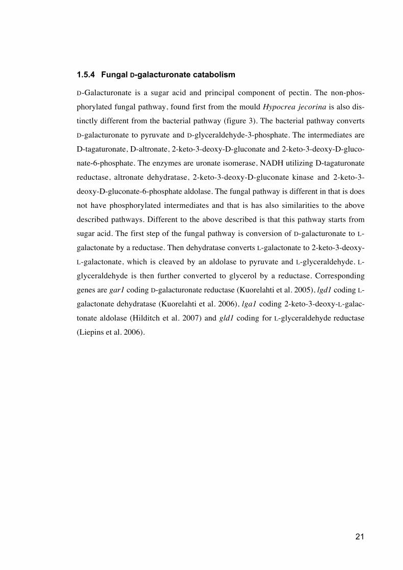
1.5.4 Fungal D-galacturonate catabolism
D-Galacturonate is a sugar acid and principal component of pectin. The non-phos-
phorylated fungal pathway, found first from the mould Hypocrea jecorina is also dis-
tinctly different from the bacterial pathway (figure 3). The bacterial pathway converts
D-galacturonate to pyruvate and D-glyceraldehyde-3-phosphate. The intermediates are
D-tagaturonate, D-altronate, 2-keto-3-deoxy-D-gluconate and 2-keto-3-deoxy-D-gluco-
nate-6-phosphate. The enzymes are uronate isomerase, NADH utilizing D-tagaturonate
reductase, altronate dehydratase, 2-keto-3-deoxy-D-gluconate kinase and 2-keto-3-
deoxy-D-gluconate-6-phosphate aldolase. The fungal pathway is different in that is does
not have phosphorylated intermediates and that is has also similarities to the above
described pathways. Different to the above described is that this pathway starts from
sugar acid. The first step of the fungal pathway is conversion of D-galacturonate to L-
galactonate by a reductase. Then dehydratase converts L-galactonate to 2-keto-3-deoxy-
L-galactonate, which is cleaved by an aldolase to pyruvate and L-glyceraldehyde. L-
glyceraldehyde is then further converted to glycerol by a reductase. Corresponding
genes are gar1 coding D-galacturonate reductase (Kuorelahti et al. 2005), lgd1 coding L-
galactonate dehydratase (Kuorelahti et al. 2006), lga1 coding 2-keto-3-deoxy-L-galac-
tonate aldolase (Hilditch et al. 2007) and gld1 coding for L-glyceraldehyde reductase
(Liepins et al. 2006).
22
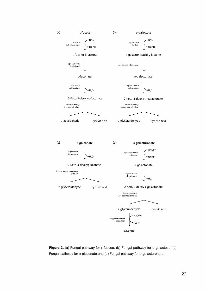
Figure 3. (a) Fungal pathway for L-fucose, (b) Fungal pathway for D-galactose, (c)
Fungal pathway for D-gluconate and (d) Fungal pathway for D-galacturonate.
23
1.6 Aim of the work
The aim of this work was to characterize some of the genes coding the catabolic path-
way of L-rhamnose in yeast. As pointed out above, the intermediates and the enzymes
of the route are known but the genes of the non-phosphorylated pathway have still not
been identified. It is important to know the corresponding gene sequences in order to
exploit L-rhamnose pathway in the engineering of S. cerevisiae or some other microbes,
which are not naturally able to utilize L-rhamnose. Thus the goal was to purify the en-
zymes in order to identify the corresponding amino acid sequences with MALDI-TOF
MS. Then the gene is to be cloned and expressed in S. cerevisiae for the kinetic studies.
A deletion of the gene is a good method to prove that there are no alternative routes for
L-rhamnose catabolism and Northern analysis would give some information about the
induction of genes.
24
2 Materials and Methods
2.1 Yeast and bacterial strains and plasmids
The wild type P. stipitis yeast strain CBS 6054 was used for all enzyme assays. Uracil
auxotroph P. stipitis strain UC7 (H2818) was used for the knock-outs. For transforming
P. stipitis pJM6 (Yang et al. 1994) based pJML223 and pJML225 plasmids were used.
The pJML223 plasmid (p2852) has a TKT1 promoter and a XYL1 terminator. The
pJML225 (p2853) plasmid has a XYL1 promoter and terminator. XYL1 is known to be
glucose repressed and TKT1 is elevated when grown on xylose medium (Jeffries et al.
2007). Xylose was used as a carbon source when these plasmids were used.
The S. cerevisiae strain H1346 used in this work is a modification of CEN.PK2
(H1346) strain (VW-1b (MATa) leu2-3/112, ura3-52, trp1-289, his3- 1, MAL2-8C,
SUC2) (Stansfield, Stark 2007). This strain was transformed with a multicopy yeast
expression vector YEplac195 (p1181) (Verho et al. 2002) containing URA3 for selec-
tion, where the PGK1 promoter and terminator were introduced. The transformed open
reading frames were released as a BamHI fragment and ligated to the BglII site of the
B1181.
For bacterial transformations E. coli strain DH5 (F-, endA1, hsdR17, recA1,
gyrA96, relA1, 80d lacZM15), XL1-Blue Supercompetent (Stratagene, USA) and
TOP10 Electrocomp (Invitrogen, USA) strains were used.
All the plasmids used in the experiments are listed in the table 1 with a short
description. Later in the text plasmids are referred to by their p-numbers.
Table 1. Plasmids used in this work. p-number, a short description and a reference.
p-number Description Reference
p1181 YEplac195 with PGK1 promoter and terminator,
2μ, URA3, Amp; 7101bp
VTT lab collection
p2852 JML223, TKT1 promoter and XYL1 terminator VTT lab collection
p2853 JML225, XYL1 promoter and terminator VTT lab collection
p3023 Nat1 wo CTG from GeneArt This work
p3025 RDH1 of P. stipitis cloned into pCR2.1-TOPO
vector
This work
25
p3026 Nat1 containing BglII fragment of B3023 ligated to
BamHI sites of B2852
This work
p3027 Nat1 containing BglII fragment of B3023 ligated to
BamHI sites of B2853
This work
p3069 BamHI cut histidine tagged (C-terminal) RDH1
fragment ligated to BglII cut B1181
This work
p3070 BamHI cut histidine tagged (N-terminal) RDH1
fragment ligated to BglII cut B1181
This work
p3090 RDH1 without CTG codon cloned into pCR2.1-
TOPO vector
This work
p3132 RDH1 without CTG codon fragment ligated to
BglII cut B1181
This work
2.2 Media and growth conditions
The nutritious growth medium (YPD) used for P. stipitis consisted of 1% Bacto-yeast
extract (BD, USA), 2% Bacto-peptone (BD, USA) and 2 % D-glucose (Sigma-Aldrich,
Germany). YPX used for P. stipitis clones transformed with pJML plasmids was similar
to YPD except D-xylose (Sigma-Aldrich, Germany) was used as a carbon source instead
of D-glucose. Solid media contained also 2% Bacto-agar (BD, USA). For the knockout
transformants 150 μg/ml nourseotrichine (Werner Bio-Agents, Germany) was added.
The synthetic complete (SC) media used for S. cerevisiae contained 6.7 g/l YNB
without amino acids (BD, USA) and amino acid/nucleotide stock to get following con-
centrations; 0.1 mM L-adenine; 2 mM L-arginine; 2 mM L-aspartic acid; 0.4 mM L-
histidine; 0.2 mM myo-inositol; 4 mM L-isoleucine; 2 mM L-leucine; 0.6 mM L-lysine;
1 mM L-methionine; 0.5 mM L-phenylalanine; 1 mM L-serine; 1 mM L-threonine; 0.4
mM L-tryptophan; 0.2 mM L-tyrosine; 0.2 mM uracil and 1 mM L-valine. Transformats
having URA3 marker for uracil selection were grown on media lacking uracil. SCD-
medium was prepared from SC-medium by adding 2 % D-glucose as carbon source.
SOC medium containing 2 % Bacto-tryptone (BD, USA), 0,5 % Bacto-yeast ex-
tract (BD, USA), 10 mM NaCl, 2 mM KCl, 10 mM MgCl2, 10 MgSO4 and 0,4 % D-
glucose (Sigma-Aldrich, Germany) was used as a recovery broth for E. coli cells after
electoporation. The E. coli cultivations were grown in LB-broth, which consisted of 1 %
Bacto-tryptone, 0.5 % Bacto-yeast extract (BD, USA) and 0.5 % NaCl. For the plasmid
26
selection 100 μg/ml ampicillin (Sigma-Aldrich, Germany) was added. Bacteria were
grown in glass test tubes or in 50 ml erlenmeyer flasks at the temperature of +37 °C and
shaken by 250 rpm.
2.3 Recombinant DNA techniques
The restriction enzymes BamHI, BglII, HindIII, Spe I , DpnI, XhoI, EcoRI (New
England BioLabs, USA), the T4 DNA-ligase (Promega, USA), and the Calf intestinal
alkaline phosphatase (Finnzymes, Finland) were used as described in the manuals of the
manufacturers. The concentrations used in the PCR-reactions were the ones recom-
mended by the manufacturers. The polymerases used in the PCR-reactions were
DyNAzyme EXT (Finnzymes, Finland), which is an optimised mixture of DyNAzyme
II DNA Polymerase and a proofreading enzyme, DyNAzyme II (Finnzymes, Finland)
and Phusion™ high fidelity polymerase (Finnzymes, Finland) was used for the PCR
reactions needed for the deletion cassette. The DNA fragments digested with the
restriction enzymes were separated by electrophoresis in 1% agarose gel (SeaKem LE
agarose by BMA, USA), and when necessary, DNA fragments were separated from the
gel by using QIAquick Gel Extraction Kit (QIAGEN GmbH, Germany) or MinElute
Gel Extraction Kit (QIAGEN GmbH, Germany) when the elution volumes were small.
GeneRulerTM 1 kb DNA Ladder (Fermentas, USA) was used as a standard size marker in
the agarose gels. TD loading buffer [20 % Ficoll 400 (Amersham Biosciences, USA),
0.0125 % bromophenol blue (Merck, Germany), 0,1 % xylene cyanol ff (Chroma-
Gesellschaft, Germany), 250 mM Tris pH 8,0 (MP Biomedicals, USA), 25 mM boric
acid (Sigma-Aldrich, Germany), 125 mM EDTA, pH 7,5 (Sigma-Aldrich, Germany),
0,5 % SDS (Sigma-Aldrich, Germany)] was used at 1/5 of the volume to all the DNA
samples ran in agarose gel.
All E. coli plasmid purifications were done by using QIAprep Spin Miniprep Kit
protocol (Qiagen GmbH, Germany). QIAprep Miniprep has been developed for purifi-
cation of plasmid DNA. Method is based on alkaline lysis of bacterial cells followed by
the absorbtion of the DNA onto silica-gel membrane in the high salt concentration
(Birnboim and Doly 1979, Vogelstein and Gillespie 1979).
All primers used in this work were manufactured by Sigma-Genosys Ltd. (UK)
(Table 2).
27
Table 2. Primers used for PCR and sequencing reactions: number, name and sequence.
Restriction sites for BamHI are underlined.
Number Name Sequence (5´to 3´) Used for
O7892 PsRDHfwdCTG CTGAAAGAGGAAATTCTCGGAC
GGTGAAAACAACGTGCTG
CTG codon mutagenesis
for P. stipitis RHA1 gene
O7893 PsRDHrevCTG GAGCAGGTTGTTTTCACCGTCC
GAGATTTCCTCTTTGAG
CTG codon mutagenesis
for P. stipitis RHA1 gene
O7871 PsRDHfwdBamHI GGATCCATCATGACTGGATTGT
TGAATGG
RHA1 amplifying from the
genomic DNA of P. stipitis
O7872 PsRDHrevBamHI GGATCCCTATTGTAAATTGACG
AACAATCCTC
RHA1 amplifying from the
genomic DNA of P. stipitis
O7885 NAT1fwd GATCTAAGCTTATGACCACTC For sequencing
O7886 XYL1rev CTCTATAAAGCAACCTTCCTG For sequencing
O7902 RDHdel5fwd GTAACGCCAGGGTTTTCCCAGT
CACGACGACTAGTGGTAGATTG
CTACATGTCTC
For constructing the
deletion cassette
O7903 RDHdel5rev TAGCATACAAAACTACTTCTGC
ATCATATCGATACTGCCAAAAT
TGGGGC
For constructing the
deletion cassette
O7904 RDHdel3fwdXYL1 TAATGTAGAACCAATTAGTGTC
TGTGGATCCTTGTATCTTCGTCC
ATACC
For constructing the
deletion cassette
O7905 RDHdel3fwdTKT1 AGATAGTTGGTTGAGTAGCATG
AACTCTGGTCTTGTATCTTCGTC
CATACC
For constructing the
deletion cassette
O7906 RDHdel3rev GCGGATAACAATTTCACACAGG
AAACAGCACTAGTCGGATTTTC
CCCCATCTCAC
For constructing the
deletion cassette
O7907 hisRDHfwdBamHI GGATCCATCATGCATCACCATC
ACCATCACGGTGGAATGACTGG
ATTGTTGAATGG
Adding the histidine tag to
the C-terminal end of the
RHA1 gene
O7996 hisRDHrevBamHI GGATCCCTAGTGATGGTGATGG
TGATGACCTCCTTGTAAATTGA
CGAACAATCCTC
Adding the histidine tag to
the N-terminal end of the
RHA1 gene
O8046 delxylprom GGAGGAACGCAGACAGAAAC For constructing the
deletion cassette
O8047 deltktprom CAACTCACGTGCATACCAATC For constructing the
deletion cassette
28
O8048 delxylterm CACCTCATCAATTCATTGC For constructing the
deletion cassette
O8049 del5middle CAGTCGCCATTGATCAACTC For sequencing
O8050 del3middle CACCAAGTGAAACTCGCTTG For sequencing
O8081 delRDHflanktark5 GAAAGCATCGTAAGTCCGTAG For sequencing
O8082 delRDHflanktark3 CAGCTGCAGTACGATAAGAG For sequencing
O8083 revNat1 ACCCATCGAGTGCCTCGATG For sequencing
M13 Universal
primer forwad
GACCGGCAGCAAAATG For sequencing
M13 Universal
primer reverse
CAGGAAACAGCTATGAC For sequencing
2.4 Transformations
2.4.1 E. coli transformations
E. coli transformations were done by electroporation. 40 μl of competent DH5 E. coli
cells, stored at –70 °C, were thawed on ice and 2 μl of plasmid DNA was added. For
electroporation the GenePulser TM (BioRad, USA) was used with following settings: 25
μF, 200 and 2.50 kV. The mixture was transferred to an ice-cold 0.2 mm
electroporation cuvette (Bio Rad, USA). Immediately after the pulse 1 ml SOC medium
was added. The suspension was incubated for one hour at +37 °C and then plated on
LB-plates with ampicillin (Sigma-Aldrich, Germany).
For transformations done according to Topo TA Cloning® kit (Invitrogen, USA)
the plates were also supplemented with 40 mg/ml X-gal (5-bromo-4-chloro-3-indolyl- -
D-galactorytanoside). X-gal is used for blue/white screening distinguishing recombinant
colonies (white) among non-recombinant ones (blue). The LacZ gene of TOPO vector
encodes -galactosidase, which hydrolyzes X-gal and forms an intense blue precipitate
and blue colonies. The fragment cloned to the TOPO cloning site interrupts the LacZ
and colonies remain white.
29
2.4.2 S. cerevisiae transformations
S. cerevisiae yeast transformations were done by using the Gietz Lab Transformation
Kit (Molecular Research Reagents Inc.) according to manufacturer’s instructions. The
transformants were plated on SCD plates lacking uracil and incubated at +30 °C for two
to three days.
2.4.3 P. stipitis transformations
For P. stipitis yeast transformation numerous different methods were tried. For the
deletion cassette transformation the method described by Dohmen et al. (1991) was
used with the improvements presented by Scruff (2005). PEG 1000 polyethylene glycol
was substituted by PEG 4000 (Sigma-Aldrich, Germany) polyethylene glycol.
2.5 Sequencing
All DNA sequencings were done according to the instructions given by Adam et al.
(2007) for the Big Dye® sequencing kit (Applied Biosystems, USA) (Platt, Woodhall
and George 2007). Capillary electrophoresis was used to analyse the reactions by ABI
Prism® 3100 Genetic Analyser (PE/Applied Biosystems, Perkin Elmer, USA).
The resulted P. stipitis DNA sequences were compared to sequences from the P.
stipitis v2.0 database (DOE Joint Genome Institute) using sequence analysis software
DNAMAN 4.1 (Lynnon BioSoft, Canada).
2.6 Enzyme purification
P. stipitis CBS 6054 was grown in 500 ml shake flasks containing YNB without amino
acids (BD, USA) and 2 % L-rhamnose (Sigma-Aldrich, Germany) as a carbon source.
Also controls where 2 % D-glucose or 1% L-rhamnose and 1% D-glucose were used as
carbon sources were grown in the same way. Flasks were shaken in +28 °C for two
days. After that yeast cells were collected by centrifugation (Hettich AHT 35R) by 3000
g for 10 minutes. Cells were washed and then resuspended into 40 ml of 5 mM sodium
phosphate pH 7.0 supplemented with Complete EDTA free protease inhibitor (Roche,
Switzerland) according to manufacturer’s instructions. The cells were kept on ice. Equal
amount of 0.4 mm of diameter glass beads (Sigma-Aldrich, Germany), fresh cell cake
and resuspension buffer were added. The cells were broken by shaking them in the
30
Mini-Beadbeater (Biospec Products) for two times one minute. The mixture was centri-
fuged in an Eppendorf microcentrifuge at full speed for 20 minutes at +4 °C. The super-
natants were collected and desalted in PD-10 Sephandex G-25M gel filtration desalting
columns (Amersham Biosciences, USA) according to manufacturer’s instructions.
2.6.1 Anion exchange and gradient
Anion exchange chromatography was used for protein purification: Fractogel TSK
DEAE–650 (M) ionexchange column material (Merck, Germany) was pipetted into the
column so that the volume of the DEAE gel was 10 ml. The column was first washed
with 75 ml of 200 mM NaCl, 5 mM sodium phosphate buffer and then with 100 ml of 5
mM sodium phosphate buffer. A peristaltic pump (P-1, Pharmacia) was adjusted to
pump 2,5 ml / 2 minutes and a fraction collector (Frac-100, Pharmacia) was used to
collect the eluent. The desalted supernatant was loaded to the column and fraction
collecting started immediately. Right after the desalted supernatant had gone through
the column it was washed with 150 ml 5 mM sodium phosphate buffer, pH 7.0. A
gradient was made by adding 100 ml of 5mM sodium phosphate buffer pH 7.0 into the
first container and 100 ml 5 mM sodium phosphate, 200 mM NaCl to the second con-
tainer. The two containers were connected so that they always had an equal volume. To
obtain a linear gradient the solution was pumped from the first container which was also
mixed with a magnetic stirrer. Fraction collection was done at +4 °C. Finally the
column was eluted with 1 M NaCl to get all the rest of the proteins. All fractions were
analyzed for the L-rhamnose dehydrogenase activity. The fractions where the enzymatic
activity was observed were combined and concentrated using Vivaspin 2 10,000
MWCO PES centrifugation columns (Vivascience Sartorius group, Germany).
2.6.2 Zymogram staining
Zymography is an electrophoretic method used to identify enzymatic activity of
enzymes separated in polyacrylamide gels. The concentrated protein samples were ran
in the non-denaturing PAGE gel (Bio-Rad 12% Tris-HCl acrylamide gel, Bio-Rad,
USA) using a buffer containing 25 mM Tris base (Sigma-Aldrich, Germany) and 192
mM glycine. As a loading buffer for the non-denaturing PAGE a mix of 0.4 M Tris-HCl
pH 6.8, 30% glycerol and 0,003% bromophenol blue was added at 1/5 of the volume to
the sample. Electrophoresis was done on ice to avoid the system to heat and the proteins
to denature. The gel was then stained in a zymogram staining solution containing 0.25
31
mM nitroblue tetrazolium (Promega, USA), 0.06 mM phenazine methosulfate (Sigma-
Aldrich, Germany), 0,5 mM NAD (Sigma-Aldrich, Germany), 200 mM Tris-HCl (pH
8.0), 100 mM L-rhamnose (Sigma-Aldrich, Germany). The only band that appeared
during the overnight incubation was cut out and the protein was eluted in 300 μl of
buffer [200 mM Tris-HCl (pH 8.0), 0.1 % SDS (Sigma-Aldrich, Germany)] during
overnight incubation. The eluted protein was concentrated using Microcon YM-10
(Millipore, USA) column according to manufacturer’s instructions. Also the saved
DEAE sample not used in the zymogram staining was concentrated in the same way.
LSB+ -Me [0.1 M Tris-HCl pH 6.8, 20% glycerol, 4% SDS (Sigma-Aldrich,
Germany), 0.02% bromphenol blue (Merck, Germany) and 10% -mercaptoethanol
(Sigma-Aldrich, Germany)] loading buffer was added at 1/4 of the volume to the
sample. To denature the proteins both samples were heated for 5 minutes at 95 °C
before they were pipetted to the SDS-PAGE gel. SDS-PAGE gel was ran using similar
buffer as with the non-denaturing PAGE except also 0.1 % SDS was added.
2.6.3 SDS PAGE
SDS-PAGE, sodium dodecyl sulfate polyacrylamide gel electrophoresis, is a technique
to separate proteins according to their size (Laemmli 1970). SDS PAGE was used to
analyze the concentrated protein samples. Commercial Bio-Rad poly-acrylamidegel
(Bio-Rad, ReadyGel, 12% Tris-HCl, Bio-Rad, USA) was used. Bio-Rad Prestained
SDS-PAGE standard, Low Range (Bio-Rad, USA), was used as a standard size marker.
Tris-glycine-SDS buffer [25 mM Tris, 192 mM glycine and 1% SDS (Sigma-Aldrich,
Germany)] was used as a running buffer. After electrophoresis the gel was stained with
Coomassie Brilliant Blue R-250 stain solution [500 mg Coomassie Brilliant Blue R-250
Serva Blau R (Serva, Germany), 150 ml ethanol, 50 ml acetic acid, 300 ml H2O] for one
hour to make the protein bands visible. The gel was then washed with destain solution I
(40% ethanol, 10 % acetic acid) for two times 15 minutes and with destain solution II (5
% ethanol, 7.5 % acetic acid). The gel was left into the destain solution II for one day
and then moved into storage solution containing 4 % glycerol.
2.7 In-gel digestion MALDI-TOF MS
The four proteins observed in the SDS-PAGE gel were cut out and in-gel digested with
trypsin and the peptides were extracted essentially according to the method of
32
Rosenfeld et al. (1992). The samples were desalted using a C-18 matrix (Eppendorf
Perfect Pure C-18 Tip). The saturated matrix solution was prepared by dissolving re-
crystallized -cyano-4-hydroxycinnamic acid (CCA, Bruker Daltonics, Germany) in a
50 % acetonitrile in 0.1 % trifluoroacetic acid solution. Equal volumes of purified pep-
tide sample or calibration standard (peptide calibration mixture II, Bruker Daltonics,
Germany) were mixed with the saturated matrix solution. 1 μl of this matrix/sample
mixture was applied onto the target (Target plate ground steel T F, Bruker Daltonics)
and let to dry at room temperature. The peptide masses were then determined by
MALDI-TOF MS using a Bruker Autoflex II mass spectrometer. FlexAnalysis software
(Bruker Daltronics) was used for the data analysis.
2.8 Site directed mutagenesis kit for changing the CTG codon
Before the RHAI was transformed to S. cerevisiae H1346 the nucleotides 166-168 of the
open reading frame were changed from CTG to TCG with the QuikChange site di-
rected mutagenesis kit (Stratagene, USA) because of the different codon usage of P.
stipitis and S. cerevisiae. The primers O7892 and O7893 were used for the mutagenesis.
The RHAI gene was amplified from genomic DNA of P. stipitis CBS 6054 using the
primers O7871 and O7872. The genomic DNA had been isolated prior to this work at
the VTT. The fragment was then cloned into the pCR®2.1-TOPO® vector (Invitrogen,
USA). Instructions given by the manufacturer were used for the thermal cycling. The
mutagenised plasmids were digested with DpnI restriction enzyme (New England
BioLabs, USA) for one hour at +37 °C. XL1-Blue Supercompetent cells (Stratagene,
USA) were thawed on ice and aliquoted to 50 μl portions which were used for trans-
formation. 1 μl of DpnI-treated DNA from each sample reaction were mixed with the
cells and then incubated for 30 minutes on ice. Transformation mixtures were heat
pulsed at +42 °C water bath for 45 seconds and then placed on ice for two minutes.
Preheated SOC broth was pipetted on the transformation reactions and they were incu-
bated at +37 °C for one hour prior to the plating on LB media with ampicillin (Sigma-
Aldrich, Germany). M13 forward and reverse primers were used for checking the se-
quences and the resulting plasmid was numbered as p3090.
33
2.9 Cloning of the RHAI
The RHAI gene without the CTG codon was amplified by PCR from p3090 using the
primers O7871 and O7872. BamHI sites had been added to the primers. The gene was
also cloned with primers having C- and N-terminal histidine tags. Six histidines were
added at the beginning or at the end of the protein. To introduce the histidine-tag at the
N-terminus a coding sequence for MHHHHHHGG was introduced before the original
start codon by using primer O7996. To introduce the histidine-tag at the C-terminus the
coding sequence for GGHHHHHH was introduced before the stop codon by using
primer O7907. The open reading frame was amplified using the following PCR pro-
gram: an initial denaturation 3 min at 94 °C, a denaturation 30 s at 94 °C, an annealing
30 s at 55 °C and an extension 1 min at 72 °C. These three subsequential steps were
carried out 25 times until the final extension 7 min at 72°C. The PCR product was
applied to gel electrophoresis and the fragment was extracted from the agarose gel by
using QIAquick Gel Extraction Kit (Qiagen, USA). The fragment was then cloned into
a pCR®2.1-TOPO® vector (Invitrogen, USA) using TOPO TA Cloning kit (Invitrogen,
USA) and transformed to the DH5 E. coli strain. BamHI digested open reading frame
was then added to the BglII sites of the p1181 yeast expression vector of S. cerevisiae
which was then transformed into S. cerevisiae strain (H1346). The picture of p1181
with the ligated RHA1 gene is shown in figure 4.
34
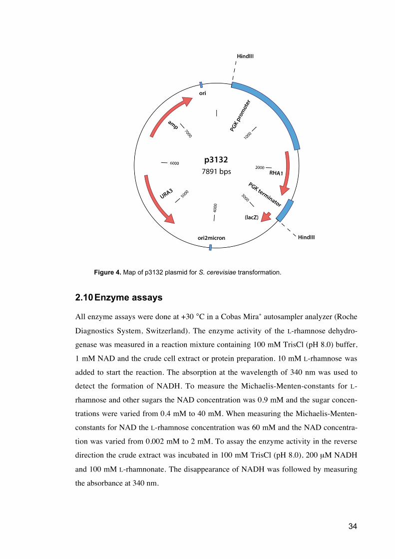
Figure 4. Map of p3132 plasmid for S. cerevisiae transformation.
2.10 Enzyme assays
All enzyme assays were done at +30 °C in a Cobas Mira+ autosampler analyzer (Roche
Diagnostics System, Switzerland). The enzyme activity of the L-rhamnose dehydro-
genase was measured in a reaction mixture containing 100 mM TrisCl (pH 8.0) buffer,
1 mM NAD and the crude cell extract or protein preparation. 10 mM L-rhamnose was
added to start the reaction. The absorption at the wavelength of 340 nm was used to
detect the formation of NADH. To measure the Michaelis-Menten-constants for L-
rhamnose and other sugars the NAD concentration was 0.9 mM and the sugar concen-
trations were varied from 0.4 mM to 40 mM. When measuring the Michaelis-Menten-
constants for NAD the L-rhamnose concentration was 60 mM and the NAD concentra-
tion was varied from 0.002 mM to 2 mM. To assay the enzyme activity in the reverse
direction the crude extract was incubated in 100 mM TrisCl (pH 8.0), 200 μM NADH
and 100 mM L-rhamnonate. The disappearance of NADH was followed by measuring
the absorbance at 340 nm.
35
The enzyme activity of L-lactaldehyde dehydrogenase was assayed by using the
same measuring conditions. The reaction mixture contained 100 mM TrisCl (pH 6.8-
9.5) buffer, 1 mM NAD (Sigma-Aldrich, Germany), 5mM MgCl2 and the crude cell
extract or protein preparation. 60 mM DL-lactaldehyde was added to start the reaction.
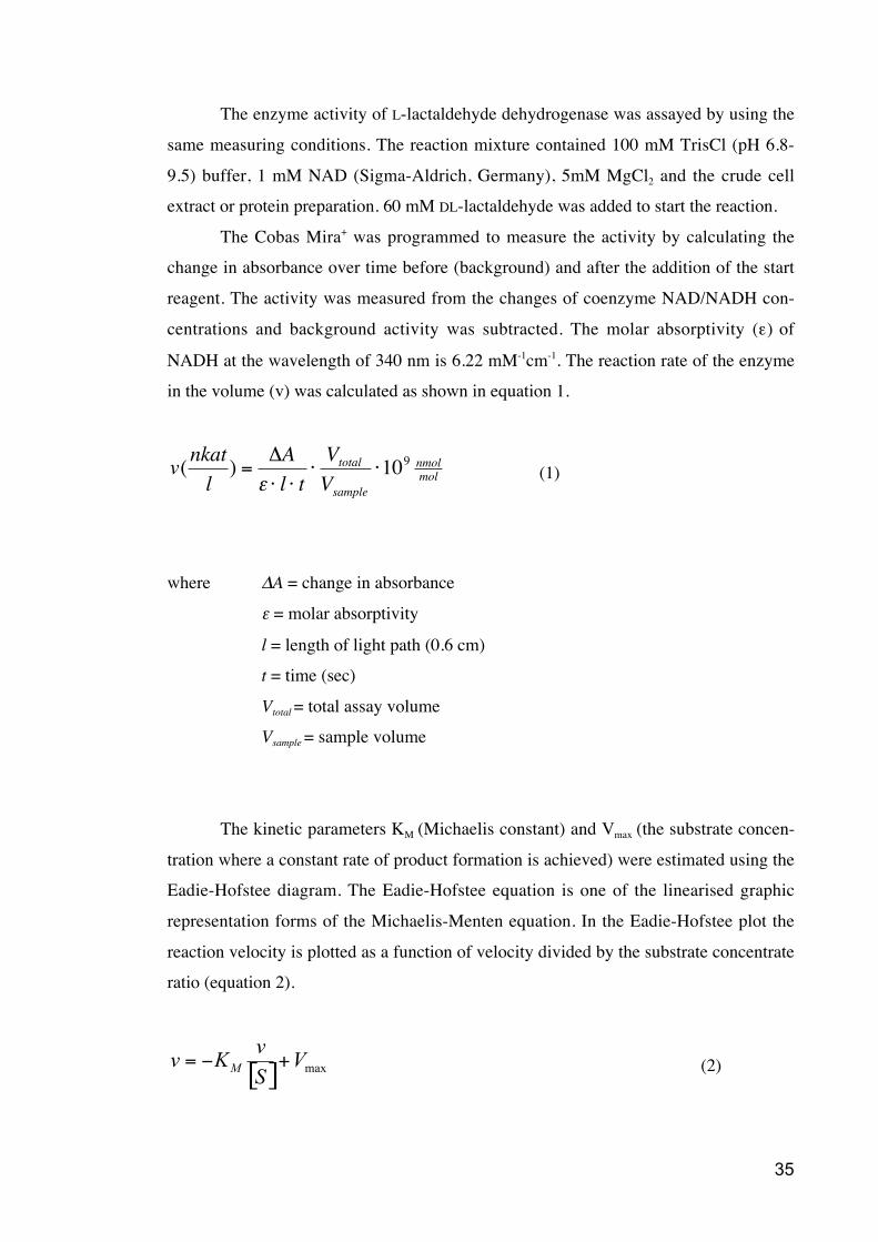
The Cobas Mira+ was programmed to measure the activity by calculating the
change in absorbance over time before (background) and after the addition of the start
reagent. The activity was measured from the changes of coenzyme NAD/NADH con-
centrations and background activity was subtracted. The molar absorptivity ( ) of
NADH at the wavelength of 340 nm is 6.22 mM-1cm-1. The reaction rate of the enzyme
in the volume (v) was calculated as shown in equation 1.
v(nkat
l) =
A
l t
Vtotal
Vsample
109 nmolmol (1)
where A = change in absorbance
= molar absorptivity
l = length of light path (0.6 cm)
t = time (sec)
Vtotal = total assay volume
Vsample = sample volume
The kinetic parameters KM (Michaelis constant) and Vmax (the substrate concen-
tration where a constant rate of product formation is achieved) were estimated using the
Eadie-Hofstee diagram. The Eadie-Hofstee equation is one of the linearised graphic
representation forms of the Michaelis-Menten equation. In the Eadie-Hofstee plot the
reaction velocity is plotted as a function of velocity divided by the substrate concentrate
ratio (equation 2).
[ ] maxV
S
vKvM
+= (2)
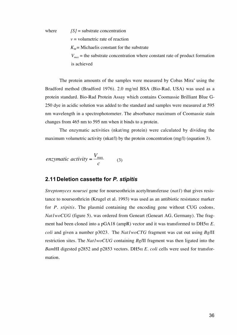
36
where [S] = substrate concentration
v = volumetric rate of reaction
KM = Michaelis constant for the substrate
Vmax = the substrate concentration where constant rate of product formation
is achieved
The protein amounts of the samples were measured by Cobas Mira+ using the
Bradford method (Bradford 1976). 2.0 mg/ml BSA (Bio-Rad, USA) was used as a
protein standard. Bio-Rad Protein Assay which contains Coomassie Brilliant Blue G-
250 dye in acidic solution was added to the standard and samples were measured at 595
nm wavelength in a spectrophotometer. The absorbance maximum of Coomassie stain
changes from 465 nm to 595 nm when it binds to a protein.
The enzymatic activities (nkat/mg protein) were calculated by dividing the
maximum volumetric activity (nkat/l) by the protein concentration (mg/l) (equation 3).
enzymatic activity =Vmaxc
(3)
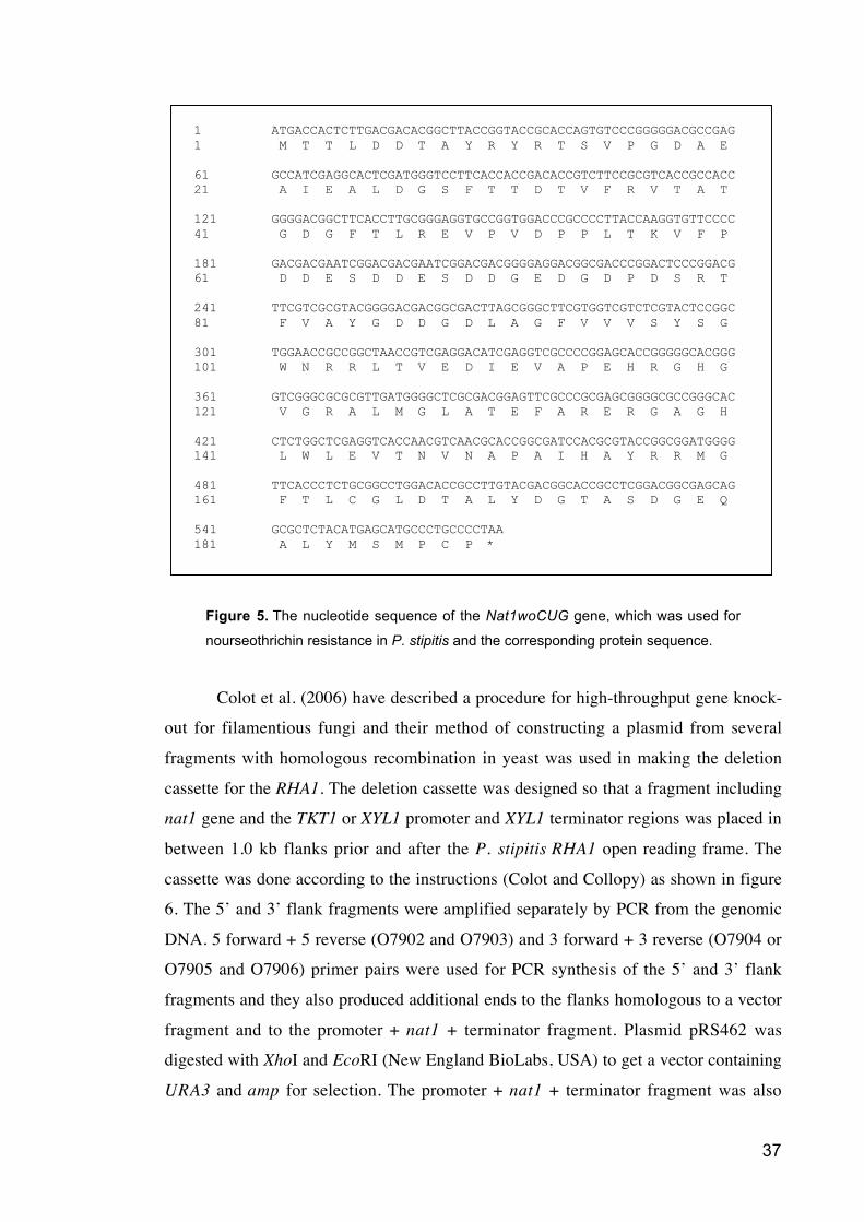
2.11 Deletion cassette for P. stipitis
Streptomyces noursei gene for nourseothricin acetyltransferase (nat1) that gives resis-
tance to nourseothricin (Krugel et al. 1993) was used as an antibiotic resistance marker
for P. stipitis. The plasmid containing the encoding gene without CUG codons,
Nat1woCUG (figure 5), was ordered from Geneart (Geneart AG, Germany). The frag-
ment had been cloned into a pGA18 (ampR) vector and it was transformed to DH5 E.
coli and given a number p3023. The Nat1woCTG fragment was cut out using BglII
restriction sites. The Nat1woCUG containing BglII fragment was then ligated into the
BamHI digested p2852 and p2853 vectors. DH5 E. coli cells were used for transfor-
mation.
37
1 ATGACCACTCTTGACGACACGGCTTACCGGTACCGCACCAGTGTCCCGGGGGACGCCGAG
1 M T T L D D T A Y R Y R T S V P G D A E
61 GCCATCGAGGCACTCGATGGGTCCTTCACCACCGACACCGTCTTCCGCGTCACCGCCACC
21 A I E A L D G S F T T D T V F R V T A T
121 GGGGACGGCTTCACCTTGCGGGAGGTGCCGGTGGACCCGCCCCTTACCAAGGTGTTCCCC
41 G D G F T L R E V P V D P P L T K V F P
181 GACGACGAATCGGACGACGAATCGGACGACGGGGAGGACGGCGACCCGGACTCCCGGACG
61 D D E S D D E S D D G E D G D P D S R T
241 TTCGTCGCGTACGGGGACGACGGCGACTTAGCGGGCTTCGTGGTCGTCTCGTACTCCGGC
81 F V A Y G D D G D L A G F V V V S Y S G
301 TGGAACCGCCGGCTAACCGTCGAGGACATCGAGGTCGCCCCGGAGCACCGGGGGCACGGG
101 W N R R L T V E D I E V A P E H R G H G
361 GTCGGGCGCGCGTTGATGGGGCTCGCGACGGAGTTCGCCCGCGAGCGGGGCGCCGGGCAC
121 V G R A L M G L A T E F A R E R G A G H
421 CTCTGGCTCGAGGTCACCAACGTCAACGCACCGGCGATCCACGCGTACCGGCGGATGGGG
141 L W L E V T N V N A P A I H A Y R R M G
481 TTCACCCTCTGCGGCCTGGACACCGCCTTGTACGACGGCACCGCCTCGGACGGCGAGCAG
161 F T L C G L D T A L Y D G T A S D G E Q
541 GCGCTCTACATGAGCATGCCCTGCCCCTAA
181 A L Y M S M P C P *
Figure 5. The nucleotide sequence of the Nat1woCUG gene, which was used for
nourseothrichin resistance in P. stipitis and the corresponding protein sequence.
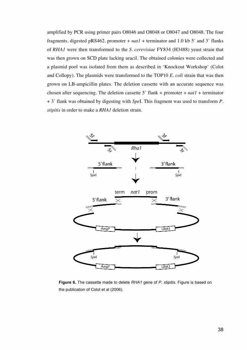
Colot et al. (2006) have described a procedure for high-throughput gene knock-
out for filamentious fungi and their method of constructing a plasmid from several
fragments with homologous recombination in yeast was used in making the deletion
cassette for the RHA1. The deletion cassette was designed so that a fragment including
nat1 gene and the TKT1 or XYL1 promoter and XYL1 terminator regions was placed in
between 1.0 kb flanks prior and after the P. stipitis RHA1 open reading frame. The
cassette was done according to the instructions (Colot and Collopy) as shown in figure
6. The 5’ and 3’ flank fragments were amplified separately by PCR from the genomic
DNA. 5 forward + 5 reverse (O7902 and O7903) and 3 forward + 3 reverse (O7904 or
O7905 and O7906) primer pairs were used for PCR synthesis of the 5’ and 3’ flank
fragments and they also produced additional ends to the flanks homologous to a vector
fragment and to the promoter + nat1 + terminator fragment. Plasmid pRS462 was
digested with XhoI and EcoRI (New England BioLabs, USA) to get a vector containing
URA3 and amp for selection. The promoter + nat1 + terminator fragment was also
38
amplified by PCR using primer pairs O8046 and O8048 or O8047 and O8048. The four
fragments, digested pRS462, promoter + nat1 + terminator and 1.0 kb 5’ and 3’ flanks
of RHA1 were then transformed to the S. cerevisiae FY834 (H3488) yeast strain that
was then grown on SCD plate lacking uracil. The obtained colonies were collected and
a plasmid pool was isolated from them as described in ‘Knockout Workshop’ (Colot
and Collopy). The plasmids were transformed to the TOP10 E. coli strain that was then
grown on LB-ampicillin plates. The deletion cassette with an accurate sequence was
chosen after sequencing. The deletion cassette 5’ flank + promoter + nat1 + terminator
+ 3’ flank was obtained by digesting with SpeI. This fragment was used to transform P.
stipitis in order to make a RHA1 deletion strain.
Figure 6. The cassette made to delete RHA1 gene of P. stipitis. Figure is based on
the publication of Colot et al (2006).
39
P. stipitis transformants were plated on YPX medium with 150 μg/ml
nourseothricin and incubated at + 30 °C for five days. Colonies were then grown in
shake flasks containing YNB without amino acids (BD, USA), amino acid/nucleotide
stock and 2 % L-rhamnose as a carbon source. Each colony was also analysed by yeast
colony PCR to find out if the deletion fragment was in the right place. Primers pairs
O7885 + O8049 and O8083 + O8050 were used. Following PCR program was used: an
initial denaturation 3 min at 94 °C, a denaturation 30 s at 94 °C, an annealing 30 s at 50
°C and an extension 2 min at 72 °C. These three sequential steps were carried out 25
times until the final extension 10 min at 72°C. The PCR product was applied to gel
electrophoresis.
2.12 Synthesis of L-rhamnoate and L-lactaldehyde
L-Rhamnoate was synthesised by Andreas Petrich (VTT) from L-rhamnose by oxidation
with bromine and purified by ion-exchange chromatography as described by Yew et al.
(2006). DL-Lactaldehyde was synthesised by Harri Setälä (VTT) using the method first
described by Durrwachter et al. (1986) and later modified by Schoevaart (2000). The
method is based on the reduction of methylglyoxal 1,1-dimethylacetal. L-Lactaldehyde
was synthesised using the method described by Huff and Rudney (1959). It was synthe-
sised by heating ninhydrin with D-threonine. After the addition of NaHCO3 the brown
precipitate was removed by filtration. Active carbon Norit A (Norit, USA) was added to
remove ninhydrin and the colored products of the reaction. Finally sodium ions and
unreacted threonine were removed by Dowex 50 (H+) (Sigma-Aldrich, Germany).
2.13 Northern analysis
Northern analysis was done using the technique described first by Alwine et al.
(Alwine, Kemp and Stark 1977). P. stipitis CBS 6054 was grown in YNB medium
supplemented with 20 g/l of six different carbon sources: L-rhamnose, D-glucose,
maltose, D-galactose, D-xylose and an ethanol/glycerol mixture. The RNA was extracted
from the yeast cells with the Trizol reagent kit (Life Technologies Inc., USA) according
to the manufacturer’s instructions. 5 μg of the total RNA per sample was used in the
analysis and the amount was checked by staining with the SYBR Green II RNA gel
stain (BMA Biomedicals, Switzerland). 3.7 μl of RNA solution containing 5 μg of
RNA was added to 2,7 μ l 6 M glyoxal, 8.0 μl dimethylsulfoxide and 1.6 μl 1,2-
40
dimyristoyl-sn-glycero-3-phosphocholine (DMPC) -NaH2PO4 (pH 7.0). Mixtures were
incubated at +50 °C for one hour and after cooling to +20 °C samples were loaded to
the RNA agarose gel (10 mM DMPC- NaH2PO4 (pH 7.0), DMPC-DDIW to get total
volume of 350 ml). All equipment and solutions used for RNA work were di-methyl-
propyl carbonate (DMPC) (Sigma-Aldrich, Germany) -treated to inactive RNAses.
The RNA gel was blotted over night to the nitrocellulose filter using capillary
method and 20x saline-sodium citrate (SSC) solution (860 g NaCl, 440 g trisodium-
citratedihydrate, H2O ad 5000 ml, pH 7.0) as a transfer buffer. After that the RNA was
UV cross-linked to the Hybond N membrane (Amersham Biosciences, USA).
10 mg/ml herring sperm DNA was added to the prehybridization mix (500 ml
deionised formamide, 100 g dextran sulphate, 1 M NaCl, 1 % SDS (Sigma-Aldrich,
Germany), H2O ad 1000 ml) and mixture was boiled in a water bath for 10 minutes. Pre-
hybridization was done in + 42 °C for one hour. As a probe for the hybridization the
open reading frame, released as a BamHI from the TOPO vector, was used. The probe
was labelled with [ -32P]dCTP (Amersham Biosciences, USA) using the randomly
primed DNA labelling kit (Roche, Switzerland). For probe purification a Sephadex G-
50 (Amersham Biosciences, USA) gel filtration resin was used. Hybridization incuba-
tion was done over night at + 42 °C. After hybridization the filter was washed with 5x,
1x and 0.1x SSPE solutions (20 x SSPE: 870 g NaCl, 138 g NaH2PO4 • H2O, 37 g
EDTA, H2O ad 5000 ml). Autoradiography was performed with Typhoon Scanner
(Amersham Biosciences, USA) and image processed by Image Quant software
(Amersham Biosciences, USA).
41
3 Results
3.1 Purification of L-rhamnose dehydrogenase
The activity of L-rhamnose dehydrogenase was first measured from the crude extract of
the P. stipitis CBS 6054 cells. The cells were harvested before the carbon source was
utilized. L-Rhamnose dehydrogenase activity of the crude extract of P. stipitis cells
grown on L-rhamnose was 14 nkat/mg of extracted protein. The activity of the crude
extract of the cells grown on D-glucose/ L-rhamnose was 2 nkat/mg. No L-rhamnose
dehydrogenase activity was detected in the crude extract of the cells grown on 2% D-
glucose. This suggests that the cells used for protein purification should be grown on L-
rhamnose. The effect of bivalent cations was tested by leaving out MgCl2 and adding
EDTA to chelate the metal ions of the reaction mixture. This did not have any influence
on the L-rhamnose dehydrogenase activity.
The purification of the L-rhamnose dehydrogenase included three steps. First the
protein extract was eluted with a salt gradient from gradient anion chromatography,
DEAE, column. The fractions with the highest activity were identified by the Cobas-
Mira+ analyzer. Those fractions were concentrated and used in a native PAGE. In the
native PAGE the active enzyme was identified using zymogram staining. Only a single
band became visible in this staining. This protein band was cut out then eluted from the
gel slice and concentrated. It was then applied to an SDS-PAGE. On the SDS-PAGE
also the concentrated active fractions from the DEAE column were loaded. The SDS-
PAGE is shown in figure 7. The fractions from the DEAE separation contained about
20 different proteins while after the second purification step including the native PAGE
only four proteins were visible in the SDS-PAGE. The estimated sizes of these proteins
were 30, 35, 52 and 70 kDa.
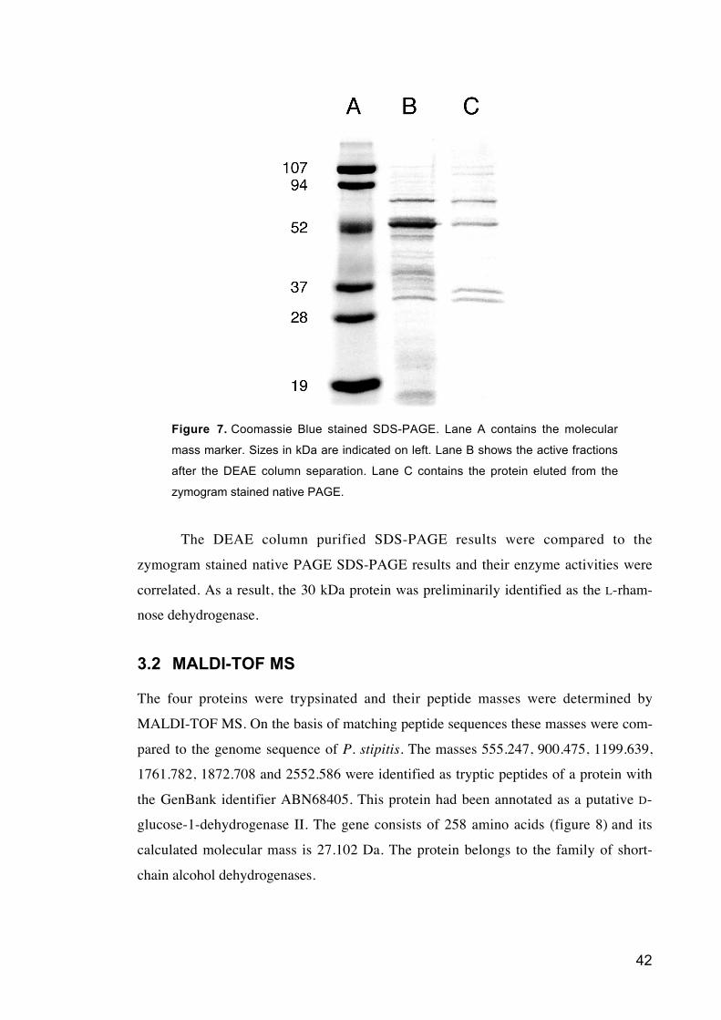
42
Figure 7. Coomassie Blue stained SDS-PAGE. Lane A contains the molecular
mass marker. Sizes in kDa are indicated on left. Lane B shows the active fractions
after the DEAE column separation. Lane C contains the protein eluted from the
zymogram stained native PAGE.
The DEAE column purified SDS-PAGE results were compared to the
zymogram stained native PAGE SDS-PAGE results and their enzyme activities were
correlated. As a result, the 30 kDa protein was preliminarily identified as the L-rham-
nose dehydrogenase.
3.2 MALDI-TOF MS
The four proteins were trypsinated and their peptide masses were determined by
MALDI-TOF MS. On the basis of matching peptide sequences these masses were com-
pared to the genome sequence of P. stipitis. The masses 555.247, 900.475, 1199.639,
1761.782, 1872.708 and 2552.586 were identified as tryptic peptides of a protein with
the GenBank identifier ABN68405. This protein had been annotated as a putative D-
glucose-1-dehydrogenase II. The gene consists of 258 amino acids (figure 8) and its
calculated molecular mass is 27.102 Da. The protein belongs to the family of short-
chain alcohol dehydrogenases.
43
1 ATGACTGGATTGTTGAATGGAAAGGTGGTTGCAATAACCGGAGGTGTCACTGGTATCGGA
1 M T G L L N G K V V A I T G G V T G I G
61 CGGGCAATTGCCATAGAAATGGCAAGGAATGGTGCCAAAGTAGTAGTGAACCATTTACCA
21 R A I A I E M A R N G A K V V V N H L P
121 TCTGAAGAGCAAGCTCAGTTGGCAAAAGAACTCAAAGAGGAAATCCTGGACGGTGAAAAC
41 S E E Q A Q L A K E L K E E I L D G E N
181 AACGTGCTCACCATTCCAGGGGACATCTCGCTCCCTGAAACAGGCAGAAGGATAGTGGAG
61 N V L T I P G D I S L P E T G R R I V E
241 CTTGCAGTTGAAAAGTTTGGCGAAATCAATGTGTTCGTTTCCAATGCTGGTGTCTGTGGG
81 L A V E K F G E I N V F V S N A G V C G
301 TTCAGAGAGTTTCTTGAAATAACTCCCGAAACTTTGTTTCAGACGGTGAATATTAACTTG
101 F R E F L E I T P E T L F Q T V N I N L
361 AATGGAGCCTTCTTCGCCATCCAAGCAGCTGCACAACAAATGGTCAAGCAGGGCAAAGGA
121 N G A F F A I Q A A A Q Q M V K Q G K G
421 GGTAGCATTATTGGAATCAGCAGCATCTCTGCCTTGGTTGGAGGAGCACACCAAACCCAT
141 G S I I G I S S I S A L V G G A H Q T H
481 TACACACCAACTAAAGCTGGAATCTTGTCCTTAATGCAATCTACAGCATGTGCTCTAGGT
161 Y T P T K A G I L S L M Q S T A C A L G
541 AAGTATGGAATCAGATGCAATGCGATCCTCCCAGGGACAATCAGTACAGCCTTGAACGAA
181 K Y G I R C N A I L P G T I S T A L N E
601 GAGGACTTGAAAGATCCAGAGAAGAGAAAGTACATGGAAGGGAGAATACCTCTAGGAAGA
201 E D L K D P E K R K Y M E G R I P L G R
661 GTCGGTGACCCCAAGGATATTGCTGGACCTGCTATCTTCTTGGCAAGTGATATGTCTAAC
221 V G D P K D I A G P A I F L A S D M S N
721 TACGTTAATGGAGCACAACTACTTGTTGATGGAGGATTGTTCGTCAATTTACAATAG
241 Y V N G A Q L L V D G G L F V N L Q *
Figure 8. The nucleotide secuence of the P. stipitis L-rhamnose dehydrogenase
and the corresponding protein sequence.
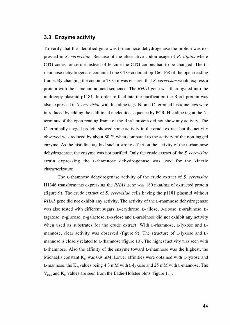
44
3.3 Enzyme activity
To verify that the identified gene was L-rhamnose dehydrogenase the protein was ex-
pressed in S. cerevisiae. Because of the alternative codon usage of P. stipitis where
CTG codes for serine instead of leucine the CTG codons had to be changed. The L-
rhamnose dehydrogenase contained one CTG codon at bp 166-168 of the open reading
frame. By changing the codon to TCG it was ensured that S. cerevisiae would express a
protein with the same amino acid sequence. The RHA1 gene was then ligated into the
multicopy plasmid p1181. In order to facilitate the purification the Rha1 protein was
also expressed in S. cerevisiae with histidine tags. N- and C-terminal histidine tags were
introduced by adding the additional nucleotide sequence by PCR. Histidine tag at the N-
terminus of the open reading frame of the Rha1 protein did not show any activity. The
C-terminally tagged protein showed some activity in the crude extract but the activity
observed was reduced by about 80 % when compared to the activity of the non-tagged
enzyme. As the histidine tag had such a strong effect on the activity of the L-rhamnose
dehydrogenase, the enzyme was not purified. Only the crude extract of the S. cerevisiae
strain expressing the L-rhamnose dehydrogenase was used for the kinetic
characterization.
The L-rhamnose dehydrogenase activity of the crude extract of S. cerevisiae
H1346 transformants expressing the RHA1 gene was 180 nkat/mg of extracted protein
(figure 9). The crude extract of S. cerevisiae cells having the p1181 plasmid without
RHA1 gene did not exhibit any activity. The activity of the L-rhamnose dehydrogenase
was also tested with different sugars. D-erythrose, D-allose, D-ribose, D-arabinose, D-
tagatose, D-glucose, D-galactose, D-xylose and L-arabinose did not exhibit any activity
when used as substrates for the crude extract. With L-rhamnose, L-lyxose and L-
mannose, clear activity was observed (figure 9). The structure of L-lyxose and L-
mannose is closely related to L-rhamnose (figure 10). The highest activity was seen with
L-rhamnose. Also the affinity of the enzyme toward L-rhamnose was the highest, the
Michaelis constant Km was 0.9 mM. Lower affinities were obtained with L-lyxose and
L-mannose, the Km values being 4.3 mM with L-lyxose and 25 mM with L-mannose. The
Vmax and Km values are seen from the Eadie-Hofstee plots (figure 11).
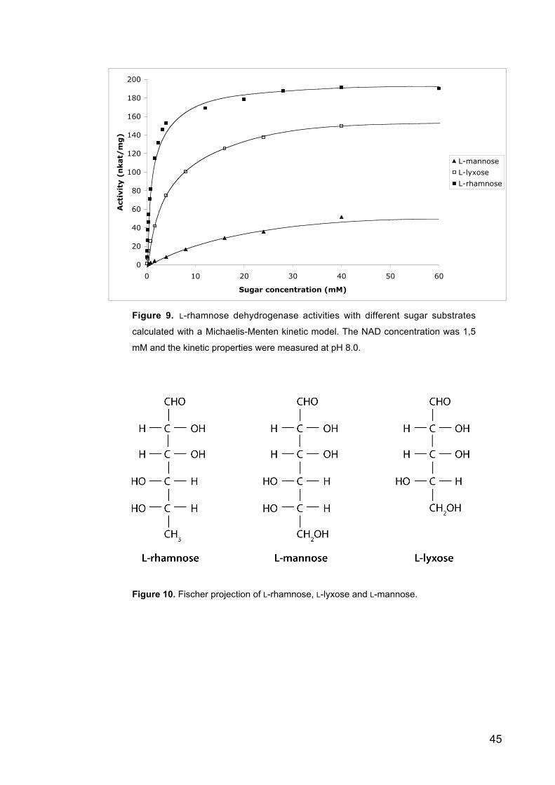
45
0
20
40
60
80
100
120
140
160
180
200
0 10 20 30 40 50 60
Sugar concentration (mM)
Acti
vit
y (
nkat/
mg
)
L-mannose
L-lyxose
L-rhamnose
Figure 9. L-rhamnose dehydrogenase activities with different sugar substrates
calculated with a Michaelis-Menten kinetic model. The NAD concentration was 1,5
mM and the kinetic properties were measured at pH 8.0.
Figure 10. Fischer projection of L-rhamnose, L-lyxose and L-mannose.
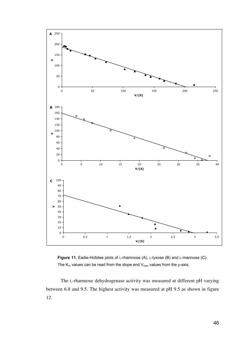
46
A
0
50
100
150
200
250
0 50 100 150 200 250
V/[S]
V
B
0
20
40
60
80
100
120
140
160
180
0 5 10 15 20 25 30 35 40
V/[S]
V
C
0
10
20
30
40
50
60
70
80
90
100
0 0,5 1 1,5 2 2,5 3 3,5
V/[S]
V
Figure 11. Eadie-Hofstee plots of L-rhamnose (A), L-lyxose (B) and L-mannose (C).
The Km values can be read from the slope and Vmax values from the y-axis.
The L-rhamnose dehydrogenase activity was measured at different pH varying
between 6.8 and 9.5. The highest activity was measured at pH 9.5 as shown in figure
12.
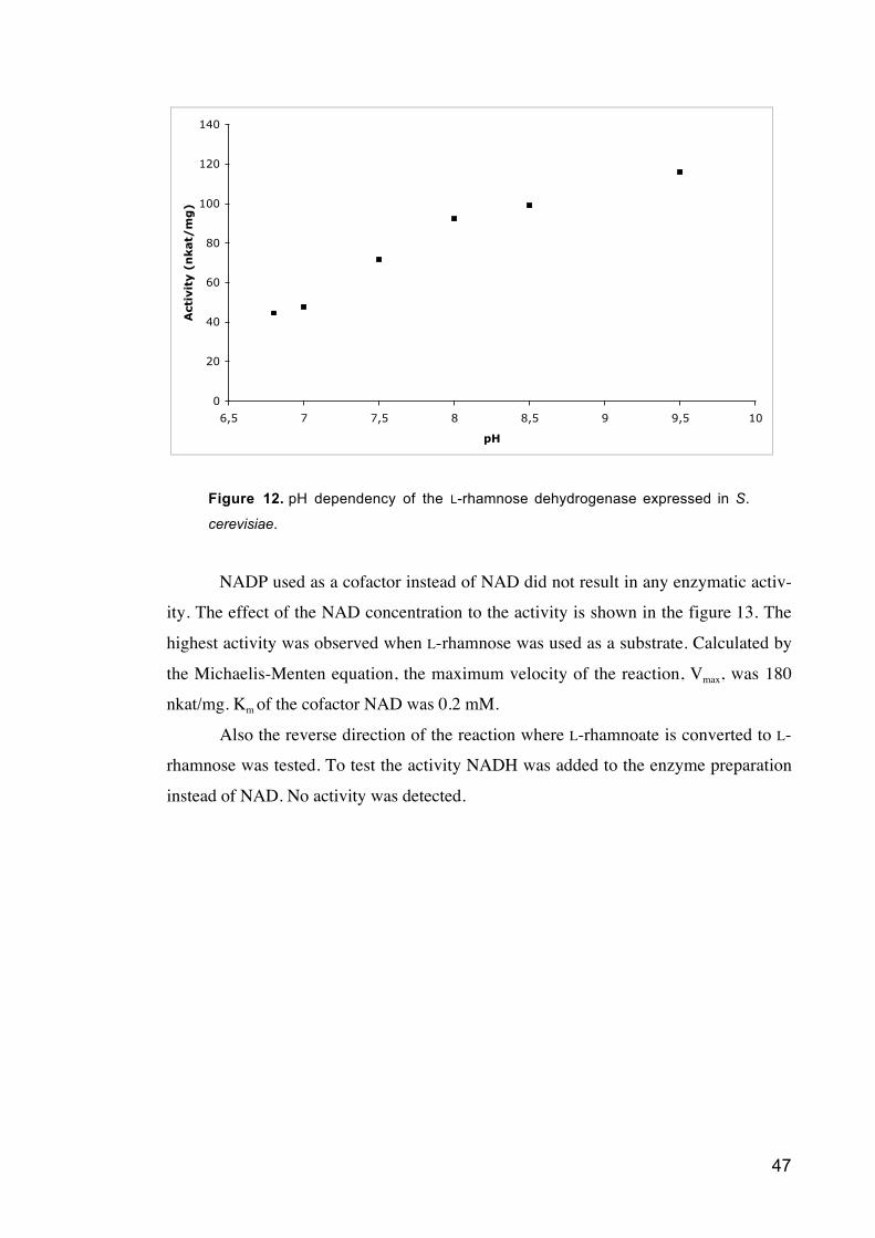
47
Figure 12. pH dependency of the L-rhamnose dehydrogenase expressed in S.
cerevisiae.
NADP used as a cofactor instead of NAD did not result in any enzymatic activ-
ity. The effect of the NAD concentration to the activity is shown in the figure 13. The
highest activity was observed when L-rhamnose was used as a substrate. Calculated by
the Michaelis-Menten equation, the maximum velocity of the reaction, Vmax, was 180
nkat/mg. Km of the cofactor NAD was 0.2 mM.
Also the reverse direction of the reaction where L-rhamnoate is converted to L-
rhamnose was tested. To test the activity NADH was added to the enzyme preparation
instead of NAD. No activity was detected.
0
20
40
60
80
100
120
140
6,5 7 7,5 8 8,5 9 9,5 10
pH
Acti
vit
y (
nkat/
mg
)
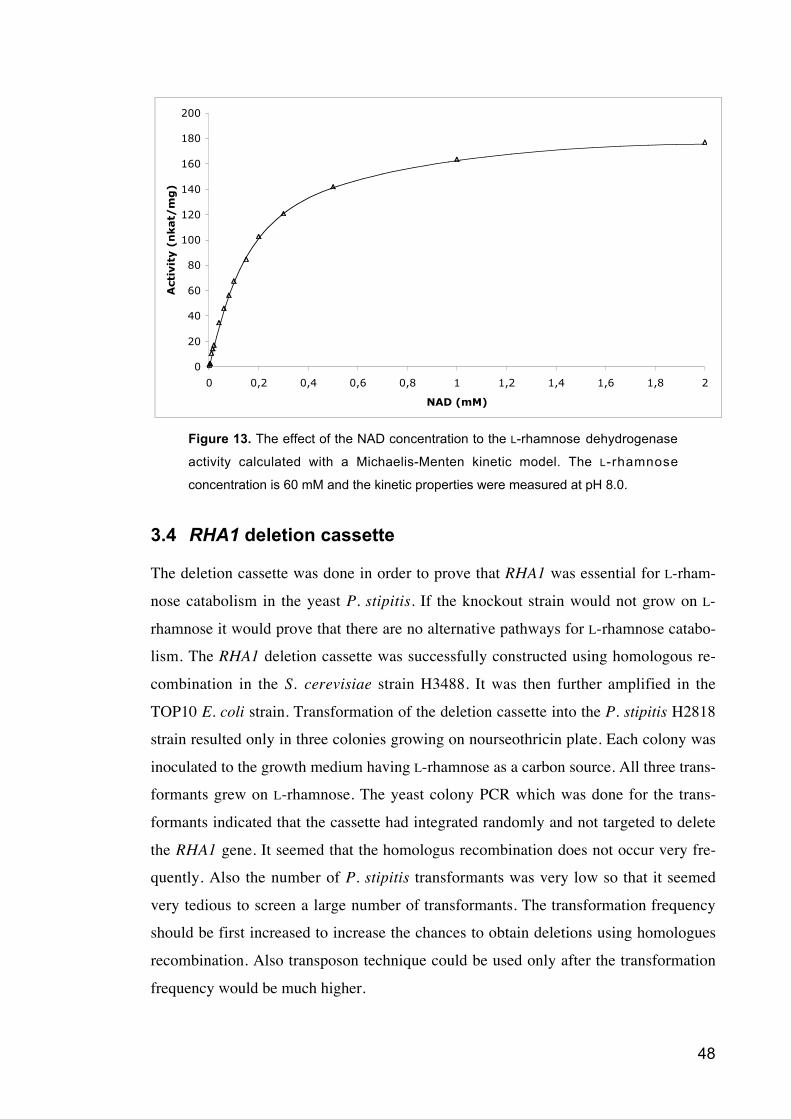
48
0
20
40
60
80
100
120
140
160
180
200
0 0,2 0,4 0,6 0,8 1 1,2 1,4 1,6 1,8 2
NAD (mM)
Acti
vit
y (
nkat/
mg
)
Figure 13. The effect of the NAD concentration to the L-rhamnose dehydrogenase
activity calculated with a Michaelis-Menten kinetic model. The L-rhamnose
concentration is 60 mM and the kinetic properties were measured at pH 8.0.
3.4 RHA1 deletion cassette
The deletion cassette was done in order to prove that RHA1 was essential for L-rham-
nose catabolism in the yeast P. stipitis. If the knockout strain would not grow on L-
rhamnose it would prove that there are no alternative pathways for L-rhamnose catabo-
lism. The RHA1 deletion cassette was successfully constructed using homologous re-
combination in the S. cerevisiae strain H3488. It was then further amplified in the
TOP10 E. coli strain. Transformation of the deletion cassette into the P. stipitis H2818
strain resulted only in three colonies growing on nourseothricin plate. Each colony was
inoculated to the growth medium having L-rhamnose as a carbon source. All three trans-
formants grew on L-rhamnose. The yeast colony PCR which was done for the trans-
formants indicated that the cassette had integrated randomly and not targeted to delete
the RHA1 gene. It seemed that the homologus recombination does not occur very fre-
quently. Also the number of P. stipitis transformants was very low so that it seemed
very tedious to screen a large number of transformants. The transformation frequency
should be first increased to increase the chances to obtain deletions using homologues
recombination. Also transposon technique could be used only after the transformation
frequency would be much higher.
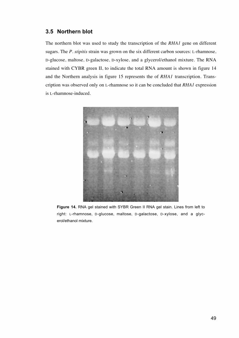
49
3.5 Northern blot
The northern blot was used to study the transcription of the RHA1 gene on different
sugars. The P. stipitis strain was grown on the six different carbon sources: L-rhamnose,
D-glucose, maltose, D-galactose, D-xylose, and a glycerol/ethanol mixture. The RNA
stained with CYBR green II, to indicate the total RNA amount is shown in figure 14
and the Northern analysis in figure 15 represents the of RHA1 transcription. Trans-
cription was observed only on L-rhamnose so it can be concluded that RHA1 expression
is L-rhamnose-induced.
Figure 14. RNA gel stained with SYBR Green II RNA gel stain. Lines from left to
right: L-rhamnose, D-glucose, maltose, D-galactose, D-xylose, and a glyc-
erol/ethanol mixture.
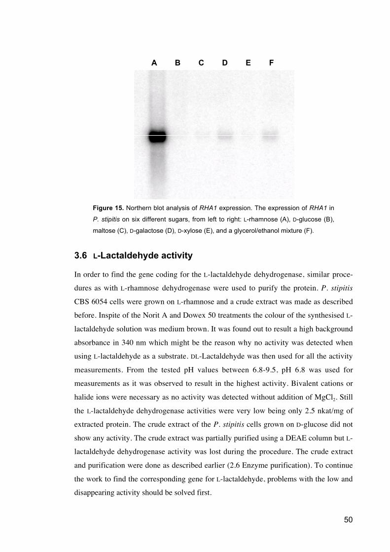
50
A EC FB D
Figure 15. Northern blot analysis of RHA1 expression. The expression of RHA1 in
P. stipitis on six different sugars, from left to right: L-rhamnose (A), D-glucose (B),
maltose (C), D-galactose (D), D-xylose (E), and a glycerol/ethanol mixture (F).
3.6 L-Lactaldehyde activity
In order to find the gene coding for the L-lactaldehyde dehydrogenase, similar proce-
dures as with L-rhamnose dehydrogenase were used to purify the protein. P. stipitis
CBS 6054 cells were grown on L-rhamnose and a crude extract was made as described
before. Inspite of the Norit A and Dowex 50 treatments the colour of the synthesised L-
lactaldehyde solution was medium brown. It was found out to result a high background
absorbance in 340 nm which might be the reason why no activity was detected when
using L-lactaldehyde as a substrate. DL-Lactaldehyde was then used for all the activity
measurements. From the tested pH values between 6.8-9.5, pH 6.8 was used for
measurements as it was observed to result in the highest activity. Bivalent cations or
halide ions were necessary as no activity was detected without addition of MgCl2. Still
the L-lactaldehyde dehydrogenase activities were very low being only 2.5 nkat/mg of
extracted protein. The crude extract of the P. stipitis cells grown on D-glucose did not
show any activity. The crude extract was partially purified using a DEAE column but L-
lactaldehyde dehydrogenase activity was lost during the procedure. The crude extract
and purification were done as described earlier (2.6 Enzyme purification). To continue
the work to find the corresponding gene for L-lactaldehyde, problems with the low and
disappearing activity should be solved first.
51
4 Discussion
4.1 L-Rhamnose dehydrogenase
There are two known pathways for L-rhamnose catabolism; the one used by prokaryotes
which has phosporylated intermediates and the one used by eukaryotes which has the
non-phosphorylated intermediates. For the latter pathway only the enzymes and inter-
mediates have been described but none of the corresponding genes have been identified.
In this work the gene of the first enzyme in this pathway, L-rhamnose dehydrogenase,
was identified. This NAD dependent gene was called RHA1.
Rigo et al. reported already in 1985 that L-rhamnose dehydrogenase is found in
the yeast like fungus Aureobasidium pullulans only when L-rhamnose is used as a car-
bon source. The same observation was done later in P. stipitis by Twerdochlib et al.
(1994) and was also confirmed in this study. No activity was detected in P. stipitis when
the cells were grown on D-glucose. Some activity was present when the cells were
grown on a mixture of D-glucose and L-rhamnose and the activity was increased
several-fold when L-rhamnose was the only source of carbon. During the purification,
the activity appeared as a single peak, indicating that there are no alternative pathways
for this step of L-rhamnose catabolism. The purified enzyme was digested with trypsin
and the masses of the peptides were identified using MALDI-TOF MS. As the genome
sequence of P. stipitis has been published the corresponding open reading frame could
be identified by the masses of the peptides.
4.1.1 Kinetic properties
To characterize the kinetic properties of the L-rhamnose dehydrogenase RHA1 was
expressed in the heterologus host S. cerevisiae. The results showed that RHA1 indeed
coded for an enzyme with this activity. In order to facilitate the purification histidine
tags were added to the N- or to the C-terminus but the activity of the tagged proteins
was much reduced or there was no activity detected. As this might be an indication that
the histidine tag is interfering with the catalytic activity the kinetic properties were
measured with the S. cerevisiae strain expressing RHA1 without any tags.
Twerdochlib et al (1994) found out that L-rhamnose dehydrogenase of P. stipitis
does not produce detectable amounts of L-rhamno- -lactone. This suggests that this
enzyme produced the more unstable L-rhamno- -lactone. The reverse reaction with L-
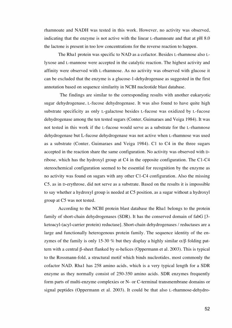
52
rhamnoate and NADH was tested in this work. However, no activity was observed,
indicating that the enzyme is not active with the linear L-rhamnoate and that at pH 8.0
the lactone is present in too low concentrations for the reverse reaction to happen.
The Rha1 protein was specific to NAD as a cofactor. Besides L-rhamnose also L-
lyxose and L-mannose were accepted in the catalytic reaction. The highest activity and
affinity were observed with L-rhamnose. As no activity was observed with glucose it
can be excluded that the enzyme is a glucose-1-dehydrogenase as suggested in the first
annotation based on sequence similarity in NCBI nucleotide blast database.
The findings are similar to the corresponding results with another eukaryotic
sugar dehydrogenase, L-fucose dehydrogenase. It was also found to have quite high
substrate specificity as only L-galactose besides L-fucose was oxidized by L-fucose
dehydrogenase among the ten tested sugars (Conter, Guimaraes and Veiga 1984). It was
not tested in this work if the L-fucose would serve as a substrate for the L-rhamnose
dehydrogenase but L-fucose dehydrogenase was not active when L-rhamnose was used
as a substrate (Conter, Guimaraes and Veiga 1984). C1 to C4 in the three sugars
accepted in the reaction share the same configuration. No activity was observed with D-
ribose, which has the hydroxyl group at C4 in the opposite configuration. The C1-C4
stereochemical configuration seemed to be essential for recognition by the enzyme as
no activity was found on sugars with any other C1-C4 configuration. Also the missing
C5, as in D-erythrose, did not serve as a substrate. Based on the results it is impossible
to say whether a hydroxyl group is needed at C5 position, as a sugar without a hydroxyl
group at C5 was not tested.
According to the NCBI protein blast database the Rha1 belongs to the protein
family of short-chain dehydrogenases (SDR). It has the conserved domain of fabG [3-
ketoacyl-(acyl-carrier protein) reductase]. Short-chain dehydrogenases / reductases are a
large and functionally heterogenous protein family. The sequence identity of the en-
zymes of the family is only 15-30 % but they display a highly similar / folding pat-
tern with a central -sheet flanked by -helices (Oppermann et al. 2003). This is typical
to the Rossmann-fold, a structural motif which binds nucleotides, most commonly the
cofactor NAD. Rha1 has 258 amino acids, which is a very typical length for a SDR
enzyme as they normally consist of 250-350 amino acids. SDR enzymes frequently
form parts of multi-enzyme complexies or N- or C-terminal transmembrane domains or
signal peptides (Oppermann et al. 2003). It could be that also L-rhamnose-dehydro-
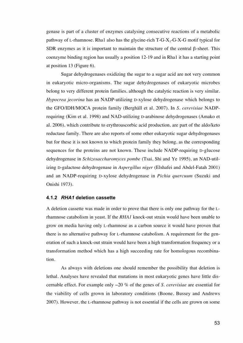
53
genase is part of a cluster of enzymes catalysing consecutive reactions of a metabolic
pathway of L-rhamnose. Rha1 also has the glycine-rich T-G-X3-G-X-G motif typical for
SDR enzymes as it is important to maintain the structure of the central -sheet. This
coenzyme binding region has usually a position 12-19 and in Rha1 it has a starting point
at position 13 (Figure 6).
Sugar dehydrogenases oxidizing the sugar to a sugar acid are not very common
in eukaryotic micro-organisms. The sugar dehydrogenases of eukaryotic microbes
belong to very different protein families, although the catalytic reaction is very similar.
Hypocrea jecorina has an NADP-utilizing D-xylose dehydrogenase which belongs to
the GFO/IDH/MOCA protein family (Berghäll et al. 2007). In S. cerevisiae NADP-
requiring (Kim et al. 1998) and NAD-utilizing D-arabinose dehydrogenases (Amako et
al. 2006), which contribute to erythroascorbic acid production, are part of the aldo/keto
reductase family. There are also reports of some other eukaryotic sugar dehydrogenases
but for these it is not known to which protein family they belong, as the corresponding
sequences for the proteins are not known. These include NADP-requiring D-glucose
dehydrogenase in Schizosaccharomyces pombe (Tsai, Shi and Ye 1995), an NAD-util-
izing D-galactose dehydrogenase in Aspergillus niger (Elshafei and Abdel-Fatah 2001)
and an NADP-requiring D-xylose dehydrogenase in Pichia quercuum (Suzuki and
Onishi 1973).
4.1.2 RHA1 deletion cassette
A deletion cassette was made in order to prove that there is only one pathway for the L-
rhamnose catabolism in yeast. If the RHA1 knock-out strain would have been unable to
grow on media having only L-rhamnose as a carbon source it would have proven that
there is no alternative pathway for L-rhamnose catabolism. A requirement for the gen-
eration of such a knock-out strain would have been a high transformation frequency or a
transformation method which has a high succeeding rate for homologous recombina-
tion.
As always with deletions one should remember the possibility that deletion is
lethal. Analyses have revealed that mutations in most eukaryotic genes have little dis-
cernable effect. For example only 20 % of the genes of S. cerevisiae are essential for
the viability of cells grown in laboratory conditions (Boone, Bussey and Andrews
2007). However, the L-rhamnose pathway is not essential if the cells are grown on some
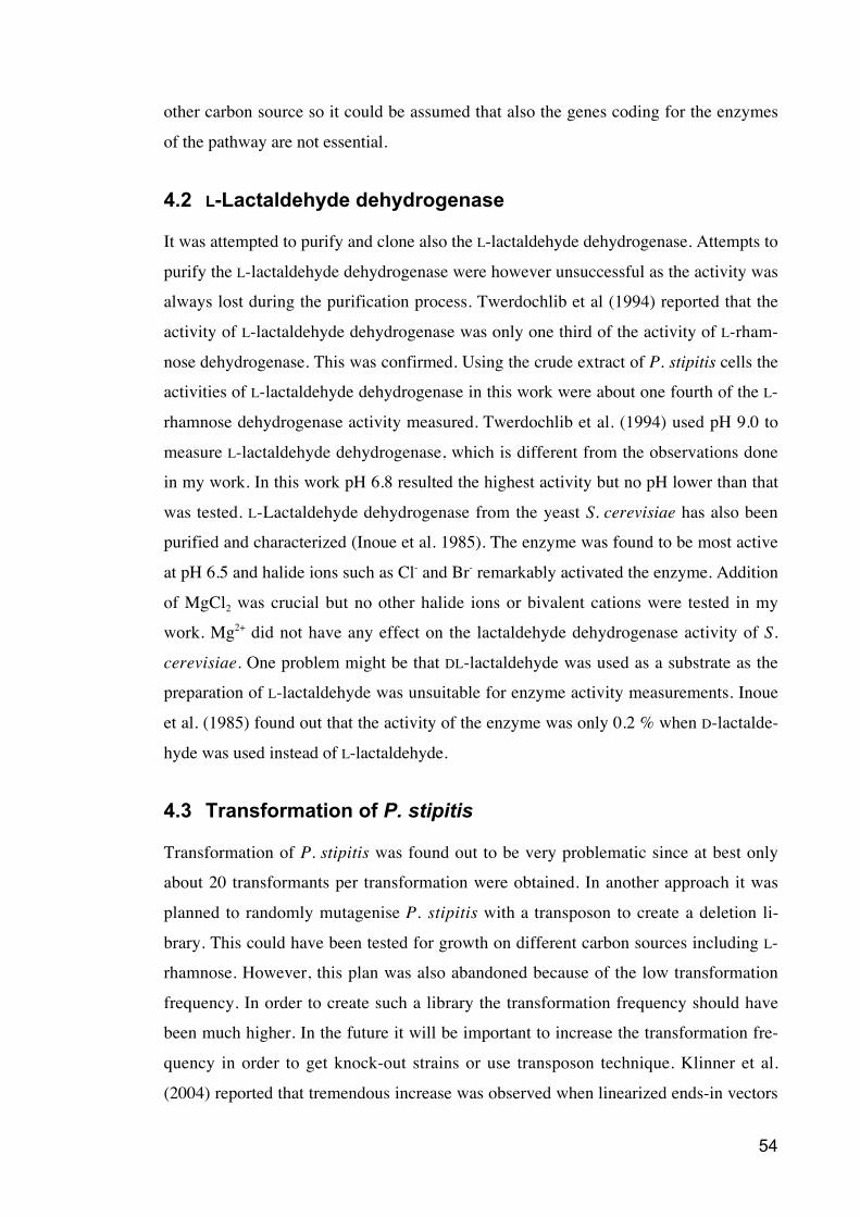
54
other carbon source so it could be assumed that also the genes coding for the enzymes
of the pathway are not essential.
4.2 L-Lactaldehyde dehydrogenase
It was attempted to purify and clone also the L-lactaldehyde dehydrogenase. Attempts to
purify the L-lactaldehyde dehydrogenase were however unsuccessful as the activity was
always lost during the purification process. Twerdochlib et al (1994) reported that the
activity of L-lactaldehyde dehydrogenase was only one third of the activity of L-rham-
nose dehydrogenase. This was confirmed. Using the crude extract of P. stipitis cells the
activities of L-lactaldehyde dehydrogenase in this work were about one fourth of the L-
rhamnose dehydrogenase activity measured. Twerdochlib et al. (1994) used pH 9.0 to
measure L-lactaldehyde dehydrogenase, which is different from the observations done
in my work. In this work pH 6.8 resulted the highest activity but no pH lower than that
was tested. L-Lactaldehyde dehydrogenase from the yeast S. cerevisiae has also been
purified and characterized (Inoue et al. 1985). The enzyme was found to be most active
at pH 6.5 and halide ions such as Cl- and Br- remarkably activated the enzyme. Addition
of MgCl2 was crucial but no other halide ions or bivalent cations were tested in my
work. Mg2+ did not have any effect on the lactaldehyde dehydrogenase activity of S.
cerevisiae. One problem might be that DL-lactaldehyde was used as a substrate as the
preparation of L-lactaldehyde was unsuitable for enzyme activity measurements. Inoue
et al. (1985) found out that the activity of the enzyme was only 0.2 % when D-lactalde-
hyde was used instead of L-lactaldehyde.
4.3 Transformation of P. stipitis
Transformation of P. stipitis was found out to be very problematic since at best only
about 20 transformants per transformation were obtained. In another approach it was
planned to randomly mutagenise P. stipitis with a transposon to create a deletion li-
brary. This could have been tested for growth on different carbon sources including L-
rhamnose. However, this plan was also abandoned because of the low transformation
frequency. In order to create such a library the transformation frequency should have
been much higher. In the future it will be important to increase the transformation fre-
quency in order to get knock-out strains or use transposon technique. Klinner et al.
(2004) reported that tremendous increase was observed when linearized ends-in vectors
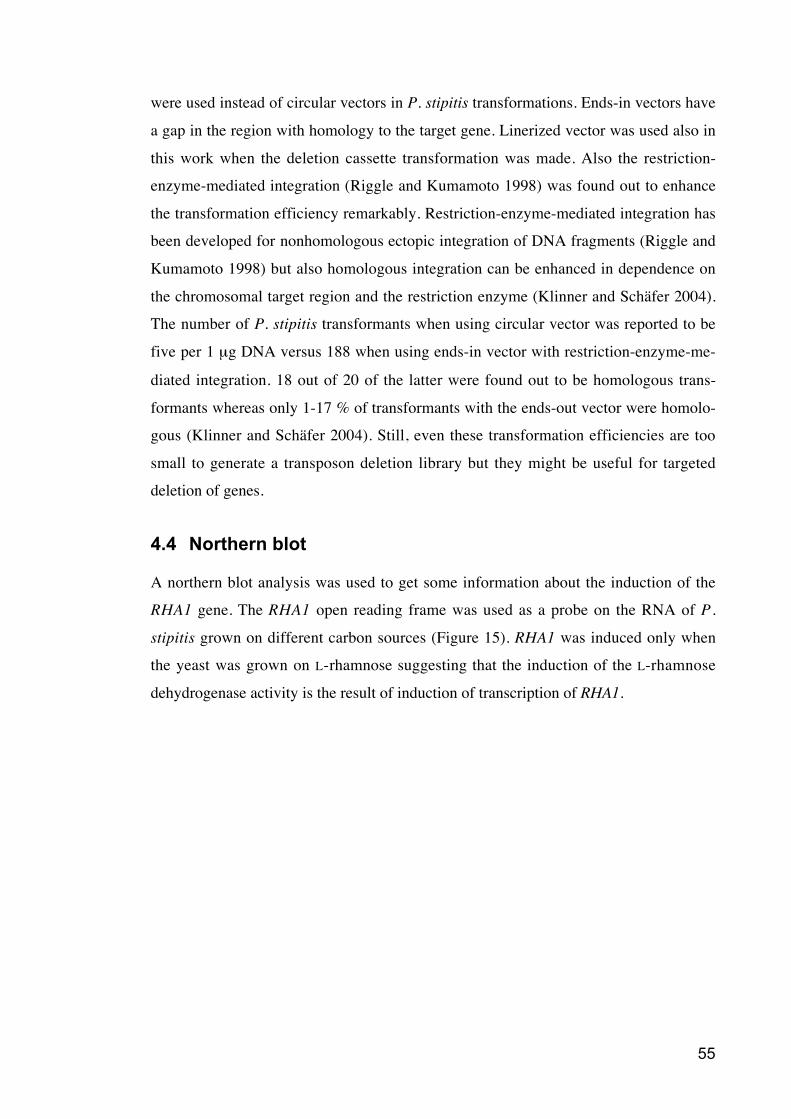
55
were used instead of circular vectors in P. stipitis transformations. Ends-in vectors have
a gap in the region with homology to the target gene. Linerized vector was used also in
this work when the deletion cassette transformation was made. Also the restriction-
enzyme-mediated integration (Riggle and Kumamoto 1998) was found out to enhance
the transformation efficiency remarkably. Restriction-enzyme-mediated integration has
been developed for nonhomologous ectopic integration of DNA fragments (Riggle and
Kumamoto 1998) but also homologous integration can be enhanced in dependence on
the chromosomal target region and the restriction enzyme (Klinner and Schäfer 2004).
The number of P. stipitis transformants when using circular vector was reported to be
five per 1 μg DNA versus 188 when using ends-in vector with restriction-enzyme-me-
diated integration. 18 out of 20 of the latter were found out to be homologous trans-
formants whereas only 1-17 % of transformants with the ends-out vector were homolo-
gous (Klinner and Schäfer 2004). Still, even these transformation efficiencies are too
small to generate a transposon deletion library but they might be useful for targeted
deletion of genes.
4.4 Northern blot
A northern blot analysis was used to get some information about the induction of the
RHA1 gene. The RHA1 open reading frame was used as a probe on the RNA of P.
stipitis grown on different carbon sources (Figure 15). RHA1 was induced only when
the yeast was grown on L-rhamnose suggesting that the induction of the L-rhamnose
dehydrogenase activity is the result of induction of transcription of RHA1.
56
4.5 Conclusions
The first enzyme of the L-rhamnose catabolic pathway in yeast, L-rhamnose 1-dehydro-
genase gene RHA1 was identified in this work. The enzyme was purified from the yeast
P. stipitis and the mass of its tryptic peptides determined using MALDI-TOF MS. This
enabled the identification of the corresponding gene, RHA1. It codes for a protein with
258 amino acids belonging to the protein family of short chain alcohol dehydrogenases.
The open reading frame was expressed in S. cerevisiae. P. stipitis has an alternative
codon usage where CUG codes for leucine instead of serine. These codons were
changed so that the same amino acid was expressed in S. cerevisiae. The NAD-specific
enzyme showed the highest activity and affinity with L-rhamnose and a lower activity
and affinity with L-mannose and L-lyxose. A Northern analysis revealed that
transcription in P. stipitis is induced during growth on L-rhamnose but not on other
carbon sources.
Further research is needed to find out the corresponding gene sequences of the
other enzymes of this pathway. This information is needed in order to transform the
pathway to different organisms as S. cerevisiae. Information about fungal sugar catabo-
lism is important in order to compose more compelling strains for the biofuel produc-
tion. Better understanding of fungal catabolic pathways and enzymes related to them is
also important for getting more comprehensive picture about fungal phylogenetics.
57
5 References
Amako, K., Fujita, K., Shimohata, T., Hasegawa, E., Kishimoto, R. and Goda, K. 2006,"NAD+-specific d-arabinose dehydrogenase and its contribution to erythroascorbicacid production in Saccharomyces cerevisiae", FEBS Letters, vol. 580, no. 27, pp.6428-6434.
Antoni, D., Zverlov, V.V. and Schwarz, W.H. 2007, "Biofuels from microbes", AppliedMicrobiology and Biotechnology, vol. 77, no. 1, pp. 23-35.
Atsumi, S., Hanai, T. and Liao, J.C. 2008, "Non-fermentative pathways for synthesis ofbranched-chain higher alcohols as biofuels", Nature, vol. 451, no. 7174, pp. 86-U13.
Berghäll, S., Hilditch, S., Penttilä, M. and Richard, P. 2007, "Identification in the mouldHypocrea jecorina of a gene encoding an NADP(+): D-xylose dehydrogenase",FEMS Microbiology Letters, vol. 277, no. 2, pp. 249-253.
Birnboim, H.C. and Doly, J. 1979, "A rapid alkaline extraction procedure for screeningrecombinant plasmid DNA", Nucleic Acids Research, vol. 7, no. 6, pp. 1513-1523.
Boone, C., Bussey, H. and Andrews, B.J. 2007, "Exploring genetic interactions andnetworks with yeast", Nature Reviews Genetics, vol. 8, no. 6, pp. 437-449.
Bradford, M.M. 1976, "A rapid and sensitive method for the quantitation of microgramquantities of protein utilizing the principle of protein-dye binding", AnalyticalBiochemistry, vol. 72, no. 1-2, pp. 248-254.
Colot, H.V. and Collopy, P.D. , Knockouts Workshop [Homepage of NeurosporaGenome Project , Dartmouth College] , [Online] . Available:http://www.dartmouth.edu/~neurosporagenome/Projects_files/Project%201/KO%20Protocols.pdf [2008, 3/31] .
Colot, H.V., Park, G., Turner, G.E., Ringelberg, C., Crew, C.M., Litvinkova, L., Weiss,R.L., Borkovich, K.A. and Dunlap, J.C. 2006, "A high-throughput gene knockoutprocedure for Neurospora reveals functions for multiple transcription factors",Proceedings of the National Academy of Sciences of the United States of America,vol. 103, no. 27, pp. 10352-10357.
Conter, P.F., Guimaraes, M.F. and Veiga, L.A. 1984, "Induction and Repression of L-Fucose Dehydrogenase of Pullularia pullulans", Canadian Journal ofMicrobiology, vol. 30, no. 6, pp. 753-757.
Dohmen, R.J., Strasser, A.W., Honer, C.B. and Hollenberg, C.P. 1991, "An efficienttransformation procedure enabling long-term storage of competent cells of variousyeast genera", Yeast (Chichester, England), vol. 7, no. 7, pp. 691-692.
58
Durrwachter, J.R., Drueckhammer, D.G., Nozaki, K., Sweers, H.M. and Wong, C.H.1986, "Enzymatic Aldol Condensation Isomerization as a Route to Unusual Sugar-Derivatives", Journal of the American Chemical Society, vol. 108, no. 24, pp. 7812-7818.
Elshafei, A.M. and Abdel-Fatah, O.M. 2001, "Evidence for a non-phosphorylated routeof galactose breakdown in cell-free extracts of Aspergillus niger", Enzyme andMicrobial Technology, vol. 29, no. 1, pp. 76-83.
Elzainy, T.A., Hassan, M.M. and Allam, A.M. 1973, "New Pathway forNonphosphorylated Degradation of Gluconate by Aspergillus niger", Journal ofBacteriology, vol. 114, no. 1, pp. 457-459.
Giraud, M. and Naismith, J.H. 2000, "The rhamnose pathway", Current Opinion inStructural Biology, vol. 10, no. 6, pp. 687-696.
Gray, K.A., Zhao, L. and Emptage, M. 2006, "Bioethanol", Current Opinion inChemical Biology, vol. 10, no. 2, pp. 141-146.
Guimaraes, M.F. and Veiga, L.A. 1990, "An Oxidative Pathway for L-FucoseDissimilation in Fungi", Arquivos de Biologia e Tecnologia, vol. 33, no. 3, pp. 645-655.
Hahn-Hägerdal, B., Karhumaa, K., Fonseca, C., Spencer-Martins, I. and Gorwa-Grauslund, M.F. 2007, "Towards industrial pentose-fermenting yeast strains",Applied Microbiology and Biotechnology, vol. 74, no. 5, pp. 937-953.
Hazelwood, L.A., Daran, J.M., van Maris, A.J., Pronk, J.T. and Dickinson, J.R. 2008,"The Ehrlich pathway for fusel alcohol production: a century of research onSaccharomyces cerevisiae metabolism", Applied and Environmental Microbiology,vol. 74, no. 8, pp. 2259-2266.
Hertz-Fowler, C. and Pain, A. 2007, "Specialist fungi, versatile genomes", Naturereviews. Microbiology, vol. 5, no. 5, pp. 332-333.
Hibbett, D.S., Binder, M., Bischoff, J.F., Blackwell, M., Cannon, P.F., Eriksson, O.E.,Huhndorf, S., James, T., Kirk, P.M., Lücking, R., Thorsten Lumbsch, H., Lutzoni,F., Matheny, P.B., McLaughlin, D.J., Powell, M.J., Redhead, S., Schoch, C.L.,Spatafora, J.W., Stalpers, J.A., Vilgalys, R., Aime, M.C., Aptroot, A., Bauer, R.,Begerow, D., Benny, G.L., Castlebury, L.A., Crous, P.W., Dai, Y., Gams, W.,Geiser, D.M., Griffith, G.W., Gueidan, C., Hawksworth, D.L., Hestmark, G.,Hosaka, K., Humber, R.A., Hyde, K.D., Ironside, J.E., Kõljalg, U., Kurtzman, C.P.,Larsson, K., Lichtwardt, R., Longcore, J., Mi_dlikowska, J., Miller, A., Moncalvo,J., Mozley-Standridge, S., Oberwinkler, F., Parmasto, E., Reeb, V., Rogers, J.D.,Roux, C., Ryvarden, L., Sampaio, J.P., Schüßler, A., Sugiyama, J., Thorn, R.G.,Tibell, L., Untereiner, W.A., Walker, C., Wang, Z., Weir, A., Weiss, M., White,M.M., Winka, K., Yao, Y. and Zhang, N. 2007, "A higher-level phylogeneticclassification of the Fungi", Mycological Research, vol. 111, no. 5, pp. 509-547.
59
Hilditch, S., Berghäll, S., Kalkkinen, N., Penttilä, M. and Richard, P. 2007, "Themissing link in the fungal D-galacturonate pathway - Identification of the L-threo-3-deoxy-hexulosonate aldolase", Journal of Biological Chemistry, vol. 282, no. 36,pp. 26195-26201.
Huff, E. and Rudney, H. 1959, "The enzymatic oxidation of 1,2-propanediol phosphateto acetol phosphate", The Journal of biological chemistry, vol. 234, no. 5, pp. 1060-1064.
Inoue, Y., Watanabe, K., Shimosaka, M., Saikusa, T., Fukuda, Y., Murata, K. andKimura, A. 1985, "Metabolism of 2-oxoaldehydes in yeasts. Purification andcharacterization of lactaldehyde dehydrogenase from Saccharomyces cerevisiae",European journal of biochemistry / FEBS, vol. 153, no. 2, pp. 243-247.
Jeffries, T.W., Grigoriev, I.V., Grimwood, J., Laplaza, J.M., Aerts, A., Salamov, A.,Schmutz, J., Lindquist, E., Dehal, P., Shapiro, H., Jin, Y.S., Passoth, V. andRichardson, P.M. 2007, "Genome sequence of the lignocellulose-bioconverting andxylose-fermenting yeast Pichia stipitis", Nature Biotechnology, vol. 25, no. 3, pp.319-326.
Jeffries, T.W. 2006, "Engineering yeasts for xylose metabolism", Current Opinion inBiotechnology, vol. 17, no. 3, pp. 320-326.
Kim, S., Huh, W., Lee, B. and Kang, S. 1998, "D-Arabinose dehydrogenase and itsgene from Saccharomyces cerevisiae", Biochimica et Biophysica Acta (BBA) -Protein Structure and Molecular Enzymology, vol. 1429, no. 1, pp. 29-39.
Klinner, U. and Schäfer, B. 2004, "Genetic aspects of targeted insertion mutagenesis inyeasts", FEMS Microbiology Reviews, vol. 28, no. 2, pp. 201-223.
Krugel, H., Fiedler, G., Smith, C. and Baumberg, S. 1993, "Sequence andtranscriptional analysis of the nourseothricin acetyltransferase-encoding gene nat1from Streptomyces noursei ", Gene, vol. 127, no. 1, pp. 127-131.
Kuorelahti, S., Jouhten, P., Maaheimo, H., Penttilä, M. and Richard, P. 2006, "L-galactonate dehydratase is part of the fungal path for D-galacturonic acidcatabolism", Molecular microbiology, vol. 61, no. 4, pp. 1060-1068.
Kuorelahti, S., Kalkkinen, N., Penttilä, M., Londesborough, J. and Richard, P. 2005,"Identification in the mold Hypocrea jecorina of the first fungal D-galacturonicacid reductase", Biochemistry, vol. 44, no. 33, pp. 11234-11240.
Kurtzman, C.,P. and Fell, J.,W. (eds) 1998, The yeasts: a taxonomic study, 4th editionedn, Elsevier Science B.V., The Netherlands.
Kuyper, M., Harhangi, H.R., Stave, A.K., Winkler, A.A., Jetten, M.S.M., de Laat,W.T.A.M., den Ridder, J.J.J., Op den Camp, H.J.M., van Dijken, J.P. and Pronk,J.T. 2003, "High-level functional expression of a fungal xylose isomerase: the keyto efficient ethanolic fermentation of xylose by Saccharomyces cerevisiae?", FemsYeast Research, vol. 4, no. 1, pp. 69-78.
60
Laemmli, U.K. 1970, "Cleavage of structural proteins during the assembly of the headof bacteriophage T4", Nature, vol. 227, no. 5259, pp. 680-685.
Laplaza, J.M., Torres, B.R., Jin, Y. and Jeffries, T.W. 2006, "Sh ble and Cre adapted forfunctional genomics and metabolic engineering of Pichia stipitis", Enzyme andMicrobial Technology, vol. 38, no. 6, pp. 741-747.
Lengeler, J.W., Drews, G. and Schlegel, H.G. (eds) 1999, Biology of the Prokaryotes,First edition edn, Blackwell Science, Germany.
Liepins, J., Kuorelahti, S., Penttilä, M. and Richard, P. 2006, "Enzymes for theNADPH-dependent reduction of dihydroxyacetone and D-glyceraldehyde and L-glyceraldehyde in the mould Hypocrea jecorina", Febs Journal, vol. 273, no. 18,pp. 4229-4235.
Moralejo, P., Egan, S.M., Hidalgo, E. and Aguilar, J. 1993, "Sequencing andcharacterization of a gene cluster encoding the enzymes for L-rhamnosemetabolism in Escherichia coli", Journal of Bacteriology, vol. 175, no. 17, pp.5585-5594.
Oliver, P.R. and Schweizer, M. (eds) 1999, Molecular fungal biology, First edition edn,Cambridge University Press, United Kingdom.
Oppermann, U., Filling, C., Hult, M., Shafqat, N., Wu, X.Q., Lindh, M., Shafqat, J.,Nordling, E., Kallberg, Y., Persson, B. and Jornvall, H. 2003, "Short-chaindehydrogenases", Chemico-biological interactions, vol. 143, pp. 247-253.
Peekhaus, N. and Conway, T. 1998, "What's for dinner? Entner-Doudoroff metabolismin Escherichia coli", Journal of Bacteriology, vol. 180, no. 14, pp. 3495-3502.
Platt, A.R., Woodhall, R.W. and George, A.L.,Jr 2007, "Improved DNA sequencingquality and efficiency using an optimized fast cycle sequencing protocol",BioTechniques, vol. 43, no. 1, pp. 58, 60, 62.
Riggle, P.J. and Kumamoto, C.A. 1998, "Genetic analysis in fungi using restriction-enzyme-mediated integration", Current Opinion in Microbiology, vol. 1, no. 4, pp.395-399.
Rigo, L.U., Marechal, L.R., Vieira, M.M. and Veiga, L.A. 1985, "Oxidative Pathwayfor L-Rhamnose Degradation in Pullularia pullulans", Canadian Journal ofMicrobiology, vol. 31, no. 9, pp. 817-822.
Romano, A.H. and Conway, T. 1996, "Evolution of carbohydrate metabolic pathways",Research in microbiology, vol. 147, no. 6-7, pp. 448-455.
Rosenfeld, J., Capdevielle, J., Guillemot, J.C. and Ferrara, P. 1992, "In-gel digestion ofproteins for internal sequence analysis after one- or two-dimensional gelelectrophoresis", Analytical Biochemistry, vol. 203, no. 1, pp. 173-179.
Sawada, H. and Takagi, Y. 1964, "The Metabolism of L-Rhamnose in Escherichia coli.3. L-Rhamulose-Phosphate Aldolase", Biochimica et biophysica acta, vol. 92, pp.26-32.
61
Schoevaart, R. 2000, Applications of aldolases in organic synthesis, Delft University ofTechnology, Netherlands.
Schruff, B.S. 2005, Zur Erfassung trans-wirksamer Komponenten der Sauerstoff-abhängigen Gärungsregulation der Hefe Pichia stipitis, RWTH AachenUniversity, Germany.
Stansfield, I. and Stark, M.,J.R. (eds) 2007, Methods in microbiology, Volume 36, YeastGene Analysis, Second edition edn, Academic Press, The Netherlands.
Suzuki, T. and Onishi, H. 1973, "Oxidation and reduction of D-xylose by cell-freeextract of Pichia quercuum", Applied Microbiology, vol. 25, no. 5, pp. 850-852.
Takagi, Y. and Sawada, H. 1964a, "The Metabolism of L-Rhamnose in Escherichiacoli. I. L-Rhamnose Isomerase", Biochimica et biophysica acta, vol. 92, pp. 10-17.
Takagi, Y. and Sawada, H. 1964b, "The Metabolism of L-Rhamnose in Escherichiacoli. Ii. L-Rhamnulose Kinase", Biochimica et biophysica acta, vol. 92, pp. 18-25.
Tsai, C.S., Shi, J.L. and Ye, H.G. 1995, "Kinetic Studies of Gluconate PathwayEnzymes from Schizosaccharomyces pombe", Archives of Biochemistry andBiophysics, vol. 316, no. 1, pp. 163-168.
Twerdochlib, A.L., Pedrosa, F.O., Funayama, S. and Rigo, L.U. 1994, "L-RhamnoseMetabolism in Pichia stipitis and Debaryomyces polymorphus", Canadian Journalof Microbiology, vol. 40, no. 11, pp. 896-902.
van Maris, A.J.A., Abbott, D.A., Bellissimi, E., van den Brink, J., Kuyper, M., Luttik,M.A.H., Wisselink, H.W., Scheffers, W.A., van Dijken, J.P. and Pronk, J.T. 2006,"Alcoholic fermentation of carbon sources in biomass hydrolysates bySaccharomyces cerevisiae: current status", Antonie Van Leeuwenhoek InternationalJournal of General and Molecular Microbiology, vol. 90, no. 4, pp. 391-418.
Verho, R., Richard, P., Jonson, P.H., Sundqvist, L., Londesborough, J. and Penttilä, M.2002, "Identification of the first fungal NADP-GAPDH from Kluyveromyceslactis", Biochemistry, vol. 41, no. 46, pp. 13833-13838.
Vogelstein, B. and Gillespie, D. 1979, "Preparative and analytical purification of DNAfrom agarose", Proceedings of the National Academy of Sciences of the UnitedStates of America, vol. 76, no. 2, pp. 615-619.
Wilson, D.M. and Ajl, S. 1957a, "Metabolism of L-rhamnose by Escherichia coli. I. L-rhamnose isomerase", Journal of Bacteriology, vol. 73, no. 3, pp. 410-414.
Wilson, D.M. and Ajl, S. 1957b, "Metabolism of L-rhamnose by Escherichia coli. II.The phosphorylation of L-rhamnulose", Journal of Bacteriology, vol. 73, no. 3, pp.415-420.
Yang, V.W., Marks, J.A., Davis, B.P. and Jeffries, T.W. 1994, "High-efficiencytransformation of Pichia stipitis based on its URA3 gene and a homologousautonomous replication sequence, ARS2", Applied and EnvironmentalMicrobiology, vol. 60, no. 12, pp. 4245-4254.
62
Yew, W.S., Fedorov, A.A., Fedorov, E.V., Rakus, J.F., Pierce, R.W., Almo, S.C. andGerlt, J.A. 2006, "Evolution of enzymatic activities in the enolase superfamily: L-fuconate dehydratase from Xanthomonas campestris", Biochemistry, vol. 45, no.49, pp. 14582-14597.