Embed Size (px)
Citation preview
Vol. 24: 179-184,1996 DISEASES OF AQUATIC ORGANISMS
Dis aquat Ory Published March 7 l
Effects of 3,N-methylglucamine lasalocid on Am yloodinium ocellatum
Daniel J. Oestmannl,*, Donald H. ~ e w i s ~
' 11 150 Beamer Rd #416. Houston. Texas 77089, USA 2Department of Veterinary Pathobiology, Texas A&M University. College Station, Texas 77843, USA
ABSTRACT: The cosmopolitan nature of Amyloodinium ocellatum insures the constant possibility of an epizootic in high density mariculture facilities. Chemotherapeutics have shown variable results against the parasite and none are currently approved for use in food fish. The ionophorous antibiotic lasalocid is currently approved for food animal use for the treatment and prophylaxis of coccidian protozoa. The sodium salt of lasalocid was converted to a water-soluble 3.N-methylglucamine salt to evaluate its ther- apeutic potential for amyloodin~osis Tomonts and trophonts were incubated in saltwater containing antibiotics (penicillin 100 IU ml-' , streptomycin 100 pg n ~ l - ' ) and 100 to 0.001 ppm concentrations of 3,N-methylglucamine lasalocld (3NMG-lasaloc~d). The 24 h toinont division rate and in vitro trophont density decreased in a dose-dependent manner when exposed to 3NMG-lasalocld concentrations between 100 and 0.001 ppm. Motile dinospores did not excyst until the 3NMG-lasalocid concentration was reduced to 0.001 ppm, and then at a concentration 44 '% that of controls. Cracked and ruptured tomonts were observed by scanmng electron m~croscopy and degenerated internal membranous struc- tures of trophonts were observed by transmission electron microscopy. Trophont infection on 0.5 to 1 g red drum Sciaenops ocellatus fry exposed to 2.5 dinospores ml-' in 6.0 1 of saltwater was reduced by 80% on gill filaments and 5 4 % mm-' surface area when treated with 0.1 ppm 3NMG-lasalocid. Infec- tion was prevented at 1.0 ppm 3NMG-lasalocid
KEY WORDS: Lasalocid . Amyloodini~~m - Ionophore . Red drum
INTRODUCTION
Management measures such as high water turnover rates, micro-filtration, and sanitation are used to con- trol Arnyloodiniurn ocellatum in mariculture facilities, but the ubiquitous dispersal of the parasite insures the possibility of an epizootic. Fresh water or forrnalin immersion bath protocols are effective management procedures for quarantined fish in laboratory and pet fish facilities (Lawler 1979, Paperna 1984a, Noga & Bower 1987), but are impractical for large production facilities. Several herbicides, disinfectants, salinity concentrations, and antiprotozoal drugs have been tested for the control of amyloodiniosls with variable results (Dempster 1955, Lawler 1977, Johnson 1984, Paperna 1984b, Lewis et al. 1988).
Dinospores have been considered the most suscep- tible stage of the 3-phase life cycle (sessile feeding trophont/encysted reproductive tomont/free-swim- rning infective dinospores) of Amyloodinium ocella- tum. Copper compounds (0.75 ppm free copper) can be used to treat an outbreak of A. ocellaturn by elim- inating the dinospore, but this dose may be harmful to most fish species (Paperna 1984a). Most fish spe- cies will survive and amyloodiniosis can be pre- vented or controlled at 0.15 ppm free copper (Demp- ster 1955, Johnson 1984). The antimalarial drug chloroquine has been demonstrated to have thera- peutic potential. Chloroquine eliminated dinospore activity in vitro after 24 h at 2.5 pg ml-l, and oral doses of 50 mg kg-' body weight produced inhibi- tory tissue drug levels (>5.0 pg drug ml- ' ) for 7 d with most of the drug disappearing from the tissue within 30 d (Lewis et al. 1988). However, due to gov- ernmental regulations and environmental concerns,
Q Inter-Research 1996 Resale of full article not permitted
180 Dis aquat Org 24: 179-184, 1996
chloroquine is unlikely to be approved for aquatic food animal use.
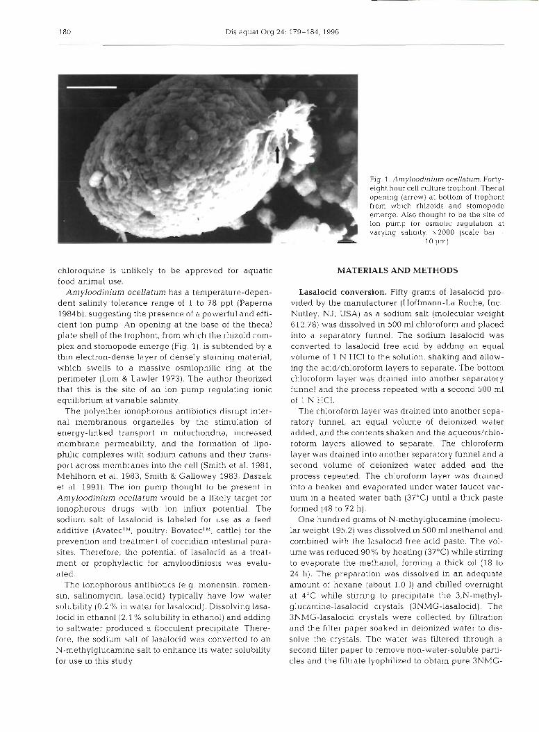
Amyloodinium ocellaturn has a temperature-depen- dent salinity tolerance range of 1 to 78 ppt (Paperna 1984b), suggesting the presence of a powerful and effi- cient ion pump. An opening at the base of the thecal plate shell of the trophont, from which the rhizoid com- plex and stomopode emerge (Fig. l), is subtended by a thin electron-dense layer of densely staining material, which swells to a massive osmiophilic ring at the perimeter (Lom & Lawler 1973). The author theorized that this is the site of an ion pump regulating ionic equilibrium at variable salinity.
The polyether ionophorous antibiotics disrupt inter- nal membranous organelles by the stimulation of energy-linked transport in mitochondria, increased membrane permeability, and the formation of lipo- philic complexes with sodium cations and their trans- port across membranes into the cell (Smith et al. 1981, Mehlhorn et al. 1983, Smith & Galloway 1983, Daszak et al. 1991). The ion pump thought to be present in Amyloodinium ocellaturn would be a likely target for ionophorous drugs with ion influx potential. The sodium salt of lasalocid is labeled for use as a feed additive (AvatecTM, poultry; BovatecTbf, cattle) for the prevention and treatment of coccidian intestinal para- sites. Therefore, the potential of lasalocid as a treat- ment or prophylactic for amyloodiniosis was evalu- ated.
The ionophorous antibiotics (e .g , monensin, romen- sin, salinomycin, lasalocid) typically have low water solubility (0.2 % in water for lasalocid). Dissolving lasa- locid in ethanol (2.1 % solubility in ethanol) and adding to saltwater produced a flocculent precipitate. There- fore, the sodium salt of lasalocid was converted to an N-methylglucamine salt to enhance its water solubility for use in this study.
Fig. 1 . Amyloodinitlrn ocellaturn Forty- eight hour cell culture trophont. Thecal opening (arrow) at bottom of trophont from which rhizoids and stomopode emerge. Also thought to be the site of ion pump for osmotic regulation at varying salinity. x2000 (scale bar =
l n l ~ m !
MATERIALS AND METHODS
Lasalocid conversion. Fifty grams of lasalocid pro- vided by the manufacturer (Hoffmann-La Roche, Inc., Nutley, NJ, USA) as a sodium salt (molecular weight 612.78) was dissolved in 500 m1 chloroform and placed into a separatory funnel. The sodium lasalocid was converted to lasalocid free acid by adding an equal volume of 1 N HC1 to the solution, shaking and allow- ing the acid/chloroform layers to separate. The bottom chloroform layer was drained into another separatory funnel and the process repeated with a second 500 m1 of 1 N HC1.
The chloroform layer was drained into another sepa- ratory funnel, an equal volume of deionized water added, and the contents shaken and the aqueous/chlo- roform layers allowed to separate. The chloroform layer was drained into another separatory funnel and a second volume of deionized water added and the process repeated. The chloroform layer was drained into a beaker and evaporated under water faucet vac- uum in a heated water bath (37°C) until a thick paste formed (48 to 72 h).
One hundred grams of N-methylglucamine (molecu- lar weight 195.2) was dissolved in 500 m1 methanol and combined with the lasalocid free acid paste. The vol- ume was reduced 90 % by heating (37OC) while stirring to evaporate the methanol, forming a thick oil (18 to 24 h). The preparation was dissolved in an adequate amount of hexane (about 1.0 1) and chilled overnight at 4°C while stirring to precipitate the 3,N-methyl- glucamine-lasalocid crystals (3NMG-lasalocid). The 3NMG-lasalocid crystals were collected by filtration and the filter paper soaked in deionized water to dis- solve the crystals. The water was filtered through a second filter paper to remove non-water-soluble parti- cles and the filtrate lyophilized to obtain pure 3NMG-
Oestmann 8. Lewis: Treatment of red drum for dmyloodiniosis 181
Table 1 Saltwater media (SM) formulations for cell culture propagation. Sterilized by 0.45 pm filtration. Based on
I02/HBSS use by Noga (1987, 1989, 1992)
SM-30 ppt SM- l0 ppt (g I l ) (g 1 - ' 1
- -
NaCl 24.29 8.10 MgSO,. 7 H 2 0 4.85 1 .G2 MgCl?. 6 H 2 0 3.49 1.16 CaCI2. 2 H 2 0 1.17 0.39 KC1 0.77 0.26 NaHCO., 0.38 0.13 Na2HP04 . 7 H 2 0 0.07 0.023 KH2P04 0.045 0.015 Glucose - 1.0 Penicillin 100 IU ml- ' 100 IU ml-' Streptomycin 100 pg ml-' 100 pg ml-' pH 7.4 7.4 Specific gravity 1.023 g ml-' 1.007 g ml-'
lasalocid. Samples of the final product, sodium lasa- locid, and N-methylglucamine were submitted to the Texas A&M University chemistry department for nuclear magnetic resonance (NMR) analysis.
Tomont treatment. The protocol for collecting microbe-free Amyloodinium ocellatum tomonts is de- scribed elsewhere (Oestmann & Lewis 1995). In brief, tomonts were collected by osmotic shock, purified by ~ercoll ' gradient centrifugatlon, enumel-ated, and dis- tributed (30000 tomonts in 30 ppt saltwater medium, SM-30; Table 1) into seven 25 cm2 cell culture flask. After 1 h settling time, the SM-30 used to prepare the tomonts was aspirated from 6 of the flasks and replaced with SM-30 supplemented with 10-fold dilu- tions of 3Nh4G-lasalocid (100 to 0.001 ppm 3NMG- lasalocid in SM-30) with the seventh flask serving as a control. At 24 h incubation the tomont division rates were assessed by observing with an inverted micro- scope. Dinospores were enumerated at 72 h incubation by aspirating an aliquot with a 37% formalin coated Pasteur pipette and loading onto a hemocytometer (Bower et al. 1987). Samples were removed from the flasks and fixed overnight a t 4OC in 5 % glutaraldehyde and 4 % paraformaldehyde in 0.1 M cacodylate buffer (Karnovsky 1965). The fixative was replaced with 0.1 M sodium cacodylate buffer the next morning in preparation for scanning electron microscopy.
3NMG-lasalocid toxicity to red drum cell culture. The toxic effect of 3NMG-lasalocid was assessed on red drum Sciaenops ocellatus dorsal fin cell (RDFC) monolayers. Three-day-old monolayers were trypsin- ized, enumerated, and their viability assessed by try- pan blue exclusion. Seven 25 cm2 cell culture flasks were prepared using 5 m1 aliquots of 1.62 X 10⁶ RDFC ml-' in L-15 (BioWhittaker, Inc., Walkersville, MD, USA) cell culture medium supplemented with 10%
fetal bovine serum, l ' % L-glutanline, and penicillin (100 IU ml- ') and streptomycin (100 pg m ] - ' ) . Conflu- ent monolayers had formed after 24 h incubation at 2G°C, at which time the media from 6 flasks were aspi- rated and replaced with L-15 containing supplements plus 10-fold dilutions of 3NMG-lasalocid (100 to 0.001 pprn 3NMG-lasalocid in L-15) with the seventh flask serving as a control. Incubation continued at 26OC for 3 d , at which time the RDFC monolayers were trypsinized, enumerated, and their viability assessed by trypan blue exclusion.
In vitro trophont treatment. The effect of 3NMG- lasalocid was evaluated on in vitl-o trophonts propa- gated on red drum dorsal fin cells grown as aggre- gates (Oestmann 1994). Dinospores emerging from microbe-free tomonts attached and transformed into trophonts on RDFC aggregates in 10 ppt saltwater media (SM-10; Table 1). Aliquots of infected RDFC aggregates were divided into 7 siliconized glass Leighton tubes. One hour after exposure to dinospores, the SM-l0 from 6 tubes was aspirated and replaced with SM-10 containing 10-fold dilutions of 3NMG- lasalocid (100 to 0.001 pprn 3NMG-lasalocid in SM-10) with the seventh tube serving as a control. The tubes were then placed in a 23°C incubator on a rotary table and gently rotated to keep the aggregates suspended At 24 and 48 h incubation trophont densities were evaluated microscopically and aggregate samples fixed as described above for tomonts and subsequently fixed in osmium tetroxide for transmission electron microscopy.
In vivo trophont treatment. Six red drum fry (0.5 to 1 g ) were placed in each of 6 small plastic tanks in 6.0 1 saltwater, 20 ppt (Fritz Supersalt; Fritz Chemical Co., Dallas, TX, USA) for 1 wk acclimation. Tank 1 was then treated with 2.5 dinospores ml- ' (15000 dino- spores), tank 2 was treated with 2.5 dinospores inl-' plus 0.1 pprn 3NMG-lasalocid, tank 3 was treated with 0.1 pprn 3NMG-lasalocid alone. Tank 4 was treated with 2.5 dinospores ml-l plus 1.0 pprn 3NMG-lasalocid while tank 5 was treated with 1.0 pprn 3NMG-lasa- locid alone. Tank 6 served as the non-treated control. After 24 h exposure, the fish were killed by cervical transection and trophont load per gill filament assessed microscopically (10 filaments per arch on each of 6 fish averaged). The surface area trophont load was also assessed on the tail, dorsal, anal, and pectoral fin clips (average mm-2).
RESULTS
The conversion of sodium lasalocid to a n N-methyl- glucamine salt is very inefficient, yielding only about 50 mg of final product from 50 g of sodium lasalocid.
Dis aquat Org 24: 179-184, 1996
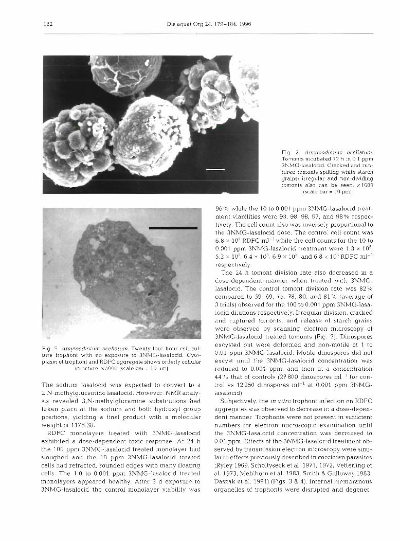
Fig. 3. Amyloodinium ocellatum. Twenty-four hour cell cul- ture trophont with no exposure to 3NMG-lasalocid. Cyto- plasm of trophont and RDFC aggregate shows orderly cellular
structure. x5000 (scale bar = l 0 pm)
The sodium lasalocid was expected to convert to a 2,N-methylglucamine lasalocid. However, NMR analy- sis revealed 3,N-methylglucamine substitutions had taken place at the sodium and both hydroxyl group positions, yielding a final product with a molecular weight of 1176.38.
RDFC monolayers treated with 3NMG-lasalocid exhibited a dose-dependent toxic response. At 24 h the 100 pprn 3NMG-lasalocid treated rnonolayer had sloughed and the 10 pprn 3NMG-lasalocid treated cells had retracted, rounded edges with many floating cells. The 1.0 to 0.001 pprn 3NMG-lasalocid treated monolayers appeared healthy. After 3 d exposure to 3NMG-lasalocid the control monolayer viabil.ity was
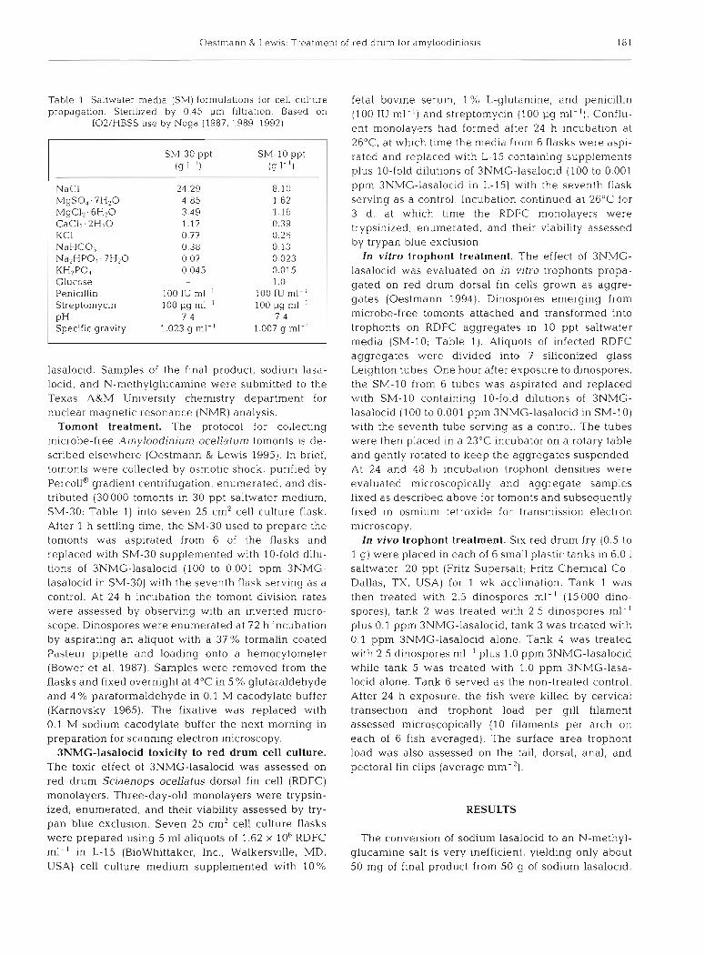
Fig. 2. Amyloodinium ocellatum. Tomonts incubated 72 h in 0.1 ppm 3NMG-lasalocid. Cracked and rup- tured tomonts spilling white starch grains: irreqular and non-dividing tomonts also can be seen. xlOOO
(scale bar = 10 pm)
96% while the 10 to 0.001 pprn 3NMG-lasalocid treat- ment viabilities were 93, 98, 98, 97, and 98% respec- tively. The cell count also was inversely proportional to the 3NMG-lasalocid dose. The control cell count was 6.8 X 10⁵ RDFC ml-l while the cell counts for the 10 to 0.001 pprn 3NMG-lasalocid treatment were 1.3 X 10⁵, 5.2 X 10⁵, 6.4 X 10⁵, 6.9 X 10⁵, and 6.8 X 10⁵RDFC ml-' respectively.
The 24 h tomont division rate also decreased in a dose-dependent manner when treated with 3NMG- lasalocid. The control tomont division rate was 82% compared to 59, 69, 75, 78, 80, and 81 % (average of 3 trials) observed for the 100 to 0.001 pprn 3NMG-lasa- locid dilutions respectively. Irregular division, cracked and ruptured tomonts, and release of starch grains were observed by scanning electron microscopy of 3NMG-lasalocid treated tomonts (Fig. 2). Dinospores excysted but were deformed and non-motile at 1 to 0.01 pprn 3NMG-lasalocid. Motile dinospores did not excyst until the 3NMG-lasalocid concentration was reduced to 0.001 ppm, and then at a concentration 44 "0 that of controls (27 800 dinospores ml-' for con- trol vs 12250 dinospores ml-' at 0.001 pprn 3NMG- lasalocid)
Subjectively, the in vitro trophont infection on RDFC aggregates was observed to decrease in a dose-depen- dent manner Trophonts were not present in sufficient numbers for electron microscopic examination until the 3NMG-lasalocid concentration was decreased to 0.01 ppm. Effects of the 3NMG-lasalocid treatment ob- served by transmission electron microscopy were simi- lar to effects previously described in coccidian parasites (Ryley 1969, Scholtyseck et al. 1971, 1972, Vetterling et al. 1973, Mehlhorn et al. 1983, Smith & Galloway 1983, Daszak et al. 1991) (Figs. 3 & 4). Internal membranous organelles of trophonts were disrupted and degener-
Oestmann & Lewls: Tredtment of red d r u n ~ tor dmyloocii~i~os~s 183
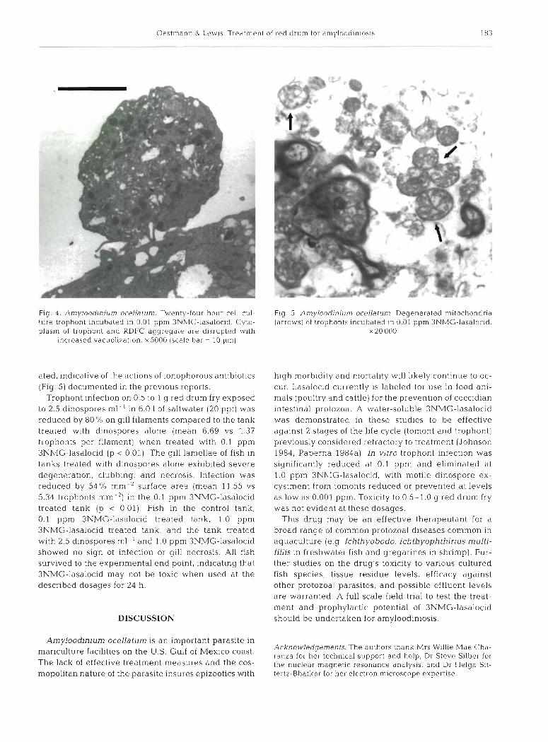
Fig. 4. Amylood~nium ocellatum. Twenty-four hour cell cul- ture trophont incubated in 0.01 pprn 3NMG-lasalocid. Cyto- plasm of trophont and RDFC aggregate are disrupted 1~1th
~ncreased vacuolization. x5000 (scale bar = 10 pm)
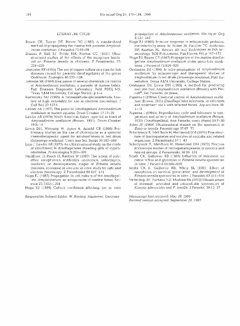
ated, indicative of the actions of ionophorous antibiotics (Fig. 5) documented in the previous reports.
Trophont infection on 0.5 to 1 g red drum fry exposed to 2.5 dinospores ml-' in 6.0 1 of saltwater (20 ppt) was reduced by 80 % on gill filaments compared to the tank treated with dinospores alone (mean 6.69 vs 1.37 trophonts per filament) when treated with 0.1 ppm 3NMG-lasalocid (p c 0.01). The gill lamellae of fish in tanks treated with dinospores alone exhibited severe degeneration, clubbing, and necrosis. Infection was reduced by 54 O/o mm-2 surface area (mean 11.55 vs 5.34 trophonts mm-2) in the 0.1 pprn 3NMG-lasalocid treated tank (p < 0.01). Fish in the control tank, 0.1 pprn 3NMG-lasalocid treated tank, 1.0 pprn 3NMG-lasalocid treated tank, and the tank treated with 2.5 dinospores ml-' and 1.0 ppm 3Nh4G-lasalocid showed no sign of infection or gill necrosis. All fish survived to the experimental end point, indicating that 3NMG-lasalocid may not be toxic when used at the described dosages for 24 h.
DISCUSSION
Amyloodinium ocellatum is an important parasite in mariculture facilities on the U.S. Gulf of Mexico coast. The lack of effective treatment measures and the cos- mopolitan nature of the parasite insures epizootics with
Fig. 5. An~yloodjnium oceUatum Degenerated mitochondr~a (arrows) of trophonts incubated in 0.01 pprn 3NMG-lasaloc~d.
X 20 000
high morbidity a.nd mortality will likely continue to oc- cur. Lasalocid currently is labeled for use in food ani- mals (poultry and cattle) for the prevention of coccidian intestinal protozoa. A water-soluble 3NMG-lasalocid was demonstrated in these studies to be effective against 2 stages of the life cycle (tomont and trophont) previously considered refractory to treatment (Johnson 1984, Paperna 1984a). 01 vitro trophont infection was s~gnificantly reduced at 0.1 pprn and eliminated at 1.0 pprn 3Nh'IG-lasalocid, with motile dinospore ex- cystment from tomonts reduced or prevented at levels as low as 0.001 ppm. Toxicity to 0.5-1.0 g red drum fry was not evident at these dosages.
This drug may be an effective therapeutant for a broad range of common protozoa1 diseases common in aquaculture (e.g Ichthyobodo, Ichth yophthirius multi- filiis in freshwater fish and gregarines in shrimp). Fur- ther studies on the drug's toxicity to various cultured fish species, tissue residue levels, efficacy against other protozoa1 parasites, and possible effluent levels are warranted. A full scale field trial to test the treat- ment and prophylactic potential of 3NMG-lasalocid should be undertaken for amyloodiniosis.
Acknowledgements. The authors thank Mrs Willie Mae Cha- ranza for her technical support and help, Dr Steve Silber for the nuclear magnetlc resona.nce analysis, and Dr Helga Sit- tertz-Bhatkar for her electron microscope expertise
184 Dis aquat Org 24: 179-184, 1996
LITERATURE CITED
Bower CE, Turner DT, Biever RC (1987) A standardized method of propagating the marine fish parasite Amylood- inium ocellatum. J Parasitol 73:85-88
Daszak P, Ball SJ, Plttilo RM, Norton CC (1991) Ultra- structural studies of the effects of the ionophore lasalo- c ~ d on Eimeria tenella in chickens. Z Parasitenkd 77. 224-229
Dempster RP (1955) The use of copper sulfate as a cure for fish diseases caused by parasitic dinoflagellates of the genus Oodinium. Zoologica 40,133-138
Johnson SK (1984) Evaluation of several chemicals for control of Amyloodinium ocellatum, a parasite of marine fishes. F ~ s h Diseases Diagnostic Laboratory Pub1 FDDL-MS. Texas A&M University, College Station, p 1-4
Karnovsky MJ (1965) A formaldehyde-glutaraldehyde fixa- tive of high osmolality for use in electron microscopy. J Cell B101 27:137A
Lawier A K (1977) Tne parasitic ainoiiageiiare Amylooainium ocellatum in marine aquaria. Drum Croaker 17:17-20
Lawler AR (1979) North American fishes reported as hosts of Amyloodinium ocellatum (Brown, 1931). Drum Croaker 19:8-14
Lewis DH, Wenxing W, Ayers A, Arnold CR (1988) Pre- llmlnary studies on the use of chloroquine as a systemic chemotherapeutic agent for amyloodiniosis in red drum (Sciaenops ocellatus). Contr mar SCI, Suppl30:183-189
Lom J, Lawler AR (1973) An ultrastructural study on the mode of attachment in dinoflagellates invading gills of cyprin- odontidae. Protistologica 9:293-309
Mehlhorn H, Pooch H, Raether W (1983) The action of poly- ether ionophorous antibiotics (monensin, salinomycln, lasalocid) on developmental stages of Eirneria tenella (coccidia, sporozoa) in vivo and in vitro: study by light and electron microscopy. Z Parasitenkd 69:457-471
Noga EJ (1987) Propagation in cell culture of the dinoflagel- late Amyloodinium, an ectoparasite of marine fishes. Sci- ence 23:1302-1304
Noga EJ (1989) Culture conditions affecting the in v~tro
Responsible Subject Editor: W. Korting, Hannover, Germany
propagation of Amyloodinium ocellatum. Dis aquat Org 6.137-143
Noga EJ (1992) Immune response to ectoparasite protozoa: the infectivity assay. In: Stolen JS, Fletcher TC, Anderson DP, Kaattari SL, Rowley AF (ed) Techniques in fish im- munology. SOS Publications, Fair Haven. NJ, p 167-175
Noga EJ, Bower CE (1987) Propagation of the marine dlnofla- gellate Amyloodin~um ocellatum under germ-free condl- tions. J Parasitol 73:924-928
Oestmann DJ (1994) In vitro propagation of Amyloodinium ocellatum for immunologic and therapeutic studies of amyloodinium in red drum (Sciaenops ocellatus). PhD dis- sertation, Texas A&kl University, College Station
Oestmann DJ, Lewis DH (1995) A method for producing mlcrobe-free Amylood~ni~~m ocellatum (Brown) with Per- toll@'. Vet Parasitol (in press)
Paperna I (1984a) Chemical control of Amyloodinium ocella- tum (Brown, 1931) (Dinoflagellida) infections: in vitro tests and treatment trials with infected fishes Aquaculture 38: i - i8
Paperna 1 (198413) Reproduction cycle and tolerance to tem- perature and salinlty of Amyloodinium ocellatum (Brown, 1931) (Dinoflagellida). Ann Parasitol comp (Paris) 59:7-30
Ryley J F (1969) Ultrastructural studies on the sporozoite of Eimeria tenella. Parasitology 37:6?-72
Scholtyseck E, Mehlhorn H, Hammond DM (1971) Fine struc- ture of macrogametes and oocytes of coccidia and related organisms. Z Parasitenkd 37:l-43
Scholtyseck E, Mehlhorn H, Hammond DM (1972) Electron microscope studies of microgametogenesis in coccidia and related groups. Z Parasitenkd 38:95-131
Smith CK, Galloway RB (1983) Influence of monensin on cation influx and glycolysis of Eimena tenella sporozoltes in vjtro. J Parasitol 69:666-670
Smith CK 11, Galloway RB, White SL (1981) Effect of ionophores on survival, penetration, and development of Eimeria tenella sporozoites in vitro. J Parasitol 67:511-516
Vetterling JM, Pacheco ND, Madden PA (1973) Ultrastructure of dormant, act~vated and intracellular sporozoites of Eimena adenoeides and E, tenella J Parasitol 59:15-27
Manuscript first received: May 29, 1995 Revised version accepted: September 20, 1995