Embed Size (px)
DESCRIPTION
Kedokteran gigi
Citation preview
Hindawi Publishing CorporationCase Reports in DentistryVolume 2013, Article ID 745959, 6 pageshttp://dx.doi.org/10.1155/2013/745959
Case ReportCombined Treatment with Laser Sintering and Zirconium:A Case Report of Dentinogenesis Imperfecta
Simel Ayyildiz,1 Cem Sahin,2 Özlem Marti Akgün,3 and Feridun Basak3
1 Department of Prosthodontics, Center of Dental Sciences, Gulhane Military Medical Academy, Etlik, 06018 Ankara, Turkey2 School of Dental Technology, Hacettepe University, 06100 Ankara, Turkey3 Department of Pediatric Dentistry, Center of Dental Sciences, Gulhane Military Medical Academy, 06018 Ankara, Turkey
Correspondence should be addressed to Simel Ayyildiz; [email protected]
Received 18 January 2013; Accepted 9 February 2013
Academic Editors: P. G. Arduino, W. L. Chai, and M. W. Roberts
Copyright © 2013 Simel Ayyildiz et al. This is an open access article distributed under the Creative Commons Attribution License,which permits unrestricted use, distribution, and reproduction in any medium, provided the original work is properly cited.
Osteogenesis imperfecta (OI) is a heterogeneous disorder of connective tissue that manifests mainly as skeletal deformity and bonefragility. Dentinogenesis imperfecta (DI) is sometimes an accompanying symptom of OI. The treatment protocol of these patientsvaries according to the clinical appearance. The case report here describes complete mouth rehabilitation of an 18-year-old malepatient with OI and DI using direct metal laser sintering (DMLS) technique of metal-ceramic restorations and zirconium all-ceramic crowns. DMLS is an additive metal fabrication technology that is simpler, more precise, and healthier than conventionalmanufacturing and can be remarkably cost effective. Moreover, the technique affords highly accurate production of fixed partialdentures with ideal marginal fit and excellent mechanical properties.The patient was treated using a multidisciplinary strategy thatfocused on controlling caries, protecting teeth from further wear, obtaining an appropriate vertical dimension, and providing softtissue support to return the facial profile to a normal appearance using new technology in the field of prosthetics.
1. Introduction
Osteogenesis imperfecta (OI), also known as “brittle bone”,disease is a genetically determined connective tissue disorderthat results frommutations of 2 genes (COL1A1 andCOL1A2)responsible for the formation of type 1 collagen [1–3].
Clinical features of OI include growth deficiency, bluesclera, bone fragility, joint hypermobility, dentinogenesisimperfect (DI), and the presence of wormian bones onskull radiographs [3, 4]. Additional radiographic findings areosteopenia, scoliosis, and thin cortices. OI is classified on thebasis of clinical and radiologic criteria established by Sillenceet al. in 1979 (Table 1) [5–7].
DI is an autosomal dominant form of mesodermal dys-plasia that affects both primary and permanent dentition.Shields [8] has identified 3 different types of DI as follows: DI-I: DI associated with OI; DI-II: the same clinical radiographicand histological features as DI-I, but without OI; and DI-III:a rare type of DI found only in the triracial population ofBrandywine, MD, USA.
DI associated with OI generally affects primary denti-tion more severely than permanent dentition. Although theenamel appears structurally normal, it is often dislodged,exposing soft, dysplastic dentin to the oral cavity and pro-voking rapid, extensive attrition [3, 9].Histologically, exposeddentin is generally characterized by irregular tubules. Radio-graphically, teeth affected by DI show cervical constriction,bulbous crowns, short roots, short pulp chambers, andobliterated canals [10]. Adult patients with OI frequentlyexhibit class III malocclusions, anterior or posterior cross-bite, posterior open-bite, and vertical height loss [1, 3, 11].
Conventional casting is the most frequently used tech-nique for manufacturing Co-Cr alloys for the fixed partialdentures. In recent years, modern computer-aided tech-nologies for manufacturing individual prostheses have beengaining popularity in the field of dental technology [12].Computer-aided design (CAD) and computer-aided man-ufacturing (CAM) technologies are used frequently in thedental field to fabricate prostheses ranging from crownsto long-span fixed partial dentures and from removable
2 Case Reports in Dentistry
Table 1: Classification of osteogenesis imperfecta.
Type DI Clinical severity Typical featuresI − Mild, nondeforming OI Normal height or mild short stature; blue sclera
II ? Perinatal lethal Multiple rib and long bone fractures at birth; pronounced deformities; broadlong bones; low density of skull bones on radiographs; and dark sclera
III + Severely deforming Very short; triangular face; severe scoliosis; and grayish scleraIV + Moderately deforming Moderately short; mild-to-moderate scoliosis; and grayish or white sclera
V − Moderately deforming Mild-to-moderate short stature; dislocation of radial head; mineralizedinterosseous membrane; hyperplastic callus; and white sclera
VI − Moderate-to-severe deforming Moderately short; scoliosis; accumulation of osteoid in bone tissue, fish-scalepattern of bone lamellation; and white sclera
VII − Moderately deforming Mild short stature; short humeri and femora; coxavara; and white scleraModified from [5, 7]. DI: Dentinogenesis Imperfecta.
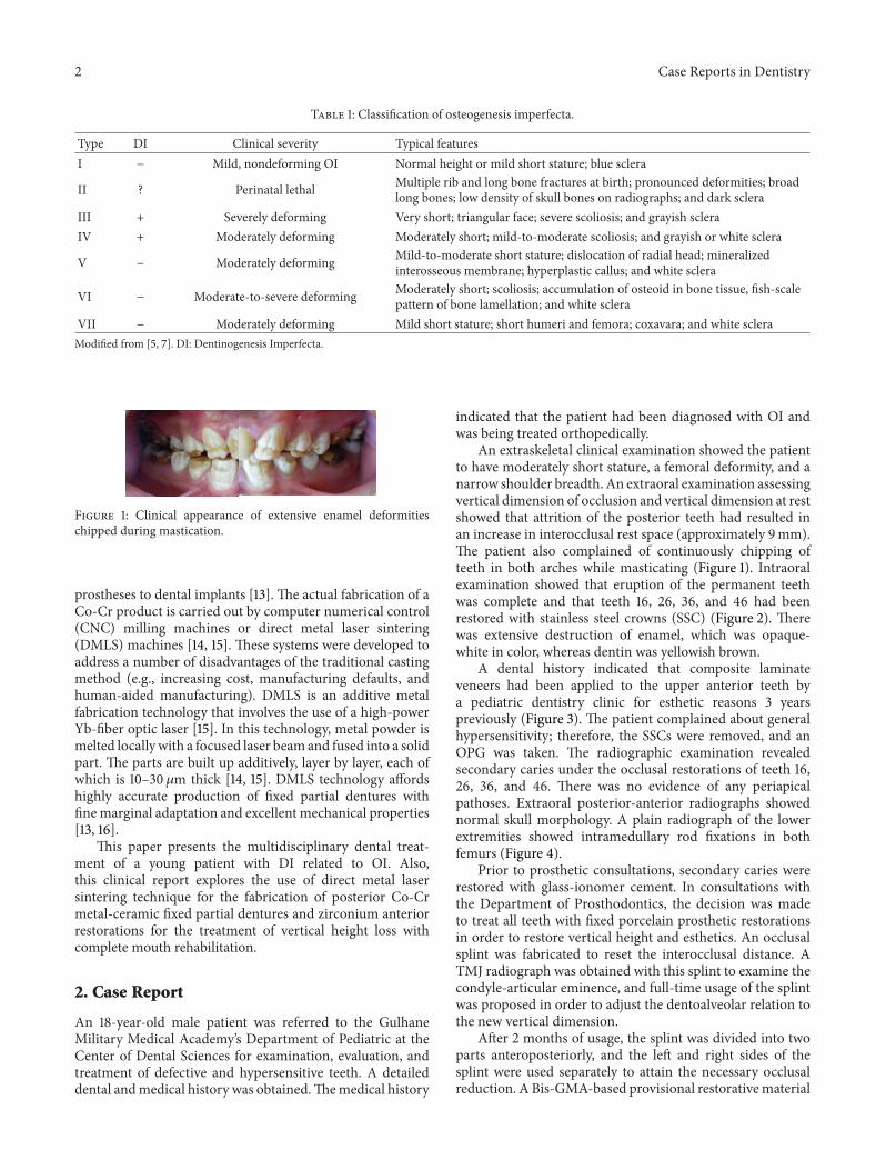
Figure 1: Clinical appearance of extensive enamel deformitieschipped during mastication.
prostheses to dental implants [13]. The actual fabrication of aCo-Cr product is carried out by computer numerical control(CNC) milling machines or direct metal laser sintering(DMLS) machines [14, 15]. These systems were developed toaddress a number of disadvantages of the traditional castingmethod (e.g., increasing cost, manufacturing defaults, andhuman-aided manufacturing). DMLS is an additive metalfabrication technology that involves the use of a high-powerYb-fiber optic laser [15]. In this technology, metal powder ismelted locallywith a focused laser beamand fused into a solidpart. The parts are built up additively, layer by layer, each ofwhich is 10–30 𝜇m thick [14, 15]. DMLS technology affordshighly accurate production of fixed partial dentures withfinemarginal adaptation and excellent mechanical properties[13, 16].
This paper presents the multidisciplinary dental treat-ment of a young patient with DI related to OI. Also,this clinical report explores the use of direct metal lasersintering technique for the fabrication of posterior Co-Crmetal-ceramic fixed partial dentures and zirconium anteriorrestorations for the treatment of vertical height loss withcomplete mouth rehabilitation.
2. Case Report
An 18-year-old male patient was referred to the GulhaneMilitary Medical Academy’s Department of Pediatric at theCenter of Dental Sciences for examination, evaluation, andtreatment of defective and hypersensitive teeth. A detaileddental andmedical history was obtained.Themedical history
indicated that the patient had been diagnosed with OI andwas being treated orthopedically.
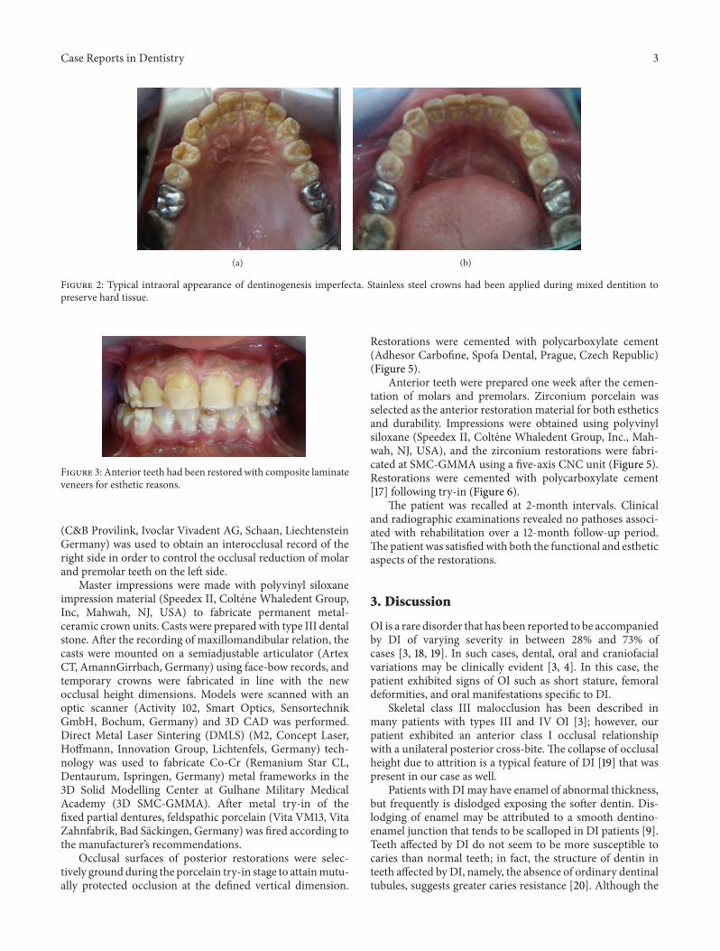
An extraskeletal clinical examination showed the patientto have moderately short stature, a femoral deformity, and anarrow shoulder breadth. An extraoral examination assessingvertical dimension of occlusion and vertical dimension at restshowed that attrition of the posterior teeth had resulted inan increase in interocclusal rest space (approximately 9mm).The patient also complained of continuously chipping ofteeth in both arches while masticating (Figure 1). Intraoralexamination showed that eruption of the permanent teethwas complete and that teeth 16, 26, 36, and 46 had beenrestored with stainless steel crowns (SSC) (Figure 2). Therewas extensive destruction of enamel, which was opaque-white in color, whereas dentin was yellowish brown.
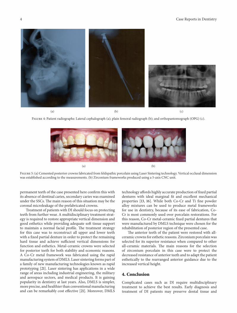
A dental history indicated that composite laminateveneers had been applied to the upper anterior teeth bya pediatric dentistry clinic for esthetic reasons 3 yearspreviously (Figure 3). The patient complained about generalhypersensitivity; therefore, the SSCs were removed, and anOPG was taken. The radiographic examination revealedsecondary caries under the occlusal restorations of teeth 16,26, 36, and 46. There was no evidence of any periapicalpathoses. Extraoral posterior-anterior radiographs showednormal skull morphology. A plain radiograph of the lowerextremities showed intramedullary rod fixations in bothfemurs (Figure 4).
Prior to prosthetic consultations, secondary caries wererestored with glass-ionomer cement. In consultations withthe Department of Prosthodontics, the decision was madeto treat all teeth with fixed porcelain prosthetic restorationsin order to restore vertical height and esthetics. An occlusalsplint was fabricated to reset the interocclusal distance. ATMJ radiograph was obtained with this splint to examine thecondyle-articular eminence, and full-time usage of the splintwas proposed in order to adjust the dentoalveolar relation tothe new vertical dimension.
After 2 months of usage, the splint was divided into twoparts anteroposteriorly, and the left and right sides of thesplint were used separately to attain the necessary occlusalreduction. A Bis-GMA-based provisional restorativematerial
Case Reports in Dentistry 3
(a) (b)
Figure 2: Typical intraoral appearance of dentinogenesis imperfecta. Stainless steel crowns had been applied during mixed dentition topreserve hard tissue.
Figure 3: Anterior teeth had been restored with composite laminateveneers for esthetic reasons.
(C&B Provilink, Ivoclar Vivadent AG, Schaan, LiechtensteinGermany) was used to obtain an interocclusal record of theright side in order to control the occlusal reduction of molarand premolar teeth on the left side.
Master impressions were made with polyvinyl siloxaneimpression material (Speedex II, Coltene Whaledent Group,Inc, Mahwah, NJ, USA) to fabricate permanent metal-ceramic crown units. Casts were preparedwith type III dentalstone. After the recording of maxillomandibular relation, thecasts were mounted on a semiadjustable articulator (ArtexCT, AmannGirrbach, Germany) using face-bow records, andtemporary crowns were fabricated in line with the newocclusal height dimensions. Models were scanned with anoptic scanner (Activity 102, Smart Optics, SensortechnikGmbH, Bochum, Germany) and 3D CAD was performed.Direct Metal Laser Sintering (DMLS) (M2, Concept Laser,Hoffmann, Innovation Group, Lichtenfels, Germany) tech-nology was used to fabricate Co-Cr (Remanium Star CL,Dentaurum, Ispringen, Germany) metal frameworks in the3D Solid Modelling Center at Gulhane Military MedicalAcademy (3D SMC-GMMA). After metal try-in of thefixed partial dentures, feldspathic porcelain (Vita VM13, VitaZahnfabrik, Bad Sackingen, Germany) was fired according tothe manufacturer’s recommendations.
Occlusal surfaces of posterior restorations were selec-tively groundduring the porcelain try-in stage to attainmutu-ally protected occlusion at the defined vertical dimension.
Restorations were cemented with polycarboxylate cement(Adhesor Carbofine, Spofa Dental, Prague, Czech Republic)(Figure 5).
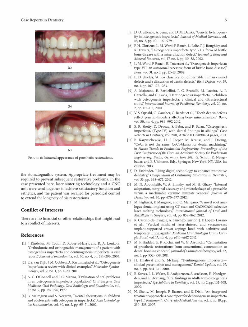
Anterior teeth were prepared one week after the cemen-tation of molars and premolars. Zirconium porcelain wasselected as the anterior restorationmaterial for both estheticsand durability. Impressions were obtained using polyvinylsiloxane (Speedex II, Coltene Whaledent Group, Inc., Mah-wah, NJ, USA), and the zirconium restorations were fabri-cated at SMC-GMMA using a five-axis CNC unit (Figure 5).Restorations were cemented with polycarboxylate cement[17] following try-in (Figure 6).
The patient was recalled at 2-month intervals. Clinicaland radiographic examinations revealed no pathoses associ-ated with rehabilitation over a 12-month follow-up period.The patient was satisfiedwith both the functional and estheticaspects of the restorations.
3. Discussion
OI is a rare disorder that has been reported to be accompaniedby DI of varying severity in between 28% and 73% ofcases [3, 18, 19]. In such cases, dental, oral and craniofacialvariations may be clinically evident [3, 4]. In this case, thepatient exhibited signs of OI such as short stature, femoraldeformities, and oral manifestations specific to DI.
Skeletal class III malocclusion has been described inmany patients with types III and IV OI [3]; however, ourpatient exhibited an anterior class I occlusal relationshipwith a unilateral posterior cross-bite. The collapse of occlusalheight due to attrition is a typical feature of DI [19] that waspresent in our case as well.
Patients with DI may have enamel of abnormal thickness,but frequently is dislodged exposing the softer dentin. Dis-lodging of enamel may be attributed to a smooth dentino-enamel junction that tends to be scalloped in DI patients [9].Teeth affected by DI do not seem to be more susceptible tocaries than normal teeth; in fact, the structure of dentin inteeth affected by DI, namely, the absence of ordinary dentinaltubules, suggests greater caries resistance [20]. Although the
4 Case Reports in Dentistry
(a) (b) (c)
Figure 4: Patient radiographs: Lateral cephalograph (a); plain femoral radiograph (b); and orthopantomograph (OPG) (c).
(a) (b)
Figure 5: (a) Cemented posterior crowns fabricated from feldspathic porcelain using Laser Sintering technology. Vertical occlusal dimensionwas established according to the measurements. (b) Zirconium frameworks produced using a 5-axis CNC unit.
permanent teeth of the case presented here confirm this withits absence of dentinal caries, secondary caries was examinedunder the SSCs. The main reason of this situation may be thecoronal microleakage of the prefabricated crowns.
Treatment of patients with DI should focus on protectingteeth from further wear. A multidisciplinary treatment strat-egy is required to restore appropriate vertical dimension andgood esthetics while providing adequate soft tissue supportto maintain a normal facial profile. The treatment strategyfor this case was to reconstruct all upper and lower teethwith a fixed partial denture in order to protect the remaininghard tissue and achieve sufficient vertical dimensions forfunction and esthetics. Metal-ceramic crowns were selectedfor posterior teeth for both stability and economic reasons.A Co-Cr metal framework was fabricated using the rapidmanufacturing systemofDMLS. Laser sintering forms part ofa family of new manufacturing technologies known as rapidprototyping [21]. Laser sintering has applications in a widerange of areas including industrial engineering, the militaryand aerospace sectors, and medical products. It is gainingpopularity in dentistry at last years. Also, DMLS is simpler,more precise, and healthier than conventionalmanufacturingand can be remarkably cost effective [21]. Moreover, DMLS
technology affords highly accurate production of fixed partialdentures with ideal marginal fit and excellent mechanicalproperties [13, 16]. While both Co-Cr and Ti fine powderalloy mixtures can be used to produce metal frameworksfor use in dentistry, because of its ease of fabrication, Co-Cr is most commonly used over porcelain restorations. Forthis reason, Co-Cr metal-ceramic fixed partial dentures thatwere manufactured by DMLS technique were chosen for therehabilitation of posterior region of the presented case.
The anterior teeth of the patient were restored with all-ceramic crowns for esthetic reasons. Zirconiumporcelainwasselected for its superior resistance when compared to otherall-ceramic materials. The main reasons for the selectionof zirconium porcelain in this case were to protect thedecreased resistance of anterior teeth and to adapt the patientesthetically to the rearranged anterior guidance due to theincreased vertical height.
4. Conclusion
Complicated cases such as DI require multidisciplinarytreatment to achieve the best results. Early diagnosis andtreatment of DI patients may preserve dental tissue and
Case Reports in Dentistry 5
(a)
(b)
(c)
Figure 6: Intraoral appearance of prosthetic restorations.
the stomatognathic system. Appropriate treatment may berequired to prevent subsequent restorative problems. In thecase presented here, laser sintering technology and a CNCunit were used together to achieve satisfactory function andesthetics, and the patient was recalled for periodical controlto extend the longevity of his restorations.
Conflict of Interests
There are no financial or other relationships that might leadto a conflict of interests.
References
[1] J. Kindelan, M. Tobin, D. Roberts-Harry, and R. A. Loukota,“Orthodontic and orthognathic management of a patient withosteogenesis imperfecta and dentinogenesis imperfecta: a casereport,” Journal of orthodontics, vol. 30, no. 4, pp. 291–296, 2003.
[2] F. S. vanDijk, J.M. Cobben, A. Kariminejad et al., “OsteogenesisImperfecta: a review with clinical examples,”Molecular Syndro-mology, vol. 2, no. 1, pp. 1–20, 2011.
[3] A. C. O’Connell and J. C. Marini, “Evaluation of oral problemsin an osteogenesis imperfecta population,” Oral Surgery, OralMedicine, Oral Pathology, Oral Radiology, and Endodontics, vol.87, no. 2, pp. 189–196, 1999.
[4] B. Malmgren and S. Norgren, “Dental aberrations in childrenand adolescents with osteogenesis imperfecta,”Acta Odontolog-ica Scandinavica, vol. 60, no. 2, pp. 65–71, 2002.
[5] D. O. Sillence, A. Senn, and D. M. Danks, “Genetic heterogene-ity in osteogenesis imperfecta,” Journal of Medical Genetics, vol.16, no. 2, pp. 101–116, 1979.
[6] F. H. Glorieux, L.M.Ward, F. Rauch, L. Lalic, P. J. Roughley, andR. Travers, “Osteogenesis imperfecta type VI: a form of brittlebone disease with a mineralization defect,” Journal of Bone andMineral Research, vol. 17, no. 1, pp. 30–38, 2002.
[7] L.M.Ward, F. Rauch, R. Travers et al., “Osteogenesis imperfectatype VII: an autosomal recessive form of brittle bone disease,”Bone, vol. 31, no. 1, pp. 12–18, 2002.
[8] E. D. Shields, “A new classification of heritable human enameldefects and a discussion of dentin defects,” Birth Defects, vol. 19,no. 1, pp. 107–127, 1983.
[9] A. Majorana, E. Bardellini, P. C. Brunelli, M. Lacaita, A. P.Cazzolla, and G. Favia, “Dentinogenesis imperfecta in childrenwith osteogenesis imperfecta: a clinical and ultrastructuralstudy,” International Journal of Paediatric Dentistry, vol. 20, no.2, pp. 112–118, 2010.
[10] V. S. Opsahl, C. Gaucher, C. Bardet et al., “Tooth dentin defectsreflect genetic disorders affecting bone mineralization,” Bone,vol. 50, no. 4, pp. 989–997, 2012.
[11] S. R. Shetty, D. Dsouza, S. Babu, and P. Balan, “Osteogenesisimperfecta, (Type IV) with dental findings in siblings,” CaseReports in Dentistry, vol. 2011, Article ID 970904, 4 pages, 2011.
[12] B. Karpuschewski, H. J. Pieper, M. Krause, and J. Doring,“CoCr is not the same: CoCr-blanks for dental machining,”in Future Trends in Production Engineering: Proceedings of theFirst Conference of the German Academic Society for ProductionEngineering, Berlin, Germany, June 2011, G. Schuh, R. Neuge-bauer, and E. Uhlmann, Eds., Springer, New York, NY, USA, 1stedition, 2013.
[13] D. Fasbinder, “Using digital technology to enhance restorativedentistry,” Compendium of Continuing Education in Dentistry,vol. 33, pp. 668–672, 2012.
[14] M. N. Aboushelib, W. A. Elmahy, and M. H. Ghazy, “Internaladaptation, marginal accuracy and microleakage of a pressableversus a machinable ceramic laminate veneers,” Journal ofDentistry, vol. 40, pp. 670–677, 2012.
[15] M. Figliuzzi, F. Mangano, and C. Mangano, “A novel root ana-logue dental implant using CT scan and CAD/CAM: selectivelaser melting technology,” International Journal of Oral andMaxillofacial Surgery, vol. 41, pp. 858–862, 2012.
[16] R. Castillo-de-Oyague, A. Sanchez-Turrion, J. F. Lopez- Lozanoet al., “Vertical misfit of laser-sintered and vacuum-castimplant-supported crown copings luted with definitive andtemporary luting agents,”Medicina Oral Patologia Oral y Ciru-gia Bucal, vol. 17, no. 4, pp. e610–e617, 2012.
[17] M. F. Haddad, E. P. Rocha, and W. G. Assuncao, “Cementationof prosthetic restorations: from conventional cementation todental bonding concept,” Journal of Craniofacial Surgery, vol. 22,no. 3, pp. 952–958, 2011.
[18] H. Dhaliwal and S. McKaig, “Dentinogenesis imperfecta—clinical presentation and management,” Dental Update, vol. 37,no. 6, pp. 364–371, 2010.
[19] R. Sæves, L. L.Wekre, E. Ambjørnsen, S. Axelsson, H. Nordgar-den, and K. Storhaug, “Oral findings in adults with osteogenesisimperfecta,” Special Care in Dentistry, vol. 29, no. 2, pp. 102–108,2009.
[20] N. Shetty, M. Joseph, P. Basnet, and S. Dixit, “An integratedtreatment approach: a case report for dentinogenesis imperfectatype II,”KathmanduUniversityMedical Journal, vol. 5, no. 18, pp.230–233, 2007.

6 Case Reports in Dentistry
[21] U. Iseri, Z. Ozkurt, and E. Kazazoglu, “Shear bond strengthsof veneering porcelain to cast, machined and laser-sinteredtitanium,” Dental Materials, vol. 30, no. 3, pp. 274–280, 2011.

Submit your manuscripts athttp://www.hindawi.com
Hindawi Publishing Corporationhttp://www.hindawi.com Volume 2014
Oral OncologyJournal of
DentistryInternational Journal of
Hindawi Publishing Corporationhttp://www.hindawi.com Volume 2014
Hindawi Publishing Corporationhttp://www.hindawi.com Volume 2014
International Journal of
Biomaterials
Hindawi Publishing Corporationhttp://www.hindawi.com Volume 2014
BioMed Research International
Hindawi Publishing Corporationhttp://www.hindawi.com Volume 2014
Case Reports in Dentistry
Hindawi Publishing Corporationhttp://www.hindawi.com Volume 2014
Oral ImplantsJournal of
Hindawi Publishing Corporationhttp://www.hindawi.com Volume 2014
Anesthesiology Research and Practice
Hindawi Publishing Corporationhttp://www.hindawi.com Volume 2014
Radiology Research and Practice
Environmental and Public Health
Journal of
Hindawi Publishing Corporationhttp://www.hindawi.com Volume 2014
The Scientific World JournalHindawi Publishing Corporation http://www.hindawi.com Volume 2014
Hindawi Publishing Corporationhttp://www.hindawi.com Volume 2014
Dental SurgeryJournal of
Drug DeliveryJournal of
Hindawi Publishing Corporationhttp://www.hindawi.com Volume 2014
Hindawi Publishing Corporationhttp://www.hindawi.com Volume 2014
Oral DiseasesJournal of
Hindawi Publishing Corporationhttp://www.hindawi.com Volume 2014
Computational and Mathematical Methods in Medicine
ScientificaHindawi Publishing Corporationhttp://www.hindawi.com Volume 2014
PainResearch and TreatmentHindawi Publishing Corporationhttp://www.hindawi.com Volume 2014
Preventive MedicineAdvances in
Hindawi Publishing Corporationhttp://www.hindawi.com Volume 2014
EndocrinologyInternational Journal of
Hindawi Publishing Corporationhttp://www.hindawi.com Volume 2014
Hindawi Publishing Corporationhttp://www.hindawi.com Volume 2014
OrthopedicsAdvances in