Embed Size (px)
Citation preview

This article was downloaded by: [University of Saskatchewan Library]On: 17 September 2012, At: 02:15Publisher: Taylor & FrancisInforma Ltd Registered in England and Wales Registered Number: 1072954 Registeredoffice: Mortimer House, 37-41 Mortimer Street, London W1T 3JH, UK
Diatom ResearchPublication details, including instructions for authors andsubscription information:http://www.tandfonline.com/loi/tdia20
KOBAYASIELLA SPECIES OF THECARPATHIAN REGION: MORPHOLOGY,TAXONOMY AND DESCRIPTION OF K.TINTINNUS SPEC. NOV.Krisztina Buczkó a , Agata Zofia Wojtal b & Regine Jahn ca Hungarian Natural History Museum, Department of Botany, P.O. Box222, 1476, Budapest, Hungaryb Department of Phycology, W. Szafer Institute of Botany, PolishAcademy of Sciences, Lubicz 46, 31-512, Kraków, Polandc Botanical Garden and Botanical Museum Berlin-Dahlem, FreieUniversität Berlin, Königin-Luise Str. 6–8, D-14195, Berlin, Germany
Version of record first published: 31 Oct 2011.
To cite this article: Krisztina Buczkó, Agata Zofia Wojtal & Regine Jahn (2009): KOBAYASIELLA SPECIESOF THE CARPATHIAN REGION: MORPHOLOGY, TAXONOMY AND DESCRIPTION OF K. TINTINNUS SPEC.NOV., Diatom Research, 24:1, 1-21
To link to this article: http://dx.doi.org/10.1080/0269249X.2009.9705780
PLEASE SCROLL DOWN FOR ARTICLE
Full terms and conditions of use: http://www.tandfonline.com/page/terms-and-conditions
This article may be used for research, teaching, and private study purposes. Anysubstantial or systematic reproduction, redistribution, reselling, loan, sub-licensing,systematic supply, or distribution in any form to anyone is expressly forbidden.
The publisher does not give any warranty express or implied or make any representationthat the contents will be complete or accurate or up to date. The accuracy of anyinstructions, formulae, and drug doses should be independently verified with primarysources. The publisher shall not be liable for any loss, actions, claims, proceedings,demand, or costs or damages whatsoever or howsoever caused arising directly orindirectly in connection with or arising out of the use of this material.

Diatom Research (2009), Volume 24 (l), 1-21
KOBAYASZELLA SPECIES OF THE CARPATHIAN REGION: MORPHOLOGY, TAXONOMY AND DESCRIPTION OF
K. TINTINNUS SPEC. NOV.
Krisztina Buczkb'
Hungarian Natural History Museum, Department of Botany, 1476 Budapest, P. 0. Box 222, Hungary
Agata ZofiaWojtal' Department of Phycology, W. Szafer Institute of Botany,
Polish Academy of Sciences, Lubicz 46, 31-512 Krakdw, Poland
Regine Jahn3 Botanical Garden and Botanical Museum Berlin-Dahlem,
Freie Universitat Berlin, Konigin-Luise Str. 6-8, D-14195 Berlin, Germany
Key words: Kobayasiella, Navicula subtilissima, Carpathian region, taxonomy, nomenclature, biogeography, morphology, nova species
The representatives of Kobayasiella Lange-Bertalot are characteristic of acidic and oligotrophic habitats. Detailed morphological observations using light (LM) and scanning electron (SEM) microscopy were performed on Carpathian populations of six species of the genus. The closely related species can easily be confused mainly due to their fine striation, but they show good distinguishing features in SEM. A comparison of the main features of the six species studied was presented, including central area, the shape of the raphe terminal fissure, the form of the kink on the raphe ~ which is the most important diagnostic criterion of Kobayasiella genus. In addition, the nomenclature and taxonomy of some European finely striated diatoms formerly belonging to the Navicula subtilissima group was reviewed. Kobayasiella tintinnus Buczk6, Wojtal & R. Jahn was described here as a new species fiom Lake Saint Anna (Romania). In order to confirm validity of Kobayasiella tintinnus sp. nov. and to provide the perfect separation from its sister species (K. micropunctata and K. parasubtilissirna) continuous morphometric characters were analysed by Linear Discriminant Analysis.
INTRODUCTION
Kobayasiella belongs to a series of genera recently split from Navicula sensu lato, and includes a morphologically and ecologically coherent group of taxa. It was first described under the name of Kobuyasia by Lange-Bertalot (1 996), but since this name appeared to be an accepted genus of Fungi belonging to the Basidiomycetes, it was modified in 1999 by Lange- Bertalot.
' [email protected]; [email protected] [email protected]
Dow
nloa
ded
by [
Uni
vers
ity o
f Sa
skat
chew
an L
ibra
ry]
at 0
2:15
17
Sept
embe
r 20
12
2 K. BUCZKO, A.Z. WOJTAL & R. JAHN
Kobayasiella representatives are characterized by delicate frustule ornamentation and predominantly linear-lanceolate, narrow valves, with rostrate or capitate apices. The striae are generally very fine, more than 30 in 10 pm, often not resolvable in the light microscope. The stria pattern is remarkable: radiate in the centre, turning abruptly convergent near the apices. The raphe is filiform, externally with slightly expanded central terminal endings and strongly hooked polar terminal endings. Internally, polar raphe terminal end with a small helictoglossae. The crest-like valve margins between the valve surface and the mantle are also general features of the genus. The unique raphe feature, noted so far only amongst Kobayasiella species, is the kink in the raphe. Other names used for this genus-specific feature is “umbilicus ” (Kobayasi & Nagumo 1988) or “notch” (Vanhoutte et al. 2004), since the term ‘‘umbilicus’’ is used for a different meaning in diatom studies, for the central part of Hyalodiscus species (Kaczmarska & Rushforth 1983). Unambiguous identification in LM of Kobayasiella taxa is difficult because the main features of the genus can only be distinguished in SEM. Especially the small, finely ornamented species can be confused with small representatives of either Adlafia, Nupela, Stauroneis or other genera.
Although Kobayasiella species are widely distributed (e.g. Haworth et al. 1988, Kobayasi & Nagumo 1988, Vanhoutte et al. 2004), their occurrence is largely confined to oligotrophic habitats (Lange-Bertalot & Metzeltin 1996, Metzeltin & Lange-Bertalot 1998, Van de Vijver et al. 2002, Siver et al. 2005).
In contrast to the extended boreal peat lands of the northern hemisphere which provide favourable conditions for the diverse development of oligotrophic diatom flora, the degrading oligotrophic areas in the Central Europe are very limited and often anthropogenically influenced, which has resulted in a significant decrease of the most sensitive diatom taxa (NCmeth 2005). In this context Kobayasiella taxa could be classified as rare and endangered taxa with high indicative value, therefore the need to study of their occurrence and distribution.
In this paper, six Kobayasiella taxa from a diversity of acidic habitats in the Carpathian basin, including the description of one new species, were studied. Discriminant analysis (DA) was used to describe character combination that best discriminate between the newly proposed taxon and its closest relatives, in order to confirm validity of our prior classification (Podani 2000). In addition, the different concepts of the European Kobayasiella taxa were revised in order to clarify their taxonomy and nomenclature.
MATERIAL AND METHODS
In order to study the occurrences of Kobayasiella taxa from Central European, 29 mires in the Carpathian region were sampled between 1999 and 2007 (Fig. l), resulting in a total of 150 collected algal samples. The pH values were below 7 at every sampling point.
The samples were treated with hot hydrogen peroxide and nitric acid, washed several times, and the cleaned material was mounted in Zrax (refractive index is 1.7). For light microscope analysis a LEICA DM LB2 was used (100 X HCX PLAN APO, and Fujifilm Digital Camera FinePix S2 Pro). Scanning electron microscopy work was performed with a Hitachi S-26OON in Budapest and a Philips 510 in Berlin (Figs 45-50,82, 85, 88,91).
To prove the separation of the three close representatives of the genus (namely K. micropunctata, K. parasubtilissima and K. tintinnus), morphometric measurements were taken on 46 valves consisting of the five populations (Podani 2000). The following eight continuous (non-categorical) parameters were measured on SEM pictures: Total Length of valve (TL); Total Width of valve (TW); Number of Striae in the Middle section of valve (NSM); Number of Striae at Third of valve (NST); Width of the valve at Changeover Point (WCP); Distance of Changeover from the pore (DC); Neck Width (NW). WCP, DC, NW were measured on both sides of the valve, and the mean values were calculated. Arithmetic
Dow
nloa
ded
by [
Uni
vers
ity o
f Sa
skat
chew
an L
ibra
ry]
at 0
2:15
17
Sept
embe
r 20
12

KOBAYASIELLA SPECIES IN CARPATHIAN REGION 3
Fig. 1. Map of the sampling localities in Central European mires. Redrawn from EDDI database (Juggins
1 Barcs Nagyberek, 2 Barcs Szuriihely, 3 Barcs Tiva-t6,4 Balatonhenye Monostori lake, 5 Balatonhenye Barkas lake, 6 Csaroda Babtava, 7 Csaroda Nyirjes, 8 Farkasfa Fekete lake, 9 Grajka, 10 KelemCr, Kis mohos, 11 KelemCr, Nagy mohos, 12 Koszeg, 13 Mariaujfalu, Ordog lake, 14 Sirok Nyirjes t6, 15 Szakonyfalu, 16 Szoce, 17 Viszak, 18 Lake Saint Anna, 19 Tusnadfirdo (Biiile Tugnad), Mohos 20 Fenyvesteto, 21 Sfoartea at 1300 m a.s.1. 22 Molhagul (Mohos) dela Ciiliiple, 23 Molhasul de la Ragca, 24 Ragca de Sus, 25 MlaStin Dimbul Negru - La Pod, 26 Dgmbul Negru, a spring fen on the plateau, 27 Spring fen on the Muntele Mare plateau, at 1685 m alt, 28 Transitional bog on the Muntele Mare plateau, at 1715-1725 ma.s.l., 29 Raised bog on the Muntele Mare, at 17161725 m a.s.1.
2001)
Fig. 2. Schematic drawing of a Kobayasiella species, showing the measured characters were using in the morphometric analysis. Length of valve (TL); Width of valve (WT); Number of striae (NSM) at middle of valve; Number of striae (NST) at third of valve; Width of the valve (WCP) at changeover point; distance of changeover (DC) from the pore; neck width (NW); angle (ANG).
Dow
nloa
ded
by [
Uni
vers
ity o
f Sa
skat
chew
an L
ibra
ry]
at 0
2:15
17
Sept
embe
r 20
12

4 K. BUCZKO. A.Z. WOJTAL & R. JAHN
mean of the angles (ANG) measured at both side at one third of the valves. The number of striae was counted on 3 4 pm and projected to 10 pm (Fig. 2).
Terminology of the diatom fmstule follows Barber & Haworth (1 98 I).
RESULTS Kobayasiella elongata Buczko et Woj tal Buczko & Wojtal2007.
(Figs 3-10,70,73,76,79)
Valves are linear-lanceolate with a somewhat gibbous middle part and subcapitate to capitate ends; 3546 pm long (41.0 f 2.8 pm) and 3.4-4.5 pm wide (4.1 f 0.2 pm) (n=45). The striae are usually hardly resolvable in LM. In SEM, the striae are radiate and slightly sigmoid in the central part, and convergent towards the apices. Striae density is 4 W 8 in 10 pm, with intercalated shorter striae in the middle part of valves (Fig. 70). Each stria consists of a single elongated areola. Externally, the valve face is flat with a hyaline marginal rim between the face and the mantle; there are no other longitudinal ribs (Figs 8-10). The axial area is narrow, linear- lanceolate (Fig. 9), there is no fascia in the central area. (Fig. 70). Raphe is straight, filiform. There is a kink in the each raphe branch, about midway between the valve central area and apices (Fig. 79). Terminal fissures are strongly bent at an obtuse angle towards the same side (Fig. 73). The central raphe terminals end in a small straight helictoglossa (Fig. 10). The central pores form a somewhat expanded depression, both externally and internally (Figs 70, 76). The distance between the central pores on the outer surface is longer than on the inner surface, about 1.2-1.3 pm and 0.8 respectively (Figs 70, 76). The mantle bears a row of poroids which exactly corresponds to the number of striae (Fig. 70). Studied material and occurrence: Lake Saint Anna (site '1 8).
Kobayasiella madumensis (Jsrgensen) Lange-Bertalot (Figs 11-21,71,74,77,80) Lange-Bertalot (1 999), Zconographia Diatomologica, 6, p. 273
Basionym: Navicula madumensis E.G.Jsrgensen, Kongel. Danske Vidensk.-Selsk. Skr. 5, p. 60, pl. 2: fig. 26. 1948.
Valves are lanceolate with narrow capitate ends (Figs 11-19, 21, 75); 36-43 pm long (38.4 f 2.5 pm) and 7-8 pm wide (7.5 f 0.70) (n=28). The striae are usually not resolvable in LM, but sometimes they are visible as a pale shadow on the valve surface (Figs 12, 14, 16). In SEM, the valve face is large with a hyaline marginal rim between the valve face and the mantle; the striae are radiate in the central part to convergent at the apices. The angle between the raphe and the transapical striae is approximately 45 degree in the central part. Striae density is 34-38 in 10 pm, less dense in the middle part of valves, where intercalated shorter striae are inserted. Each stria consists of a single elongated areola; there are no longitudinal ribs. Externally, the valve face is flat with a hyaline marginal rim between the face and the mantle. The mantle bears a row of poroids which exactly correspond to the number to the striae (Fig. 77). The axial area is narrow, linear-lanceolate, the central area lanceolate. Raphe is straight, filiform. There is a kink in each raphe branch, about midway between the valve central area and the apices (Figs 17, 21, 80). Terminal fissures are bent to the same side in an obtuse angle. The polar raphe terminals end in a small straight helictoglossa. The distance between the central terminal endings on the outer surface is much longer than on the inner surface, 1 . 0 - 1 . 1 pm (Figs 17,20, 71) and only 0.05 pm (Figs 18,77).
Studied material and occurrence: Sirok (site 14), Lake Saint Anna (site 18).
Dow
nloa
ded
by [
Uni
vers
ity o
f Sa
skat
chew
an L
ibra
ry]
at 0
2:15
17
Sept
embe
r 20
12
KOBAYASIELLA SPECIES IN CARPATHIAN REGION 5
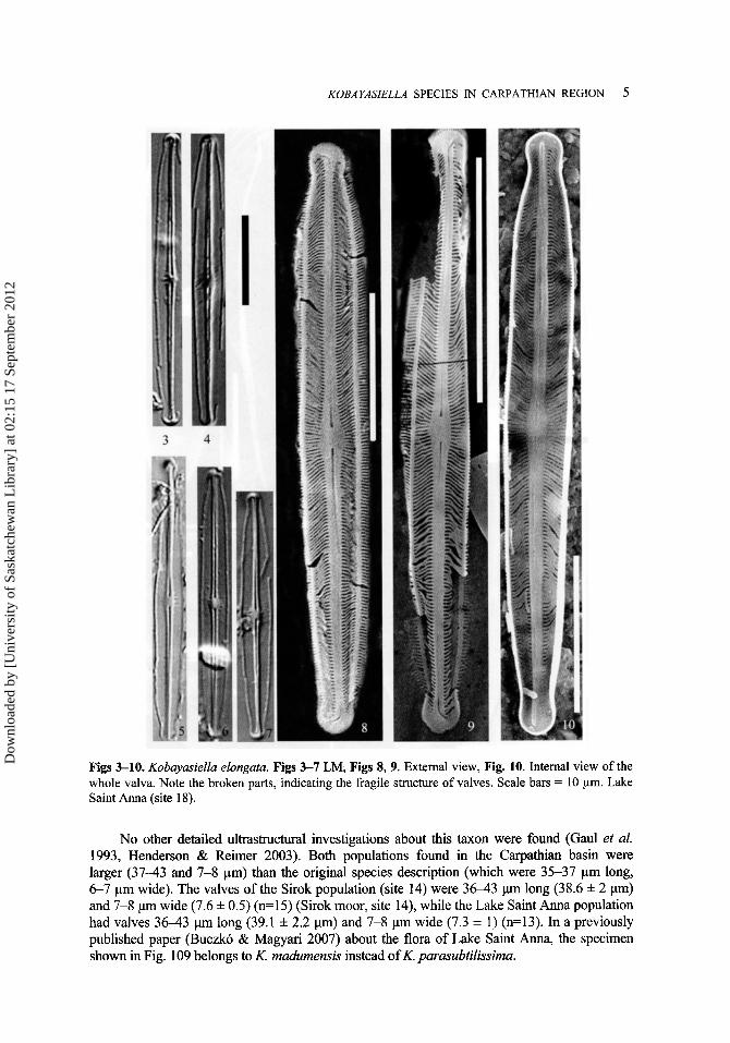
Figs 3-10. Kobuyusiellu elongutu. Figs 3-7 LM, Figs 8, 9. External view, Fig. 10. Internal view of the whole valva. Note the broken parts, indicating the fragile structure of valves. Scale bars = 10 pm. Lake Saint Anna (site 18).
No other detailed ultrastructural investigations about this taxon were found (Gaul et al. 1993, Henderson & Reimer 2003). Both populations found in the Carpathian basin were larger (37-43 and 7-8 pm) than the original species description (which were 35-37 pm long, 6-7 pm wide). The valves of the Sirok population (site 14) were 3 6 4 3 p long (38.6 f 2 pm) and 7-8 pm wide (7.6 f 0.5) (n=15) (Sirok moor, site 14), while the Lake Saint Anna population had valves 3 6 4 3 p long (39.1 f 2.2 pm) and 7-8 pm wide (7.3 f 1) (n=13). In a previously published paper (Buczko & Magyari 2007) about the flora of Lake Saint Anna, the specimen shown in Fig. 109 belongs to K. madumensis instead of K. parasubtilissima.
Dow
nloa
ded
by [
Uni
vers
ity o
f Sa
skat
chew
an L
ibra
ry]
at 0
2:15
17
Sept
embe
r 20
12

6 K. BUCZKO, A.Z. WOJTAL & R. JAHN
Figs 11-21. Kobuyusiellu rnadumensis. Fig. 11. Lectotype by Jorgensen (1948). Figs 12-16 LM, Figs 17, 19 SEM external view of the valves. Fig. 18. Internal view, Fig. 20. Central area of the valva with short visible intercalated striae. Fig. 21. Kink on the raphe (white arrow). Figs 17, 19-21 SEM external view. Scale bars = 10 pm (Figs 11-19); 2 pm (Figs 20,21). Figs 12, 13, 17-21 Lake Saint Anna (site 18), Figs 14-16 Sirok (site 14).
KobayasieZZa micropunctata (H. Germain) Lange-Bertalot (Figs 22-36,72,75,78,81) Lange-Bertalot 1999, Iconographia Diatomologica, 6, p. 273. Basionym: Navicula subtilissima var. micropunctata H. Germain, Flore Diatomkes eaux
douces et saumbtres, p. 234, fig. 169: 4-5. 1981.
Dow
nloa
ded
by [
Uni
vers
ity o
f Sa
skat
chew
an L
ibra
ry]
at 0
2:15
17
Sept
embe
r 20
12

KOBAYASIELLA SPECIES IN CARPATHIAN REGION 7
Figs 22-36. Kobayasiella micropunctata. Figs 22-29 LM, Figs 30,32. The external view of the raphe end. Note the fish-hook shaped polar terminal endings. Fig. 31. The external view of the central portion of valva. Note the different number of rows of poroids on the mantle and the striae. Figs 33-35. The external view of the whole valves. Fig. 36. The internal view of the raphe with helictoglossa. Scale bars = 10 pm (Figs 22-29,33-35); 2 pm (Figs 3&32,36).
Synonym: Navicula micropunctata (H. Germain) H. Kobayasi & Nagumo, Bot. Mag. Tokyo,
Synonym?: Navicula cumbriensis f. minor Haworth & Atkinson, nom. inval., Proceedings of 101, p. 247, figs 38-55. 1988.
the 9th International Diatom Symposium, p. 21, figs 15-16. 1988.
Dow
nloa
ded
by [
Uni
vers
ity o
f Sa
skat
chew
an L
ibra
ry]
at 0
2:15
17
Sept
embe
r 20
12

8 K. BUCZKO, A.Z. WOJTAL & R. JAHN
Valves are narrowly linear-lanceolate with capitate ends. The length of the valve varies between 17.5 and 23 pm (20.4 f 1.5), the width between 3.54.2 ((3.9 f 0.2) (n=14). In SEM, the valve face is almost flat (Figs 33-35), the valve mantle is deep (1.6-2 pm) with a hyaline marginal rim between the face and the mantle (Figs 30, 33). The raphe is straight, externally the central raphe terminal endings are straight (Figs 31, 72), distance between 0.8-1.2 pm externally and 1 pm internally. There is a kink on the raphe (Fig. 81). The polar raphe terminal endings are bent to the same side, they are characteristically fish-hook shaped (Figs 30, 32-35, 75) The raphe forms internally a small helictoglossa on the distal end (Fig. 36). The axial area is hyaline. Striae are radiate. The number of striae is 36-42 per 10 pm, each stria consists of a single, elongated areolae. The mantle bears a row of poroids that do not correspond in number to the transapical striae in the central part of valve. On the mantle, the number of rows is higher than the number of transapical striae (Fig. 31).
According to Kobayasi & Nagumo (1988) K. micropunctata is characterized by three features, namely the fish-hooked shaped terminal fissures, the non-corresponding numbers of transapical striae and poroids on the mantle at the central part of the valve. These two features are clearly visible in the studied population. The third criterion - “continuous striae closed externally by a hymenate pore occlusion with perforations arranged in a hexagonal array” can be observed only partially. The continuous striae are closed. The perforations can only be studied with TEM. Studied material: Mtii Gil5ului (Gyalui Havasok). NW part of the Muntele Mare (Oreghavas)
plateau (site 28). Occurrence: Grajka brook (site 9), NW part of the Muntele Mare (Oreghavas) plateau,
(site 28) and Mlastin Dfimbul Negru, La Pod (site 25).
In spite of the characteristic features of this species, clear discrimination of K. micropunctata between K. parasubtilissima is impossible without the aid of TEM and SEM. In LM, only the smaller valves with overlapping dimensions could identification be put in doubt. The co-occurrence andor coexistence of different Kobayasiella species have often been noted (i.e. Kobayasi & Nagumo 1988), causing difficulties in the other area of diatom research (e.g. palaeolimnological studies). More details about the morphological variability of K. micropunctata/parasubtilissima were discussed in Buczk6 (2007).
Kobaymit?hpasub&sima (H. Kobayasi & Nagumo) hge-Bertalot (Figs 37-50,82,85,88,91) Lange-Bertalot 1999, Iconographia Diatomologica, 6, p. 274. Basionym: Navicula parasubtilissima H. Kobayasi & Nagumo, Bot. Mag. Tokyo 101, p. 245,
Synonym pro parte: Navicula cumbriensis Haworth et Atkinson, nom. inval., Proceedings of the 9th International Diatom Symposium, p. 21, figs 12-14. 1988.
Valves are linear-lanceolate with capitate ends. The length of the valves varied between 22 and 31 pm, the width 4.0-4.7 pm. In SEM, the valve face is flat, with a hyaline marginal rim between the face and the mantle. The valve mantle is deep, up to 3.5 pm (Fig. 47). The raphe is straight with the kink somewhat in the middle (Figs 45, 91). The internal central raphe terminates in a T-shaped depression (Fig. 88); distally, the raphe ends in a small straight helictoglossa (Figs 46, 48). Terminal fissures are strongly bent at an obtuse angle to the same side, but have not the fish-hook shape (Figs 45, 49, 50, 85). The striae are usually not resolvable in LM. In SEM, the striae are radiate in the central part, and convergent at the apices. Stria density is 3 8 4 4 in 10 pm, with 3 W O in 10 pm in the middle, along with shorter intercalated striae. The mantle bears a row of poroids that clearly corresponds in number to
figs 19-37.1988.
Dow
nloa
ded
by [
Uni
vers
ity o
f Sa
skat
chew
an L
ibra
ry]
at 0
2:15
17
Sept
embe
r 20
12

KOBAYASIELLA SPECIES IN CARPATHIAN REGION 9
the transapical striae in the central part of valve. The central raphe terminal endings forms a somewhat expanded depression (Figs 45, 82), externally while T-shaped internally (Fig. 88). The external distance between the central raphe endings is longer, 1.2-1.3 pm (Figs 45, 82), than on the inner surface, 0.7-0.8 pm (Figs 46,88).
Studied material: Fenyves-teto, Romania, (site 20). Raised bog, with filamentous green algae and Drepanocladus (C. Muller) G . Roth. The studied populations corresponded well to the description by Kobayasi & Nagumo (1 988).
Occurrences: Sirok (site 14), Fenyves-teto (site 20), Mohos (site 19), Lake Saint Anna (site 18).
Figs 37-50. Kobayasiella parasubtilissima, Figs 37-44 L M , Fig. 45. External view of whole valva the arrows mark the kinks on the raphe. Fig. 46. Internal view of the whole valva. Fig. 47. Gridle view. Fig. 48. Internal view of the polar raphe terminal and helictoglossa. Fig. 49. Obtuse angle of polar raphe terminal (external view). Fig. 50. The broken valves allow an insight view into the frustule. Figs 3740. Belis 41-50 Fenyvesteto. Scale bars = 10 pm (Figs 3747); 2 pm (Figs 48-50).
Dow
nloa
ded
by [
Uni
vers
ity o
f Sa
skat
chew
an L
ibra
ry]
at 0
2:15
17
Sept
embe
r 20
12

10 K. BUCZKO, A.Z. WOJTAL & R. JAHN
Kobuyusiellu subtilissima (Cleve) Lange-Bertalot Lange-Bertalot, Iconographia Diatomologica, 6, p. 268. 1999. Basionym: Navicula subtilissima Cleve, Act. SOC. Fauna Flora Fennica, 8, p. 37, fig. 2: 35. 1891. Lectotype of Navicula subtilissima is by Kobayasi & Nagumo, Bot. Mag. Tokyo, 101, p. 240,
Synonym: Navicula simsii in Haworth et al., nom. inval. Proceedings of the 9th International
(Figs 51-58,83,86,89,92)
figs 1-18. 1988.
Diatom Symposium, fig. 17, 1988.
Figs 51-58. Kobayasiella subtilissirna. Figs 51-54. LM, Fig. 55. The central portion of valva showing the internal striation and the raphe terminal endings. Fig. 56. Polar raphe terminal endings and the row of poroids on the mantle. Figs 57, 58. The whole valves in SEM. White arrow shows the kink on the raphe. Figs 55-57. External views in SEM. Fig. 58. Internal view in SEM. Scale bars = 10 pm (Figs 51-54, 57, 58); 2 pm (Figs 55, 56). Fig. 51 Sirok (site 14), Figs 52-58 Lake Saint Anna (site 18).
Dow
nloa
ded
by [
Uni
vers
ity o
f Sa
skat
chew
an L
ibra
ry]
at 0
2:15
17
Sept
embe
r 20
12

KOBAYASIELLA SPECIES IN CARPATHIAN REGION 11
Valves are linear-lanceolate with capitate ends, the length 26-32 pm, width 5-6 pm. In the SEM, the valve face is flat, with a hyaline marginal rim between the face and the mantle (Figs 56, 57). The raphe is straight with the kink somewhere in the middle (arrow in Figs 57, 92). Internally, the raphe ends in a small T-shaped depression at centre (Fig. 89), and a small straight helictoglossa in the distal part (Fig. 58). Terminal fissures are bent at an obtuse angle towards the same side (Fig. 57). The axial area is smooth and hyaline externally (Fig. 57) and raised internally (Fig. 58). The external distance between the central raphe pores on the outer surface is longer 1.2-1.3 pm, (Figs 55, 57) than internally, 0.8 pm (Figs 58, 83). The striae in LM are clearly resolved (Figs 51-54), the valve appears mottled. In SEM, the striae are strongly radiate in the centre part, and convergent towards the apices (Fig. 57). Striae density 3 4 3 6 in 10 pm, 30-32 in the centre. Each stria consists of a single row of mostly four to six areolae (Figs 55-58, 83, 86, 89, 92). The striae pattern is the most important diagnostic criteria for this species. The mantle bears a row of poroids that exactly corresponds in number to the transapical striae in the central part of valve. Studied material and occurrences: Sirok (site 14), Lake Saint Anna (site 18).
(1988). The studied populations exactly fit the detailed description of Kobayasi & Nagumo
Kobayasiella tintinnus Buczkb, Wojtal & R. Jahn spec. nov. Diagnosis:
(Figs 59-69,84,87,90,93)
Frustula solitaria. Structura chromatophororum incognita. Valvae lanceolatae vel lineari-lanceolatae, cum apicibus rostratis vel subcapitatis. Longitudo 17-20 pm (18.4 f 0.8 pm) et latitudo 3 . 2 4 pm (3.7 f 0.2 pm) (n=15). LongitudinisAatitudinis ratio 4.3-5.3 est. Striae transapicales in microscopio optico invisibiles. In microscopio electronico striae 4 0 4 4 in 10 pm visibiles, radiatae in media parte vehementer in apicibus convergentes.
Striae in media parte breviores additivae (inter longiores insertae) occurrunt. Striae areolis anguste elongatis compositae. Externa facies plana cum crista hyalina inter limbum et frontem ornament0 privata. Area axialis angusta, lineari-lanceolata, area centralis lanceolata. Raphe filiformis, recta, cum umbilico in tractu dimidio inter aream centralem et apices sito. Fissurae terminales valde retroflexae, bibcate. Helictoglossae minutae ad extrema terminalia raphis sunt.
Holotype: BP 1057 Department of Botany, Hungarian Natural History Museum (Fig. 66) Isotypes: B 40 0040 623 Berlin, IB TO02 Department of Phycology, W. Szafer Institute of
Locus typicus: Lake Saint Anna sediment, in the Ciomatu Massif of the Hargita Mountain,
Description:
Botany, Polish Academy of Sciences.
Romania.
Frustules solitary. Plastid structure unknown. Valves lanceolate or linear-lanceolate (Figs 5942, 66, 67) with rostrate (Figs 63, 68, 87) or sometimes subcapitate ends (Figs 64,66). Valves 17-20 pm long (18.4 f 0.8 pm), 3 . 2 4 pm wide (3.7 * 0.2 pm) ( ~ 1 5 ) . Lengtldwidth ratio varies between 4.3 and 5.3.
Striae usually not resolvable in LM. In SEM, striae radiate becoming abruptly strongly convergent at the apices. Striae density 4 0 4 4 in 10 pm. Often in the middle part of the valves shorter striae are intercalated (Figs 65, 66 white arrows), no longitudinal ribs. Each stria consists of a single elongated areola. Externally, valve face flat (Fig. 66) with a hyaline marginal rim between the face and the
Dow
nloa
ded
by [
Uni
vers
ity o
f Sa
skat
chew
an L
ibra
ry]
at 0
2:15
17
Sept
embe
r 20
12

12 K. BUCZKO. A.Z. WOJTAL & R. JAHN
mantle (Figs 65, 66, 87). Axial area narrow, linear-lanceolate. Raphe straight, filiform, with a kink in each raphe branch, about midway between the valve central area and apices (Figs 66, 67 black arrows, 93). Internally, the distal raphe-branches end in a small straight helictoglossa (Figs 62, 68). The distance between the central terminal endings is short, on the outer surface 0.67 pm (n=6), and 0.2 pm on the inner surface. They form a somewhat expanded depression externally but not internally. The mantle bears a row of poroids corresponding in number to the striae (including the shorter ones).
Figs 59-69. Kobayasiella tintinnus n. sp. Figs 59-61. LM, Fig. 62. The whole valva. Figs 63, 64. Raphe polar terminal. Fig. 65. Hyaline rib between the surface and the mantle. Note the number of rows of poroid equals to number of striae. Fig. 66. The whole valva with intercalated short striae (white arrows) and kinks (black arrows). Fig. 67. The valva and the mantle. The surface of valve is partly corroded. Fig. 68. The helictoglossa and polar raphe terminal. Fig. 69. The pattern of striae. Figs 62, 68, 69. Internal views in SEM. Figs 6347. External views in SEM. Scale bars = 10 pm (Figs 59-61,67); 5 pm (Figs 62,66); 2 pm (Figs 65,68,69).
Dow
nloa
ded
by [
Uni
vers
ity o
f Sa
skat
chew
an L
ibra
ry]
at 0
2:15
17
Sept
embe
r 20
12

KOBAYASZELLA SPECIES IN CARPATHIAN REGION 13
Figs 70-81. Comparison of different Kobayasiella species. Figs 70,73, 76,79. K. elongata. Figs 71,74, 77,80. K. madurnensis. Figs 72,75,78,81. K. micropunctata. Figs 70-72. The central portion of valves, with the slightly expanded central raphe terminal endings (Figs 70, 71), and without expansion. Fig 72. Note the different number of rows of poroid on the mantle comparing the number of striae. Figs 73-75. The ends of the valves. Note the different shapes of the polar raphe terminal endings. Figs 76-78. The central parts of valves with the central polar terminal endings. Figs 79-81. The kinks of the central part of raphe. Scale bars = 2 pm. Figs 70-75, 81. External views in SEM. Figs 76-80. Internal views in SEM. Figs 70, 71, 73, 74, 76, 77, 79, 80. Lake Saint Anna (site 18) Figs 72, 75,78, 81 Muntele Mare (Oreghavas) plateau (site 28).
Diferential diagnosis : The external ending of the terminal fissures and the angle it forms with the raphe of this new
taxon are similar only to the terminal fissure and angle of K. subtilissima. But they differ in their dimensions and striae pattern: in K. subtilissima each stria is composed of four-six areolae, while K. tintinnus has single elongated areolae. The measurements of the newly described species partly overlap only with only one European species, namely K. micropunctata. There are two characteristic features distinguishing K. micropunctata from K. tintinnus: the terminal fissure of K. micropunctata has a fish-hook shape (Figs 30,32,75) and the number of rows of poroids on the mantle are always more dense in the middle part of the valves (Fig. 31) [= partial discrepancy (Kobayasi & Nagumo 1988)], while in K. tinintinnus they exactly correspond (Fig. 65).
Dow
nloa
ded
by [
Uni
vers
ity o
f Sa
skat
chew
an L
ibra
ry]
at 0
2:15
17
Sept
embe
r 20
12

14 K. BUCZKO, A.Z. WOJTAL & R. JAHN
Discriminant analysis (DA) was used to describe character combination that best discriminated between target taxon, K. tintinnus and its closest relatives, K. micropunctata and K. parasubtilissima. For analysis, valves were assigned to three groups apriori on the basis of the traditional features (e.g. the shape of terminal fissure, correspondence between the number of striae and number of poroids in the middle part of the valves). Altogether eight continuous characters (Fig. 2) were analyzed by DA in order to confirm the validity of our a priori classification. Figure 95 shows the clear separation of canonical scores of the three studied Kobayasiella species on a scatter plot.
Combination of three variables (TL = Total Length of valve; NSM = Number of striae at the middle of valve; ANG = Angle of striae) were most significant in the model. It provided the best, loo%, separation between K tintinnus and the other two species, K. micropunctata and K. parasubtilissima. Root 1 (Table 3) perfectly separates K tintinnus from its sister species (Fig. 94). On the other hand, the best achievable discrimination between K. micropunctata and K. parasubtilissima was only 87%. Using all eight characters (TL, TW, NSM, NST, WCP, DC, NW and ANG) did not increase the discriminative power of DA. On the basis of the DA results K. tintinnus proved to be a separate entity from its closest relatives, K. micropunctata and K. parasubtilissima.
The dimensions of K. tintinnus partly overlap with those of the following non-European Kobayasiella species: K. subantarctica Van de Vijver, K. tasmanica Vyverman and K. neocaledonica (Moser, Metzeltin & Lange-Bertalot) Lange-Bertalot. The important diagnostic criteria of K. subantarctica are the typical fascia and the accompanying short striae, which are missing in K. tintinnus. K. tasmanica is the only known Kobayasiella species with distinct multiple longitudinal ridges on the valve surface which give it its characteristic appearance. Finally, K. neocaledonica has a single longitudinal rib crossing the striae, whereas K. tintinnus has no ribs at all. Distribution: So far known only from the type locality. It was rare in the sediment of Lake
Etymology: The epithet refers to the small size of this species, “tiny”; It is so far the shortest
Note: This species was earlier published under a wrong name as K. pseudosubtilissima
Saint Anna.
known European Kobayasiella species.
(Buczkb & Magyari 2007, Figs 103, 104).
DISCUSSION
In the 1980s intensive studies were conducted in different parts of the world on acidophilous/acidobiontic taxa, especially on how they could be used as pH indicators, as lake acidification (and other problems caused by acid rain) became an environmental issue of international significance (e.g. Battarbee et al. 1999). In order to collect reliable and comparable data in different laboratories, intercalibration of species was necessary. In the mid-80s, independent and parallel studies were conducted on the type material of Navicula subtilissima Cleve. Cleve’s original material, collected from the shore of lake Imandra, Lapland (Cleve 1891) was studied in the Stockholm Museum of Natural History independently by Elizabeth Haworth (Haworth, pers. comm.) and by Hiromu Kobayasi (Kobayasi & Nagumo 1988). Both of them concluded that at least three different, finely striated forms could be distinguished in the material.
Kobayasi & Nagumo (1 988) lectotypified Navicula subtilissima and split off two other taxa: Navicula micropunctata (= N. subtilissima var. rnicropunctata Germain 198 l), which was raised to species rank, and a new species Navicula parasubtilissima, which was described based on the basis of TEM and SEM investigations. During the period between the study and
Dow
nloa
ded
by [
Uni
vers
ity o
f Sa
skat
chew
an L
ibra
ry]
at 0
2:15
17
Sept
embe
r 20
12

KOBA YASIELLA SPECIES IN CARPATHIAN REGION 15
the publication of the paper on the reinvestigation of Cleve’s type material by Kobayasi & Nagumo (1 988), Haworth et al. (1 988) presented the results on diatoms living in Cumbrian waters. They distinguished six different taxa belonging to the Navicula subtilissima group (five species and one forma) in their own material. They were documented using SEM images and supported by short descriptions for LM identification. The names ‘Navicula cumbriensis ’ and “. cumbriensis f. minor’ were introduced in the paper, but not formally described. In addition, the name Navicula simsii, can be found in the legend of the figures (Haworth et al. 1988, fig. 17), but not mentioned in the text. Haworth prepared a note at the end of their paper in the last proof (Haworth, pers. comm.) that the lectotype of Navicula subtilissima by Kobayasi & Nagumo (1988) corresponded to Navicula hoej7eri Cholnoky & Schindler (Ross & Sims 1978). It was also stated that “whilst I do not entirely agree with his (Kobayasi’s) choice, because the form depicted by Hustedt appears in the majority, I believe that we have to agree on one of the forms (Haworth et al. 1988).” In the Haworth et al. (1990) paper on the diatom distribution in Cumbria waters, Kobayasi’s names and synonyms of the Navicula subtilissima group were not mentioned.
Figs 82-93. Comparison of different Kobayasiella species. Figs 82, 85, 88, 91. K. parasubtilissima. Figs 83,86,89,92. K. subtilissima. Figs 84,87,90,93. K. tintinnus. Figs 82-84. The central portion of valves, with the slightly expanded central raphe terminal endings. Figs 85-87. The ends of the valves showing the shape of the polar raphe terminal endings. Figs 86, 87. Note the bifurcate raphe terminal fissures, Figs 88-90. The central parts of valves with the central raphe terminal endings. Figs 91-93. The kinks on the raphe. Figs 82-87,91-93. External views in SEM. Figs 88-90. Internal views in SEM. Scale bars = 2 pm. Figs 82, 85, 88,91 Fenyvesteto (site 20), Figs 83, 84, 86, 87, 89,90,92,93 Lake Saint Anna (site 18).
Dow
nloa
ded
by [
Uni
vers
ity o
f Sa
skat
chew
an L
ibra
ry]
at 0
2:15
17
Sept
embe
r 20
12

16 K. BUCZKO, A.Z. WOJTAL & R. JAHN
Table 1. Study site characteristics and detailed locations where Kobayasiella species were found. (see the map in Fig. 1).
Site Site name Latitude Longitude Altitude (m) Peatland type pH No of number samples on map
9 Grajka N46"53'26" E 16" 14' 19" 275 BasinMoodplain 6-6.5 6
14 Sirok N 47" 56' E20'11' 280 Raised bog 4.2 30 18 Lake Saint N 46" 13' E 25 O 89' 950 Crater lake 4.2-6.5 20
fen
Anna 19 Mohos N 46 08' E 25 O 54' 1050 Raised bog 3.6 1
25 Mlastin Dambu N 46" 41 ' 28" E 23 01 ' 53" 1093 Raised bog 4.3 2
27 W end of the N 46" 29' 30.9" E 3'12' 31.4" 1685 Transition 5 1
20 Fenyves-teto N 47 O 40' E 24" 02' 1340 Raised bog 3,9 6
Negru
Muntele Mare bog (Oreghavas) plateau
28 Muntele Mare N 46" 29' 36.4" E 23' 12' 50.2" 1715 - 1725 Transition spring 4.8 2 (Oreghavas) bog plateau
Table 2. Variables in the model. Three of them (TL, NSM, ANG) were significant in the model. In discriminant D(3) analysis these three characters are used. For abbreviations of characters see Fig. 2.
Wilks' p-level Toler.
TL TW NSM NST WCP DC NW ANG
0.060 0.037 0.068 0.035 0.036 0.038 0.039 0.052
< 0.001 ns. < 0.001 ns . n.s. n.s. n.s. = 0.001
0.319 0.691 0.801 0.726 0.512 0.368 0.733 0.953
Table 3. Classification matrix. Correctly classified cases (in percentages) and the discriminant scores achieved for the two roots of three species (K. tintinnus sp. n., K. micropuncatata and K. parusubtilissimu) based on discriminant D(3) analysis values: mean f SD, [min, max] values. For discriminant hc t ions see Fig. 94.
Correct (%) Root 1 Root 2
K. tintinnus (n=17) 100.0 4.196 f 1.04 [2.482,5.881] 0.033 f 0.79 [-1.268, 1.1901 K. micropuncututu (n=14) 85.7 -2.130 f 1.08 [-4.221, -0.4571 -1.230 f 0.92 [-2.771,0.800] K.purusubtiZissimu (n=16) 87.5 -2.332 f 0.88 [-3.928, -1.0781 1.043 f 1.23 [-1.197,3.666] Total 91.3
Dow
nloa
ded
by [
Uni
vers
ity o
f Sa
skat
chew
an L
ibra
ry]
at 0
2:15
17
Sept
embe
r 20
12

Tab
le 4
. Mor
phol
ogic
al m
easu
rem
ents
of K
obay
asie
lla s
peci
es in
the
Car
path
ian
regi
on. C
orre
spon
danc
e re
fers
to th
e nu
mbe
r of s
triae
and
the
num
ber o
f por
oids
on
the
man
tle.
Leng
th o
f val
ves
axon
nam
e
Wid
th o
f val
ves
. mic
ropu
ncta
ta
num
ber o
f st
riae
[in lO
pm]
desc
riptio
n
40-4
8
40-4
5
36-4
0
40-4
4 br-
. pa
rasu
btili
ssim
a
Num
ber
of st
riae
at
mid
dle
part
of v
alve
[in
lOpm
] thi
s st
udy
38-4
6
30-3
6
34-3
8
36-4
0
b. subt
iliss
ima
35-4
6
28-3
6
19-2
2.5
22-3
4
tintin
nus
bc
35-4
6 3.
4-4.
5 3.
4-4.
5
36-4
3 6-
7 7-
9
17.5
-23
3-4
3.5-
4.2
22-3
1 3.
5-4.
5 4-
4.7
desc
riptio
n I this
stud
y I descri
ptio
n --I this
stu
dy
20-3
1 I 2
6-32
I
5-6
1 5-
6
I 17-
20
1 I 3
.2-4
34-3
6 I
30-3
2
I 48
-55
Num
ber o
f st
riae
at th
ird
part
of v
alve
[in
lOpm
] in
this
stu
dy
40-4
8
34-4
5
36-4
2
38-4
4
Term
inal
fiss
ure
obtu
se a
ngle
obtu
se a
ngle
fish
hook
shap
e
obtu
se a
ngle
Cor
resp
onda
nce
no
34-3
6 I ri
ght a
ngle
I
Yes
40-4
4 I r
ight
angl
e I
Yes
Dow
nloa
ded
by [
Uni
vers
ity o
f Sa
skat
chew
an L
ibra
ry]
at 0
2:15
17
Sept
embe
r 20
12

18 K. BUCZKO, A.Z. WOJTAL & R. JAHN
Although Haworth et al.'s (1988) concept of the Navicula subtilissima group was based on invalidly described names, it has been widely used in Europe especially in paleolimnology (e.g. Juggins 2001, Larsen 2000, Virkanen et al. 1997). Five taxa: N. cumbriensis Haworth et Atkinson nom. inval., N. cumbriensis f. minor Haworth et Atkinson nom. inval., N. hoefleri Cholnoky & Schindler (Ross & Sims 1978), N. madurnensis Jarrgensen and N. subtilissima sensu Hustedt, non sensu Kobayasi & Nagumo (e.g. Larsen 2000, Virkanen et al. 1997) occur commonly, with Navicula cumbriensis being the most common and abundant finely striated diatom in NW Europe.
6
5
4
3
2 c.l 0
K
c.
0 '
0
-1
-2
-3
-4
tintinnus micropunctata
A parasubtilissima A
,*-* J \
I $ I \
/ \ A
A\ ' A A \ I A A A t
" I
-6 -4 -2 0 2 4 6 8
Root 1
Fig. 94. Scatterplot of canonical scores of the three Kobayasiella morphospecies based on discriminant D(3) analysis. Ellipsis: 95 % range. Number of valves observed by species respectively: Kobayasiella tintinnus (n = 17), K. micropunctata (n = 14), K. parasubtilissima (n = 16). Unstandardized discriminant D(3) functions for roots (1, 2) are as follows. D(3) Root 1 = - 0.1 1340 TL + 0.24241 NSM - 0.17079 ANG - 1.76528, D(3) Root 2 = 0.4386 TL + 0.1913 NSM + 0.0389 ANG - 18.6578.
Since earlier published identifications were poorly described, their data must be handled with caution and their taxonomy carefully reviewed. In this study, six Kobayasiella species from acidic habitats in the Carpathian basin were carefully investigated. However, the occurrence of Kobayasiella taxa was sporadic and unpredictable. Of the 29 mires or peat bogs studied, only seven supported some Kobayasiella taxa. For the applied sciences (biomonitoring, palaeolimnological studies), the characteristic and distinguishing features of these taxa were summarized, since they can easily be confused due to invisible in LM striation:
Dow
nloa
ded
by [
Uni
vers
ity o
f Sa
skat
chew
an L
ibra
ry]
at 0
2:15
17
Sept
embe
r 20
12

KOBAYASIELLA SPECIES IN CARPATHIAN REGION 19
K. parasubtilissima (Figs 37-50, 82, 85, 88, 91) is the most frequent representative of the genus, which can be identified in LM by its narrow, linear shape, which gradually narrows into capitate ends.
K. subtilissima (Figs 51-58, 83, 86, 89,92) is the most characteristic Kobayasiella taxon. The shape of the valves are linear, with abrupt shoulders and capitate ends. At least the central striae are clearly visible in LM. The whole valves appear lace-like or “mottled” (Haworth et al. 1988) due to breaks in the striae.
K. madumensis (Figs 11-21, 71, 74, 77, 80) is larger than the other representatives of European Kobayasiella taxa. The valve is lanceolate with small capitate ends, strongly radiate, hardly visible in LM striae pattern. The axial area is narrow; the central area is slightly expanded.
K. micropunctata (Figs 22-36, 72, 75, 78, 81) is impossible to differentiate from K. parasubtilissima (Figs 37-50,82, 85,88,91) in LM. Only the presence of the smaller K. micropunctata individuals can be indicative of the presence of this taxon (due to the overlap in dimensions with K. parasbtilissima). The result of DA confirmed the overlap of these species (Figs 94, Table 3).
K. elongata (Figs 3-10,70,73,76,79) has a characteristic slender shape with a gibbous central area.
K. tintinnus (Figs 59-69,84, 87,90,93) is the shortest Kobayasiella species, only the lanceolate shape of the valves with rostrate ends can be seen in LM. The distance between the central raphe endings is very short. The valves are usually thin and weakly silicified.
Multivariate methods such as discriminant analysis (DA) have widely been used in taxonomy (Csosz et al. 2007, Neustupa & Nemcovh 2007, Paull et al. 2008) as tools to prove separation of morpho-species. In the genus Kobuyasiellu a single morphometric character can separate all valves from sister species, hence a combination of the significant characters were provided by the results of linear discriminant functions.
Kobayasiella taxa are good indicators of the degree of acidification in Central Europe. Recently, their frequencies have changed in connection with the decrease in the impact of acid rain (van Dam & Mertens 2007). But, many features used to distinguish Kobayasiella species from other fine ornamented diatoms e.g. Adlafia are only observable by the electron microscope (SEM, TEM). It is regretable that the representatives of Kobayasiella genus are a member of continually growing groups of diatoms, in which identification to species-level and even genus-level must use the SEM (e.g. Paull et al. 2008). Therefore, further detailed investigations of the genus Kobayasiella are necessary.
ACKNOWLEDGEMENTS
We would like to express our special thanks to Elizabeth Haworth for her personal advice and anonymous reviewers for their very valuable comments and suggestions. Thanks to Tam& Pocs, the prominent bryologist, who had drawn our attention to the mires in the Romanian Western Carpathians (M6i. Apuseni), and played a significant role in the exciting hunt for Kobayasiella species. The DA analysis was performed by Shndor Csosz. The first author’s visit in the Botanic Garden and Botanical Museum Berlin-Dahlem was supported by the S m E S Y s DE-TAF-28 grant. The collecting trip was partly supported by OTKA grant no. 43078.
Dow
nloa
ded
by [
Uni
vers
ity o
f Sa
skat
chew
an L
ibra
ry]
at 0
2:15
17
Sept
embe
r 20
12

20 K. BUCZKO, A.Z. WOJTAL & R. JAHN
REFERENCES
BARBER, H. G. & HAWORTH, E. Y. (1981). A guide to the morphology of the diatom frustule with a key to the British freshwater genera. ScientiJc publication, 44, 1-1 12. Freshwater Biological Association, Cumbria.
BATTARBEE, R. W., CHARLES, D. F., DIXIT, S. S., RENBERG, I. (1999). Diatoms as indicators of surface water acidity. In: The Diatoms: Applications for the Environmental and Earth Sciences (E.F. Stoermer & J.P. Smol, eds), 85-127. Cambridge University Press, Cambridge.
BUCZKO, K. (2007). The morphological variability of Kobayasiella parasubtilissima and K. micropunctata in the Carpathian basin. In: Proceedings of the 1st Central-European Diatom Meeting, Berlin (W.-H. Kusber & R. Jahn, eds), 19-23. Botanic Garden and Botanical Museum Berlin-Dahlem, FU-Berlin.
BUCZKO, K. & MAGYARI, E. (2007). The Holocene diatom flora of Lake Saint Ana (Eastern Carpathians, Europe). Algological Studies, 124, 1-28.
BUCZKO, K. & WOJTAL, A. (2007). A new Kobayasiella species (Bacillariophyceae) from Lake Saint Anna’s sub-recent deposits in Eastern Carpathian Mountains, Europe. Nova Hedwigia, 84,
CHOLNOKY, J. B. & SCHINDLER, H. (1953). Die Diatomeengesellschaften der Ramsuer Torfmoore. Sitzungsberichte Osterreichischen Akademie der Wissenschaften Mathematisch-Naturwissen- schaftliche Klasse, 162, 597-624.
155-166.
CLEVE, P. T. (1891). The diatoms of Finland. Acta Societatis Fauna Flora Fennica, 8, 1-63. CSOSZ, S., RADCHENKO, A. & SCHULZ, A. (2007): Taxonomic revision of the Palaearctic
Tetramorium chefketi species complex (Hymenoptera: Formicidae). Zootaua, 1405, 1-38. DAM, H. VAN & MERTENS, A. (2008): Monitoring of moorland pools 1978-2006: Effects of climatic
change and decrease of acidification [in Dutch]. Report. Grontmij, Aquasense and Herman van Dam, Adviseur Water en Natuur, Amsterdam 1-100. http://library.wur.nVebooks/1866010.pdf
GAUL, U., GEISSLER, U., HENDERSON, M., MAHONEY, R. & REIMER, C. W. (1993). Bibliography on the Fine-Stucture of diatom frustules (Bacillariophyceae). Proceedings of the Academy of Natural Sciences of Philadelphia, 144,69-238.
GERMAIN, H. (1981). Flore des diatomkes. DiatomophycCes. SociCtC Nouvelle des Editions BoubCe. Paris. 1444.
HAWORTH, E. Y., ATKINSON, K. M. & NEWELL, P. S. (1988). Distribution of certain diatom taxa in Cumbrian waters. In: Proceedings of the 9th International Diatom Symposium, Bristol, 1986 (F.E. Round, ed.), 17-28. Biopress Ltd. & Koeltz Scientific Books, Konigstein.
HAWORTH, E. Y., ATKINSON, K. M. & CARRICK, T. R. (1990). The preliminary assessment of diatom distribution in the waterbodies of Cumbria, NW England. In: Proceedings of the Tenth International Diatom Symposium, Joensuu, 1988 (H. Simola, ed.), 459470. Koeltz Scientific Books.
HENDERSON, M. & REIMER, C. W. (2003). Bibliography on the Fine Structure of Diatom Frustules (Bacillariophyceae), 11. & Deletions, Addenda and Corrigenda for Bibliography I. Diatom Monographs, 3, 1-372.
JORGENSEN, E. G. (1948). Diatom communities in some Danish lakes and ponds. Det Kongelige Danske Videnskabernes Selskab. Biologiske Skrijier, 5, 1-140.
JUGGINS, S. (2001). The European Diatom Database, User Guide, Version 1.0. 77 pp. http://craticula.ncl.ac.u!dEddi/docs/Eddi Guide.pdf [accessed on 19 March 20071.
KACZMARSKA I. & RUSHFORTH S. R. (1983). Notes on a rare inland Hyalodiscus. - Bacillaria, 6,
KOBAYASI, H. & NAGUMO, T. (1988). Examination of the type materials of Navicula subtilissima
LANCE-BERTALOT, H. (1 996). Kobayasia bicuneus gen. et spec. nov. Iconographia Diatomologica,
LANCE-BERTALOT, H. (1999). Kobayasiella nov. nom. Ein neuer Gattungsname f i r Kobayasia
LANCE-BERTALOT, H. & METZELTIN, D. (1996). Indicators of oligotrophy. 800 taxa representative of
157-167.
Cleve (Bacillariophyceae). Botanical Magazine Tokyo, 101,239-253.
4,277-287.
Lange-Bertalot 1996. Iconographia Diatomologica, 6,266-269.
three ecologically distinct lake types. Iconographia Diatomologica, 2, 1-390.
Dow
nloa
ded
by [
Uni
vers
ity o
f Sa
skat
chew
an L
ibra
ry]
at 0
2:15
17
Sept
embe
r 20
12
KOBA YASIELLA SPECIES IN CAWATHIAN REGION 2 1
LARSEN, J. (2000). Recent changes in diatom-inferred pH, heavy metals, and spheroidal carbonaceous particles in lake sediments near an oil refinery at Mongstad, Western Norway. Journal of Paleolimnology, 23,343-363.
METZELTIN, D. & LANCE-BERTALOT, H. (1998). Tropical diatoms of South America I. Zconographia Diatomologica, 5, 1-672.
NEMETH, J. (2005). Red list of algae in Hungary. Acta Botanica Hungarica, 47, 379417. NEUSTRUPA, J. & NEMCOVA, Y. (2007). A geometric morphometric study of the variation in scales
of Mallomonas striata (Synuraphyceae, Heterokontophyta). Phycologia, 46, 123-1 30. PAULL, T. M., HAMILTON, P. B., GAJEWSKI, K. & LEBLANC, M. (2008). Numerical analysis of
small Arctic diatoms (Bacillariophyceae) representing the Staurosira and Staurosirella species complexes. Phycologia, 47,2 13-224.
PODANI, J. (2000). Introduction to the exploration of multivariate biological data. Backhuys Publishers, Leiden. The Netherlands. 407 pp.
ROSS, R. & SIMS, P. A. (1978). Notes on some diatoms from the Isle of Mull, and other Scottish locallties. Bacillaria, 1, 15 1-168.
SIVER, P. A., HAMILTON, P. B., STACHURA-SUCHOPLES, K. & KOCIOLEK, J. P. (2005). Freshwater Diatom Flora of North America: Cape Cod, Massachussetts, U.S.A. Zconographia Diatomologica, 14, 1 4 6 3 .
VAN DE VIJVER B., FRENOT, Y. & BEYENS, L. (2002). Freshwater diatoms from Ile de la Possession (Crozet Archipelago, Subantarctica). Bibliotheca Diatomologica, 46, 1 4 12.
VANHOUTTE, K., VERLEYEN, E., VYVERMAN, W., CHEPURNOV, V. & SABBE, K. (2004). The freshwater diatom genus Kobayasiella (Bacillariophyta) in Tasmania, Australia. Australian Systematic Botany, 17,483496.
VIRKANEN, J., KORHOLA, A., TIKKANEN, M. & BLOM, T. (1997). Recent environmental changes in a naturally acidic rocky lake in southern Finland, as reflected in its sediment geochemistry and biostratigraphy. Journal of Paleolimnology, 17, 19 1-2 13.
Manuscript received August 2007; accepted for publication October 2008
Dow
nloa
ded
by [
Uni
vers
ity o
f Sa
skat
chew
an L
ibra
ry]
at 0
2:15
17
Sept
embe
r 20
12