Embed Size (px)
Citation preview
Thangudu et al. BMC Bioinformatics 2010, 11:365http://www.biomedcentral.com/1471-2105/11/365
Open AccessR E S E A R C H A R T I C L E
Research articleKnowledge-based annotation of small molecule binding sites in proteinsRatna R Thangudu, Manoj Tyagi, Benjamin A Shoemaker, Stephen H Bryant, Anna R Panchenko* and Thomas Madej*
AbstractBackground: The study of protein-small molecule interactions is vital for understanding protein function and for practical applications in drug discovery. To benefit from the rapidly increasing structural data, it is essential to improve the tools that enable large scale binding site prediction with greater emphasis on their biological validity.
Results: We have developed a new method for the annotation of protein-small molecule binding sites, using inference by homology, which allows us to extend annotation onto protein sequences without experimental data available. To ensure biological relevance of binding sites, our method clusters similar binding sites found in homologous protein structures based on their sequence and structure conservation. Binding sites which appear evolutionarily conserved among non-redundant sets of homologous proteins are given higher priority. After binding sites are clustered, position specific score matrices (PSSMs) are constructed from the corresponding binding site alignments. Together with other measures, the PSSMs are subsequently used to rank binding sites to assess how well they match the query and to better gauge their biological relevance. The method also facilitates a succinct and informative representation of observed and inferred binding sites from homologs with known three-dimensional structures, thereby providing the means to analyze conservation and diversity of binding modes. Furthermore, the chemical properties of small molecules bound to the inferred binding sites can be used as a starting point in small molecule virtual screening. The method was validated by comparison to other binding site prediction methods and to a collection of manually curated binding site annotations. We show that our method achieves a sensitivity of 72% at predicting biologically relevant binding sites and can accurately discriminate those sites that bind biological small molecules from non-biological ones.
Conclusions: A new algorithm has been developed to predict binding sites with high accuracy in terms of their biological validity. It also provides a common platform for function prediction, knowledge-based docking and for small molecule virtual screening. The method can be applied even for a query sequence without structure. The method is available at http://www.ncbi.nlm.nih.gov/Structure/ibis/ibis.cgi.
BackgroundThe physical interactions between proteins and othermolecules in protein crystal structures provide crucialinsights into protein function. It is precisely these struc-tures that enable researchers to study interactions inatomic detail, and find out, for example, how a specificmutation in a protein affects its function, or how a fewatom modifications in a small molecule might lead to amore effective drug. With the large number of availablecrystal structures (nearly 60,000 currently in the RCSB
Protein Data Bank), it is of great importance to improvethe tools available for study of these interactions.
Moreover, a powerful method of inference can be usedto predict function and interactions. It is based on theobservation that homologous proteins have similar func-tions and often interact with their small molecules in asimilar manner. Thus it is possible to infer protein-smallmolecule interactions even if there are no crystal struc-tures available for a particular protein of interest, as longas there are structures of sufficiently close homologs.Recent estimates suggest that the majority of Entrez Pro-tein sequences have homologs with a known structure[1,2], thereby providing a reasonable chance to find rele-vant interactions via structures for protein sequences.
* Correspondence: [email protected], [email protected] Center for Biotechnology Information, 8600 Rockville Pike, Building 38A, Bethesda, MD 20894 USAFull list of author information is available at the end of the article
© 2010 Thangudu et al; licensee BioMed Central Ltd. This is an Open Access article distributed under the terms of the Creative Com-mons Attribution License (http://creativecommons.org/licenses/by/2.0), which permits unrestricted use, distribution, and reproduc-tion in any medium, provided the original work is properly cited.

Thangudu et al. BMC Bioinformatics 2010, 11:365http://www.biomedcentral.com/1471-2105/11/365
Page 2 of 12
Homology inference methods, although powerful, havecertain limitations. Common descent does not necessar-ily imply similarity in function or interactions; and anno-tations transferred from one protein to a homolog mayresult in incorrect functional or interolog assignment atlarger evolutionary distances [3-6]. To verify and guideannotations, it is often essential to ensure close evolu-tionary relationships, and at the same time characterizethe details of interactions in terms of binding site similar-ity. Current binding site prediction methods can be sub-divided into several major categories: those which useevolutionary conservation of binding site motifs [7-9],those which use information about a structure of a com-plex [10-12], and docking and other methods [13,14].Structure-based methods use detailed knowledge of theprotein structure to identify binding sites on the basis ofthe physico-chemical properties of individual residues,their electrostatic contribution, and their location in the3D structure [15-26].
A number of methods and servers have been developedfor predicting protein function by identifying similaritiesin sequence and structural features of binding pockets inhomologous proteins, or evolutionary constraints on res-idues [27], or by using threading and other approaches[20,28-39]. The main goal of these methods is to providefunctional annotation for proteins out to the most distanthomology relationships. FINDSITE [40], for example,looks for structural templates with bound small mole-cules for a query protein using threading. The templatesare superimposed and the centers of mass of the boundsmall molecules are clustered to annotate putative bind-ing sites on the query. Threading based methods,although capable of recognizing distant functional rela-tions, are limited by the complexity of model building andlow reliability of function transfer associated with distanthomology [41,42].
Firestar [31] predicts functionally important residuesbased on PSI-BLAST [43] alignments between the querysequence and structures with functional informationderived from the PDB and the Catalytic Site Atlas [44].PHUNCTIONER [20] uses sequence profiles based onclustered sequences with matching GO [45] terms;potential binding sites are detected from sequence con-servation. This method is capable of inferring the loca-tion of highly conserved small molecule binding sites, butmight be questionable if the conservation of sites iscaused by factors other than binding.
Transitive annotation of small molecule binding sites isalso possible by detection of functional domains in thequery protein sequence through BLAST heuristics andmapping the functionally important residues and/or fea-tures from the domain family members [30,46].
There are a few other methods that directly detectsmall molecule binding sites via geometric analysis ofprotein structures. These methods include LIGSITEcsc
[29], CAST [47], PASS [48], SURFNET [49], SCREEN[50], and ConCavity [51]. All of these algorithms attemptto identify solvent-accessible pockets formed by surfaceresidues on the protein, and to rank those pockets (forexample by volume), in order to assign the most highlyranked pockets as the predicted/putative small moleculebinding sites. LIGSITEcsc, SURFNET, and ConCavity usea more complex ranking function that takes into accountresidue conservation of binding site residues. These geo-metric methods are reasonably accurate, achieving suc-cess rates of 60-70% in correctly identifying smallmolecule binding sites. In their evaluation of LIGSITEcsc,the authors showed that their algorithm outperformedthe other three methods on a test set of 48 structures [29].The SCREEN method identifies binding sites geometri-cally, and also computes feature vectors that are used bymachine learning techniques. SCREEN is included in asuite of powerful modeling tools for functional annota-tion [52]
Recently we have developed a new database andmethod called "IBIS" (Inferred Biomolecular InteractionServer [53], http://www.ncbi.nlm.nih.gov/Structure/ibis/ibis.html) which enables researchers to convenientlystudy biomolecular interactions that have been observedin protein structures and through inference by homologyto formulate predictions/hypotheses for biomolecularinteractions, even if the data for specific biomolecules isnot available. Therefore, IBIS can be considered aresource for functional annotation of proteins that haverelevant homologs in the PDB [54]. An input proteinsequence may or may not have a structure itself; if not, itis assigned to the most closely related structure(s) usingBLAST. IBIS can identify and infer a protein's interactionpartners together with the locations of the correspondingbinding sites on the protein query. It provides annota-tions of binding sites for proteins, small molecules(chemicals), nucleic acids, peptides and ions. In thispaper we describe the method used in IBIS to annotateprotein-small molecule interactions. To ensure biologicalrelevance of binding sites, IBIS clusters similar bindingsites found in homologous proteins based on conserva-tion of sequence and structure of the binding site resi-dues. Binding sites which appear evolutionarilyconserved among non-redundant sets of homologousproteins are given higher priority. Additionally, bindingsite clusters are validated by comparing them with avail-able binding site annotations from a manually curatedsubset of the CDD database [55,56], and sites with non-biological small molecules are excluded. After binding
Thangudu et al. BMC Bioinformatics 2010, 11:365http://www.biomedcentral.com/1471-2105/11/365
Page 3 of 12
sites are clustered, position specific score matrices(PSSMs) are constructed from the corresponding bindingsite alignments. Together with other measures, thePSSMs are subsequently used to rank binding sites toassess how well they match the query, and to gauge thebiological relevance of binding sites with respect to thequery.
A critical difference between our method and others isthat IBIS pays particular attention to ensuring the biolog-ical relevance of binding sites, and homology between theunknown query sequence and the known structures ofprotein complexes. Our method might miss some remotesimilarities which could be detectable, for example byFINDSITE, but in exchange IBIS's top ranked annota-tions should be considered highly reliable. Unlike othermethods, IBIS does not filter out similar structures tospeed up the search process, but accounts for all struc-tures so that interesting small molecule binding com-plexes are easily accessible. Our method derives theactual binding sites from observed structures, and groupsthem to account for variations in the binding site residuesdue to differences in small molecule size and conforma-tions. This is essential for proteins which are importantdrug targets, as they have often been co-crystallized witha great variety of inhibitors. The clustering (grouping) ofbinding sites by similarity is very important because itidentifies the distinct binding modes and allows for aneasier interpretation of the results, despite the greatgrowth in the amount of structure data over the last sev-eral years. As we have shown, it is possible to do the clus-tering automatically and in a biologically meaningful way.
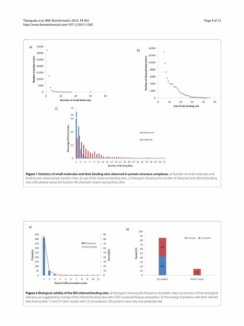
ResultsAnnotation of protein chains with observed and inferred binding sitesThere are about 28000 PDB entries with observed smallmolecule binding sites and about 56000 protein chains.The total number of observed small molecule bindingsites is about 91000 and approximately 67000 of theserepresent biologically relevant small molecules(i.e.around 24000 small molecules represent crystallizationagents). Small molecule binding is a specific feature thatplays a crucial role in the protein function. About 64% ofprotein chains in the PDB bind to a single small moleculeand 95% bind to no more than four small molecules (Fig-ure 1a). Likewise, the binding site pockets are rathersmall compared to the size of their functional domains.The binding sites are usually less than 25 residues and55% of the binding sites in the current study are smallerthan 10 residues (Figure 1b). Our algorithm inferredbinding sites for 92000 protein chains and the overallaverage number of binding site clusters inferred per chainis 6.5 (Figure 1c) whereas the average number of biologi-
cally relevant binding site clusters inferred per chain isabout 4.
One of the important features of this method is that itdoes not exclude redundant sequences bound to differentsmall molecules. For example, to account for all specificinteractions of various drugs targeting the Kinase ATPbinding site, it is imperative to consider all the proteinsequences even if they are identical.
We validated the IBIS method by comparing theobtained annotations to the manually curated CDDannotations and to other different methods which usegeometry of binding pockets and/or sequence conserva-tion of binding sites. It should be mentioned that sincethe IBIS method is based on different types of structuralevidence, the notion of false positives might not be validin many cases.
Validation of the IBIS method using the Conserved Domain DatabaseTo test the ability of our method to successfully infer thebiologically relevant binding sites, a validation procedurewas implemented using the manually curated ConservedDomain Database (CDD) [56] alignments and the func-tional features recorded in it as a standard of truth. Man-ually curated functional site annotations in CDD havebeen extracted from the published literature or derivedfrom manual interpretation of individual three-dimen-sional structures. Altogether 49% of the proteins withobserved small molecule binding sites have CDD smallmolecule binding site conserved annotations whereasover 55% of the proteins with inferred binding sites haveat least one site overlapping with CDD annotated bindingsite annotation.
In our analysis we used the CDD release 2.16 contain-ing 4092 protein chains. We chose representative proteinchains purged at the 25% sequence identity level. In thisjackknifing experiment, the query protein and its identi-cal homologs are omitted from clustering. Altogether 486representative chains had at least one structurally similarnon-identical homolog which had observed protein-smallmolecule binding sites. Figure 2a shows how well ourmethod can retrieve the CDD annotated binding sites atthe top ranks by calculating the fraction of true positives(sensitivity) or percentage of correctly annotated bindingsites (overlap between CDD and IBIS annotated bindingsites should be at least 50%). For 207 of these there wasonly one inferred binding site (cluster) detected, and bydefault these will always be ranked first. There remain279 examples which have at least two IBIS binding sites,209 (75%) of these were ranked first, and 49 were rankedsecond, so that 258 (92%) were ranked either first or sec-ond (Figure 2b).
Thangudu et al. BMC Bioinformatics 2010, 11:365http://www.biomedcentral.com/1471-2105/11/365
Page 4 of 12
Figure 1 Statistics of small molecules and their binding sites observed in protein structure complexes. a) Number of small molecules and binding sites observed per protein chain, b) size of the observed binding sites, c) histogram showing the number of observed and inferred binding sites with plotted versus the fraction (%) of protein chains having these sites.
Figure 2 Biological validity of the IBIS inferred binding sites. a) Histogram showing the frequency of protein chains as function of their biological relevancy as suggested by overlap of the inferred binding sites with CDD conserved feature annotation. b) Percentage of proteins with their inferred sites having their 1st and 2nd rank clusters with CD annotations; 165 proteins have only one predicted site.

Thangudu et al. BMC Bioinformatics 2010, 11:365http://www.biomedcentral.com/1471-2105/11/365
Page 5 of 12
Since there are a number of proteins which do not haveCDD annotations, IBIS inferred binding sites may be bio-logically relevant in these cases.
Validation of ranking scheme: discriminating between biological and non-biological chemicalsWe used the same set of protein queries (604 chains) toevaluate our method using structures which containedboth biological and non-biological small molecules (seeAdditional file 1 Table S1). Our goal is to assess how wellour ranking scheme distinguishes between the twogroups of binding sites: those containing biological versusnon-biological small molecules. If all the bound smallmolecules in an inferred binding site are non-biological,it is deemed as non-biological. To address this, we applieda linear discriminant analysis which constructs a discrim-inant function that divides the parameter space intoregions so as to separate the groups as distinctly as possi-ble. The method computes the posterior probability ofgroup membership for each observation, and assigns theobservation to the group that has the highest probability.As a result, a classification matrix is produced, whichgives the fraction of observations correctly assigned toeach group by the discriminant function. In our case, agood classification would be quantified by high fractionsfor both correctly predicted biological binding site clus-ters and correctly predicted non-biological binding siteclusters. We found that our method correctly classifies87% of biological clusters and 85% of non-biological clus-ters.
Validation of IBIS method by comparison with - geometric methodsTo further validate the prediction ability of our methodwe compared it with several widely used geometry andenergy-based approaches discussed in a recent study [57]
which includes LIGSITEcsc [29], PASS [48], Q-SiteFinder[28], Surfnet [49]. We used 44 out of 48 proteins from thispaper which have structure homologs with at least 30%sequence identity and also have both small molecule-bound and unbound structures.
For each method tested, the top ranked predicted sitesfor the unbound structure are compared with theobserved binding sites in the bound structure of a pro-tein-small molecule complex of a homolog. Table 1 showsthe sensitivity of retrieval of the true observed sites at thetop three ranks. To measure the sensitivity of retrieval ofbound structures at different levels of similarity betweenthe unbound query and bound structure from the data-base, we selected from the test set only those unbound-bound pairs which are within a given similarity range (nomore than 80, 90, or 100% identity) and denoted themIBIS80, IBIS90 and IBIS100. For example, the IBIS90 datasetcontains unbound query proteins for which the averagesequence identity between the unbound protein andmembers of the binding site clusters containing thebound homolog is no more than 90%. It is difficult if notimpossible to define false positives in our case since thereare many binding site clusters which are biologically rele-vant (for example have a significant overlap with themanually curated CDD functional annotations) but at thesame time do not match the binding site of the boundform of the protein from the test set. This happens if, forexample, there are multiple binding sites/pockets in theprotein which bind different small molecules and havedistinct functions. As can be seen from this table IBISperformance is similar to the LIGSITEcs method whichuses sequence conservation and reaches about 72% sensi-tivity. Overall we found that a total of 31 proteins (70%)from the test set have at least one of their IBIS predictedsites overlap with CDD binding site annotation. This sug-gests that IBIS successfully uses the knowledge of thestructure complexes of homologs to predict and rank therelevant sites. The complete details of the predictionresults can be seen in Additional file 1 Table S2.
All of these approaches, although they perform reason-ably well, are limited by the requirement of differentiatingtrue positives from false positives. Introducing sequenceconservation need not necessarily improve the predictionaccuracy and could be a source of error, leading to overprediction of the binding site area [58]. IBIS on the otherhand predicts only a handful of small molecule bindingsites with high probability of being biologically relevant.On average our method predicts 4 'biologically relevant'binding sites per protein chain and over half of all pre-dicted sites map to CDD curator annotations.
Knowledge-based docking using IBIS, an exampleTo demonstrate the effectiveness of IBIS as a knowledge-based prediction system, we compared our method withan established reverse docking approach. Cai and
Table 1: Prediction sensitivity (%) of the top three predictions by different geometric approaches and their comparison to IBIS.
Method* Top1 Top2 Top3
IBIS100 73 89 89
IBIS90 75 91 91
IBIS80 72 88 88
LIGSITEcs 71 79 85
PASS 58 67 75
Q-SiteFinder 52 60 75
SURFNET 42 58 62
*IBIS100, IBIS90, IBIS80 dataset contains unbound query proteins for which the average sequence identity between the unbound protein and members of the binding site clusters containing the bound homolog is no more than 100%, 90% and 80% respectively.
Thangudu et al. BMC Bioinformatics 2010, 11:365http://www.biomedcentral.com/1471-2105/11/365
Page 6 of 12
coworkers [59] employed a reverse docking method tofind a potential target protein for a natural product: N-trans-caffeoyltyramin in the genome of Helicobacterpyroli. Initially all potential binding proteins of N-trans-caffeoyltyramin were screened from a database of poten-tial drug targets with known structures from the ProteinData Bank using the reverse docking approach TarFis-Dock [60]. Only two proteins from the H.pyroli genomewere found by the TarFisDock method: diaminopimelatedecarboxylase (DC) and Peptide deformylase (PDF).After enzymatic validation, only the PDF protein wasfound to be a probable drug target. The crystal structurecomplex of N-trans-caffeoyltyramin with PDF suggesteda highly selective binding in the PDF binding pocket.
We attempted to identify the binding sites of N-trans-caffeoyltyramin on the PDF protein sequence. The closesthomolog for H.pyroli PDF is P.aeruginosa PDF which has45% sequence identity and has been used as a templatefor inferring the interactions by IBIS. The top ranked andhighly conserved inferred binding site of P.aeruginosaPDF when mapped onto H.pyroli PDF is in completeagreement with the native/experimentally determinedbinding site of the N-trans-caffeoyltyramin - PDF com-plex (Figure 3).
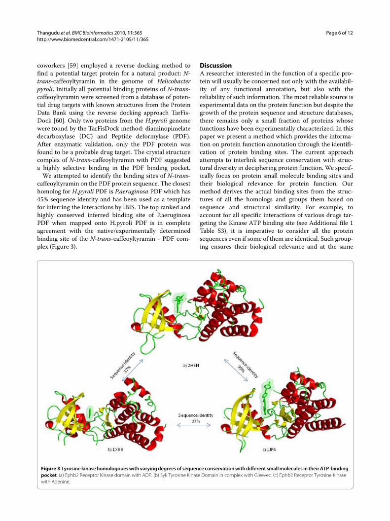
DiscussionA researcher interested in the function of a specific pro-tein will usually be concerned not only with the availabil-ity of any functional annotation, but also with thereliability of such information. The most reliable source isexperimental data on the protein function but despite thegrowth of the protein sequence and structure databases,there remains only a small fraction of proteins whosefunctions have been experimentally characterized. In thispaper we present a method which provides the informa-tion on protein function annotation through the identifi-cation of protein binding sites. The current approachattempts to interlink sequence conservation with struc-tural diversity in deciphering protein function. We specif-ically focus on protein small molecule binding sites andtheir biological relevance for protein function. Ourmethod derives the actual binding sites from the struc-tures of all the homologs and groups them based onsequence and structural similarity. For example, toaccount for all specific interactions of various drugs tar-geting the Kinase ATP binding site (see Additional file 1Table S3), it is imperative to consider all the proteinsequences even if some of them are identical. Such group-ing ensures their biological relevance and at the same
Figure 3 Tyrosine kinase homologoues with varying degrees of sequence conservation with different small molecules in their ATP-binding pocket. (a) Ephb2 Receptor Kinase domain with ADP. (b) Syk Tyrosine Kinase Domain in complex with Gleevec. (c) Ephb2 Receptor Tyrosine Kinase with Adenine.

Thangudu et al. BMC Bioinformatics 2010, 11:365http://www.biomedcentral.com/1471-2105/11/365
Page 7 of 12
time accounts for variations in the binding site residuesdue to differences in small molecule sizes and conforma-tions. By using all available structures of close homologs,IBIS provides a great opportunity for analyzing the diver-sity of binding modes. Figure 4, for example, shows theconserved tyrosine kinase fold with varying degrees ofsequence similarity but sharing a highly conserved ATPbinding site occupied by different small molecules.
Recently, it was estimated that over two-thirds of allprotein sequences in the GenBank database have at leastone structure homolog [1,2]. As the on-going structuralgenomics initiative continues to close the sequence-structure gap, our method might be very useful for anno-tating proteins with unknown function and structure.Moreover, the location of putative binding sites providesguidance for the protein docking methods for drugdesign. We have assessed the reliability of our method bydirect comparison with the binding site annotations fromliterature and manual curation and have shown that inthe great majority of cases, the method detects and ranksthe manually annotated binding site cluster at the first orsecond rank. This is achievable for a number of reasons,such as using a sufficient level of similarity between theunknown query and its homologs with the known bind-ing sites, accurate clustering of small molecule bindingsites using a reasonable similarity measure, and applyinga deliberately designed ranking scheme that distinguishesthe non-biological from the biologically relevant bindingsites.
We have also compared our method with several widelyused geometry and energy-based approaches to predictsmall molecule binding sites. As we have shown, the per-formance of our prediction method is very similar topopular geometric approaches. Moreover, one of the
advantages is that our method can be applied even for aquery sequence without structure, which is not the casefor those binding site prediction methods which explicitlyrely on the specific features of binding pocket geometry.
Using remote homology for functional inference isoften based on the general assumption that there is a neg-ative correlation between small molecule binding sitesimilarity and overall sequence similarity. However, smallmolecule binding site similarity is much more compli-cated with many examples of strikingly similar bindingsites with low (<30%) overall sequence identity and alsovery weakly similar binding sites with high overallsequence identity [61]. Likewise, the similarities of smallmolecule binding sites across different protein folds,although providing new insights, leads to new challengesin deciphering the functional relevance. Large-scale auto-mated function prediction methods are often limited bythe lack of sufficient understanding of biological functionand also by the quality of structure data. Hence, throughthe IBIS approach, we strive to limit the false positive rateby employing a conservative sequence similarity thresh-old of at least 30% over the structurally superimposedregions of homologs. It is often possible that the protein-small molecule crystal state may correspond to a globalminimum of free energy where biologically relevantinteractions are difficult to distinguish from non-specificcontacts. For example, a recent estimate suggests some20% of dimeric structures in PDB may be crystallizationartifacts [62]. The elaborate scoring scheme of ourmethod based on recurrence and evolutionary conserva-tion, along with the list of non-biological small molecules,tends to de-emphasize the artifactual interactions andranks such sites near or at the bottom of the list.
Figure 4 Mapping of the inferred binding site. Inferred binding site of peptide deformylase P.aeruginosa (PDB:1IX1) mapped onto the sequence of Helicobacter pyroli and its agreement with the observed binding site in N-trans-caffeoyltyramin-PDF complex (PDB: 1EW5). MMDB residue number-ing is used which starts from the beginning of the corresponding GenBank protein sequence.
Thangudu et al. BMC Bioinformatics 2010, 11:365http://www.biomedcentral.com/1471-2105/11/365
Page 8 of 12
Furthermore, the chemical properties of small mole-cules bound to the inferred binding sites can be used as astarting step in small molecule virtual screening. ThePubChem compound database [63] mapping of IBISsmall molecules accomplishes a preliminary step in smallmolecule virtual screening by clustering the similarchemicals into structurally unique compounds. The func-tional groups of the small molecules binding in a com-mon binding site of evolutionarily related proteins arelikely conserved. Recently it was shown that sequenceand structure conservation of the binding site residuescontacting these anchor functional groups is significantlyhigher than those contacting variable regions [40]. IBIS,thus provides a common platform for function predic-tion, knowledge-based docking and also for small mole-cule virtual screening.
ConclusionsFinding small molecule binding sites that specify proteinfunction is of great importance in drug development.Here we proposed a method to decipher the function ofan unknown protein by interlinking sequence conserva-tion with structural diversity of its homologs. To facilitatevalidation of the inferred binding sites from homologs,we developed an elaborate scoring scheme that can accu-rately distinguish biologically relevant sites. The methodhas been implemented as a web server, IBIS (InferredBiomolecular Interaction Server - http://www.ncbi.nlm.nih.gov/Structure/ibis/ibis.cgi) to facilitate accurate, effi-cient and high-throughput function prediction.
MethodsWe used the NCBI Molecular Modeling Database(MMDB) [64] as a source of data on protein complexes.The automated MMDB processing of PDB files includessteps such as deposition of the protein sequences intoGenBank [65], deposition of small molecules into Pub-Chem [63], addition of corresponding links to these data-bases in the MMDB records, also links to citations andreferences in PubMed, and Entrez indexing for quicksearching.
Below we describe different steps of processing, includ-ing defining observed interactions from structures,inferred interactions from homologs, clustering bindingsites and their ranking in terms of biological relevancewith respect to the query protein.
Defining observed interactionsIn the current release of the Molecular Modeling Data-base (MMDB) [64], there are about 28000 entries withbound small molecules. The resulting 39000 small mole-cules are bound to about 56000 protein chains in total. Asmall molecule is defined as any non-polypeptide, non-nucleic acid molecule in the structure complex or any
molecule with a sufficient number of non-standardamino acid/nucleic acids and without an assigned Gen-Bank identifier from NCBI. All the small molecules arestandardized in the PubChem database [63] and havevalid substance and compound identifiers. In this workwe do not consider small molecules that are smaller than5 heavy atoms or those having molecular weight outsidethe range of 70-800 Da. Small molecules such as metalions often play role as crystallization agents, and there-fore ions are not considered in this paper.
The filters by atom count and molecular weight onlypartially remove non-biological small molecules (i.e. buf-fers, salts, detergents, solvents, and ions added for thepurpose of crystallization and/or purification). Thesenon-biological molecules sometimes mimic natural smallmolecules and tend to bind in functional/active sites ofproteins. For validation purposes we used a list of poten-tial non-biological small molecules which has been col-lected from the literature (see Additional file 1 Table S1)[30,66,67].
We define a protein residue to be in contact with asmall molecule if there is at least one (heavy) atom of theresidue within 4.0Å of some atom from the small mole-cule. For most pairs of atoms, this threshold correspondsto the sum of their van der Waals radii plus a tolerance ofabout 0.5Å to allow for coordinate errors in structuredetermination. For manual curation of the ConservedDomain Database (CDD) a similar contact definition isused for defining protein-small molecule contacts. Weretain only those protein-small molecule complexeswhich have at least five interacting protein residues. Wedefine "binding site" as a set of residues on a given proteinchain which are in contact with a given small molecule.Each MMDB entry is analyzed, and all pairs of biomole-cules consisting of a protein chain and small molecule incontact with that chain are retained for further analysis.
It should be mentioned that a small molecule can bebound to a single domain or multiple domains whichcould come from more than one protein chain in the PDBrecord. Almost half of all the small molecules in the PDBare bound by more than one domain with <75% of allcontacts to any single domain [66]. However, usingdomains as structural units would necessitate automaticdomain decomposition methods in many cases [68,69],and the domain boundaries chosen could affect theresults. To circumvent the potential technical difficultiesin using domains as the structural unit in recording theobserved/physical interactions, we use only completeprotein chains for defining protein-small molecule inter-actions. Small molecules binding to multiple proteinchains entail even more technical difficulties. For exam-ple, simultaneous superposition of multiple chains wouldneed to be checked to ensure similarity of binding sites.Therefore, when multiple chains are involved in a binding
Thangudu et al. BMC Bioinformatics 2010, 11:365http://www.biomedcentral.com/1471-2105/11/365
Page 9 of 12
site, if one of the chains includes 75% or more of the con-tacts, then we define only one binding site and assign itonly to that particular chain. Otherwise, we define sepa-rate binding sites on each of the chains. The latter situa-tion is relatively rare as only about 15% of the proteins inthe current PDB release have small molecule interactionsthat fall into this category.
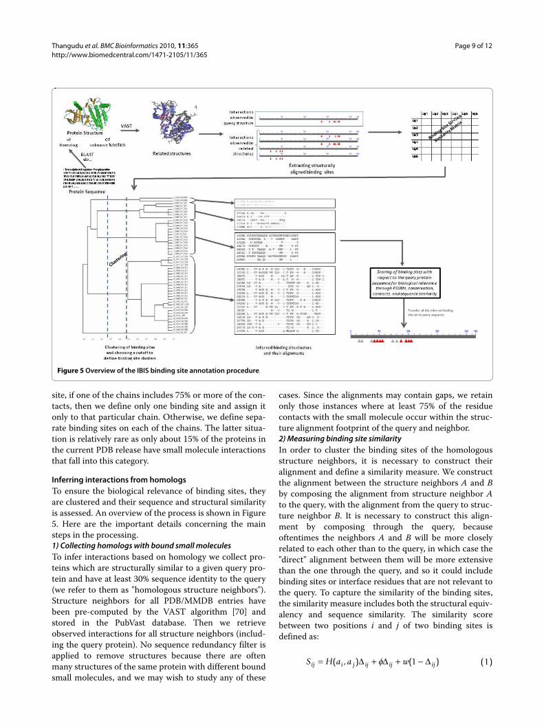
Inferring interactions from homologsTo ensure the biological relevance of binding sites, theyare clustered and their sequence and structural similarityis assessed. An overview of the process is shown in Figure5. Here are the important details concerning the mainsteps in the processing.1) Collecting homologs with bound small moleculesTo infer interactions based on homology we collect pro-teins which are structurally similar to a given query pro-tein and have at least 30% sequence identity to the query(we refer to them as "homologous structure neighbors").Structure neighbors for all PDB/MMDB entries havebeen pre-computed by the VAST algorithm [70] andstored in the PubVast database. Then we retrieveobserved interactions for all structure neighbors (includ-ing the query protein). No sequence redundancy filter isapplied to remove structures because there are oftenmany structures of the same protein with different boundsmall molecules, and we may wish to study any of these
cases. Since the alignments may contain gaps, we retainonly those instances where at least 75% of the residuecontacts with the small molecule occur within the struc-ture alignment footprint of the query and neighbor.2) Measuring binding site similarityIn order to cluster the binding sites of the homologousstructure neighbors, it is necessary to construct theiralignment and define a similarity measure. We constructthe alignment between the structure neighbors A and Bby composing the alignment from structure neighbor Ato the query, with the alignment from the query to struc-ture neighbor B. It is necessary to construct this align-ment by composing through the query, becauseoftentimes the neighbors A and B will be more closelyrelated to each other than to the query, in which case the"direct" alignment between them will be more extensivethan the one through the query, and so it could includebinding sites or interface residues that are not relevant tothe query. To capture the similarity of the binding sites,the similarity measure includes both the structural equiv-alency and sequence similarity. The similarity scorebetween two positions i and j of two binding sites isdefined as:
S H a a wij i j ij ij ij= + + −( , ) ( )Δ Δ Δf 1 (1)
Figure 5 Overview of the IBIS binding site annotation procedure.
Thangudu et al. BMC Bioinformatics 2010, 11:365http://www.biomedcentral.com/1471-2105/11/365
Page 10 of 12
where H is the element of the BLOSUM62 matrix cor-responding to the aligned amino acids in positions i and j;Δij is equal to 1 if two positions are aligned and 0 other-wise. θ is an additional weight of "+1" added to eachstructurally equivalent position. w is a gap penalty of "-4",to mimic the most unfavorable substitution score fromBLOSUM62 matrix, which showed the best performancein our preliminary studies. The overall similarity scorebetween two binding sites is calculated by summing up Sijover all positions in the gapped alignment. To facilitatecomparison of scores from different alignments, the rawscore is converted to a bit score with the statisticalparameters λ and K previously defined in the BLOSUMscoring system.
The similarity score is then converted into a conserva-tion score CS by dividing by the maximum of the bitscores when the binding sites are scored against them-selves.
3) Clustering of binding sitesBased on the calculated conservation score CS, the bind-ing sites of the homologs are clustered using a complete-linkage clustering algorithm, which considers the dis-tance between two clusters to be equal to the maximumdistance between their members. A distance cutoff valueto define the clusters is chosen using a free energy func-tion defined previously. This function F is formulated tomaximize the mean similarity of members within a clus-ter and minimize the complexity of the description pro-vided by cluster membership [71].
where T is the temperature factor, S(i, j) is the similarityscore between binding site i and binding site j in eachcluster, C represents a cluster, |C| is the number of bind-ing sites in the cluster C, and N is the total number ofbinding sites clustered. The temperature T is a parameter(constant) that is chosen so as to correctly balance theenergy-like and entropy-like terms in the function [71].
Biological relevance of binding sites and their ranking with respect to the query proteinAll binding site clusters are ranked in terms of their pre-dicted biological relevance and similarity to the query.First we assess the evolutionary conservation of bindingsite clusters. Those sites which reoccur in diverse enoughprotein complexes are ranked higher. Clusters that haveonly one non-redundant member (after members withmore than 90% identity are removed) are considered "sin-gletons" and are not assigned any score (ranked at thebottom of the list). A "conservation score" is computed inorder to measure the diversity of cluster members andhow well the binding site is conserved across thehomologs. To do this, positional conservation in thebinding site multiple sequence alignment is calculatedusing the Shannon entropy measure with the Henikoff-Henikoff sequence weights. Sequence weights are esti-mated using the complete sequences of neighbors alignedwith the query protein.
To account for evolutionary closeness of a givenbinding site cluster to the query we use the sequence-PSSM score and the average sequence identity betweenthe query and all cluster members calculated over thewhole structure-structure alignment (not just bindingsites). A position specific score matrix (PSSM) is con-structed based on the binding site multiple alignmentusing the implicit pseudo-count method of Gribskov,McLachlan and Eisenberg [72]. The aligned bindingsite region of the query protein is then scored againstthe PSSM and a sequence-PSSM score is calculated. Ahigher sequence-PSSM score points to a higher proba-bility of this site being a biologically relevant site forthe query.
To rank the larger interfaces more highly we also cal-culate the average number of interfacial contactswhich the binding site makes in the complex of thecorresponding homolog. All components of the rank-ing score are then normalized and all clusters areranked with respect to the Z-scores. Any cluster withall members binding non-biological small molecules isdisregarded.
The Z-score is calculated for each of these four corre-sponding terms (i.e. conservation score, PSSM-score,contact count, and percent sequence identity to query) inthe ranking scheme by subtracting the mean value anddividing by the standard deviation obtained from thescore distribution of other binding site clusters for a givenquery protein. The coefficients in front of each term inthe ranking score were calculated empirically. The com-bined score is designed to rank the most biologically rele-vant sites at the top.
SS K
’ln
ln= −l
2(2)
CSS A B
Max S A A S B B=
′( )( , )
( , ) ( , )(3)
FN
CS i j
T C C
TN N
i j CC
C
= +
−
⎧
⎨
⎪⎪⎪⎪
⎩
⎪⎪⎪⎪
⎫
⎬
⎪⎪⎪
∈∑∑
∑1
1| |
( , )
| | log | |
log
,
⎪⎪
⎭
⎪⎪⎪⎪
(4)
Z Z Z Z Zcomb pssm conserv contact pcnt= ( ) + ( ) + ( ) + (0 4 0 4 0 1 0 1. * . * . * . * )){ }(5)
Thangudu et al. BMC Bioinformatics 2010, 11:365http://www.biomedcentral.com/1471-2105/11/365
Page 11 of 12
Additional material
Authors' contributionsARP, BAS, SHB, and TM conceived the project. RRT, MT, and BAS implementedthe database and analysis programs. RRT, ARP, and TM wrote the paper. Allauthors contributed to the underlying ideas of the method and the analysis. Allauthors read and approved the final manuscript.
AcknowledgementsThe authors are grateful to Aron Märchler-Bauer, Dachuan Zhang, and Jessica Fong. This research was supported by the Intramural Research Program of the NIH, National Library of Medicine.
Author DetailsNational Center for Biotechnology Information, 8600 Rockville Pike, Building 38A, Bethesda, MD 20894 USA
References1. Wang Y, Addess KJ, Chen J, Geer LY, He J, He S, Lu S, Madej T, Marchler-
Bauer A, Thiessen PA, et al.: MMDB: annotating protein sequences with Entrez's 3D-structure database. Nucleic Acids Res 2007, 35(Database issue):D298-300.
2. Fukuchi S, Homma K, Sakamoto S, Sugawara H, Tateno Y, Gojobori T, Nishikawa K: The GTOP database in 2009: updated content and novel features to expand and deepen insights into protein structures and functions. Nucleic Acids Res 2009, 37(Database issue):D333-337.
3. Bork P, Koonin EV: Predicting functions from protein sequences--where are the bottlenecks? Nat Genet 1998, 18(4):313-318.
4. Gerlt JA, Babbitt PC: Can sequence determine function? Genome Biol 2000, 1(5):REVIEWS0005.
5. Hegyi H, Gerstein M: The relationship between protein structure and function: a comprehensive survey with application to the yeast genome. J Mol Biol 1999, 288(1):147-164.
6. Yu H, Luscombe NM, Lu HX, Zhu X, Xia Y, Han JD, Bertin N, Chung S, Vidal M, Gerstein M: Annotation transfer between genomes: protein-protein interologs and protein-DNA regulogs. Genome Res 2004, 14(6):1107-1118.
7. Capra JA, Singh M: Predicting functionally important residues from sequence conservation. Bioinformatics 2007, 23(15):1875-1882.
8. Zhang T, Zhang H, Chen K, Shen S, Ruan J, Kurgan L: Accurate sequence-based prediction of catalytic residues. Bioinformatics 2008, 24(20):2329-2338.
9. Fischer JD, Mayer CE, Soding J: Prediction of protein functional residues from sequence by probability density estimation. Bioinformatics 2008, 24(5):613-620.
10. Burgoyne NJ, Jackson RM: Predicting protein interaction sites: binding hot-spots in protein-protein and protein-ligand interfaces. Bioinformatics 2006, 22(11):1335-1342.
11. Ota M, Kinoshita K, Nishikawa K: Prediction of catalytic residues in enzymes based on known tertiary structure, stability profile, and sequence conservation. J Mol Biol 2003, 327(5):1053-1064.
12. Liang S, Zhang C, Liu S, Zhou Y: Protein binding site prediction using an empirical scoring function. Nucleic Acids Res 2006, 34(13):3698-3707.
13. Campbell SJ, Gold ND, Jackson RM, Westhead DR: Ligand binding: functional site location, similarity and docking. Curr Opin Struct Biol 2003, 13(3):389-395.
14. Thibert B, Bredesen DE, del Rio G: Improved prediction of critical residues for protein function based on network and phylogenetic analyses. BMC Bioinformatics 2005, 6:213.
15. Bray T, Chan P, Bougouffa S, Greaves R, Doig AJ, Warwicker J: SitesIdentify: a protein functional site prediction tool. BMC Bioinformatics 2009, 10(1):379.
16. Brylinski M, Prymula K, Jurkowski W, Kochanczyk M, Stawowczyk E, Konieczny L, Roterman I: Prediction of functional sites based on the fuzzy oil drop model. PLoS Comput Biol 2007, 3(5):e94.
17. Brylinski M, Skolnick J: A threading-based method (FINDSITE) for ligand-binding site prediction and functional annotation. Proc Natl Acad Sci USA 2008, 105(1):129-134.
18. Jones S, Thornton JM: Analysis of protein-protein interaction sites using surface patches. J Mol Biol 1997, 272(1):121-132.
19. Landgraf R, Xenarios I, Eisenberg D: Three-dimensional cluster analysis identifies interfaces and functional residue clusters in proteins. J Mol Biol 2001, 307(5):1487-1502.
20. Pazos F, Sternberg MJ: Automated prediction of protein function and detection of functional sites from structure. Proc Natl Acad Sci USA 2004, 101(41):14754-14759.
21. Teichmann SA, Murzin AG, Chothia C: Determination of protein function, evolution and interactions by structural genomics. Curr Opin Struct Biol 2001, 11(3):354-363.
22. Panchenko AR, Kondrashov F, Bryant S: Prediction of functional sites by analysis of sequence and structure conservation. Protein Sci 2004, 13(4):884-892.
23. Bartlett GJ, Porter CT, Borkakoti N, Thornton JM: Analysis of catalytic residues in enzyme active sites. J Mol Biol 2002, 324(1):105-121.
24. Bate P, Warwicker J: Enzyme/non-enzyme discrimination and prediction of enzyme active site location using charge-based methods. J Mol Biol 2004, 340(2):263-276.
25. Greaves R, Warwicker J: Active site identification through geometry-based and sequence profile-based calculations: burial of catalytic clefts. J Mol Biol 2005, 349(3):547-557.
26. Marti-Renom MA, Rossi A, Al-Shahrour F, Davis FP, Pieper U, Dopazo J, Sali A: The AnnoLite and AnnoLyze programs for comparative annotation of protein structures. BMC Bioinformatics 2007, 8(Suppl 4):S4.
27. Chelliah V, Chen L, Blundell TL, Lovell SC: Distinguishing structural and functional restraints in evolution in order to identify interaction sites. J Mol Biol 2004, 342(5):1487-1504.
28. Laurie AT, Jackson RM: Q-SiteFinder: an energy-based method for the prediction of protein-ligand binding sites. Bioinformatics 2005, 21(9):1908-1916.
29. Huang B, Schroeder M: LIGSITEcsc: predicting ligand binding sites using the Connolly surface and degree of conservation. BMC Struct Biol 2006, 6:19.
30. Snyder KA, Feldman HJ, Dumontier M, Salama JJ, Hogue CW: Domain-based small molecule binding site annotation. BMC Bioinformatics 2006, 7:152.
31. Lopez G, Valencia A, Tress ML: firestar--prediction of functionally important residues using structural templates and alignment reliability. Nucleic Acids Res 2007:W573-577.
32. Qin S, Zhou HX: meta-PPISP: a meta web server for protein-protein interaction site prediction. Bioinformatics 2007, 23(24):3386-3387.
33. Skolnick J, Brylinski M: FINDSITE: a combined evolution/structure-based approach to protein function prediction. Brief Bioinform 2009.
34. Hernandez M, Ghersi D, Sanchez R: SITEHOUND-web: a server for ligand binding site identification in protein structures. Nucleic Acids Res 2009:W413-416.
35. Talavera D, Laskowski RA, Thornton JM: WSsas: a web service for the annotation of functional residues through structural homologues. Bioinformatics 2009, 25(9):1192-1194.
36. Ivanisenko VA, Pintus SS, Grigorovich DA, Kolchanov NA: PDBSiteScan: a program for searching for active, binding and posttranslational modification sites in the 3 D structures of proteins. Nucleic Acids Res 2004:W549-554.
37. Chang DT, Weng YZ, Lin JH, Hwang MJ, Oyang YJ: Protemot: prediction of protein binding sites with automatically extracted geometrical templates. Nucleic Acids Res 2006:W303-309.
38. Jambon M, Andrieu O, Combet C, Deleage G, Delfaud F, Geourjon C: The SuMo server: 3 D search for protein functional sites. Bioinformatics 2005, 21(20):3929-3930.
39. Shulman-Peleg A, Nussinov R, Wolfson HJ: SiteEngines: recognition and comparison of binding sites and protein-protein interfaces. Nucleic Acids Res 2005:W337-341.
40. Brylinski M, Skolnick J: FINDSITE: a threading-based approach to ligand homology modeling. PLoS Comput Biol 2009, 5(6):e1000405.
41. Wilson CA, Kreychman J, Gerstein M: Assessing annotation transfer for genomics: quantifying the relations between protein sequence,
Additional file 1 Table S1: The most common non-biological small molecules found in protein structure complexes. Table S2: Summary of the IBIS predictions and CDD annotation validation for the 44 bound and unbound structures used as test set to compare with existing geometric approaches. Table S3: Variety of small molecules binding in the ATP binding pocket of tyrosine kinase homologs.
Received: 16 March 2010 Accepted: 1 July 2010 Published: 1 July 2010This article is available from: http://www.biomedcentral.com/1471-2105/11/365© 2010 Thangudu et al; licensee BioMed Central Ltd. This is an Open Access article distributed under the terms of the Creative Commons Attribution License (http://creativecommons.org/licenses/by/2.0), which permits unrestricted use, distribution, and reproduction in any medium, provided the original work is properly cited.BMC Bioinformatics 2010, 11:365
Thangudu et al. BMC Bioinformatics 2010, 11:365http://www.biomedcentral.com/1471-2105/11/365
Page 12 of 12
structure and function through traditional and probabilistic scores. J Mol Biol 2000, 297(1):233-249.
42. Rost B: Enzyme function less conserved than anticipated. J Mol Biol 2002, 318(2):595-608.
43. Altschul SF, Madden TL, Schaffer AA, Zhang J, Zhang Z, Miller W, Lipman DJ: Gapped BLAST and PSI-BLAST: a new generation of protein database search programs. Nucleic Acids Res 1997, 25(17):3389-3402.
44. Porter CT, Bartlett GJ, Thornton JM: The Catalytic Site Atlas: a resource of catalytic sites and residues identified in enzymes using structural data. Nucleic Acids Res 2004, 32(Database issue):D129-133.
45. Harris MA, Clark J, Ireland A, Lomax J, Ashburner M, Foulger R, Eilbeck K, Lewis S, Marshall B, Mungall C, et al.: The Gene Ontology (GO) database and informatics resource. Nucleic Acids Res 2004, 32(Database issue):D258-261.
46. Marchler-Bauer A, Bryant SH: CD-Search: protein domain annotations on the fly. Nucleic Acids Res 2004:W327-331.
47. Liang J, Edelsbrunner H, Woodward C: Anatomy of protein pockets and cavities: measurement of binding site geometry and implications for ligand design. Protein Sci 1998, 7(9):1884-1897.
48. Brady GP Jr, Stouten PF: Fast prediction and visualization of protein binding pockets with PASS. J Comput Aided Mol Des 2000, 14(4):383-401.
49. Laskowski RA: SURFNET: a program for visualizing molecular surfaces, cavities, and intermolecular interactions. J Mol Graph 1995, 13(5):323-330. 307-328
50. Nayal M, Honig B: On the nature of cavities on protein surfaces: application to the identification of drug-binding sites. Proteins 2006, 63(4):892-906.
51. Capra JA, Laskowski RA, Thornton JM, Singh M, Funkhouser TA: Predicting Protein Ligand Binding Sites by Combining Evolutionary Sequence Conservation and 3 D Structure. PLoS Computational Biology 2009. accepted for publication
52. Petrey D, Fischer M, Honig B: Structural relationships among proteins with different global topologies and their implications for function annotation strategies. Proc Natl Acad Sci USA 2009, 106(41):17377-17382.
53. Shoemaker BA, Zhang D, Thangudu RR, Tyagi M, Fong JH, Marchler-Bauer A, Bryant SH, Madej T, Panchenko AR: Inferred Biomolecular Interaction Server--a web server to analyze and predict protein interacting partners and binding sites. Nucl Acids Res 2010, 38(Database issue):D518-524.
54. Berman H, Henrick K, Nakamura H, Markley JL: The worldwide Protein Data Bank (wwPDB): ensuring a single, uniform archive of PDB data. Nucleic Acids Res 2007, 35(Database issue):D301-303.
55. Marchler-Bauer A, Anderson JB, DeWeese-Scott C, Fedorova ND, Geer LY, He S, Hurwitz DI, Jackson JD, Jacobs AR, Lanczycki CJ, et al.: CDD: a curated Entrez database of conserved domain alignments. Nucleic Acids Res 2003, 31(1):383-387.
56. Marchler-Bauer A, Anderson JB, Chitsaz F, Derbyshire MK, DeWeese-Scott C, Fong JH, Geer LY, Geer RC, Gonzales NR, Gwadz M, et al.: CDD: specific functional annotation with the Conserved Domain Database. Nucleic Acids Res 2009, 37(Database issue):D205-210.
57. Huang B: MetaPocket: a meta approach to improve protein ligand binding site prediction. OMICS 2009, 13(4):325-330.
58. Wass MN, Sternberg JEM: Prediction of ligand binding sites using homologous structures and conservation at CASP8. Proteins: Structure, Function, and Bioinformatics 2009, 77(S9):147-151.
59. Cai J, Han C, Hu T, Zhang J, Wu D, Wang F, Liu Y, Ding J, Chen K, Yue J, et al.: Peptide deformylase is a potential target for anti-Helicobacter pylori drugs: reverse docking, enzymatic assay, and X-ray crystallography validation. Protein Sci 2006, 15(9):2071-2081.
60. Li H, Gao Z, Kang L, Zhang H, Yang K, Yu K, Luo X, Zhu W, Chen K, Shen J, et al.: TarFisDock: a web server for identifying drug targets with docking approach. Nucleic Acids Res 2006:W219-224.
61. Kinjo AR, Nakamura H: Comprehensive structural classification of ligand-binding motifs in proteins. Structure 2009, 17(2):234-246.
62. Krissinel E: Crystal contacts as nature's docking solutions. J Comput Chem 31(1):133-143.
63. Wang Y, Xiao J, Suzek TO, Zhang J, Wang J, Bryant SH: PubChem: a public information system for analyzing bioactivities of small molecules. Nucleic Acids Res 2009:W623-633.
64. Chen J, Anderson JB, DeWeese-Scott C, Fedorova ND, Geer LY, He S, Hurwitz DI, Jackson JD, Jacobs AR, Lanczycki CJ, et al.: MMDB: Entrez's 3D-structure database. Nucleic Acids Res 2003, 31(1):474-477.
65. Sayers EW, Barrett T, Benson DA, Bryant SH, Canese K, Chetvernin V, Church DM, DiCuccio M, Edgar R, Federhen S, et al.: Database resources of the National Center for Biotechnology Information. Nucleic Acids Res 2009, 37(Database issue):D5-15.
66. Bashton M, Nobeli I, Thornton JM: Cognate ligand domain mapping for enzymes. J Mol Biol 2006, 364(4):836-852.
67. Wang R, Fang X, Lu Y, Yang CY, Wang S: The PDBbind database: methodologies and updates. J Med Chem 2005, 48(12):4111-4119.
68. Koczyk G, Berezovsky IN: Domain Hierarchy and closed Loops (DHcL): a server for exploring hierarchy of protein domain structure. Nucleic Acids Res 2008:W239-245.
69. Holland TA, Veretnik S, Shindyalov IN, Bourne PE: Partitioning protein structures into domains: why is it so difficult? J Mol Biol 2006, 361(3):562-590.
70. Madej T, Gibrat JF, Bryant SH: Threading a database of protein cores. Proteins 1995, 23(3):356-369.
71. Slonim N, Atwal GS, Tkacik G, Bialek W: Information-based clustering. Proc Natl Acad Sci USA 2005, 102(51):18297-18302.
72. Gribskov M, McLachlan AD, Eisenberg D: Profile analysis: detection of distantly related proteins. Proc Natl Acad Sci USA 1987, 84(13):4355-4358.
doi: 10.1186/1471-2105-11-365Cite this article as: Thangudu et al., Knowledge-based annotation of small molecule binding sites in proteins BMC Bioinformatics 2010, 11:365


![[XLS] · Web viewdmy 0005506 // iron ion binding // inferred from electronic annotation /// 0005515 // protein binding // inferred from electronic annotation /// 0008475 // procollagen-lysine](https://img.pdfslide.us/doc/110x75/5ae8e2d37f8b9ab24d8b5343/xls-viewdmy-0005506-iron-ion-binding-inferred-from-electronic-annotation.jpg)