Embed Size (px)
Citation preview
Yao et al. Cancer Cell Int (2019) 19:41 https://doi.org/10.1186/s12935-019-0765-6
PRIMARY RESEARCH
Knockdown of AGGF1 inhibits the invasion and migration of gastric cancer via epithelial–mesenchymal transition through Wnt/β-catenin pathwayHan‑Hui Yao1†, Ya‑Jun Zhao1†, Yi‑Fu He2, Da‑Bing Huang2 and Wei Wang2*
Abstract
Background: Angiogenic factor with G‑patch and FHA domain 1 (AGGF1), as a newly identified human angiogenic factor, is overexpressed in some types of malignant tumors and closely associated with patient’s prognosis. However, the mechanisms involved in the regulation of AGGF1 in gastric cancer (GC) still remain unclear.
Methods: In this study, AGGF1 level in GC tissues and cell lines was analyzed by western blot and quantitative real‑time polymerase chain reaction (qRT‑PCR). After knockdown of AGGF expression by RNA interference in GC cell lines MKN‑45 and MGC‑803, wound healing and transwell assays were conducted to examine the effects of AGGF1 on migration and invasion. Tumor growth was assessed in a mouse xenograft model in vivo. Furthermore, expression levels of epithelial–mesenchymal transition (EMT) biomarkers and involvement of the Wnt/β‑catenin pathway were detected by western blot and qRT‑PCR.
Results: Compared to those in normal groups, the protein and mRNA of AGGF1 expression levels were signifi‑cantly higher both in GC tissues and cell lines (all P < 0.05). Knockdown of AGGF1 dramatically inhibited the invasion and migration of MKN‑45 and MGC‑803 cells (all P < 0.01) in vitro, and suppressed the tumor growth of nude mice xenograft model in vivo. Western blot revealed alterations in EMT biomarkers, suggesting the role of AGGF1 in EMT. Moreover, we found that downregulated expression of AGGF1 attenuated Wnt/β‑catenin related protein expression.
Conclusions: Collectively, knockdown of AGGF1 inhibits the invasion and migration of gastric cancer via epithelial–mesenchymal transition through Wnt/β‑catenin pathway.
Keywords: Gastric cancer, AGGF1, Epithelial mesenchymal transition, Wnt/β‑catenin pathway, Invasion and migration
© The Author(s) 2019. This article is distributed under the terms of the Creative Commons Attribution 4.0 International License (http://creat iveco mmons .org/licen ses/by/4.0/), which permits unrestricted use, distribution, and reproduction in any medium, provided you give appropriate credit to the original author(s) and the source, provide a link to the Creative Commons license, and indicate if changes were made. The Creative Commons Public Domain Dedication waiver (http://creat iveco mmons .org/publi cdoma in/zero/1.0/) applies to the data made available in this article, unless otherwise stated.
BackgroundGastric cancer (GC) is one of the most common malig-nant gastrointestinal tumors in the world [1, 2]. Although its mortality has decreased significantly over the past
20 years, its morbidity and mortality are still at the fore-front of malignant tumors in China [3, 4]. Obviously, the major obstacle for GC treatment failure is tumor metastasis, during which invasion and migration are the pivotal steps. In recent years, epithelial–mesenchy-mal transition (EMT) has become a research hotspot of tumor metastasis. EMT is a key process during embry-onic morphogenesis, heart development, wound healing, and cancer metastasis [5, 6]. During EMT, epithelial cells lose their junctions and apical-basal polarity, reorganize their cytoskeleton and undergo a change in the signal-ing programmes. This ultimately increases the motility of
Open Access
Cancer Cell International
*Correspondence: [email protected] †Han‑Hui Yao and Ya‑Jun Zhao contributed equally as co‑first authors2 Department of Medical Oncology, The First Affiliated Hospital of USTC, Division of Life Sciences and Medicine, University of Science and Technology of China, No. 17 Lujiang Road, Hefei Anhui,, 230001, P.R. ChinaFull list of author information is available at the end of the article
Page 2 of 10Yao et al. Cancer Cell Int (2019) 19:41
individual cells and enables the development of an inva-sive phenotype. Therefore, through exploration of the molecular mechanism of EMT in GC, it not only guide a new research direction for the biological behavior of GC metastasis, but also provide a potential strategy for the treatment of GC.
Angiogenic factor with G-patch and FHA domain 1 (AGGF1 or VG5Q), as a newly identified human angio-genic factor, was first reported by Tian et al. [7] in 2004. Recent studies have found that AGGF1 is expressed in some types of malignant tumors and is closely related to tumor angiogenesis [8–11]. Besides, our previous study has revealed that AGGF1 expression was significantly associated with the lymph node metastasis, invasion depth and TNM stage of GC patients [12]. Moreover, high expression of AGGF1 could be used as an independ-ent factor to predict poor postoperative survival of GC patients [12]. However, the detailed regulatory mecha-nism of AGGF1 in the invasion and metastasis of GC still remains unclear. Interestingly, Major et al. [13] have identified the new regulators of Wnt/β-catenin signaling by using integrative molecular screening and character-ized AGGF1 as a nuclear chromatin-associated protein that participates in β-catenin-mediated transcription in human colon cancer cells. Moreover, Wnt/β-catenin signaling is one of the most important signaling path-ways involved in EMT of malignancies including GC [14–16]. Therefore, the issue whether AGGF1 can regu-late the EMT of GC through Wnt/β-catenin signaling has attracted our great interest and concern.
In this study, we used in vitro and in vivo approaches to demonstrate that whether knockdown of AGGF1 could inhibit EMT and whether the regulatory effects of AGGF1 on the EMT were partially attributed to the Wnt/β-catenin signaling pathway in GC.
MethodsClinical specimensForty cases of fresh gastric cancer samples and adja-cent noncancerous tissues were collected from patients that underwent curative gastric cancer resection at the Department of General Surgery in our hospital. Samples were dissected from resected specimens by a patholo-gist, and immediately snap-frozen in individual vials using liquid nitrogen. Frozen specimens were stored at − 70 °C in a tumor bank until further AGGF1 expression detection by western blot and qRT-PCR. Written consent was obtained from all patients, and all experiments were performed in accordance with The Code of Ethics of the World Medical Association (Declaration of Helsinki). The study (including the collection and use of patients’ samples) was approved by the Ethics Committee of the First Affiliated Hospital of USTC.
Cell cultureFour human gastric cancer cell lines (SGC-7901, MGC-803, MKN-45 and AGS) and one normal human gastric epithelium cell line (GES-1) were purchased from Onco-gene Biotechnology Company (Yangzhou, P. R. China), and cultured in RPMI-1640 or DMEM medium. All cell lines were maintained at 37 °C, 5% CO2. The character-istics and origin of GC cell lines used in the study were summarized in Additional file 1: Table S1.
RNA interference and plasmid constructThe small interfering RNA (siRNA) was used for the knockdown of endogenous AGGF1 in MKN-45 and MGC-803 GC cells. The target sequence was: 5′-CGA ATG AAG ATC ATC AAG AAT-3′, and a non-targeting sequence was used as a normal control (NC). Cells with depleted endogenous AGGF1 expression were selected by being cultured in puromycin at the final selec-tion concentration of 2 μg/mL. To construct β-catenin overexpression in GC cells, PCR product covering β-catenin open reading frame (Gene Bank Accession No. NM001012329) was cloned into the pcDNA3.1 vec-tor. MKN-45 GC cells after AGGF1 knockdown were transfected with the β-catenin overexpression vector by Lipofectamine 3000 (Invitrogen, USA) according to man-ufacturer’s protocol.
Wound‑healing assayScratch was made by a pipette tip after the formation of a monolayer of cells. Then, cells were incubated in DMEM medium containing 10% FBS at 37 °C. Gap size was measured at 0 and 24 h later. Besides, the migration distance was calculated at each time point in three inde-pendent samples.
Cell invasion and migration assaysTranswell cell migration assays were performed in 24-well plates with 8.0 µm permeable polycarbonate membrane. Matrigel at high concentration and Growth-factor lowered the Matrigel (BD Biosciences, San Diego, CA, USA) through the spread of each bottom of transwell chamber. Subsequently, cells were diluted by serum-free basic culture medium into 5 × 10⁵/mL, followed by trans-ferring 0.5–1 mL to each transwell using pipette guns. Contrarily, we filled the lower wells by culture media with 10% FBS as a chemoattractant. Thereafter, the wells were incubated at 37 °C for 24 h in a moistened cell culture incubator. The non-invading cells on the membrane’s top side were removed by scrubbing. The invading cells on the migrated side were fixed in 10% formalin for 10 min, followed by staining with 0.1% crystal violet.
Page 3 of 10Yao et al. Cancer Cell Int (2019) 19:41
RNA isolation and qRT‑PCRTotal RNA was extracted from the cell lines using TRIzol (Invitrogen, Carlsbad, CA, USA), according to manufac-turer’s instructions. The absorbance of RNA was meas-ured at 260 nm using a NanoDrop spectrophotometer (ND-1000, Thermo Scientific, Waltham, MA, USA), in order to determine the total RNA concentration. Reverse transcription of 2 µg total RNA was conducted using the Prime Script RT reagent kit, gDNA Eraser (TaKaRa, Japan). Based on the manufacturer’s protocol, the ABI 7500 fast real-time PCR system (Applied Biosystems, Foster City, CA, USA) together with SYBR Green PCR Master Mix (Applied Biosystems, Foster City, CA, USA) were used with a first step at 95 °C for 10 min followed by 40 cycles with 95 °C for 15 s and 60 °C for 1 min, with a fluorescent reading at the end of this step to amplify the specific genes. The primers used in PCR amplification were listed in Table 1. GAPDH was used as the interval control to calculate the relative expression level of testing genes using the comparative delta Cq (2−ΔΔCq) method. Moreover, independent determination of each sample was performed thrice, and the mean value of the expres-sion levels was calculated.
In vivo tumor xenograft assaysNOD/SCID mice (6–8 weeks old) were purchased from Model Animal Research Center of Nanjing University. A lentiviral shRNA vector targeting AGGF1 was gener-ated by inserting stranded oligonucleotides (shAGGF1, forward sequence 5′-CCG GAT GGG TAG TGG AGC CTA ATT TCT CGA GAA ATT AGG CTC CAC TAC CCA TTT TTTG-3′) into pLKO-puro Vector (Sigma-Aldrich). The cells were infected with AGGF1 shRNA vectors and selected with puromycin (5 μg/mL). Each mouse was injected subcutaneously into the dorsal of the mice with empty vector-transfected cells (4 × 10⁶) or with sh-AGGF1 cells (4 × 10⁶). Mice were euthanized, and the tumors were excised after 40 days. All animal
experiments complied with the ARRIVE guidelines and were carried out in accordance with the National Insti-tutes of Health guide for the care and use of Laboratory animals (NIH Publications No. 8023, revised 1978) and the guidelines of the First Affiliated Hospital of USTC.
Western blottingAll cells were lysed completely in lysis buffer at 4 °C. Then, total protein concentration was calculated using the BCA protein assay kit (Beyotime, Jiangsu, China). To break the structure of protein, the protein was heated at 100 °C for 10 min. Following that, equivalent amount of total protein was added into each well of 10% polyacryla-mide gels. Subsequent to electrophoresis, the protein was transferred to nitrocellulose membranes, followed by soaking the same in 5% bovine serum albumin (BSA) or 5% non-fat milk diluted in Tris Buffered Saline Tween (TBST) for 2 h. Thereafter, the blocked membranes were incubated in primary antibodies AGGF1 (1:1000; Pro-teintech, IL, USA), E-cadherin (1:1500; Cell Signaling Technology Danvers, MA, USA), vimentin (1:1000; Cell Signaling Technology Danvers, MA, USA), snail (1:1000; Cell Signaling Technology Danvers, MA, USA), β-catenin (1:1000; Cell Signaling Technology Danvers, MA, USA), GSK-3β (1:1000; Cell Signaling Technology Danvers, MA, USA), Phosphorylated GSK-3β (1:1000; Cell Signaling Technology Danvers, MA, USA), and GAPDH (1:1000; Abmart, Shanghai, China) at 4 °C overnight. Subsequent to washing the membranes with TBST, the membranes were incubated with a secondary antibody (dilution, 1: 5000; Cat. No. KC-RB-035; KangCheng Bio-tech, Shang-hai, China) for a period of approximately 60 min at room temperature. After washing the membranes with TBST, the membranes were ameliorated through the ECL kit (Thermo Scientific), followed by capturing the emitted signals using KODAK X-OMAT BT Film (Kodak, Roch-ester, NY). The gray value of the protein was measured by performing the ImageJ software (National Institutes of Health, Bethesda, MD, USA).
Statistical analysisAll data were analyzed using GraphPad Prism 5.0 (GraphPad Software Inc., La Jolla, CA, USA). Two-tailed Student’s t-test was used to determine the differences between groups. P < 0.05 was considered statistically significant.
ResultsAGGF1 was upregulated in GC tissue samples and cell linesInitially, the protein and mRNA levels of AGGF1 in 40 pairs of frozen GC and corresponding normal tissues
Table 1 The primer sequences for PCR used in the study
Genes Forward (from 5′ to 3′) Reverse (from 5′ to 3′)
AGGF1 GCA TCA CAC AGA ACG GCT GTA
TCA TTT CTC CCA CGT TGG AGTAT
E‑Cadherin CGA GAG CTA CAC GTT CAC GG GGG TGT CGA GGG AAA AAT AGG
β‑Catenin ACG GAG GAA GGT CTG AGG AG
AGC CGC TTT TCT GTC TGG TT
Snail TCC AGA GTT TAC CTT CCA GCA
CTT TCC CAC TGT CCT CAT CTG
Vimentin GGA CCA GCT AAC CAA CGA CA
AAG GTC AAG ACG TGC CAG AG
GAPDH CGT CCC GTA GAC AAA ATG GT TTG ATG GCA ACA ATC TCC AC
Page 4 of 10Yao et al. Cancer Cell Int (2019) 19:41
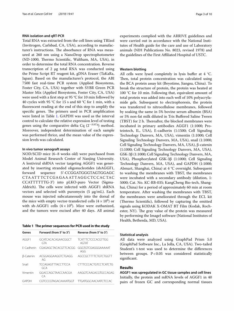
were detected by western blot and qRT-PCR, respec-tively. As shown in Fig. 1a, AGGF1 protein levels were upregulated in GC tissues compared with the matched adjacent normal tissues (P < 0.001). Besides, AGGF1 mRNA levels were increased in GC tissues compared with the matched adjacent normal tissues (P < 0.01, Fig. 1b). Then, AGGF1 expression levels in 4 GC cell lines (SGC-7901, MGC-803, MKN-45 and AGS) and GES-1 (normal control) were examined. The results showed that both mRNA and protein levels of AGGF1 were upregu-lated in GC cell lines compared with GES-1 (all P < 0.05, Fig. 1c, d).
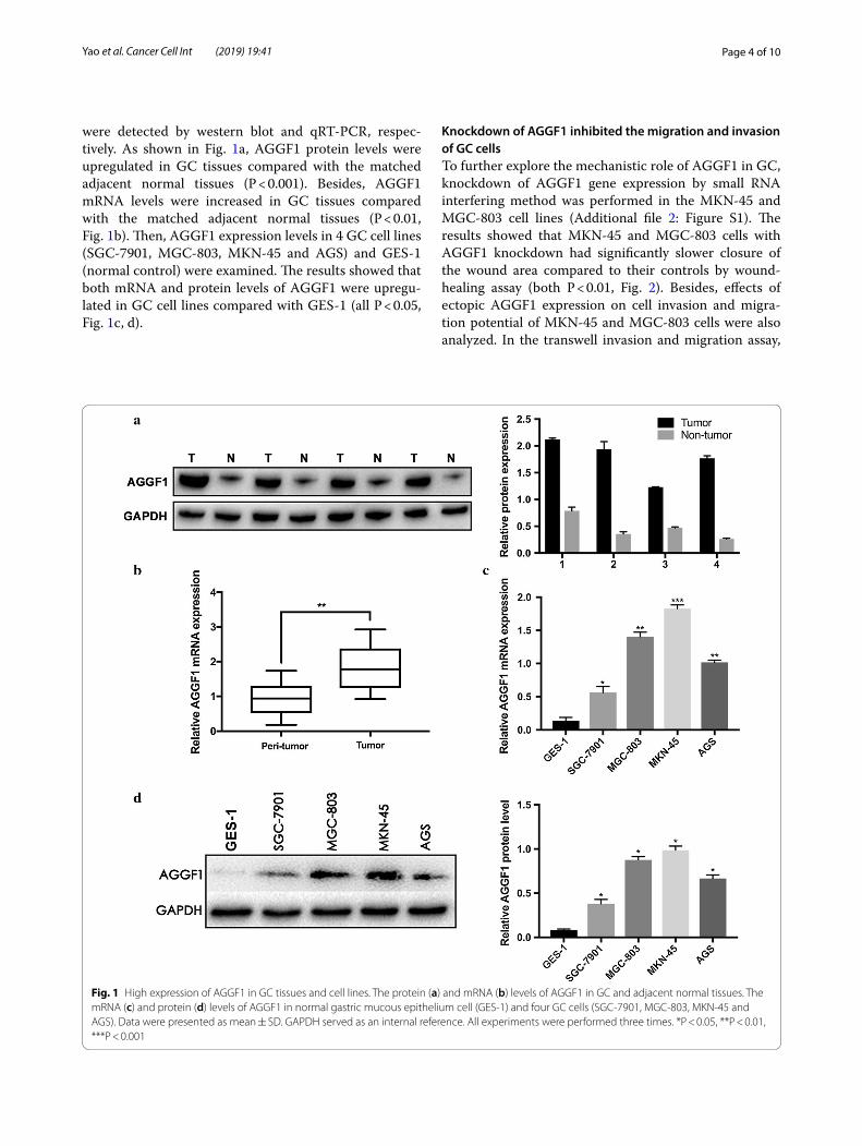
Knockdown of AGGF1 inhibited the migration and invasion of GC cellsTo further explore the mechanistic role of AGGF1 in GC, knockdown of AGGF1 gene expression by small RNA interfering method was performed in the MKN-45 and MGC-803 cell lines (Additional file 2: Figure S1). The results showed that MKN-45 and MGC-803 cells with AGGF1 knockdown had significantly slower closure of the wound area compared to their controls by wound-healing assay (both P < 0.01, Fig. 2). Besides, effects of ectopic AGGF1 expression on cell invasion and migra-tion potential of MKN-45 and MGC-803 cells were also analyzed. In the transwell invasion and migration assay,
Fig. 1 High expression of AGGF1 in GC tissues and cell lines. The protein (a) and mRNA (b) levels of AGGF1 in GC and adjacent normal tissues. The mRNA (c) and protein (d) levels of AGGF1 in normal gastric mucous epithelium cell (GES‑1) and four GC cells (SGC‑7901, MGC‑803, MKN‑45 and AGS). Data were presented as mean ± SD. GAPDH served as an internal reference. All experiments were performed three times. *P < 0.05, **P < 0.01, ***P < 0.001
Page 5 of 10Yao et al. Cancer Cell Int (2019) 19:41
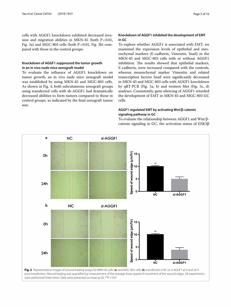
cells with AGGF1 knockdown exhibited decreased inva-sion and migration abilities in MKN-45 (both P < 0.01, Fig. 3a) and MGC-803 cells (both P < 0.01, Fig. 3b) com-pared with those in the control groups.
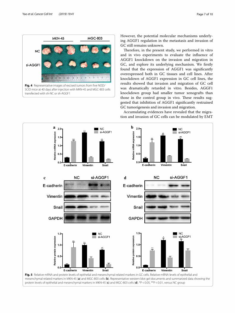
Knockdown of AGGF1 suppressed the tumor growth in an in vivo nude mice xenograft modelTo evaluate the influence of AGGF1 knockdown on tumor growth, an in vivo nude mice xenograft model was established by using MKN-45 and MGC-803 cells. As shown in Fig. 4, both subcutaneous xenograft groups using transfected cells with sh-AGGF1 had dramatically decreased abilities to form tumors compared to those in control groups, as indicated by the final xenograft tumor size.
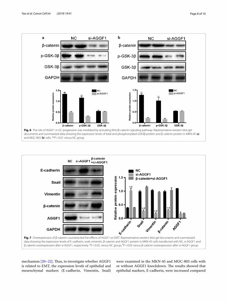
Knockdown of AGGF1 inhibited the development of EMT in GCTo explore whether AGGF1 is associated with EMT, we examined the expression levels of epithelial and mes-enchymal markers (E-cadherin, Vimentin, Snail) in the MKN-45 and MGC-803 cells with or without AGGF1 inhibition. The results showed that epithelial markers, E-cadherin, were increased compared with the controls, whereas mesenchymal marker Vimentin and related transcription factors Snail were significantly decreased in MKN-45 and MGC-803 cells with AGGF1 knockdown by qRT-PCR (Fig. 5a, b) and western blot (Fig. 5c, d) analyses. Consistently, gene silencing of AGGF1 retarded the development of EMT in MKN-45 and MGC-803 GC cells.
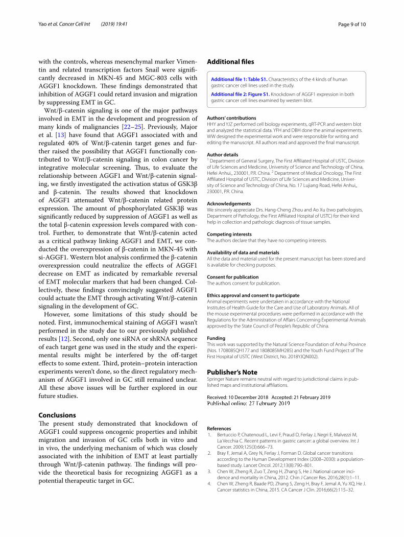
AGGF1 regulated EMT by activating Wnt/β‑catenin signaling pathway in GCTo evaluate the relationship between AGGF1 and Wnt/β-catenin signaling in GC, the activation status of GSK3β
Fig. 2 Representative images of wound‑healing assays for MKN‑45 cells (a) and MGC‑803 cells (b) transfected si‑NC or si‑AGGF1 at 0 and 24 h post‑transfection. Wound healing was quantified by measurement of the average linear speed of movement of the wound edges. All experiments were performed three times. Data were presented as mean ± SD. **P < 0.01
Page 6 of 10Yao et al. Cancer Cell Int (2019) 19:41
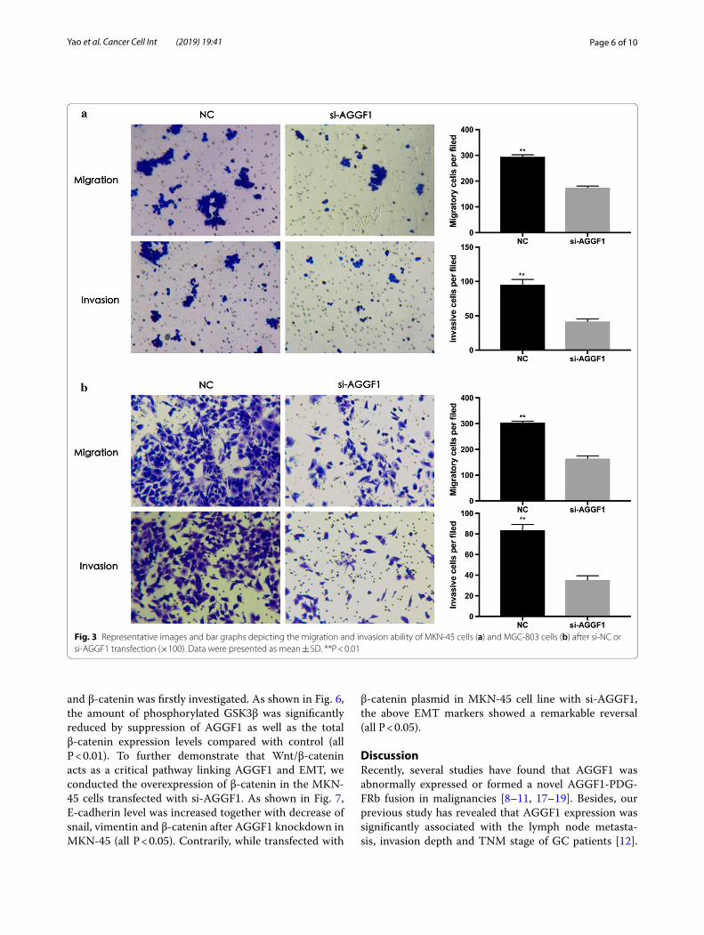
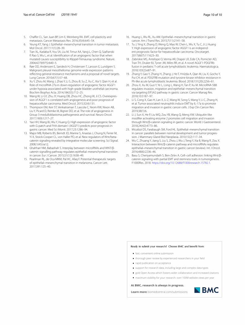
and β-catenin was firstly investigated. As shown in Fig. 6, the amount of phosphorylated GSK3β was significantly reduced by suppression of AGGF1 as well as the total β-catenin expression levels compared with control (all P < 0.01). To further demonstrate that Wnt/β-catenin acts as a critical pathway linking AGGF1 and EMT, we conducted the overexpression of β-catenin in the MKN-45 cells transfected with si-AGGF1. As shown in Fig. 7, E-cadherin level was increased together with decrease of snail, vimentin and β-catenin after AGGF1 knockdown in MKN-45 (all P < 0.05). Contrarily, while transfected with
β-catenin plasmid in MKN-45 cell line with si-AGGF1, the above EMT markers showed a remarkable reversal (all P < 0.05).
DiscussionRecently, several studies have found that AGGF1 was abnormally expressed or formed a novel AGGF1-PDG-FRb fusion in malignancies [8–11, 17–19]. Besides, our previous study has revealed that AGGF1 expression was significantly associated with the lymph node metasta-sis, invasion depth and TNM stage of GC patients [12].
Fig. 3 Representative images and bar graphs depicting the migration and invasion ability of MKN‑45 cells (a) and MGC‑803 cells (b) after si‑NC or si‑AGGF1 transfection (×100). Data were presented as mean ± SD. **P < 0.01
Page 7 of 10Yao et al. Cancer Cell Int (2019) 19:41
However, the potential molecular mechanisms underly-ing AGGF1 regulation in the metastasis and invasion of GC still remains unknown.
Therefore, in the present study, we performed in vitro and in vivo experiments to evaluate the influence of AGGF1 knockdown on the invasion and migration in GC, and explore its underlying mechanism. We firstly found that the expression of AGGF1 was significantly overexpressed both in GC tissues and cell lines. After knockdown of AGGF1 expression in GC cell lines, the results showed that invasion and migration of GC cell was dramatically retarded in vitro. Besides, AGGF1 knockdown group had smaller tumor xenografts than those in the control group in vivo. These results sug-gested that inhibition of AGGF1 significantly restrained GC tumorigenesis and invasion and migration.
Accumulating evidences have revealed that the migra-tion and invasion of GC cells can be modulated by EMT
Fig. 4 Representative images of excised tumors from five NOD/SCID mice at 40 days after injection with MKN‑45 and MGC‑803 cells transfected with sh‑NC or sh‑AGGF1
Fig. 5 Relative mRNA and protein levels of epithelial and mesenchymal related markers in GC cells. Relative mRNA levels of epithelial and mesenchymal related markers in MKN‑45 (a) and MGC‑803 cells (b). Representative western blot gel documents and summarized data showing the protein levels of epithelial and mesenchymal markers in MKN‑45 (c) and MGC‑803 cells (d). *P < 0.05, **P < 0.01, versus NC group
Page 8 of 10Yao et al. Cancer Cell Int (2019) 19:41
mechanism [20–22]. Thus, to investigate whether AGGF1 is related to EMT, the expression levels of epithelial and mesenchymal markers (E-cadherin, Vimentin, Snail)
were examined in the MKN-45 and MGC-803 cells with or without AGGF1 knockdown. The results showed that epithelial markers, E-cadherin, were increased compared
Fig. 6 The role of AGGF1 in GC progression was mediated by activating Wnt/β‑catenin signaling pathway. Representative western blot gel documents and summarized data showing the expression levels of total and phosphorylated GSK3β protein and β‑catenin protein in MKN‑45 (a) and MGC‑803 (b) cells. **P < 0.01 versus NC group
Fig. 7 Overexpression of β‑catenin counteracted the effects of AGGF1 on EMT. Representative western blot gel documents and summarized data showing the expression levels of E‑cadherin, snail, vimentin, β‑catenin and AGGF1 protein in MKN‑45 cells transfected with NC, si‑AGGF1 and β‑catenin overexpression after si‑AGGF1, respectively. *P < 0.05, versus NC group; #P < 0.05 versus β‑catenin overexpression after si‑AGGF1 group
Page 9 of 10Yao et al. Cancer Cell Int (2019) 19:41
with the controls, whereas mesenchymal marker Vimen-tin and related transcription factors Snail were signifi-cantly decreased in MKN-45 and MGC-803 cells with AGGF1 knockdown. These findings demonstrated that inhibition of AGGF1 could retard invasion and migration by suppressing EMT in GC.
Wnt/β-catenin signaling is one of the major pathways involved in EMT in the development and progression of many kinds of malignancies [22–25]. Previously, Major et al. [13] have found that AGGF1 associated with and regulated 40% of Wnt/β-catenin target genes and fur-ther raised the possibility that AGGF1 functionally con-tributed to Wnt/β-catenin signaling in colon cancer by integrative molecular screening. Thus, to evaluate the relationship between AGGF1 and Wnt/β-catenin signal-ing, we firstly investigated the activation status of GSK3β and β-catenin. The results showed that knockdown of AGGF1 attenuated Wnt/β-catenin related protein expression. The amount of phosphorylated GSK3β was significantly reduced by suppression of AGGF1 as well as the total β-catenin expression levels compared with con-trol. Further, to demonstrate that Wnt/β-catenin acted as a critical pathway linking AGGF1 and EMT, we con-ducted the overexpression of β-catenin in MKN-45 with si-AGGF1. Western blot analysis confirmed the β-catenin overexpression could neutralize the effects of AGGF1 decrease on EMT as indicated by remarkable reversal of EMT molecular markers that had been changed. Col-lectively, these findings convincingly suggested AGGF1 could actuate the EMT through activating Wnt/β-catenin signaling in the development of GC.
However, some limitations of this study should be noted. First, immunochemical staining of AGGF1 wasn’t performed in the study due to our previously published results [12]. Second, only one siRNA or shRNA sequence of each target gene was used in the study and the experi-mental results might be interfered by the off-target effects to some extent. Third, protein–protein interaction experiments weren’t done, so the direct regulatory mech-anism of AGGF1 involved in GC still remained unclear. All these above issues will be further explored in our future studies.
ConclusionsThe present study demonstrated that knockdown of AGGF1 could suppress oncogenic properties and inhibit migration and invasion of GC cells both in vitro and in vivo, the underlying mechanism of which was closely associated with the inhibition of EMT at least partially through Wnt/β-catenin pathway. The findings will pro-vide the theoretical basis for recognizing AGGF1 as a potential therapeutic target in GC.
Additional files
Additional file 1: Table S1. Characteristics of the 4 kinds of human gastric cancer cell lines used in the study.
Additional file 2: Figure S1. Knockdown of AGGF1 expression in both gastric cancer cell lines examined by western blot.
Authors’ contributionsHHY and YJZ performed cell biology experiments, qRT‑PCR and western blot and analyzed the statistical data. YFH and DBH done the animal experiments. WW designed the experimental work and were responsible for writing and editing the manuscript. All authors read and approved the final manuscript.
Author details1 Department of General Surgery, The First Affiliated Hospital of USTC, Division of Life Sciences and Medicine, University of Science and Technology of China, Hefei Anhui,, 230001, P.R. China. 2 Department of Medical Oncology, The First Affiliated Hospital of USTC, Division of Life Sciences and Medicine, Univer‑sity of Science and Technology of China, No. 17 Lujiang Road, Hefei Anhui,, 230001, P.R. China.
AcknowledgementsWe sincerely appreciate Drs. Hang‑Cheng Zhou and Ao Xu (two pathologists, Department of Pathology, the First Affiliated Hospital of USTC) for their kind help in collection and pathologic diagnosis of tissue samples.
Competing interestsThe authors declare that they have no competing interests.
Availability of data and materialsAll the data and material used for the present manuscript has been stored and is available for checking purposes.
Consent for publicationThe authors consent for publication.
Ethics approval and consent to participateAnimal experiments were undertaken in accordance with the National Institutes of Health Guide for the Care and Use of Laboratory Animals. All of the mouse experimental procedures were performed in accordance with the Regulations for the Administration of Affairs Concerning Experimental Animals approved by the State Council of People’s Republic of China.
FundingThis work was supported by the Natural Science Foundation of Anhui Province (Nos. 1708085QH177 and 1808085MH285) and the Youth Fund Project of The First Hospital of USTC (West District, No. 2018YJQN002).
Publisher’s NoteSpringer Nature remains neutral with regard to jurisdictional claims in pub‑lished maps and institutional affiliations.
Received: 10 December 2018 Accepted: 21 February 2019
References 1. Bertuccio P, Chatenoud L, Levi F, Praud D, Ferlay J, Negri E, Malvezzi M,
La Vecchia C. Recent patterns in gastric cancer: a global overview. Int J Cancer. 2009;125(3):666–73.
2. Bray F, Jemal A, Grey N, Ferlay J, Forman D. Global cancer transitions according to the Human Development Index (2008–2030): a population‑based study. Lancet Oncol. 2012;13(8):790–801.
3. Chen W, Zheng R, Zuo T, Zeng H, Zhang S, He J. National cancer inci‑dence and mortality in China, 2012. Chin J Cancer Res. 2016;28(1):1–11.
4. Chen W, Zheng R, Baade PD, Zhang S, Zeng H, Bray F, Jemal A, Yu XQ, He J. Cancer statistics in China, 2015. CA Cancer J Clin. 2016;66(2):115–32.
Page 10 of 10Yao et al. Cancer Cell Int (2019) 19:41
• fast, convenient online submission
•
thorough peer review by experienced researchers in your field
• rapid publication on acceptance
• support for research data, including large and complex data types
•
gold Open Access which fosters wider collaboration and increased citations
maximum visibility for your research: over 100M website views per year •
At BMC, research is always in progress.
Learn more biomedcentral.com/submissions
Ready to submit your research ? Choose BMC and benefit from:
5. Chaffer CL, San Juan BP, Lim E, Weinberg RA. EMT, cell plasticity and metastasis. Cancer Metastasis Rev. 2016;35(4):645–54.
6. Yeung KT, Yang J. Epithelial–mesenchymal transition in tumor metastasis. Mol Oncol. 2017;11(1):28–39.
7. Tian XL, Kadaba R, You SA, Liu M, Timur AA, Yang L, Chen Q, Szafranski P, Rao S, Wu L, et al. Identification of an angiogenic factor that when mutated causes susceptibility to Klippel‑Trenaunay syndrome. Nature. 2004;427(6975):640–5.
8. Røe OD, Anderssen E, Sandeck H, Christensen T, Larsson E, Lundgren S. Malignant pleural mesothelioma: genome‑wide expression patterns reflecting general resistance mechanisms and a proposal of novel targets. Lung Cancer. 2010;67(1):57–68.
9. Xu Y, Zhou M, Wang J, Zhao Y, Li S, Zhou B, Su Z, Xu C, Xia Y, Qian H, et al. Role of microRNA‑27a in down‑regulation of angiogenic factor AGGF1 under hypoxia associated with high‑grade bladder urothelial carcinoma. Biochim Biophys Acta. 2014;1842(5):712–25.
10. Wang W, Li GY, Zhu JY, Huang DB, Zhou HC, Zhong W, Ji CS. Overexpres‑sion of AGGF1 is correlated with angiogenesis and poor prognosis of hepatocellular carcinoma. Med Oncol. 2015;32(4):131.
11. Thompson EM, Keir ST, Venkatraman T, Lascola C, Yeom KW, Nixon AB, Liu Y, Picard D, Remke M, Bigner DD, et al. The role of angiogenesis in Group 3 medulloblastoma pathogenesis and survival. Neuro Oncol. 2017;19(9):1217–27.
12. Yao HH, Wang BJ, Wu Y, Huang Q. High expression of angiogenic factor with G‑patch and FHA domain1 (AGGF1) predicts poor prognosis in gastric cancer. Med Sci Monit. 2017;23:1286–94.
13. Major MB, Roberts BS, Berndt JD, Marine S, Anastas J, Chung N, Ferrer M, Yi X, Stoick‑Cooper CL, von Haller PD, et al. New regulators of Wnt/beta‑catenin signaling revealed by integrative molecular screening. Sci Signal. 2008;1(45):ra12.
14. Ghahhari NM, Babashah S. Interplay between microRNAs and WNT/β‑catenin signalling pathway regulates epithelial–mesenchymal transition in cancer. Eur J Cancer. 2015;51(12):1638–49.
15. Pearlman RL, de Oca MKM, Pal HC, Afaq F. Potential therapeutic targets of epithelial–mesenchymal transition in melanoma. Cancer Lett. 2017;391:125–40.
16. Huang L, Wu RL, Xu AM. Epithelial–mesenchymal transition in gastric cancer. Am J Transl Res. 2015;7(11):2141–58.
17. Tu J, Ying X, Zhang D, Weng Q, Mao W, Chen L, Wu X, Tu C, Ji J, Huang Y. High expression of angiogenic factor AGGF1 is an independ‑ent prognostic factor for hepatocellular carcinoma. Oncotarget. 2017;8(67):111623–30.
18. Zabriskie MS, Antelope O, Verma AR, Draper LR, Eide CA, Pomicter AD, Tran TH, Druker BJ, Tyner JW, Miles RR, et al. A novel AGGF1‑PDGFRb fusion in pediatric T‑cell acute lymphoblastic leukemia. Haematologica. 2018;103(2):e87–91.
19. Zhang Y, Gao Y, Zhang H, Zhang J, He F, Hnízda A, Qian M, Liu X, Gocho Y, Pui CH, et al. PDGFRB mutation and tyrosine kinase inhibitor resistance in Ph‑like acute lymphoblastic leukemia. Blood. 2018;131(20):2256–61.
20. Zhou X, Xu M, Guo Y, Ye L, Long L, Wang H, Tan P, Xu M. MicroRNA‑588 regulates invasion, migration and epithelial–mesenchymal transition via targeting EIF5A2 pathway in gastric cancer. Cancer Manag Res. 2018;10:5187–97.
21. Li S, Cong X, Gao H, Lan X, Li Z, Wang W, Song S, Wang Y, Li C, Zhang H, et al. Tumor‑associated neutrophils induce EMT by IL‑17a to promote migration and invasion in gastric cancer cells. J Exp Clin Cancer Res. 2019;38(1):6.
22. Li J, Sun X, He P, Liu WQ, Zou YB, Wang Q, Meng XW. Ubiquitin‑like modifier activating enzyme 2 promotes cell migration and invasion through Wnt/β‑catenin signaling in gastric cancer. World J Gastroenterol. 2018;24(42):4773–86.
23. Micalizzi DS, Farabaugh SM, Ford HL. Epithelial–mesenchymal transition in cancer: parallels between normal development and tumor progres‑sion. J Mammary Gland Biol Neoplasia. 2010;15(2):117–34.
24. Wu C, Zhuang Y, Jiang S, Liu S, Zhou J, Wu J, Teng Y, Xia B, Wang R, Zou X. Interaction between Wnt/β‑catenin pathway and microRNAs regulates epithelial–mesenchymal transition in gastric cancer (review). Int J Oncol. 2016;48(6):2236–46.
25. Basu S, Cheriyamundath S, Ben‑Ze’ev A. Cell–cell adhesion: linking Wnt/β‑catenin signaling with partial EMT and stemness traits in tumorigenesis. F1000Res. 2018. https ://doi.org/10.12688 /f1000 resea rch.15782 .1.