Embed Size (px)
Citation preview
®
®
®
®
®
®
®
®
®
®
®
®
®
®
®
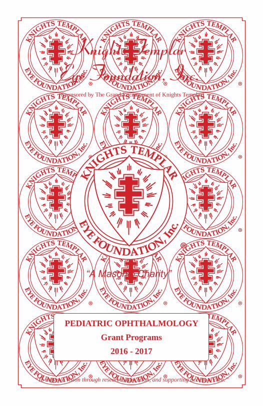
Knights TemplarEye Foundation, Inc.
(Sponsored by The Grand Encampment of Knights Templar)
“To improve vision through research, education, and supporting access to care.”
PEDIATRIC OPHTHALMOLOGYGrant Programs
2016 - 2017
®

2
The MissionThe mission of the Knights Templar Eye Foundation, Inc., is “to improve vision through research, education, and supporting access to care.”
To that end, the Knights Templar Eye Foundation, Inc. annually announces its call for research grant applications. The Foundation invites eligible investigators to submit applications for pediatric ophthalmology research grants for the award period which normally runs from July 1 to June 30.
From the applications received the Scientific Advisory Committee recommends to the trustees which requests should be funded.
To date over $24 million has been spent on research.
Note: For more information on the Knights Templar Eye Foundation, Inc. pediatric ophthalmology grant programs go to our web site:
http://www.knightstemplar.org/ktef/grantinfo.html
Inquiries & requests for materials regarding the Knights Templar Eye Foundation, Inc. may be directed to:
Robert W. Bigley
Office Administrator/Assistant Secretary
Knights Templar Eye Foundation, Inc. 1033 Long Prairie Road, Suite 5 Flower Mound, TX 75022-4230
Phone: (214) 888-0220 Fax: (214) 888-0230
E-mail: [email protected] Website: www.knightstemplar.org/ktef
The report of the Knights Templar Eye Foundation, Inc. as of August 2016.
$148 million has been spent on research, patient care, and education.
3
Officers President Vice President Vice President Treasurer Secretary Assistant Secretary & Office Administrator
Duane L. Vaught, Bloomington, IN Jeffrey N. Nelson, Bismarck, ND Michael B. Johnson, Crowheart, WY Bobby B. Simmons, Bonaire, GA Lawrence E. Tucker, Bellaire, TX Robert W. Bigley, Flower Mound, TX email: [email protected]
General Counsel
*Trustee Emeritus
James C. Herndon, Blackfoot, ID
Trustees David M. Dryer, Indianola, IA Ned E. Dull, Springboro, OH Kenneth B. Fischer, Friendswood, TX David D. Goodwin, Vestal, NY James C. Herndon, Blackfoot, ID Michael B. Johnson, Crowheart, WY William Jackson Jones D.D.S., Villa Grove, IL James N. Karnegis M.D., Ph.D., Elkhorn, NE William H. Koon, II, Columbus Grove, OH
David J. Kussman, Anaheim, CA Rodney A. Mann, Shelbyville, IN Jeffrey N. Nelson, Bismarck, ND Terry L. Plemons, Chattanooga, TN W. Bruce Pruitt, Roseville, CA Bobby B. Simmons, Bonaire, GA
*Herbert D. Sledd, Winchester, KY *Donald H. Smith, Richmond, KY Lawrence E. Tucker, Bellaire, TX Duane L. Vaught, Bloomington, IN James M. Ward, Valle, MS
Who are the Knights Templar?...Today’s organization known as the Knights Templar does not claim to be a direct descendant of the ancient order of Knights Templar that was founded during the crusades in the 12th century. The purpose of those crusader knights was to protect pilgrims from danger when on their way to the Holy Land. These men took vows of poverty, chastity, and obedience, and were renowned for their courage in battle. In 1118 A.D., nineteen years after the successful crusade, these Poor Fellow Soldiers of Christ and the Temple of Jerusalem, as they termed themselves, were officially recognized, sanctioned, and given, for their headquarters, a building on Mount Moriah, the site of the Temple of King Solomon. Consequently, they became known as Knights of the Temple, or Knights Templar.
What are Knights Templar doing today?...Eight centuries after the crusades, the current organization is still dedicated to assisting those in need and in using its efforts for the prevention of blindness. Because sight is a most precious gift, The Knights Templar Eye Foundation is often referred to as “A Great Humanitarian Charity.”
4
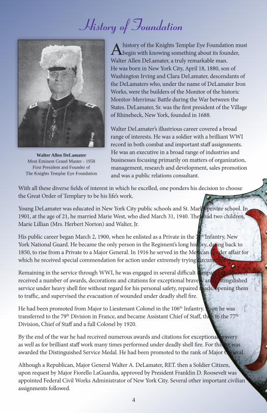
Walter Allen DeLamater Most Eminent Grand Master - 1958
First President and Founder of The Knights Templar Eye Foundation
History of Foundation
A history of the Knights Templar Eye Foundation must begin with knowing something about its founder,
Walter Allen DeLamater, a truly remarkable man. He was born in New York City, April 18, 1880, son of Washington Irving and Clara DeLamater, descendants of the DeLamaters who, under the name of DeLamater Iron Works, were the builders of the Monitor of the historic Monitor-Merrimac Battle during the War between the States. DeLamater, Sr. was the first president of the Village of Rhinebeck, New York, founded in 1688.
Walter DeLamater’s illustrious career covered a broad range of interests. He was a soldier with a brilliant WWI record in both combat and important staff assignments. He was an executive in a broad range of industries and businesses focusing primarily on matters of organization, management, research and development, sales promotion and was a public relations consultant.
With all these diverse fields of interest in which he excelled, one ponders his decision to choose the Great Order of Templary to be his life’s work.
Young DeLamater was educated in New York City public schools and St. Mark’s private school. In 1901, at the age of 21, he married Marie West, who died March 31, 1940. They had two children, Marie Lillian (Mrs. Herbert Norton) and Walter, Jr.
His public career began March 2, 1900, when he enlisted as a Private in the 71st Infantry, New York National Guard. He became the only person in the Regiment’s long history, dating back to 1850, to rise from a Private to a Major General. In 1916 he served in the Mexican Border affair for which he received special commendation for action under extremely trying circumstances.
Remaining in the service through WWI, he was engaged in several difficult campaigns in France, received a number of awards, decorations and citations for exceptional bravery and distinguished service under heavy shell fire without regard for his personal safety, repaired roads, opening them to traffic, and supervised the evacuation of wounded under deadly shell fire.
He had been promoted from Major to Lieutenant Colonel in the 106th Infantry. Soon he was transferred to the 79th Division in France, and became Assistant Chief of Staff, then to the 77th Division, Chief of Staff and a full Colonel by 1920.
By the end of the war he had received numerous awards and citations for exceptional bravery as well as for brilliant staff work many times performed under deadly shell fire. For this he was awarded the Distinguished Service Medal. He had been promoted to the rank of Major General.
Although a Republican, Major General Walter A. DeLamater, RET. then a Soldier Citizen, upon request by Major Fiorello LaGuardia, approved by President Franklin D. Roosevelt was appointed Federal Civil Works Administrator of New York City. Several other important civilian assignments followed.
5
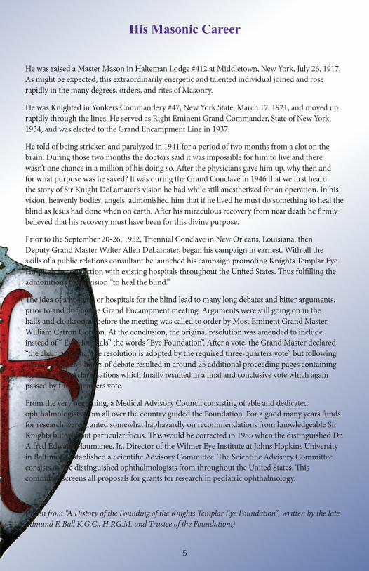
His Masonic Career
He was raised a Master Mason in Halteman Lodge #412 at Middletown, New York, July 26, 1917. As might be expected, this extraordinarily energetic and talented individual joined and rose rapidly in the many degrees, orders, and rites of Masonry.
He was Knighted in Yonkers Commandery #47, New York State, March 17, 1921, and moved up rapidly through the lines. He served as Right Eminent Grand Commander, State of New York, 1934, and was elected to the Grand Encampment Line in 1937.
He told of being stricken and paralyzed in 1941 for a period of two months from a clot on the brain. During those two months the doctors said it was impossible for him to live and there wasn’t one chance in a million of his doing so. After the physicians gave him up, why then and for what purpose was he saved? It was during the Grand Conclave in 1946 that we first heard the story of Sir Knight DeLamater’s vision he had while still anesthetized for an operation. In his vision, heavenly bodies, angels, admonished him that if he lived he must do something to heal the blind as Jesus had done when on earth. After his miraculous recovery from near death he firmly believed that his recovery must have been for this divine purpose.
Prior to the September 20-26, 1952, Triennial Conclave in New Orleans, Louisiana, then Deputy Grand Master Walter Allen DeLamater, began his campaign in earnest. With all the skills of a public relations consultant he launched his campaign promoting Knights Templar Eye Hospitals in connection with existing hospitals throughout the United States. Thus fulfilling the admonitions of his vision “to heal the blind.”
The idea of a hospital or hospitals for the blind lead to many long debates and bitter arguments, prior to and during the Grand Encampment meeting. Arguments were still going on in the halls and cloakrooms before the meeting was called to order by Most Eminent Grand Master William Catron Gordon. At the conclusion, the original resolution was amended to include instead of “ Eye Hospitals” the words “Eye Foundation”. After a vote, the Grand Master declared “the chair rules that the resolution is adopted by the required three-quarters vote”, but following a break another 3 hours of debate resulted in around 25 additional proceeding pages containing resolutions and clarifications which finally resulted in a final and conclusive vote which again passed by three-quarters vote.
From the very beginning, a Medical Advisory Council consisting of able and dedicated ophthalmologists from all over the country guided the Foundation. For a good many years funds for research were granted somewhat haphazardly on recommendations from knowledgeable Sir Knights but without particular focus. This would be corrected in 1985 when the distinguished Dr. Alfred Edward Maumanee, Jr., Director of the Wilmer Eye Institute at Johns Hopkins University in Baltimore, established a Scientific Advisory Committee. The Scientific Advisory Committee consists of five distinguished ophthalmologists from throughout the United States. This committee screens all proposals for grants for research in pediatric ophthalmology.
(taken from “A History of the Founding of the Knights Templar Eye Foundation”, written by the late Edmund F. Ball K.G.C., H.P.G.M. and Trustee of the Foundation.)
6
Career-Starter Research Grants – up to $65,000 per grant. Applicants for these grants must be at the beginning of their academic careers and must have received an M.D., Ph.D. or equivalent degree.
Competitive Renewal Grants - up to $65,000 per grant to extend the original grant project for one additional year when the data collected from the original grant is compelling enough to apply.
Training Mentors for Developing Countries (TMDC) Fellowship – Annual stipend of $60,000 - The Scientific Advisory Committee for the Knights Templar Eye Foundation has identified a significant need for well-trained pediatric ophthalmology faculty (mentors) in developing countries. As a result, the Foundation has created a one-year fellowship to help meet that training need. Those receiving a fellowship have agreed in writing to return to their native country immediately following the fellowship, to practice pediatric ophthalmology for a minimum of five years and, to the extent possible, be directly involved in the training of residents during those five years.
Pediatric Ophthalmology Grants
The Knights Templar Eye Foundation, Inc. is committed to support research that can help launch the careers of clinical and basic researchers focused on the prevention and cure of
potentially blinding diseases in infants and children. Grants supported by the Knights Templar Eye Foundation, Inc. are awarded to impact the care of infants, children, and adults. Clinical and basic research on conditions that may be potentially preventable or correctable such as amblyopia, cataract, glaucoma, optic nerve hypoplasia, nystagmus, retinopathy of prematurity, and hereditary diseases that occur at birth or within early childhood, such as retinoblastoma, is encouraged. Proposals for support of basic research on eye and visual system development also are welcome.
Each year the Knights Templar Eye Foundation, Inc., invites eligible investigators to submit applications for pediatric ophthalmology research grants:
To hear a message from the Grand Master and the Chair of the
Knights Templar Eye Foundation Scientific Advisory Committee scan the QR code. (free QR readers are available at your phone’s app store)
7
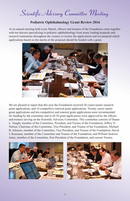
Scientific Advisory Committee MeetingPediatric Ophthalmology Grant Review 2016
At an annual meeting held every March, officers and trustees of the Foundation come together with ten doctors specializing in pediatric ophthalmology from many leading hospitals and research institutions throughout the country to review the applications and recommend which applications based on the merits of the proposal should be funded with a grant.
We are pleased to report that this year the Foundation received 50 career-starter research grant applications, and 16 competitive renewal grant applications. Twenty career starter grant applications and ten competitive and renewal grant applications were recommended for funding by the committee and in all 30 grant applications were approved by the officers and trustees serving on the Scientific Advisory Committee. This committee consists of Duane L. Vaught, member of the Committee, President, and Trustee of the Foundation; Jeffrey N. Nelson, Chairman of the Committee, Vice President, and Trustee of the Foundation; Michael B. Johnson, member of the Committee, Vice President, and Trustee of the Foundation; David J. Kussman, member of the Committee and Trustee of the Foundation; and William Jackson Jones, member of the Committee, Past President of the Foundation, and current Trustee.
8
Endowed Professorship Awarded
In 2011, the Board explored the feasibility and desirability of establishing an endowed professorship program at a leading research university or teaching hospital focusing on
ophthalmic education. Preliminary groundwork proved positive and in 2012 the President formed a committee of Board members to further explore this idea. Advantages to the Foundation of endowing a professorship identified by the committee included the fact that an endowed professorship would be consistent with the Foundation’s mission, it would provide a perpetual benefit to the Foundation from a one-time investment, it would promote visibility of the Foundation, and it would create a new partnership legacy for the Foundation. Advantages to the institution identified by the committee included the fact that an endowed professorship would provide the institution with a financial resource, it would be consistent with the institution’s mission statement, and it would provide publicity for the institution. In August 2013, the committee recommended, and the Board subsequently approved, committing $2 million, matched dollar for dollar by the Mayo Clinic to establish the first endowed professorship to be named:
“Knights Templar Eye Foundation Inc., Professor of Ophthalmology Research” to
Michael Brodsky, M.D. at
The Mayo Clinic campuses in Rochester, MN; Phoenix, AZ; and Jacksonville, FL
In August 2015, the comittee again recommended, and the Board subsequently approved, committing another $2 million, matched dollar for dollar by Johns Hopkins to establish the
second endowed professorship to be named:
“Knights Templar Eye Foundation Inc., Professor of Ophthalmology” to
Thomas McCarthy Bosley, M.D. at
The Wilmer Eye Institute of Johns Hopkins University Baltimore, MD
9
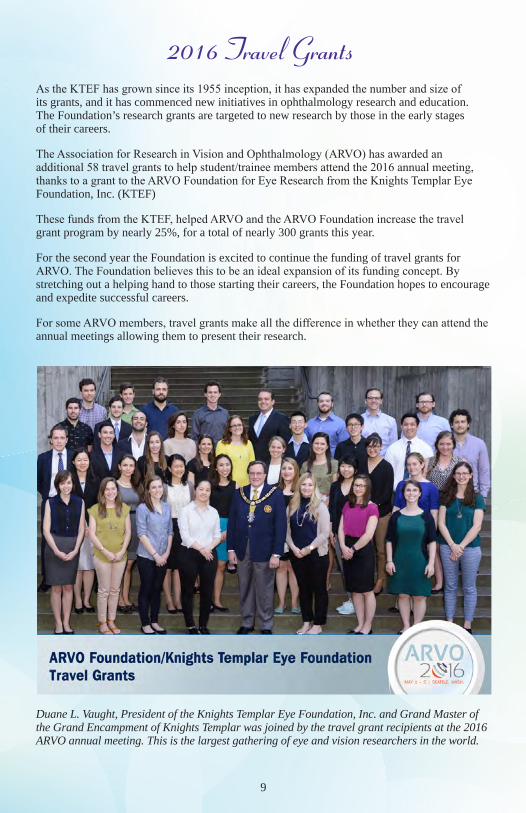
2016 Travel GrantsAs the KTEF has grown since its 1955 inception, it has expanded the number and size of its grants, and it has commenced new initiatives in ophthalmology research and education. The Foundation’s research grants are targeted to new research by those in the early stages of their careers.
The Association for Research in Vision and Ophthalmology (ARVO) has awarded an additional 58 travel grants to help student/trainee members attend the 2016 annual meeting, thanks to a grant to the ARVO Foundation for Eye Research from the Knights Templar Eye Foundation, Inc. (KTEF)
These funds from the KTEF, helped ARVO and the ARVO Foundation increase the travel grant program by nearly 25%, for a total of nearly 300 grants this year.
For the second year the Foundation is excited to continue the funding of travel grants for ARVO. The Foundation believes this to be an ideal expansion of its funding concept. By stretching out a helping hand to those starting their careers, the Foundation hopes to encourage and expedite successful careers.
For some ARVO members, travel grants make all the difference in whether they can attend the annual meetings allowing them to present their research.
Duane L. Vaught, President of the Knights Templar Eye Foundation, Inc. and Grand Master of the Grand Encampment of Knights Templar was joined by the travel grant recipients at the 2016 ARVO annual meeting. This is the largest gathering of eye and vision researchers in the world.
10
Mary Elizabeth Hartnett, M.D.Professor of Ophthalmology, Adjunct Professor of Pediatrics,
University of Utah - Department of Ophthalmology, John A. Moran Eye Center
The Impact of KTEF FundingI have the great honor of serving on the Knights Templar Eye Foundation Scientific Advisory Committee, where I have the opportunity to review scientific proposals designed to understand causes and find treatments for blinding pediatric eye diseases.
As a clinician scientist and vitreoretinal specialist, I understand the predicament that M.D.s and scientists find themselves in when beginning their research careers, namely how to obtain resources to get started, develop preliminary data, and put together laboratories and laboratory teams. The Knights Templar Eye Foundation provides grant funding and support for scientists
in their early careers to obtain preliminary data necessary to refine scientific questions, start laboratories, and successfully compete for later funding through organizations, including the National Institutes of Health.
In my situation, I was a practicing vitreoretinal surgeon with specialty training and expertise in pediatric retinal diseases. I had always wanted to pursue science from the time of high school, but I was concerned that taking time out of my career to pursue a Ph.D. would cause a gap between my surgical training and the start of a medical practice and interfere with my ability to provide the best care and treatment for my patients as a physician and surgeon. Therefore, I pursued a postdoctoral fellowship as a practicing M.D. and was able to learn many of the techniques and ways of approaching questions as a scientist. When I was ready to start my independent research program, I found that funding organizations required preliminary data and publication before ever considering funding. I well recognize how important it is to have funding sources at early stages in one’s career in order to pursue science. I fortunately was able to successfully obtain support. Also important in developing one’s research program is mentorship. One of my mentors was John Penn, now the chair of the Scientific Advisory Committee, who had developed an animal model that mimicked many of the features of human retinopathy of prematurity. John was very helpful in helping me get the model up and running in my own laboratory. Although I did not apply for funding from the Knights Templar Eye Foundation, I have mentored others, including my own laboratory members, and have had one research assistant professor who has been successfully funded through the Knights Templar Eye Foundation.
Now as a Scientific Advisory Committee member, I think back to the difficult times in my early career and how beneficial it is to have research support early in one’s career. I also remember the importance of mentors and I try to reach out and offer support to other scientists and clinicians beginning their research programs.
The Knights Templar Eye Foundation provides needed support to scientists and clinician scientists at the beginning of their research careers and is one of the only, if not the only, organizations that specifically provides funding for pediatric eye disease. Pediatric eye research is such an important and needed area of research support. I believe that through the support of the Knights Templar Eye Foundation, the Scientific Advisory Committee is able to help clinician scientists and scientists develop worthwhile careers to improve the outcomes and quality of life of children and infants with blinding eye diseases.
11
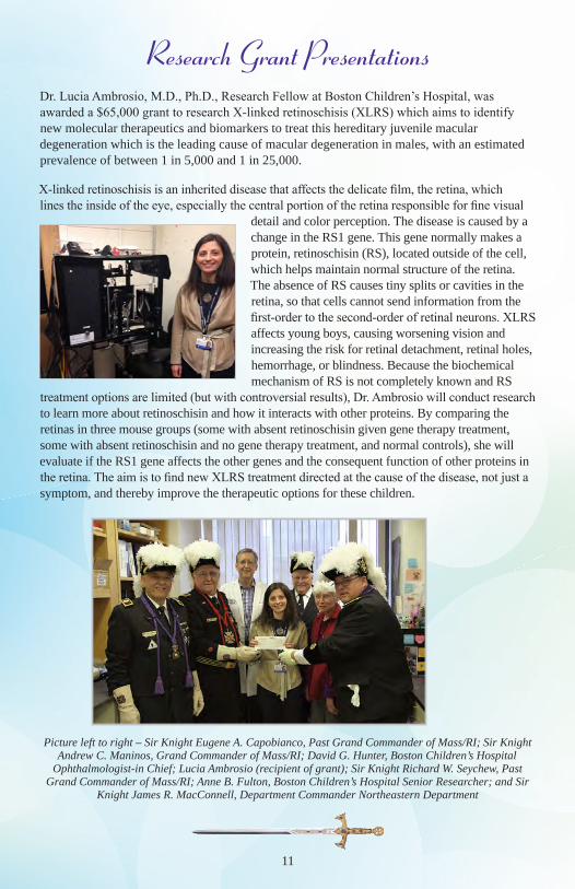
Research Grant PresentationsDr. Lucia Ambrosio, M.D., Ph.D., Research Fellow at Boston Children’s Hospital, was awarded a $65,000 grant to research X-linked retinoschisis (XLRS) which aims to identify new molecular therapeutics and biomarkers to treat this hereditary juvenile macular degeneration which is the leading cause of macular degeneration in males, with an estimated prevalence of between 1 in 5,000 and 1 in 25,000.
X-linked retinoschisis is an inherited disease that affects the delicate film, the retina, which lines the inside of the eye, especially the central portion of the retina responsible for fine visual
detail and color perception. The disease is caused by a change in the RS1 gene. This gene normally makes a protein, retinoschisin (RS), located outside of the cell, which helps maintain normal structure of the retina. The absence of RS causes tiny splits or cavities in the retina, so that cells cannot send information from the first-order to the second-order of retinal neurons. XLRS affects young boys, causing worsening vision and increasing the risk for retinal detachment, retinal holes, hemorrhage, or blindness. Because the biochemical mechanism of RS is not completely known and RS
treatment options are limited (but with controversial results), Dr. Ambrosio will conduct research to learn more about retinoschisin and how it interacts with other proteins. By comparing the retinas in three mouse groups (some with absent retinoschisin given gene therapy treatment, some with absent retinoschisin and no gene therapy treatment, and normal controls), she will evaluate if the RS1 gene affects the other genes and the consequent function of other proteins in the retina. The aim is to find new XLRS treatment directed at the cause of the disease, not just a symptom, and thereby improve the therapeutic options for these children.
Picture left to right – Sir Knight Eugene A. Capobianco, Past Grand Commander of Mass/RI; Sir Knight Andrew C. Maninos, Grand Commander of Mass/RI; David G. Hunter, Boston Children’s Hospital
Ophthalmologist-in Chief; Lucia Ambrosio (recipient of grant); Sir Knight Richard W. Seychew, Past Grand Commander of Mass/RI; Anne B. Fulton, Boston Children’s Hospital Senior Researcher; and Sir
Knight James R. MacConnell, Department Commander Northeastern Department
12
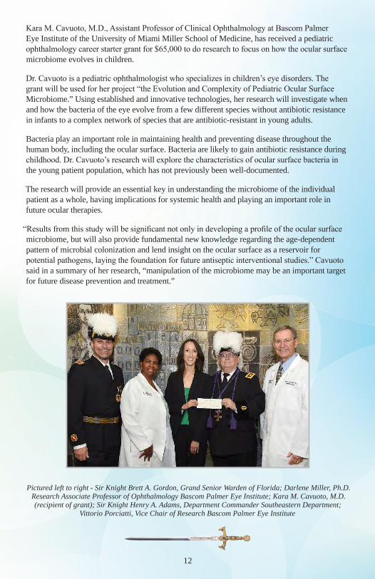
Kara M. Cavuoto, M.D., Assistant Professor of Clinical Ophthalmology at Bascom Palmer Eye Institute of the University of Miami Miller School of Medicine, has received a pediatric ophthalmology career starter grant for $65,000 to do research to focus on how the ocular surface microbiome evolves in children.
Dr. Cavuoto is a pediatric ophthalmologist who specializes in children’s eye disorders. The grant will be used for her project “the Evolution and Complexity of Pediatric Ocular Surface Microbiome.” Using established and innovative technologies, her research will investigate when and how the bacteria of the eye evolve from a few different species without antibiotic resistance in infants to a complex network of species that are antibiotic-resistant in young adults.
Bacteria play an important role in maintaining health and preventing disease throughout the human body, including the ocular surface. Bacteria are likely to gain antibiotic resistance during childhood. Dr. Cavuoto’s research will explore the characteristics of ocular surface bacteria in the young patient population, which has not previously been well-documented.
The research will provide an essential key in understanding the microbiome of the individual patient as a whole, having implications for systemic health and playing an important role in future ocular therapies.
“Results from this study will be significant not only in developing a profile of the ocular surface microbiome, but will also provide fundamental new knowledge regarding the age-dependent pattern of microbial colonization and lend insight on the ocular surface as a reservoir for potential pathogens, laying the foundation for future antiseptic interventional studies.” Cavuoto said in a summary of her research, “manipulation of the microbiome may be an important target for future disease prevention and treatment.”
Pictured left to right - Sir Knight Brett A. Gordon, Grand Senior Warden of Florida; Darlene Miller, Ph.D. Research Associate Professor of Ophthalmology Bascom Palmer Eye Institute; Kara M. Cavuoto, M.D. (recipient of grant); Sir Knight Henry A. Adams, Department Commander Southeastern Department;
Vittorio Porciatti, Vice Chair of Research Bascom Palmer Eye Institute
13
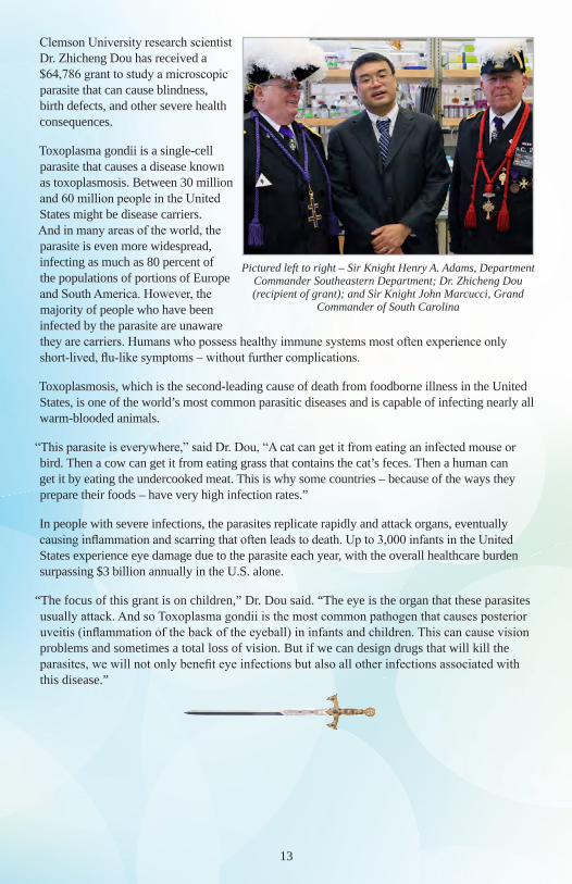
Clemson University research scientist Dr. Zhicheng Dou has received a $64,786 grant to study a microscopic parasite that can cause blindness, birth defects, and other severe health consequences.
Toxoplasma gondii is a single-cell parasite that causes a disease known as toxoplasmosis. Between 30 million and 60 million people in the United States might be disease carriers. And in many areas of the world, the parasite is even more widespread, infecting as much as 80 percent of the populations of portions of Europe and South America. However, the majority of people who have been infected by the parasite are unaware they are carriers. Humans who possess healthy immune systems most often experience only short-lived, flu-like symptoms – without further complications.
Toxoplasmosis, which is the second-leading cause of death from foodborne illness in the United States, is one of the world’s most common parasitic diseases and is capable of infecting nearly all warm-blooded animals.
“This parasite is everywhere,” said Dr. Dou, “A cat can get it from eating an infected mouse or bird. Then a cow can get it from eating grass that contains the cat’s feces. Then a human can get it by eating the undercooked meat. This is why some countries – because of the ways they prepare their foods – have very high infection rates.”
In people with severe infections, the parasites replicate rapidly and attack organs, eventually causing inflammation and scarring that often leads to death. Up to 3,000 infants in the United States experience eye damage due to the parasite each year, with the overall healthcare burden surpassing $3 billion annually in the U.S. alone.
“The focus of this grant is on children,” Dr. Dou said. “The eye is the organ that these parasites usually attack. And so Toxoplasma gondii is the most common pathogen that causes posterior uveitis (inflammation of the back of the eyeball) in infants and children. This can cause vision problems and sometimes a total loss of vision. But if we can design drugs that will kill the parasites, we will not only benefit eye infections but also all other infections associated with this disease.”
Pictured left to right – Sir Knight Henry A. Adams, Department Commander Southeastern Department; Dr. Zhicheng Dou (recipient of grant); and Sir Knight John Marcucci, Grand
Commander of South Carolina
14
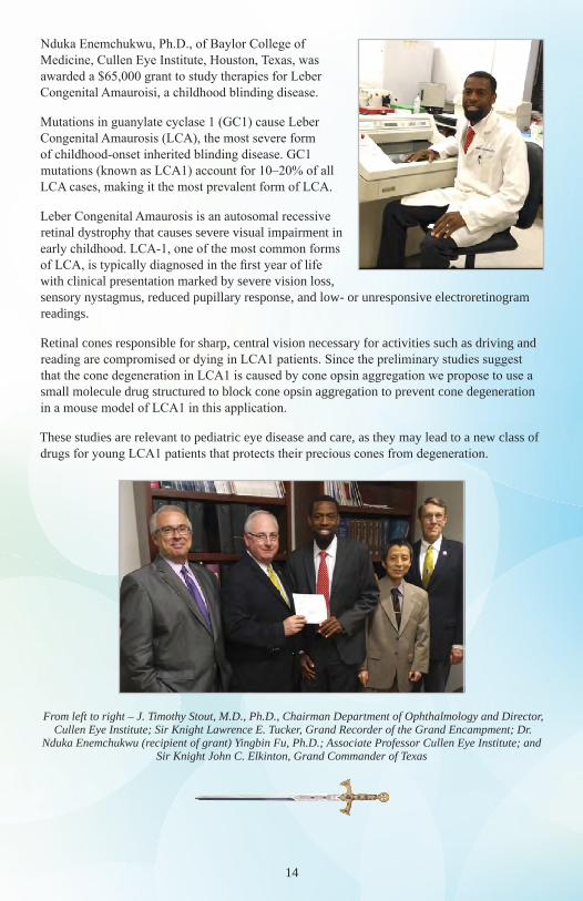
Nduka Enemchukwu, Ph.D., of Baylor College of Medicine, Cullen Eye Institute, Houston, Texas, was awarded a $65,000 grant to study therapies for Leber Congenital Amauroisi, a childhood blinding disease.
Mutations in guanylate cyclase 1 (GC1) cause Leber Congenital Amaurosis (LCA), the most severe form of childhood-onset inherited blinding disease. GC1 mutations (known as LCA1) account for 10–20% of all LCA cases, making it the most prevalent form of LCA.
Leber Congenital Amaurosis is an autosomal recessive retinal dystrophy that causes severe visual impairment in early childhood. LCA-1, one of the most common forms of LCA, is typically diagnosed in the first year of life with clinical presentation marked by severe vision loss, sensory nystagmus, reduced pupillary response, and low- or unresponsive electroretinogram readings.
Retinal cones responsible for sharp, central vision necessary for activities such as driving and reading are compromised or dying in LCA1 patients. Since the preliminary studies suggest that the cone degeneration in LCA1 is caused by cone opsin aggregation we propose to use a small molecule drug structured to block cone opsin aggregation to prevent cone degeneration in a mouse model of LCA1 in this application.
These studies are relevant to pediatric eye disease and care, as they may lead to a new class of drugs for young LCA1 patients that protects their precious cones from degeneration.
From left to right – J. Timothy Stout, M.D., Ph.D., Chairman Department of Ophthalmology and Director, Cullen Eye Institute; Sir Knight Lawrence E. Tucker, Grand Recorder of the Grand Encampment; Dr.
Nduka Enemchukwu (recipient of grant) Yingbin Fu, Ph.D.; Associate Professor Cullen Eye Institute; and Sir Knight John C. Elkinton, Grand Commander of Texas
15
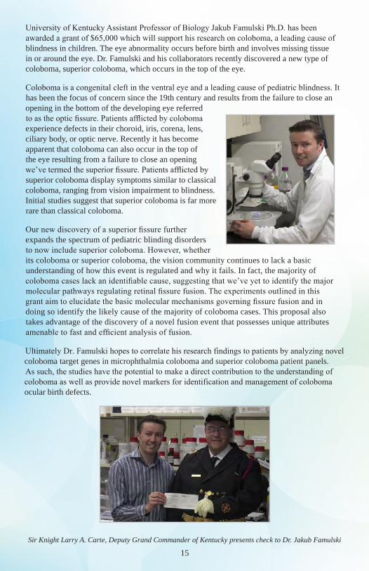
University of Kentucky Assistant Professor of Biology Jakub Famulski Ph.D. has been awarded a grant of $65,000 which will support his research on coloboma, a leading cause of blindness in children. The eye abnormality occurs before birth and involves missing tissue in or around the eye. Dr. Famulski and his collaborators recently discovered a new type of coloboma, superior coloboma, which occurs in the top of the eye.
Coloboma is a congenital cleft in the ventral eye and a leading cause of pediatric blindness. It has been the focus of concern since the 19th century and results from the failure to close an opening in the bottom of the developing eye referred to as the optic fissure. Patients afflicted by coloboma experience defects in their choroid, iris, corena, lens, ciliary body, or optic nerve. Recently it has become apparent that coloboma can also occur in the top of the eye resulting from a failure to close an opening we’ve termed the superior fissure. Patients afflicted by superior coloboma display symptoms similar to classical coloboma, ranging from vision impairment to blindness. Initial studies suggest that superior coloboma is far more rare than classical coloboma.
Our new discovery of a superior fissure further expands the spectrum of pediatric blinding disorders to now include superior coloboma. However, whether its coloboma or superior coloboma, the vision community continues to lack a basic understanding of how this event is regulated and why it fails. In fact, the majority of coloboma cases lack an identifiable cause, suggesting that we’ve yet to identify the major molecular pathways regulating retinal fissure fusion. The experiments outlined in this grant aim to elucidate the basic molecular mechanisms governing fissure fusion and in doing so identify the likely cause of the majority of coloboma cases. This proposal also takes advantage of the discovery of a novel fusion event that possesses unique attributes amenable to fast and efficient analysis of fusion.
Ultimately Dr. Famulski hopes to correlate his research findings to patients by analyzing novel coloboma target genes in microphthalmia coloboma and superior coloboma patient panels. As such, the studies have the potential to make a direct contribution to the understanding of coloboma as well as provide novel markers for identification and management of coloboma ocular birth defects.
Sir Knight Larry A. Carte, Deputy Grand Commander of Kentucky presents check to Dr. Jakub Famulski
16
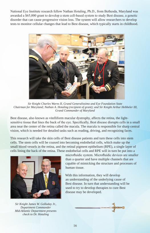
National Eye Institute research fellow Nathan Hotaling, Ph.D., from Bethesda, Maryland was awarded a $65,000 grant to develop a stem cell-based system to study Best disease, a genetic disorder that can cause progressive vision loss. The system will allow researchers to develop tests to monitor cellular changes that lead to Best disease, which typically starts in childhood.
Sir Knight Charles Warns II, Grand Generalissimo and Eye Foundation State Chairman for Maryland; Nathan A. Hotaling (recipient of grant); and Sir Knight Arthur Hebbeler III,
Grand Commander of Maryland
Best disease, also known as vitelliform macular dystrophy, affects the retina, the light-sensitive tissue that lines the back of the eye. Specifically, Best disease disrupts cells in a small area near the center of the retina called the macula. The macula is responsible for sharp central vision, which is needed for detailed tasks such as reading, driving, and recognizing faces.
This research will take the skin cells of Best disease patients and turn these cells into stem cells. The stem cells will be coaxed into becoming endothelial cells, which make up the small blood vessels in the retina, and the retinal pigment epithelium (RPE), a single layer of cells lining the back of the retina. These endothelial cells and RPE will in turn be put into a
microfluidic system. Microfluidic devices are smaller than a quarter and have multiple channels that are capable of mimicking the structure and processes of human tissue.
With this information, they will develop an understanding of the underlying cause of Best disease. In turn that understanding will be used to try to develop therapies to cure Best disease may be developed.
Sir Knight James W. Golladay Jr., Department Commander
Mid-Atlantic Department presents check to Dr. Hotaling
17
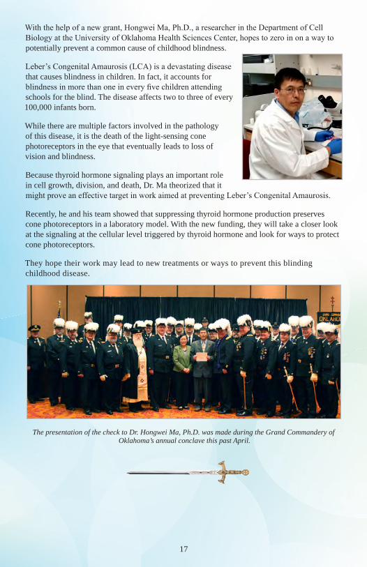
With the help of a new grant, Hongwei Ma, Ph.D., a researcher in the Department of Cell Biology at the University of Oklahoma Health Sciences Center, hopes to zero in on a way to potentially prevent a common cause of childhood blindness.
Leber’s Congenital Amaurosis (LCA) is a devastating disease that causes blindness in children. In fact, it accounts for blindness in more than one in every five children attending schools for the blind. The disease affects two to three of every 100,000 infants born.
While there are multiple factors involved in the pathology of this disease, it is the death of the light-sensing cone photoreceptors in the eye that eventually leads to loss of vision and blindness.
Because thyroid hormone signaling plays an important role in cell growth, division, and death, Dr. Ma theorized that it might prove an effective target in work aimed at preventing Leber’s Congenital Amaurosis.
Recently, he and his team showed that suppressing thyroid hormone production preserves cone photoreceptors in a laboratory model. With the new funding, they will take a closer look at the signaling at the cellular level triggered by thyroid hormone and look for ways to protect cone photoreceptors.
They hope their work may lead to new treatments or ways to prevent this blinding childhood disease.
The presentation of the check to Dr. Hongwei Ma, Ph.D. was made during the Grand Commandery of Oklahoma’s annual conclave this past April.
18
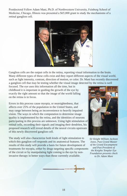
Postdoctoral Fellow Adam Mani, Ph.D. of Northwestern University, Feinberg School of Medicine, Chicago, Illinois was presented a $65,000 grant to study the mechanisms of a retinal ganglion cell.
Ganglion cells are the output cells in the retina, reporting visual information to the brain. Many different types of these cells exist and they report different aspects of the visual world, such as light intensity, contrast, direction of motion, or color. Dr. Mani has recently discovered a ganglion cell that may be testing whether the visual image detected by the retina is well focused. The eye uses this information all the time, but in childhood it is important in guiding the growth of the eye by exactly the right amount so that the image of the world falling on the retina is in focus.
Errors in this process cause myopia, or nearsightedness, that affects over 25% of the population in the United States, and may range between being an inconvenience to heavily impaired vision. The ways in which the computation to determine image quality is implemented by the retina, and the identities of neurons participating in this process are unknown. Using light stimulation of retinal cells, recording their signals and imaging their dendrites, his proposed research will reveal details of the neural circuits upstream of this newly discovered ganglion cell.
The study will also characterize both kinds of light stimulation to which this ganglion cell responds and its anatomical features. The results of this study will provide a basis for future development of treatments for myopia, either by drugs targeting specific components of the circuit or by manipulating light coming to the eye by non-invasive therapy in better ways than those currently available.
Sir Knight William Jackson Jones, Past Grand Master of the Grand Encampment
and Past President of the Knights Templar Eye
Foundation presents check to Dr. Adam Mani
19
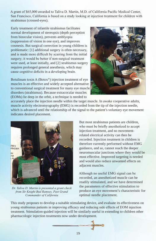
A grant of $65,000 awarded to Taliva D. Martin, M.D. of California Pacific Medical Center, San Francisco, California is based on a study looking at injection treatment for children with strabismus (crossed-eyes).
Early treatment of infantile strabismus facilitates normal development of stereopsis (depth perception from binocular vision), prevents amblyopia (suppression of vision in one eye), and improves cosmesis. But surgical correction in young children is problematic: [1] additional surgery is often necessary, and is made more difficult by scarring from the initial surgery; it would be better if non-surgical treatment were used, at least initially, and [2] strabismus surgery requires prolonged general anesthesia, which may cause cognitive deficits in a developing brain.
Botulinum toxin A (Botox®) injection treatment of eye muscles is an effective and widely accepted alternative to conventional surgical treatment for many eye muscle disorders (strabismus). Because extracocular muscles (EOMs) lie deep in the orbit, a technique is needed to accurately place the injection needle within the target muscle. In awake cooperative adults, muscle activity electromyography (EMG) is recorded from the tip of the injection needle, which is advanced until the relationship of the signal to the patient’s voluntary eye movement indicates desired placement.
But most strabismus patients are children, who must be briefly anesthetized to accept injection treatment, and no movement-related electrical activity can then be recorded. Injection treatment in children is therefore currently performed without EMG guidance, and so, cannot reach the deeper neuromuscular junctions where they would be most effective. Improved targeting is needed and would also reduce unwanted effects on adjacent muscles.
Although no useful EMG signal can be recorded, an anesthetized muscle can be readily stimulated, and we have determined the parameters of effective stimulation to produce an eye movement’s characteristic for optimal needle placement.
This study proposes to develop a suitable stimulating device, and evaluate its effectiveness on young strabismus patients in improving efficacy and reducing side effects of EOM injection treatment. Stimulation-guided injection will be similarly useful in extending to children other pharmacologic injection treatments now under development.
20
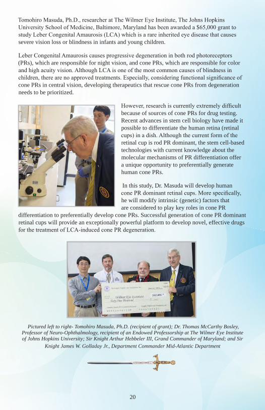
Tomohiro Masuda, Ph.D., researcher at The Wilmer Eye Institute, The Johns Hopkins University School of Medicine, Baltimore, Maryland has been awarded a $65,000 grant to study Leber Congenital Amaurosis (LCA) which is a rare inherited eye disease that causes severe vision loss or blindness in infants and young children.
Leber Congenital Amaurosis causes progressive degeneration in both rod photoreceptors (PRs), which are responsible for night vision, and cone PRs, which are responsible for color and high acuity vision. Although LCA is one of the most common causes of blindness in children, there are no approved treatments. Especially, considering functional significance of cone PRs in central vision, developing therapeutics that rescue cone PRs from degeneration needs to be prioritized.
However, research is currently extremely difficult because of sources of cone PRs for drug testing. Recent advances in stem cell biology have made it possible to differentiate the human retina (retinal cups) in a dish. Although the current form of the retinal cup is rod PR dominant, the stem cell-based technologies with current knowledge about the molecular mechanisms of PR differentiation offer a unique opportunity to preferentially generate human cone PRs.
In this study, Dr. Masuda will develop human cone PR dominant retinal cups. More specifically, he will modify intrinsic (genetic) factors that are considered to play key roles in cone PR
differentiation to preferentially develop cone PRs. Successful generation of cone PR dominant retinal cups will provide an exceptionally powerful platform to develop novel, effective drugs for the treatment of LCA-induced cone PR degeneration.
Pictured left to right- Tomohiro Masuda, Ph.D. (recipient of grant); Dr. Thomas McCarthy Bosley, Professor of Neuro-Ophthalmology, recipient of an Endowed Professorship at The Wilmer Eye Institute of Johns Hopkins University; Sir Knight Arthur Hebbeler III, Grand Commander of Maryland; and Sir
Knight James W. Golladay Jr., Department Commander Mid-Atlantic Department
21
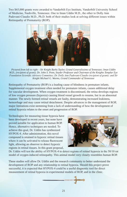
Two $65,000 grants were awarded to Vanderbilt Eye Institute, Vanderbilt University School of Medicine, Nashville, Tennessee. One to Iman Uddin M.D., the other to Dolly Ann Padovani-Claudio M.D., Ph.D. both of their studies look at solving different issues within Retinopathy of Prematurity (ROP).
Pictured from left to right – Sir Knight Burks Taylor, Grand Generalissimo of Tennessee; Iman Uddin M.D., (recipient of grant); Dr. John S. Penn, Snyder Professor and Chairman of the Knights Templar Eye Foundation Scientific Advisory Committee; Dr. Dolly Ann Padovani-Claudio (recipient of grant); and Sir
Knight Michael G. Weems, Grand Commander of Tennessee
Retinopathy of Prematurity (ROP) is a leading cause of blindness in premature infants. Supplemental oxygen treatment often needed for premature infants, causes additional delay for vascular development. When oxygen treatment is discontinued, the retina develops regions of low oxygen pressure (hypoxia) causing blood vessel growth to resume, but in an abnormal manner. The newly formed retinal vessels are faulty, demonstrating increased leakiness, hemorrhage and may cause retinal detachment. Despite advances in the management of ROP, major limitations exist stemming from a lack of understanding of how the development of retinal hypoxia relates to the onset and progression of ROP.
Technologies for measuring tissue hypoxia have been developed in recent years, but none have proved suitable for application to human ROP. Hence, alternative techniques are needed. To achieve this goal, Dr. Uddin has synthesized HYPOX-4. After administration, this novel compound is retained in hypoxic retinal tissues where it can be activated to release fluorescent light, allowing an observer to detect hypoxic regions in retinal tissues. In this grant proposal, Dr. Uddin will test the ability of HYPOX-4 to detect regions of retinal hypoxia in the 50/10 rat model of oxygen-induced retinopathy. This animal model very closely resembles human ROP.
These studies will allow Dr. Uddin and the research community to better understand the development of ROP and any relationship to retinal hypoxia. Should this project prove successful, it is expected that HYPOX-4 could be a useful diagnostic tool for direct measurement of retinal hypoxia in experimental models of ROP, and in the clinic.
22
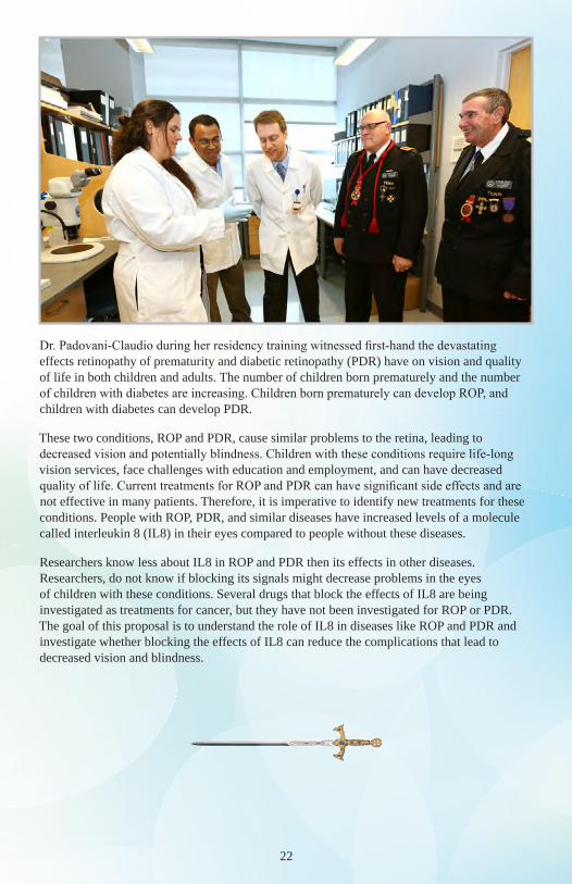
Dr. Padovani-Claudio during her residency training witnessed first-hand the devastating effects retinopathy of prematurity and diabetic retinopathy (PDR) have on vision and quality of life in both children and adults. The number of children born prematurely and the number of children with diabetes are increasing. Children born prematurely can develop ROP, and children with diabetes can develop PDR.
These two conditions, ROP and PDR, cause similar problems to the retina, leading to decreased vision and potentially blindness. Children with these conditions require life-long vision services, face challenges with education and employment, and can have decreased quality of life. Current treatments for ROP and PDR can have significant side effects and are not effective in many patients. Therefore, it is imperative to identify new treatments for these conditions. People with ROP, PDR, and similar diseases have increased levels of a molecule called interleukin 8 (IL8) in their eyes compared to people without these diseases.
Researchers know less about IL8 in ROP and PDR then its effects in other diseases. Researchers, do not know if blocking its signals might decrease problems in the eyes of children with these conditions. Several drugs that block the effects of IL8 are being investigated as treatments for cancer, but they have not been investigated for ROP or PDR. The goal of this proposal is to understand the role of IL8 in diseases like ROP and PDR and investigate whether blocking the effects of IL8 can reduce the complications that lead to decreased vision and blindness.
23
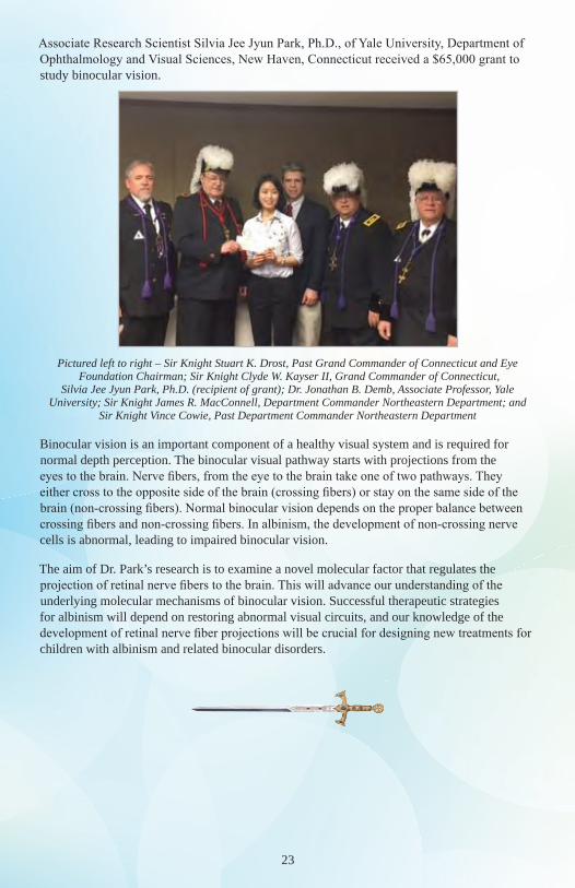
Associate Research Scientist Silvia Jee Jyun Park, Ph.D., of Yale University, Department of Ophthalmology and Visual Sciences, New Haven, Connecticut received a $65,000 grant to study binocular vision.
Pictured left to right – Sir Knight Stuart K. Drost, Past Grand Commander of Connecticut and Eye Foundation Chairman; Sir Knight Clyde W. Kayser II, Grand Commander of Connecticut,
Silvia Jee Jyun Park, Ph.D. (recipient of grant); Dr. Jonathan B. Demb, Associate Professor, Yale University; Sir Knight James R. MacConnell, Department Commander Northeastern Department; and
Sir Knight Vince Cowie, Past Department Commander Northeastern Department
Binocular vision is an important component of a healthy visual system and is required for normal depth perception. The binocular visual pathway starts with projections from the eyes to the brain. Nerve fibers, from the eye to the brain take one of two pathways. They either cross to the opposite side of the brain (crossing fibers) or stay on the same side of the brain (non-crossing fibers). Normal binocular vision depends on the proper balance between crossing fibers and non-crossing fibers. In albinism, the development of non-crossing nerve cells is abnormal, leading to impaired binocular vision.
The aim of Dr. Park’s research is to examine a novel molecular factor that regulates the projection of retinal nerve fibers to the brain. This will advance our understanding of the underlying molecular mechanisms of binocular vision. Successful therapeutic strategies for albinism will depend on restoring abnormal visual circuits, and our knowledge of the development of retinal nerve fiber projections will be crucial for designing new treatments for children with albinism and related binocular disorders.
24
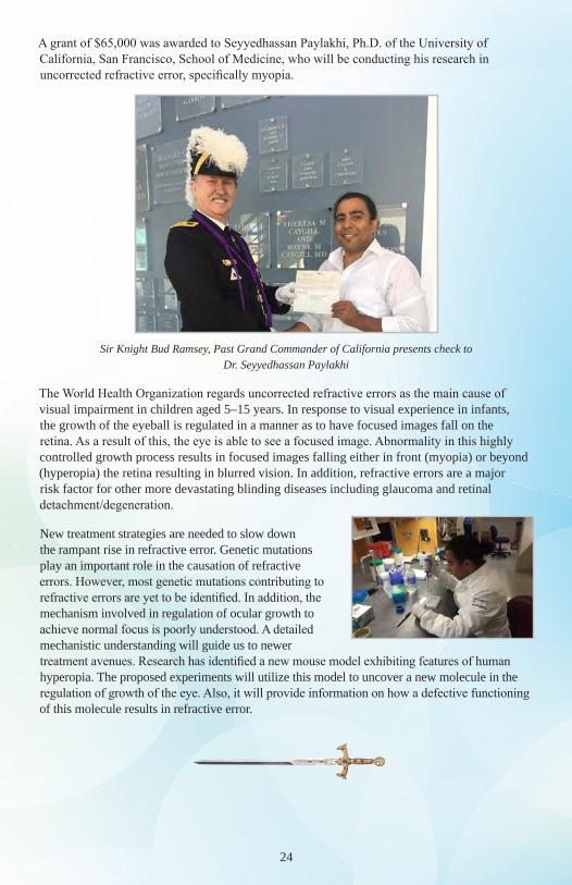
A grant of $65,000 was awarded to Seyyedhassan Paylakhi, Ph.D. of the University of California, San Francisco, School of Medicine, who will be conducting his research in uncorrected refractive error, specifically myopia.
Sir Knight Bud Ramsey, Past Grand Commander of California presents check to Dr. Seyyedhassan Paylakhi
The World Health Organization regards uncorrected refractive errors as the main cause of visual impairment in children aged 5–15 years. In response to visual experience in infants, the growth of the eyeball is regulated in a manner as to have focused images fall on the retina. As a result of this, the eye is able to see a focused image. Abnormality in this highly controlled growth process results in focused images falling either in front (myopia) or beyond (hyperopia) the retina resulting in blurred vision. In addition, refractive errors are a major risk factor for other more devastating blinding diseases including glaucoma and retinal detachment/degeneration.
New treatment strategies are needed to slow down the rampant rise in refractive error. Genetic mutations play an important role in the causation of refractive errors. However, most genetic mutations contributing to refractive errors are yet to be identified. In addition, the mechanism involved in regulation of ocular growth to achieve normal focus is poorly understood. A detailed mechanistic understanding will guide us to newer treatment avenues. Research has identified a new mouse model exhibiting features of human hyperopia. The proposed experiments will utilize this model to uncover a new molecule in the regulation of growth of the eye. Also, it will provide information on how a defective functioning of this molecule results in refractive error.
25
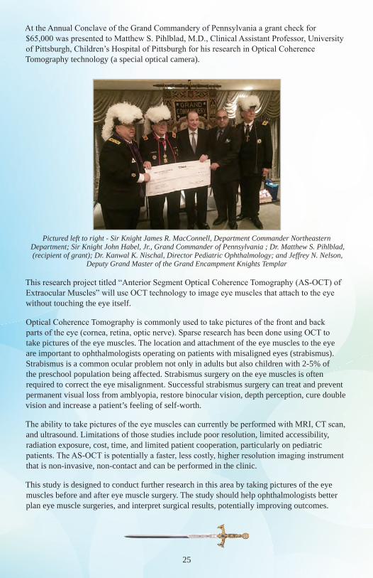
At the Annual Conclave of the Grand Commandery of Pennsylvania a grant check for $65,000 was presented to Matthew S. Pihlblad, M.D., Clinical Assistant Professor, University of Pittsburgh, Children’s Hospital of Pittsburgh for his research in Optical Coherence Tomography technology (a special optical camera).
Pictured left to right - Sir Knight James R. MacConnell, Department Commander Northeastern Department; Sir Knight John Habel, Jr., Grand Commander of Pennsylvania ; Dr. Matthew S. Pihlblad, (recipient of grant); Dr. Kanwal K. Nischal, Director Pediatric Ophthalmology; and Jeffrey N. Nelson,
Deputy Grand Master of the Grand Encampment Knights Templar
This research project titled “Anterior Segment Optical Coherence Tomography (AS-OCT) of Extraocular Muscles” will use OCT technology to image eye muscles that attach to the eye without touching the eye itself.
Optical Coherence Tomography is commonly used to take pictures of the front and back parts of the eye (cornea, retina, optic nerve). Sparse research has been done using OCT to take pictures of the eye muscles. The location and attachment of the eye muscles to the eye are important to ophthalmologists operating on patients with misaligned eyes (strabismus). Strabismus is a common ocular problem not only in adults but also children with 2-5% of the preschool population being affected. Strabismus surgery on the eye muscles is often required to correct the eye misalignment. Successful strabismus surgery can treat and prevent permanent visual loss from amblyopia, restore binocular vision, depth perception, cure double vision and increase a patient’s feeling of self-worth.
The ability to take pictures of the eye muscles can currently be performed with MRI, CT scan, and ultrasound. Limitations of those studies include poor resolution, limited accessibility, radiation exposure, cost, time, and limited patient cooperation, particularly on pediatric patients. The AS-OCT is potentially a faster, less costly, higher resolution imaging instrument that is non-invasive, non-contact and can be performed in the clinic.
This study is designed to conduct further research in this area by taking pictures of the eye muscles before and after eye muscle surgery. The study should help ophthalmologists better plan eye muscle surgeries, and interpret surgical results, potentially improving outcomes.
26
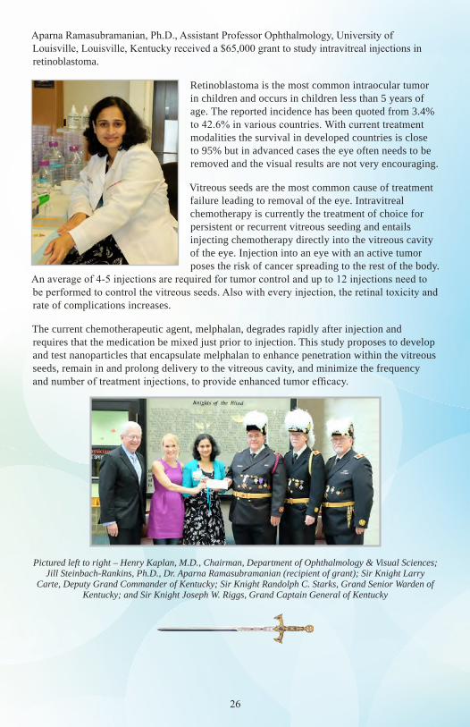
Aparna Ramasubramanian, Ph.D., Assistant Professor Ophthalmology, University of Louisville, Louisville, Kentucky received a $65,000 grant to study intravitreal injections in retinoblastoma.
Retinoblastoma is the most common intraocular tumor in children and occurs in children less than 5 years of age. The reported incidence has been quoted from 3.4% to 42.6% in various countries. With current treatment modalities the survival in developed countries is close to 95% but in advanced cases the eye often needs to be removed and the visual results are not very encouraging.
Vitreous seeds are the most common cause of treatment failure leading to removal of the eye. Intravitreal chemotherapy is currently the treatment of choice for persistent or recurrent vitreous seeding and entails injecting chemotherapy directly into the vitreous cavity of the eye. Injection into an eye with an active tumor poses the risk of cancer spreading to the rest of the body.
An average of 4-5 injections are required for tumor control and up to 12 injections need to be performed to control the vitreous seeds. Also with every injection, the retinal toxicity and rate of complications increases.
The current chemotherapeutic agent, melphalan, degrades rapidly after injection and requires that the medication be mixed just prior to injection. This study proposes to develop and test nanoparticles that encapsulate melphalan to enhance penetration within the vitreous seeds, remain in and prolong delivery to the vitreous cavity, and minimize the frequency and number of treatment injections, to provide enhanced tumor efficacy.
Pictured left to right – Henry Kaplan, M.D., Chairman, Department of Ophthalmology & Visual Sciences; Jill Steinbach-Rankins, Ph.D., Dr. Aparna Ramasubramanian (recipient of grant); Sir Knight Larry
Carte, Deputy Grand Commander of Kentucky; Sir Knight Randolph C. Starks, Grand Senior Warden of Kentucky; and Sir Knight Joseph W. Riggs, Grand Captain General of Kentucky
27
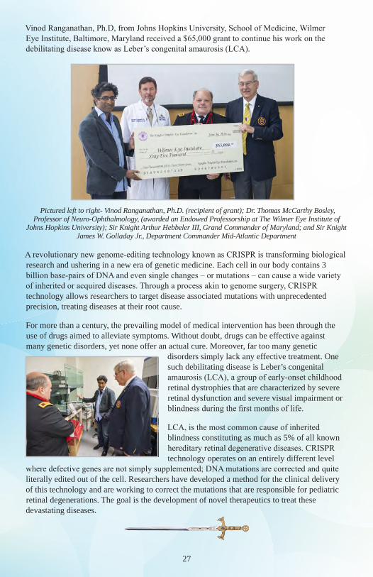
Vinod Ranganathan, Ph.D, from Johns Hopkins University, School of Medicine, Wilmer Eye Institute, Baltimore, Maryland received a $65,000 grant to continue his work on the debilitating disease know as Leber’s congenital amaurosis (LCA).
Pictured left to right- Vinod Ranganathan, Ph.D. (recipient of grant); Dr. Thomas McCarthy Bosley, Professor of Neuro-Ophthalmology, (awarded an Endowed Professorship at The Wilmer Eye Institute of
Johns Hopkins University); Sir Knight Arthur Hebbeler III, Grand Commander of Maryland; and Sir Knight James W. Golladay Jr., Department Commander Mid-Atlantic Department
A revolutionary new genome-editing technology known as CRISPR is transforming biological research and ushering in a new era of genetic medicine. Each cell in our body contains 3 billion base-pairs of DNA and even single changes – or mutations – can cause a wide variety of inherited or acquired diseases. Through a process akin to genome surgery, CRISPR technology allows researchers to target disease associated mutations with unprecedented precision, treating diseases at their root cause.
For more than a century, the prevailing model of medical intervention has been through the use of drugs aimed to alleviate symptoms. Without doubt, drugs can be effective against many genetic disorders, yet none offer an actual cure. Moreover, far too many genetic
disorders simply lack any effective treatment. One such debilitating disease is Leber’s congenital amaurosis (LCA), a group of early-onset childhood retinal dystrophies that are characterized by severe retinal dysfunction and severe visual impairment or blindness during the first months of life.
LCA, is the most common cause of inherited blindness constituting as much as 5% of all known hereditary retinal degenerative diseases. CRISPR technology operates on an entirely different level
where defective genes are not simply supplemented; DNA mutations are corrected and quite literally edited out of the cell. Researchers have developed a method for the clinical delivery of this technology and are working to correct the mutations that are responsible for pediatric retinal degenerations. The goal is the development of novel therapeutics to treat these devastating diseases.
28
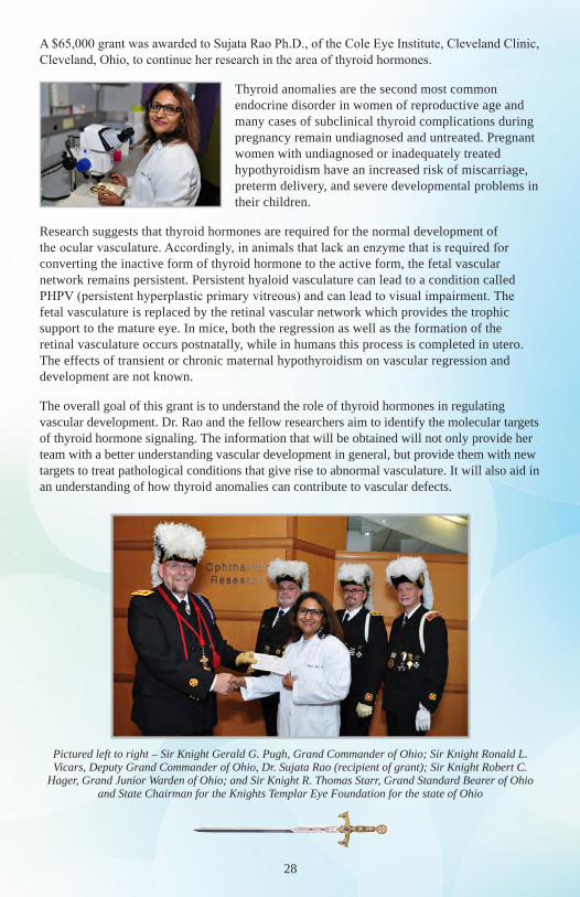
A $65,000 grant was awarded to Sujata Rao Ph.D., of the Cole Eye Institute, Cleveland Clinic, Cleveland, Ohio, to continue her research in the area of thyroid hormones.
Thyroid anomalies are the second most common endocrine disorder in women of reproductive age and many cases of subclinical thyroid complications during pregnancy remain undiagnosed and untreated. Pregnant women with undiagnosed or inadequately treated hypothyroidism have an increased risk of miscarriage, preterm delivery, and severe developmental problems in their children.
Research suggests that thyroid hormones are required for the normal development of the ocular vasculature. Accordingly, in animals that lack an enzyme that is required for converting the inactive form of thyroid hormone to the active form, the fetal vascular network remains persistent. Persistent hyaloid vasculature can lead to a condition called PHPV (persistent hyperplastic primary vitreous) and can lead to visual impairment. The fetal vasculature is replaced by the retinal vascular network which provides the trophic support to the mature eye. In mice, both the regression as well as the formation of the retinal vasculature occurs postnatally, while in humans this process is completed in utero. The effects of transient or chronic maternal hypothyroidism on vascular regression and development are not known.
The overall goal of this grant is to understand the role of thyroid hormones in regulating vascular development. Dr. Rao and the fellow researchers aim to identify the molecular targets of thyroid hormone signaling. The information that will be obtained will not only provide her team with a better understanding vascular development in general, but provide them with new targets to treat pathological conditions that give rise to abnormal vasculature. It will also aid in an understanding of how thyroid anomalies can contribute to vascular defects.
Pictured left to right – Sir Knight Gerald G. Pugh, Grand Commander of Ohio; Sir Knight Ronald L. Vicars, Deputy Grand Commander of Ohio, Dr. Sujata Rao (recipient of grant); Sir Knight Robert C.
Hager, Grand Junior Warden of Ohio; and Sir Knight R. Thomas Starr, Grand Standard Bearer of Ohio and State Chairman for the Knights Templar Eye Foundation for the state of Ohio
29
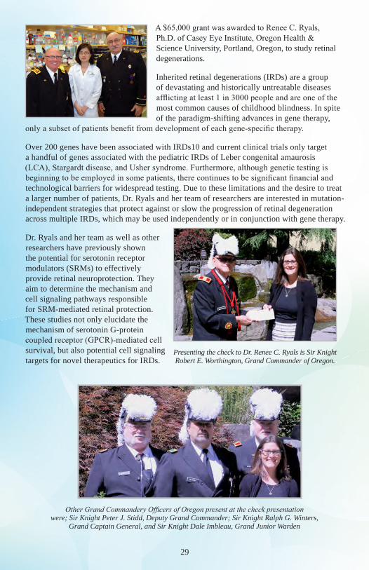
A $65,000 grant was awarded to Renee C. Ryals, Ph.D. of Casey Eye Institute, Oregon Health & Science University, Portland, Oregon, to study retinal degenerations.
Inherited retinal degenerations (IRDs) are a group of devastating and historically untreatable diseases afflicting at least 1 in 3000 people and are one of the most common causes of childhood blindness. In spite of the paradigm-shifting advances in gene therapy,
only a subset of patients benefit from development of each gene-specific therapy.
Over 200 genes have been associated with IRDs10 and current clinical trials only target a handful of genes associated with the pediatric IRDs of Leber congenital amaurosis (LCA), Stargardt disease, and Usher syndrome. Furthermore, although genetic testing is beginning to be employed in some patients, there continues to be significant financial and technological barriers for widespread testing. Due to these limitations and the desire to treat a larger number of patients, Dr. Ryals and her team of researchers are interested in mutation-independent strategies that protect against or slow the progression of retinal degeneration across multiple IRDs, which may be used independently or in conjunction with gene therapy.
Dr. Ryals and her team as well as other researchers have previously shown the potential for serotonin receptor modulators (SRMs) to effectively provide retinal neuroprotection. They aim to determine the mechanism and cell signaling pathways responsible for SRM-mediated retinal protection. These studies not only elucidate the mechanism of serotonin G-protein coupled receptor (GPCR)-mediated cell survival, but also potential cell signaling targets for novel therapeutics for IRDs.
Presenting the check to Dr. Renee C. Ryals is Sir Knight Robert E. Worthington, Grand Commander of Oregon.
Other Grand Commandery Officers of Oregon present at the check presentation were; Sir Knight Peter J. Stidd, Deputy Grand Commander; Sir Knight Ralph G. Winters,
Grand Captain General, and Sir Knight Dale Imbleau, Grand Junior Warden
30
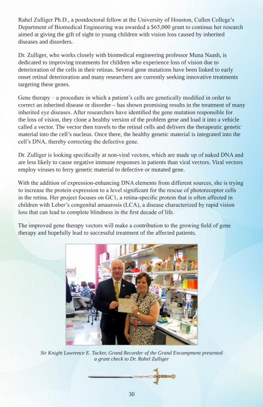
Rahel Zulliger Ph.D., a postdoctoral fellow at the University of Houston, Cullen College’s Department of Biomedical Engineering was awarded a $65,000 grant to continue her research aimed at giving the gift of sight to young children with vision loss caused by inherited diseases and disorders.
Dr. Zulliger, who works closely with biomedical engineering professor Muna Naash, is dedicated to improving treatments for children who experience loss of vision due to deterioration of the cells in their retinas. Several gene mutations have been linked to early onset retinal deterioration and many researchers are currently seeking innovative treatments targeting these genes.
Gene therapy – a procedure in which a patient’s cells are genetically modified in order to correct an inherited disease or disorder – has shown promising results in the treatment of many inherited eye diseases. After researchers have identified the gene mutation responsible for the loss of vision, they clone a healthy version of the problem gene and load it into a vehicle called a vector. The vector then travels to the retinal cells and delivers the therapeutic genetic material into the cell’s nucleus. Once there, the healthy genetic material is integrated into the cell’s DNA, thereby correcting the defective gene.
Dr. Zulliger is looking specifically at non-viral vectors, which are made up of naked DNA and are less likely to cause negative immune responses in patients than viral vectors. Viral vectors employ viruses to ferry genetic material to defective or mutated gene.
With the addition of expression-enhancing DNA elements from different sources, she is trying to increase the protein expression to a level significant for the rescue of photoreceptor cells in the retina. Her project focuses on GC1, a retina-specific protein that is often affected in children with Leber’s congenital amaurosis (LCA), a disease characterized by rapid vision loss that can lead to complete blindness in the first decade of life.
The improved gene therapy vectors will make a contribution to the growing field of gene therapy and hopefully lead to successful treatment of the affected patients.
Sir Knight Lawrence E. Tucker, Grand Recorder of the Grand Encampment presented a grant check to Dr. Rahel Zulliger
31
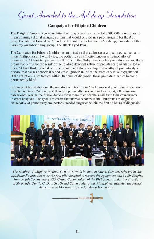
Grant Awarded to the Apl.de.ap FoundationCampaign for Filipino Children
The Knights Templar Eye Foundation board approved and awarded a $95,000 grant to assist in purchasing a digital imaging system that would be used in a pilot program for the Apl.de.ap Foundation formed by Allan Pineda Lindo better known as Apl.de.ap, a member of the Grammy Award-winning group, The Black Eyed Peas.
The Campaign for Filipino Children is an initiative that addresses a critical medical concern in the Philippines and worldwide, the pediatric eye affliction known as retinopathy of prematurity. At least ten percent of all births in the Philippines involve premature babies, these premature births are the result of the relative deficient nature of prenatal care available to the poor. At least thirty percent of these premature babies develop retinopathy of prematurity, a disease that causes abnormal blood vessel growth in the retina from excessive oxygenation. If the affliction is not treated within 48 hours of diagnosis, these premature babies become permanently blind.
In four pilot hospitals alone, the initiative will train from 6 to 10 medical practitioners from each hospital, a total of 24 to 40, and therefore potentially prevent blindness for 4,380 premature babies each year. In the future, doctors from these pilot hospitals will train their counterparts in other hospitals. The goal is to create the internal capacity in the Philippines to diagnose retinopathy of prematurity and perform needed surgeries within the first 48 hours of diagnosis.
The Southern Philippine Medical Center (SPMC) located in Davao City was selected by the Apl.de.ap Foundation to be the first pilot hospital to receive the equipment and 34 Sir Knights
from Rajah Commandery #20, Grand Commandery of the Philippines, under the direction of Sir Knight Danilo C. Datu Sr., Grand Commander of the Philippines, attended the formal
dedication as VIP guests of the Apl.de.ap Foundation.
V5-2016-GRANT
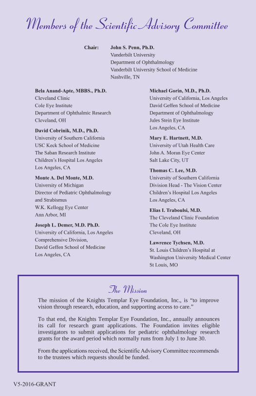
Members of the Scientific Advisory Committee
Bela Anand-Apte, MBBS., Ph.D. Cleveland Clinic Cole Eye Institute Department of Ophthalmic Research Cleveland, OH
David Cobrinik, M.D., Ph.D. University of Southern California USC Keck School of Medicine The Saban Research Institute Children’s Hospital Los Angeles Los Angeles, CA
Monte A. Del Monte, M.D. University of Michigan Director of Pediatric Ophthalmology and Strabismus W.K. Kellogg Eye Center Ann Arbor, MI
Joseph L. Demer, M.D. Ph.D. University of California, Los Angeles Comprehensive Division, David Geffen School of Medicine Los Angeles, CA
Michael Gorin, M.D., Ph.D. University of California, Los Angeles David Geffen School of Medicine Department of Ophthalmology Jules Stein Eye Institute Los Angeles, CA
Mary E. Hartnett, M.D. University of Utah Health Care John A. Moran Eye Center Salt Lake City, UT
Thomas C. Lee, M.D. University of Southern California Division Head - The Vision Center Children’s Hospital Los Angeles Los Angeles, CA
Elias I. Traboulsi, M.D. The Cleveland Clinic Foundation The Cole Eye Institute Cleveland, OH
Lawrence Tychsen, M.D. St. Louis Children’s Hospital at Washington University Medical Center St Louis, MO
Chair: John S. Penn, Ph.D. Vanderbilt University Department of Ophthalmology Vanderbilt University School of Medicine Nashville, TN
The MissionThe mission of the Knights Templar Eye Foundation, Inc., is “to improve vision through research, education, and supporting access to care.”
To that end, the Knights Templar Eye Foundation, Inc., annually announces its call for research grant applications. The Foundation invites eligible investigators to submit applications for pediatric ophthalmology research grants for the award period which normally runs from July 1 to June 30.
From the applications received, the Scientific Advisory Committee recommends to the trustees which requests should be funded.