Embed Size (px)
Citation preview
Grand Valley State UniversityScholarWorks@GVSU
Masters Theses Graduate Research and Creative Practice
1998
Knee Flexion Angle and its Influence on VMO:VLRatios During Isometric Quadriceps ContractionJeffrey P. HendraGrand Valley State University
William D. AllanGrand Valley State University
Follow this and additional works at: http://scholarworks.gvsu.edu/theses
Part of the Physical Therapy Commons
This Thesis is brought to you for free and open access by the Graduate Research and Creative Practice at ScholarWorks@GVSU. It has been acceptedfor inclusion in Masters Theses by an authorized administrator of ScholarWorks@GVSU. For more information, please [email protected].
Recommended CitationHendra, Jeffrey P. and Allan, William D., "Knee Flexion Angle and its Influence on VMO:VL Ratios During Isometric QuadricepsContraction" (1998). Masters Theses. 396.http://scholarworks.gvsu.edu/theses/396
KNEE FLEXION ANGLE AND ITS INFLUENCE ON VM OiVL RATIOS
DURING ISOMETRIC QUADRICEPS CONTRACTION
By
Jeffrey P. Hendra SPT William D. Allan SPT
T H E S IS
Submitted to the D epartm ent of Physical Therapy at Grand Valley S ta te University
Allendale, Michigan in partial fulfillment of th e requirements
for th e degree of
MASTER OF SCIENCE IN PHYSICAL THERAPY
1998
THESIS COMMITTEE APPROVAL:/I
Ch'air. Timothy Striidkler Ph.D. Date
Memoer: Jane Toot Ph.D., PT Date
4 /^3 /Member: Justine Ritchie Ph.D. Date
KNEE FLEXION ANGLE AND ITS INFLUENCE ON VMOrVL RATIOS DURING ISOMETRIC QUADRICEPS CONTRACTION
ABSTRACT
Patellofemoral Pain Syndrome (PFPS) is a major cause of knee pain
and is caused by lateral patellar tracking. T reatm ent consists of
strengthening th e vastus medialis obliquus (VMO). While many
exercises streng then th e VMO, simultaneous vastus lateralis (VL)
strengthening often occurs and patellar malalignment remains. The
VMO must, therefore, be strengthened independently or to a greater
extent than the VL. Thus, the VMOrVL ratio of activity must be
considered rather than VMO activity alone. This study compared
isometric knee extension electromyographically a t 0, 20, 60, and 90
degree angles to determ ine which angle produced th e g rea test
VMOrVL ratio of activity. Results of this study indicated no
significant differences among angles te s ted for VMOrVL ratio during
isometric knee extension. Further research is needed to investigate
the possibility of significant associations betw een VMOrVL ratios
and gender, pathology, and angles not included in this study.
ACKNOWLEDGMENTS
The investigators would like to thank th e following individuals
for their time and assistance: Dr. Tim Strickler and Dr. Jane Toot
for direction, input and support, Dr. Justine Ritchie, Dr. Neal Rogness
and Todd Uvergood for making sense of a thousand different numbers
with no apparent meaning. We would also like to thank Dr. John Peck
for ordering supplies, Jim S co tt for providing assis tance in th e lab,
and Kathy “The Connection” Johnstone and Kathy “Wiz” Wagner for
introducing us to Myosoft. Special thanks to Sheila Allan for her
com puter expertise and our friends and families for providing
support, patience, and understanding throughout th e entirety of this
pro ject.
11
TABLE OF CONTENTS
Page
ABSTRACT...............................................................................................................i
ACKNOWLEDGMENTS......................................................................................... ii
PREFACE..................................................................................................................V
Definition o f Term s................................................................. v
LIST OF TABLES...................................................................................................vii
LIST OF FIGURES.............................................................................................v iii
CHAPTER
INTRODUCTION..................................................................................1
2. LITERATURE REVIEW..................................................................... 4
Prevalence and Causes o f PFPS..............................................4Anatomy o f the P ate llo fem oral J o i n t .................................7Role o f th e VMO..........................................................................9S elective R ecruitm ent o f th e VMO....................................10
3. METHODOLOGY...............................................................................20
S u b je c ts .................................................................................... 20D esign.........................................................................................20P ro ced u re .................................................................................. 21Instrum entation ........................................................................22V alidity and R eliability ......................................................... 23
4. RESULTS/DATA ANALYSIS........................................................24
i i i
T echn iques................................................................................ 24H ypothesis/R esearch Question............................................ 26Other Findings and O bservations o f In te re s t .................. 27
5. D iscussion and Im p lic a tio n s .................................................... 30
D iscussion o f Findings............................................................30A pplication o f P rac tice ..........................................................31L im ita tio n s ...............................................................................32Suggestions For Further Research...................................... 34C onclusion .................................................................................35
REFERENCES....................................................................................................... 37
APPENDIX A - INFORMED CONSENT FORM................................................... 42
APPENDIX B - DATA COLLECTION SHEET..................................................... 45
APPENDIX C -PROPOSAL SUMMARY FOR HSRC.......................................... 47
I V
PREFACE
Definitions of Terms
Patellofemoral Pain Syndrome (PFPSVpain resulting from a dysfunction of the patella’s ability to track in th e femoral groove
isom etric quadriceps se ts -a contraction of th e quadriceps in which tension is developed but no joint movement or change in muscle length occurs. This contraction is performed a t terminal knee extension
short-arc quadriceps se ts -an exercise where th e leg is moved through the last 35 degrees of extension
isometric knee extension-a contraction of th e quadriceps in which tension is developed but no joint movement or change in muscle length occurs. This contraction can be performed a t any angle.
0 -an g le-the acute angle formed betw een intersecting lines drawn from th e anterior-superior iliac spine through th e mid-portion of th e patella, and from th e anterior tibial tuberosity through the mid patella. Normally 15 degrees
subluxatlon-an incomplete or partial dislocation
pes planus-condition of th e foot in which th e medial longitudinal arch is decreased. Also known as pronated or flat foot
genu recurvatum-a condition characterized by a hyperextension of the knee
biofeedback-training technique utilizing visual and audio ou tput regarding muscle activity via electromyography. Subjects are, therefore, trained to selectively stim ulate or inhibit a specific
m uscle
electromyography fEMGVtechnique in which m uscle activity is de tec ted and recorded through an electrical medium
hip adduction-movement of th e fem ur tow ard th e midline of th e body
goniom eter-a device used to m easure jo in t angles
VI

LIST OF TABLES
Table Page
1. D escriptive S ta t i s t ic s Table For Avg VMOiVLM easurem ents.................................................................................. 27
2. D escriptive S ta t i s t ic s Table For Avg VMO:VLby G ender........................................................................................... 28
VI I

LIST OF FIGURES
Table Page
1. Sample o f Noraxon Myosoft O u tp u t................................................24
VIII
CHAPTER. 1 INTRODUCTION
According to Malek and Mangine,i “Patellofemoral Pain
Syndrome (PFPS) is the number one cause of knee pain in a majority
of th e knee clinics in sports medicine centers around th e country.”
Many researchers agree th a t th e major underlying cause of PFPS is a
malalignment of the patellofemoral joint causing th e patella to
track laterally in the patellofemoral groove. 27 Malalignment is due
to a variety of causes. These causes include abnormal architecture
and articulation of the patellofemoral joint, 2.4-6 a tight lateral
retinaculum , 2,4-6,a and a muscular imbalance between the vastus
medialis obliquus (VMO) and th e vastus lateralis (VL). 2,5.6.9 The
result of th ese abnormalities is a laterally tracking patella causing
pain for th e patient.
The VMO has been iden tified as the only s tru c tu re w ith the
ability to pull the patella medially, lo.n Theoretically, strengthening
of th e VMO will correct malalignment thus relieving pain. Exercises
recommended by researchers and therapists to strengthen the VMO
include isometric quadriceps s e ts , 1,12-17 straight leg raises, 1.12-15.17-
1
19 short-arc quadriceps se ts , i.i8,i9 isometric knee extension, is-zo and
various types of squats and s tep ups. Many of th e se protocols
have been found to successfully strengthen th e VMO. However, while
strengthening the VMO, these exercises may also strengthen th e VL
to th e same degree. Even though th e VMO is strengthened, it may
still no t be able to pull the patella medially because of the
sim ultaneous strengthening of th e VL pulling the patella laterally.
For an exercise to be efficient in altering malalignment and reducing
pain, it is necessary to strengthen th e VMO independent of the VL or
a t least strengthen it to a significantly g reater degree than th e VL.
When identifying an exercise th a t corrects malalignment, the
researcher should look a t th e VMOiVL ratio of activity rather than
simply VMO activity.
Finding an exerc ise th a t can sp e c if ica lly s treng then th e VMO
and thus relieve pain would be of g reat benefit to many people. It
has been estim ated th a t the incidence of PFPS in th e general
population is as high as one in four. 21 A proven protocol in
combating PFPS would also save th e therapist much tim e and
anguish and possibly influence reim bursem ent by third party payors.
Several au thors have recommended an iso m etric contraction of
th e quadriceps to facilitate strengthening of the VMO. Many believe
th a t this contraction should be performed a t terminal knee
extension. 1,12-17 Others believe th a t isom etric quadriceps
contractions should be performed a t 20 degrees of flexion. 1 s i9 More
recently researchers have suggested th a t th ese isometric quadriceps
contractions should be performed between the range of 60 and 90
degrees of knee f le x io n .i 9.22,23 The problem is th a t there is little
scientific data th a t indicates the VMO is strengthened to a greater
degree than th e VL in any of these ranges of motion (ROM) during an
isometric contraction. The purpose of this study is to compare the
activity of th e VMO relative to the activity of the VL a t various
points betw een 0 and 90 degrees of knee flexion during isometric
contraction. Our hypothesis is th a t isom etric quadriceps
contractions between 60 and 90 degrees of flexion will result in
larger VMOiVL ratios than a t other ranges of motion in the knees of
healthy college age males and females.
CHAPTER 2 LITERATURE REVIEW
The majority of this literature review relates to th e exercises
and theories surrounding the selective strengthening of the VMO. For
a b e tte r understanding of th ese exercises and theories, it is
important tha t the reader have a background in the prevalence and
causes of PFPS, the anatomy of the patellofemoral Joint, and the
role of the VMO.
Prevalence and Causes of PFPS
Patellofem oral pain syndrome (PFPS) p resen ts a s a s ign ifican t
problem for many ath letes involved in sporting activities and is
classified as a dysfunction of the patella's ability to track properly
in the femoral groove.? PFPS has been called the number one cause of
knee pain in a majority of the knee clinics in sports medicine
centers around the c o u n t r y ,1.2.8,21,24 and its incidence in th e general
population is as high as one in fou r .z i This syndrome is
characterized by pain in th e anterior portion of the knee and can be
attributed to many different causes. The end result, however,
appears to be excessive lateral pressure on th e patellofemoral (PF)
articulation.6
One o f the m ajor causes o f PFPS I s m alalignm ent o f th e PF
jo in t .1-6.8 Kramer 6 lists the four main causes of malalignment as 1)
architectural abnormalities of articulation, 2) muscle imbalance
betw een VMO and VL, 3) retlnacular abnormalities, and 4) an
increased Q-angle. Kettlekamps agrees with these sam e four causes
adding laxity of the medial retinaculum and atrophy of th e VMO as
underlying causes of malalignment. Fulkersons s ta te s th a t PF
malalignment may cause repeated mild to moderate subluxation and
relocation of th e patella which causes chronic strain and tightening
of th e lateral retinaculum. This in turn increases the Q-angle which
can lead to PFPS.
O ther causes o f an increased Q-angle include foo t pronation,
pes planus, genu recurvatum, and wide hips.6 Women, therefore, are
more prone to PFPS due to larger pelvic width.
O ther m ajor conditions leading to PFPS include degenerative
disease, repeated microtrauma, acu te severe trauma,i iliotibial band
tigh tness,8 and medial retinacular rupture.^ These conditions may
cause lateral tightness and medial weakness of connective tissue
surrounding the patella predisposing th e patient to a bony
malalignment betw een th e femur and tibia. This bony malalignment
will cause th e patella to track improperly in th e patellar groove.
Thus, it is evident th a t a proper balance betw een medial and lateral
structu res m ust be maintained for maximal alignment of th e PF
jo in t.
A balance o f s tren g th betw een th e VMO and VL m uscles i s
imperative for this balance to be maintained.^ Individuals affected
by PFPS differ from healthy individuals in regards to their VMOiVL
activation patterns. While th e VMOiVL activation patterns of the
healthy individual are close to equal, th e patterns of PFPS patients
show decreased activation of the VMO during EMG assessm ent.24.25
Conservative trea tm en t is, therefore, aimed a t selective
strengthening of the VMO.
Due to th e prevalence o f th is syndrom e i t may be assum ed th a t
an effective exercise protocol is currently in widespread use. In
truth, however, no such protocol exists due to lack of evidence of
exercises th a t selectively ta rg e t the VMO. Thus, PFPS also poses a
problem for th e clinician attem pting to tre a t this disorder.
Anatomy- of the Jatelloffimocal-Joint
The anatom y o f th e PF jo in t a lso m akes th is syndrome
particularly difficult to tre a t. According to Norkin and L e v a n g ie ,2 6
"to g e th er with th e femoral surface on which it sits, th e
patellofemoral joint is...the least congruent joint in th e body."
T hese authors also define th e patella as th e largest sesamoid bone
in th e body which functions as an anatomical pulley to reduce
friction betw een th e quadriceps tendon and the femoral condyles. In
order to perform this function without limiting knee motion, th e
patella m ust slide on th e femoral condyles while remaining sea ted
betw een them in the intercondylar g r o o v e .2 6 This groove corresponds
to th e vertical ridge on th e posterior aspect of the patella thereby
providing a tracking mechanism for th e PF joint to maintain proper
alignment. This mechanism is under control of two restraining
mechanisms crossing a t right angles: a group of transverse
stabilizers and a group of longitudinal stabilizers. For simplicity,
the longitudinal stabilizers will be defined as the quadriceps tendon
superiorly and the patellar tendon in fer iorly .26 Norkin and L ev a n g ie2 6
have described th e transverse stabilizers as the medial and lateral
8
extensor (patellar) retinaculae. In general, th e function of the
retinaculae are to connect th e patella to dense connective tissues
around th e knee. Laterally th ese include th e fascia lata and iliotibial
band as well as th e vastus lateralis muscle. Similarly, the medial
retinaculum functions to connect dense connective tissue and the
vastus medialis to the medial patellar border .26 Lieb and Perry”
found the pull of the VL to be 12 to 15 degrees lateral to the long
axis of th e femur, with the distal fibers even more angulated. The
angulation of th e VM pull depends on which portion of the muscle is
assessed. The upper fibers of th e VM are angulated 15 to 18 degrees
medially to th e femoral shaft, while the distal fibers are angulated
approximately 50 to 55 degrees medially. It is because of this
drastic difference in fiber orientation of th e upper and lower
portions of th e VM muscle th a t it is categorized into two
com ponents: vastus medialis longus (VML) whose fibers originate
from th e intertrochanteric line and medial lip of the linea aspera of
the femur and vastus medialis oblique (VMO) 26 whose fibers
originate primarily from the adductor magnus tendon.27.2s This
difference in fiber orientation between the VMO and VL may be a
reason why difficulties in trea tm en t have been experienced.
Role of th e VMQ
The ro le of the VMO as a dynamic m edial s ta b ilize r providing
proper patellar alignment is strongly supported in the
lite ra tu re .10.11.27.29.30 Lieb and Perryn s ta te th a t the only selective
function attributable to th e VMO is patellar alignment.
A second and questionable role o f th e VMO i s term inal knee
extension. Fiebert e t al3i claim th a t th e VMO becomes m ost active
near terminal knee extension. Basmajianio also found th a t the VMO
appears to increase activity toward th e end of unweighted
extension, but found no significant difference between the VMO and
o ther quadriceps com ponents during squatting exercises. This is
supported by other research th a t claims th e best exercises for PFPS
are ones performed with full knee extension.12.17.19
Despite these claim s, o ther research ind icates the VMO does
not account for the last few degrees of terminal extension .22 Lieb
and Perry 11 report th a t all four quadriceps heads increase activity
in terminal extension. They s ta te th e VMO helps perform terminal
extension but does not individually account for the final 15 degrees.
10
In another study by Brownstein e t a l,23 it was found th a t th e VMO is
actually least active in the extended position. Finally, a study by
Ingersoll and Knight32 showed th a t terminal extension exercises do
no t produce medial relocation of th e patella. Interestingly, th ey
reported th a t th ese exercises actually seem to predispose patien ts
to PFPS.
Selective Recruitment of the VMO
Currently, biofeedback has th e m ost prom ise in fa c ilita tin g th e
selective recruitm ent of the VMO. Wise e t al.33 studied th e effects of
biofeedback in conjunction with exercise (straight leg raise, quad
s e ts , and terminal knee extension) and functional activities
(ambulation, bicycling, and sta ir climbing). Results of this study
showed th a t biofeedback is effective in producing significantly
g reater activation of the VMO than VL in all activities.
Ingersol and Knightaz used a protocol established by Wise e t
al.33 and compared th e effects of biofeedback to the effects of a
progressive resistance routine using a short arc quadriceps exercise.
Their study suggests tha t, “quadriceps group strength changes are
no t enough to fully rehabilitate patellar tracking dysfunctions. The
n
use of EMG biofeedback training to selectively strengthen the vastus
medialis obliquus appears to be essential in correcting faulty
patellar tracking." Thus it appears from th e literature th a t the
incorporation of biofeedback into a rehabilitation program is
extremely advantageous. Biofeedback, however, is not often
available for a home program, and research into the effectiveness of
exercises w ithout biofeedback is still needed.
The McConnell taping and ex e rc ise pro tocol i s another avenue
in the rehabilitation of PFPS which holds potential.^ In this protocol,
th e patella is taped into proper alignment. Once taping is completed,
th e patien t performs a number of weightbearing exercises with
emphasis on th e eccentric contraction of th e quadriceps group.
During th e contractions the patient is instructed to contract the
VMO and to relax th e lateral hamstrings and th e VL as much as
possible. The hip is also adducted during exercise to, theoretically,
aid in selective VMO recruitment. The use of McConnell taping to
increase VMO activity is validated by King e t al..34 King e t al. found
th a t mechanically altering the alignment o f th e patella facilitates
the neural drive of th e VMO and reduces th e activity of the VL. At
12
this tim e, however, few therapists are well versed in patellar
taping techniques. Also, more research needs to be done to verify
McConnell’s claim of a success rate of over 90%.
Because o f th e VMO’s a ttachm ent to th e adductor magnus
tendon, many researchers believe th a t hip adduction can be used to
help selectively strengthen the VMO. They believe this can be
accomplished through either isolated hip adduction or hip adduction
accompanied by a quadriceps contraction. Hanten and Schulthiesss
had sub jec ts perform maximal isometric hip adduction against a
resistance pad. Measurements of VMO and VL activity were taken and
results were expressed as a percentage of th e maximal voluntary
isom etric contraction (MVIC). Significantly g rea te r activity was
found in th e VMO (61.75% ) compared to the VL(45.63%). From this
research it appears th a t isometric hip adduction selectively
streng thens th e VMO relative to the VL. Westfall and Worrell,20
however, criticize th ese findings questioning th e high ratio of VMO
activity without knee extension. Thus, more research needs to be
done to confirm these findings and determ ine whether a hip
adduction protocol can increase the VMOiVL ratio with any
13
clinical significance.
The influence o f hip adduction on th e VMO during quadriceps
contraction is less clear. King e t al.34 te s te d patients concentrically
and eccentrically a t 30 degrees per second over a range of motion of
10 to 100 degrees with a Kincom dynamometer. Testing subjects
with and without concurrent hip adduction they found th a t 1 ) hip
adduction had no effect on the activity of the VMO and 2) the VMO:VL
ratio remained unchanged.
Grabiner e t al.36 monitored VMO and VL activity of subjects
under two se ts of conditions. First, they simultaneously performed
hip adduction and an isometric quadriceps contraction a t 20 degrees
of flexion. Second, they performed th e isometric contraction without
hip adduction. The researchers found th a t th e effect of hip adduction
was no t significant.
Hodges and Richardsons? refute the notion th a t hip adduction
has no influence on selective strengthening of the VMO. In their
research, “hip adduction was superimposed onto the contraction of
the quadriceps femoris in a weight bearing and a non-weight bearing
position a t th ree levels of hip adduction force.” They found VMO

14
activity increased relatively more than the VL with th e addition of
each level of hip adduction in weight bearing. The results were not
as dramatic in non-weight bearing with differences being seen only
a t maximal hip adduction. Still, their research indicates th a t hip
adduction is advantageous to VMO strengthening.
Obviously, w ith all th e d iscrepancies in the li te ra tu re , more
research is needed to determine th e actual role of hip adduction.
Even though th e current body of literature is limited, research
seem s to indicate th a t the effec ts of hip adduction may differ
depending on th e type of exercise being performed.
Isok inetic knee extension i s another exerc ise o ften prescribed
in th e treatm ent of PFPS. Sczepanski e t al.38 investigated th e
effects of arc of motion, angular velocity, and type of contraction on
VMO:VL ratio during isokinetic knee extension. They found a
significant difference in VMOrVL ratio depending on th e arc of
motion. A significantly greater VMO:VL EMG ratio occurred a t 60-85
degrees of flexion than at 35-60 or 10-35 degrees of flexion.
Results also indicated concentric contractions produce a g rea ter
VMOrVL ratio than eccentric contractions and faster velocities (120
15
degrees per second) enhance VMO:VL ratio b e tte r than slower
velocities (60 degrees per second). These findings were supported by
the work of Boucher e t al.22 who compared th e VMO and VL activity
betw een 0 and 90 degrees. They specifically m easured EMG activity
at 15, 30, and 90 degrees and concluded th a t VMO:VL ratios are
g rea te s t a t 90 degrees of knee flexion and th e VMO is facilitated in
th a t specific ROM.
W hile dynamic e x e rc ise s a re usually p resc rib ed in th e la te r
stages of rehabilitation, many researchers have recom m ended a
protocol consisting of isom etric quadriceps contractions during th e
initial s ta g e s . 1,12.14.18-20 The belief is th a t isom etric quadriceps
contractions will maintain or enhance VMO stren g th during the
period when dynamic exercise may still be too painful to perform.
As Woodall and Welshis s ta te , “th e initial phase o f rehabilitation is
to increase joint motion, and to g e t the VMO under good, active,
voluntary control, and to decrease patellofem oral jo int irritation.”
Many th e ra p is ts o f th e p as t have a ttem p ted to s treng then the
VMO by isometric quadriceps contractions a t full knee extension.
This was based on older research which claimed th a t the VMO was
16
chiefly responsible for the last 10 to 15 degrees of knee
ex tensions 1.39 Using am putated limbs and an elaborate pulley system
to replicate th e force vectors of th e quadriceps heads, Lieb and
P erry 11 dem onstrated tha t the VMO did not contribute any more than
th e other quadriceps heads to knee extension. They found th a t the
only selective function attributable to th e VMO was patellar
alignment. This research was more recently supported by an
electromyographical study by Basmajian.io He concurs that the only
selective function of th e VMO is patellar alignment. Basmajian also
found th a t th e VMO appeared to increase activity toward the end of
extension in unweighted limbs, bu t th is activity was not greatly
different from th a t of the other heads of th e quadriceps. This
indicates th a t even though VMO activity is increased, isometric
quadriceps contraction a t 0 to 15 degrees of knee flexion is not an
efficient exercise for PFPS because th e VMO:VL ratio is not changed
s ign ifican tly .
O ther au thors have prescribed iso m e tr ic quadriceps
contractions a t 20 degrees of flexion.is.i9 This position is chosen
because, “it is here th a t the patella actively begins to centralize in
17
th e femoral sulcus. With more flexion, about 45 degrees, th e main
arthritic zones of the patella are present so this area is avoided,
especially if pain or specific pathology is p resen t.”i9 While this
position is successful a t reducing pain, there is no research to
indicate th a t it is efficient a t increasing the VMO:VL ratio.
W estfall and Worrell2o recommend isometric quadriceps
contractions in the range of 60 to 85 degrees of flexion based on
research by Brownstein e t a l.23 and Boucher e t aL.22 Brownstein e t
al. a ttem pted to locate the position of peak torque of the quadriceps
complex to establish a normalization protocol. Subjects perform ed
maximal isometric contractions every 10 degrees from 10 to 90
degrees of knee flexion. The researchers recorded the torque of each
individual quadriceps head as well as the torque of the quadriceps
complex as a whole. The study showed peak VMO torque occurred
betw een 60 and 90 degrees of knee flexion. While Brownstein e t al.
recorded the torque of the VMO and VL, they neglected to calculate
the VMOiVL ratios. Boucher e t al.22 performed an isokinetic study and
found, similar to the Brownstein e t al.23 isometric study, th a t peak
torque occurred at 90 degrees of flexion. Although Brownstein
18
e t al. did not figure the VMO:VL ratio, and the Boucher e t al. study
was done isokinetically, Westfall and Worrelizo still believe th a t 60
to 90 degrees of knee flexion is the optimal position for selectively
strengthening the VMO during an isometric quadriceps contraction.
Biofeedback and McConnell Taping hold prom ise in the
trea tm en t of PFPS. Research also shows th a t dynamic exercises
such as isokinetic knee extension and weight bearing eccentric
exercises may selectively strengthen the VMO. Thus, th ese exercises
would be useful in th e later stages of PFPS trea tm en t.
T raditionally , isom etric exerc ises have been prescribed in th e
early s tag es of PFPS rehabilitation. The literature shows there is no
consensus on th e m ost beneficial position to perform these
isometric quadriceps contractions. During this literature review the
only research found on VMO:VL ratio during isom etric quadriceps
exercise was a study by Cerny.24 Cerny compared VMO and VL
activity a t 15, 45, and 60 degrees of knee flexion. Results indicated
th a t maximal VMO and VL activity occurred a t 45 degrees. There
was, however, no significant difference in the VMOiVL ratio a t any of
the th ree positions.
19
Based on th e l i te ra tu re , th is study exam ined VMO:VL ra tio s a t
0, 20, 60, and 90 degrees of knee flexion and compared obtained
values. It is th e in tent of this study to add to the negligible body of
research on VMO:VL activity and to further define th e b e s t position
for selectively strengthening th e VMO during isometric contraction.
CHAPTER 3 METHODOLOGY
SuWects
S ub jec ts fo r th is study consisted of 49 m ales and fem ales
ranging from 21 to 37 years of age with no history of hip or knee
problems. The subjects, 31 females and 18 males, were selected via
a sample of convenience using physical therapy s tu d en ts enrolled a t
Grand Valley S tate University. All testing was performed in the
Human Performance Lab of the physical therapy departm ent a t the
Allendale cam pus of Grand Valley S ta te University.
Qfisiga
Sim ultaneous EMG readings w ere taken o f th e VMO and VL
during isometric contraction a t angles of 0, 20, 60, and 90 degrees
of knee flexion. From the EMG values, a VMO:VL ratio was established
for each trial. These ratios were then averaged to establish a mean
VMO-.VL ratio for each of the designated te s t angles. Finally, a
repeated m easures analysis of variance te s t was used to determine
if a statistically significant difference exists among the VMO:VL
ratios of the four angles.
20
21
Procedure
Following approval by th e Human Subjects Review Board o f
GVSU, subjects were asked to sign a consent form informing them
of their role in this study. Consenting subjects were te s te d
isometrically a t arcs of 0, 20, 60, and 90 degrees of knee flexion. To
reduce any effects of fatigue or learning, the order in which th e
angles were te s ted was selected randomly by each subject. This was
accomplished by writing each angle on a separate piece of paper.
The papers were folded to conceal the number, and all four pieces of
paper were placed into a hat. The subject was then instructed to
take one piece of paper out of th e hat and read the number written on
it. The order the numbers were pulled out of the hat by th e subject
w as th e order in which each angle w as tested .
Once th e order o f angles to be te s te d w as se lec ted , the
sub ject's leg was shaved over th e belly of the VMO and distal VL
using a disposable razor. Each shaved area was then rubbed clean
with an isopropyl alcohol swab to sanitize the skin and maximize
electrode adherence. While th e subject was seated in a CYBEX te s t
chair, the researcher placed self-adherent surface electrodes to
22
each of these designated areas. The electrodes were then plugged
into lead wires connected to a com puter and th e EMG data fed into
MYOSOFT EMG software for Windows. The subject's knee was then
positioned a t the first randomly se lected angle using a standard
goniometer. The padded resistance bar from th e CYBEX was placed
tw o finger widths proximal to the medial malleolus a t th e anterior
ankle of th e te s t leg for resistance and to maintain the knee a t the
designated te s t angle. When instructed, th e subject performed a
maximal isometric contraction of th e quadriceps femoris for th ree
seconds against th e padded resistance bar. Subjects performed three
consecutive trials a t each angle with a one minute rest between
tr ia ls .
Instrumentation
A Noraxon Myosystem 1200 Research EMG unit w as used fo r the
collection of raw data. Disposable silver-silver chloride self-
adhering electrodes measuring one and th ree quarters inches were
used on all subjects. Measurements of knee flexion were taken using
a standard goniometer. The goniometer was aligned to specific
anatomical landmarks suggested by Norkin and White with the
23
fulcrum of th e goniom eter over the lateral epicondyle of the femur,
the proximal arm aligned with the g reater trochan ter and the distal
arm aligned with th e lateral malleolus.
Validity and Reliability
R esearch has shown a linear re la tionsh ip betw een m uscle
tension and EMG microvolt output.^ws The validity of using EMG
surface electrodes as a measurem ent of isom etric muscle tension is
also well estab lished in th e Iiterature.i7.23
To fam ilia r iz e th e te s te r s w ith the Noraxon Myosoft 1200
Research EMG unit, a pilot study with 18 sub jects was performed.
During th e actual study, th e same te s te r positioned electrodes,
measured angles of knee flexion, and gave instruction to all 49
subjects. The order of th e testing angles was se lec ted randomly to
decrease th e e ffec ts of fatigue and improve reliability.
CHAPTER 4 RESULTS/DATA ANALYSIS
Ifichniques
EMG data w as co llec ted on 31 fem ales and 18 m ales using th e
Noraxon Myosoft 1200. Param eters for the unit were se t with a
trigger level of 10 microvolts, an onset time of 100 milliseconds,
and a subsist tim e of 20 milliseconds. The Myosoft software package
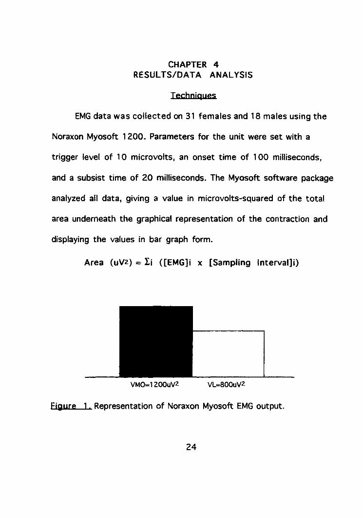
analyzed all data, giving a value in microvolts-squared of th e to ta l
area underneath th e graphical representation of the contraction and
displaying the values in bar graph form.
Area (uVz) = Xi ([EMG]i x [Sam pling ln terval]i)
VM0=1 ZOOuW VL=800uV2
Figure 1. Representation of Noraxon Myosoft EMG output.
24
25
These values were then transferred by hand onto a data collection
sh ee t (see Appendix B). The data were analyzed by a studen t from the
GVSU Statistics Departm ent using a repeated m easures analysis of
variance te s t on SAS softw are.
A one-way w ith in su b je c ts ANOVA w as conducted w ith the
factor being angle and th e dependent variable being the average ratio
of VMO:VL The descriptive sta tis tics for the dependent variable
broken down by the angle are presented in Table 1. One of the
assumptions for the one-way within-subjects ANOVA is th a t the
dependent variable is normally distributed in th e population for
each level of the within-subjects factor (angle). This was not
satisfied at each of the angles (all tended to follow a right-skewed
distribution). To adjust for th e violation of this assumption, the
square root transformation of th e dependent variable was used. This
means that the dependent variable used in the one-way within-
subjects ANOVA was the square root of the average ratio of VMO:VL.
Another assum ption fo r th is one-way w ith in -su b je c ts ANOVA
is th e homogeneity of variance assumption which, according to
Portney and Watkins'^ “s ta te s th a t the variances within each of
26
th e se se ts of difference scores will be relatively equal and
correlated with each o ther.” According to Mauchly’s criterion, this
assumption was not satisfied.
To ad ju st fo r th e f a c t th a t th is assum ption w as no t sa tis f ie d ,
th e degrees of freedom for th e F-dlstrlbutlon were adjusted by the
Greenhouse-Gelsser correction factor-^. The results for th e repeated
measures ANOVA, therefore. Indicate that there Is not a significant
angle effect (F= 2.26, dfl = 2.14, df2 = 102.73, Adj G-G value = .106).
Therefore, there Is not sufficient evidence a t th e 0.05 level to say
th a t there Is a difference In the average VMO:VL ratio among the
angles studied.
Hypothesis/Research Question
Our original hypothesis th a t VMOiVL ra tio s would be la rg e s t I n
the sixty to ninety degree range was not supported by th e statistical
analysis. Testing th e null hypothesis, th a t there would be no
difference between mean VMO:VL ratios between angles, a P-value of
0 .106 was calculated. Because this value Is greater than .05, there
Is not sufficient evidence to conclude tha t there Is a difference In
the mean average VMO:VL ratio among the angles studied. This may
27
be due to the fact th a t a larger sample size is necessary to d e tec t
small differences between the VMO:VL ratios. A larger sample size
would reduce th e standard deviation (SD) and b e tte r determine if the
small differences are statistically significant. These small
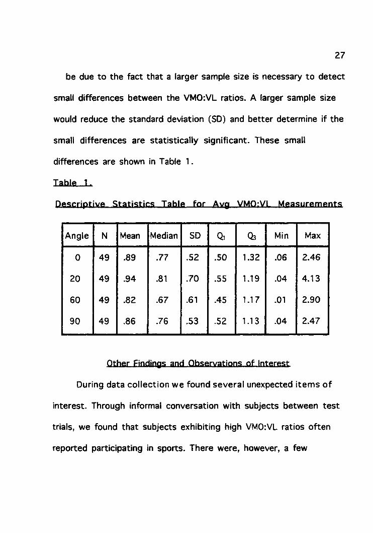
differences are shown in Table 1.
Table 1.
D escrip tive S ta tis t ic s Table fo r Avg VMQ:VL M easu rem en ts
Angle N Mean Median SD Qi 03 Min Max
0 49 .89 .77 .52 .50 1.32 .06 2.46
20 49 .94 .81 .70 .55 1.19 .04 4.13
60 49 .82 .67 .61 .45 1.17 .01 2.90
90 49 .86 .76 .53 .52 1.13 .04 2.47
Other Findings and .Observations of Interest
During data collection we found sev era l unexpected ite m s o f
in terest. Through informal conversation with subjects betw een te s t
trials, we found th a t subjects exhibiting high VMOiVL ratios often
reported participating in sports. There were, however, a few
28
subjects who were not involved in sporting activities who displayed
VMOrVL ratios similar to “athletic” sub jec ts .
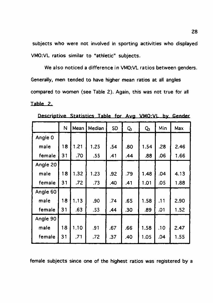
We a lso noticed a d ifference in VMOrVL ra tio s betw een genders.
Generally, men tended to have higher mean ratios a t all angles
compared to women (see Table 2). Again, this was not true for all
Table 2.
D escrip tive S ta tis t ic s Table for Avg VMOrVL by G ender
N Mean Median SD Qi 03 Min Max
Angle 0
m ale
fem ale
18
31
1.21
.70
1.25
.55
.54
.41
.80
.44
1.54
.88
.28
.06
2.46
1.66
Angle 20
m ale
fem ale
18
31
1.32
.72
1.23
.73
.92
.40
.79
.41
1.48
1.01
.04
.05
4.13
1.88
Angle 60
m ale
fem ale
18
31
1.13
.63
.90
.53
.74
.44
.65
.30
1.58
.89
.11
.01
2.90
1.52
Angle 90
m ale
fem ale
18
31
1.10
.71
.91
.72
.67
.37
.66
.40
1.58
1.05
.10
.04
2.47
1.55
female sub jects since one of the highest ratios was registered by a
29
twenty-four year old female who had not been active in sports for an
eleven month period. These differences in VMO:VL ratio may explain
why more women than men are trea ted in the clinic for PFPS. i.is
We also observed many su b je c ts had d ifficu lty performing
isometric contractions a t 0 degrees. These subjects, however, often
had their highest VMO:VL ratios a t this angle. Why this occurred is
unknown.
CHAPTER 5 DISCUSSION AND IMPLICATIONS
Discussion of Findings
Although our findings did not support our hypothesis, we feel
this study is significant for several reasons. First, it adds to th e
literature regarding knee flexion angle and VMO strengthening.
According to our literature review, some researchers hypothesize
th a t selective strengthening of th e VMO may be achieved
isometrically a t terminal extension (0 degrees). Still, o ther studies
suggest the VMO may be strengthened isometrically a t larger angles
of knee flexion including 20, 60, and 90 degrees. Due to these
inconsistencies, all research pertaining to selective VMO
strengthening is beneficial inasmuch as it adds to th e small body of
knowledge in this area.
Second, our findings and much of the existing l i te ra tu re
suggest th a t there is no single angle th a t may be universally
prescribed to selectively strengthen the VMO (See Table 1). Our data
suggests th a t m ost people have a specific angle for optimally
strengthening th e VMO.
30
31
Although many subjects had similar VMOiVL ratios a t all four te s t
angles, o ther sub jec ts displayed distinctly higher values a t certain
angles. We believe this occurs as a result of differences in muscle
recruitm ent p a tte rn s betw een individuals.
Third, th is study i s helpful c lin ically because i t provides a
starting point for developing an isometric protocol for PFPS.
Because EMG is necessary in determining the optimal angle for
isometric strengthening, an EMG study m ust be performed on each
patien t to determ ine which angle produces th e highest VMOiVL ratio.
Isometrically streng then ing a t this angle will individualize
trea tm en t and provide th e g rea tes t benefit for th e patient.
Application of Practice
Considering th a t PFPS i s very commoni, It would benefit
clinicians to have an established protocol for this disorder. As
mentioned above, this study provides a foundation for establishing a
protocol exclusive to each patien t by first analyzing individual
muscle recru itm ent pa ttern s. Thus, individual characteristics
pertaining to recruitm ent pa ttern s must be considered before
issuing isom etric exercises. Not only will the patien t benefit from
32
this approach but third party payors will also be more likely to
reim burse for a research-based protocol.
Lim itations
The first limitation of this stu d y is in th e m easurem ent of th e
angles of knee flexion. Goniometry Is a very subjective m easurem ent
prone to human error. Norkin and White^o refer to 3 to 5 degrees of
error when measuring angles as “accep tab le .” These small
differences between the actual angle and our measured angle may
have caused som e discrepancies in our data.
A nother possib le lim ita tio n due to human e rro r is the
placem ent of th e electrodes. It is virtually Impossible to place the
electrodes in th e exact sam e sp o t on every subject. While a trained
researcher should be able to properly place electrodes with great
consistency, subtle variations in anatom ical landmarks betw een
su b jec ts make this difficult.
A th ird lim ita tio n i s th a t only fou r angles w ere te s te d due to
tim e constra in ts and th e possibility of sub ject fatigue. This limited
our study because the VMO:VL ratio may be g rea test a t an angle other
than 0, 20, 60, or 90 degrees.
33
Also, only su b je c ts betw een th e ages o f 21 and 37 years o f age
with healthy knees were used in this study. This limits th e use of
our results. The results gained with this research may not be
applicable to populations outside th e 21 to 37 year old age bracket
or to subjects with knee pathology.
A nother lim ita tion i s th a t although th e te s tin g o rder of jo in t
angles was randomized, this did not mean the effects of fatigue and
learning were eliminated or minimized. It is possible th a t low third
trial values were due to fatigue. On th e other hand, high third trial
values may be due to learning effects. Fatigue and learning
associated with the three trial te s t design may, therefore, be a
limitation of this study.
A final shortcom ing o f th is s tudy w as th e inab ility o f some
subjects to maintain an isometric contraction above the EMG
threshold level for 3 seconds. When this occurred, the Noraxon
Myosoft software recorded a separate muscle contraction each time
a sub jec t’s muscle activity dropped below the threshold. As a result,
a single trial was recorded as multiple contractions and data
accuracy was compromised.
34
Suggestions for-Eutlher Research
Our study w as one a tte m p t to devise a way to se lec tiv e ly
recruit the VMO during isometric exercise using four different
angles. Although analysis revealed no significant difference in
average VMO:VL ratios betw een th e angles te s ted , several findings
noted in our study may have significant impact on further research.
One area w arran ting fu rth e r study i s recording VMO:VL ra tio s
of subjects with PFPS. Our study was limited to “normal” knees of
young, healthy subjects with no previous history of injury, disease,
or surgery. A study of pathologic knees may reveal significant
differences between average VMOrVL ratios a t different angles in
comparison to healthy knees. It is possible th a t subjects with PFPS
experience pain secondary to a significant imbalance in VMOrVL
ratios. Further research is necessary to determ ine if this is true.
A second area fo r fu rth e r research involves studying VMOrVL
ratios at angles other than those studied here. Although we believe
our study was thorough by involving angles throughout th e full range
of motion, it is possible th a t angles different than those in our
study could produce statistically significant results. This could be
35
achieved by testing VMO:VL ratio a t every 5 degrees. The problem
here, however, would be fatigue from testing so many angles a t once.
Thus, future studies may Involve greater numbers of subjects to
allow each 5 degree increment between 0 and 90 degrees to be
measured.
A th ird recom m endation fo r fu rth e r research i s a study sim ila r
to ours but involving only female subjects or only male subjects.
Through informal observation during data collection we noticed
differences in recruitm ent patterns between men and women.
Because of th ese observations and because women are anatomically
more susceptible to PFPSs, we conducted further analysis seperating
VMOiVL ratios by gender. This analysis showed th a t men have a
g reater VMOiVL ratio than women (see Table 2). The difference in
VMOiVL ratio by gender is a possible explanation for th e lack of
significant differences in average VMOiVL ratios among the angles
te s ted . We did not, however, analyze the data to determ ine if an
optimal angle for VMO strengthening exists for each gender.
Conclusions
This study investiga ted th e possib ility of a re la tio n sh ip
36
betw een knee angle and VMO and VL activity. The findings of this
study indicated there is no significant difference among th e
te s te d knee angles for the VMOiVL atio during isometric knee
extension. Generalizations from th ese data, however, should be made
carefully because of the characteristics of our study group. Further
research is needed to investigate the possibility of significant
associations between VMOiVL ratios and gender, pathology, and
angles o ther than those te s te d in this study.
REFERENCES
1. Malek MM, Mangine RE. P ate llo fem oral pain syndromes: a com prehensive and conservative approach. The Journal o f Orthopaedic and Sports Physical Therapy. 1981 ; 2:108-116.
2. Fulkerson JP , Hungerford DS. Disorders o f the Patellofemoral Joint. Baltimore, Maryland: Williams and Wilkins; 1990.
3. Fulkerson JP. The etiology o f pate llo fem ora l pain in young, ac tive patien ts: a prospective study. Clinical Orthopaedics and Related Research. 1983; 179:129-133.
4. Insall J. Current concepts review : p a te lla r pain. The Journal o f Bone and Joint Surgery. 1982; 64 :147-152.
5. Kettelkamp DB. Current concepts review : m anagem ent o f p a te lla r malalignment. The Journal o f Bone and Joint Surgery. 1981; 63:1344-1347.
6 . Kramer PG. P ate lla m alalignm ent syndrome: ra tio n a le to reduce excessive la te ra l pressure. The Journal o f Orthopaedic and Sports Physical Therapy. 1986; 8 :301-309.
7. Wise HH, F iebert lA, K ates JL. EMG biofeedback a s a tre a tm e n t fo r patellofem oral pain syndrome. The Journal o f Orthopaedic and Sports Physical Therapy. 1984; 6:95-103.
8 . Puniello MS. Illio tib ia l band tig h tn e ss and m edial p a te lla r glide in p a tien ts w ith patello fem oral dysfunction. The Journal o f Orthopaedic and Sports Physical Therapy. 1993; 17:144-148.
9. McConnell J. The managem ent o f chondrom alacia patellae: a long te rm solution. The Australian Journal o f Physiotherapy. 1986; 32:215-222.
37
38
10. Basm ajian JV. Muscles Alive, Their Function Revealed by Electromyography. Baltim ore, Maryland: W averly P re ss Inc.; 1979:250-257.
11. Lieb FJ, P erry J. Quadriceps function: an anatom ical and m echanical study using am putated lim bs. The Journal o f Bone and Joint Surgery. 1968; 50-A:l 535-1548.
12. Pevsner DN, Johnson JR, Blazina ME. The pate llo fem oral jo in t and i t s im p lica tions in the rehab ilita tion o f th e knee. Physical Therapy. 1979; 59:869-894.
13. Dehaven KR, Dolan WA, Mayer PJ. Chondromalacia pa te lla i n a th le tes . The American Journal o f Sports Medicine. 1979;7:511.
14. Henry JH, Crosland JW. Conservative tre a tm e n t o f patellofem oral subluxation. The American Journal o f Sports Medicine. 1979; 7:12-14.
15. Sm illie IS. Injuries o f the Knee Joint. Baltimore, Maryland: The W illiam s and W ilkins Co; 1971:2.
16. Soderberg GL, McCock T. An electrom yographic analysis o f quadriceps fem oris m uscle se ttin g and s tr a ig h t leg raising. Physical Therapy. 1983; 63:1434-1438.
17. Wild J J , Franklin TD, Woods GW. P a te lla r pain and quadriceps rehabilita tion : an EMG study. The American Journal o f Sports Medicine. 1982; 10:12-15.
18. Doucette SA. The e f fe c t of exercise on p a te lla r tracking i n la te ra l p a te lla r com pression syndrome. The American Journal o f Sports Medicine. 1992; 20:434-440.
19. Woodall W, Welsh J. A biomechanical b as is fo r rehab ilita tion program s involving th e PF jo in t. The Journal o f Orthopaedic and
39
Sports Physical Therapy. 1990; 11:535-542.
20. W estfall DC, W orrell TW. A nterior knee pain syndrome: ro le o f the vastus m edialis oblique. Journal o f Sports Rehabilitation. 1992; 1:317-325.
21. Levine J. Chondromalacia patellae. The Physician and Sports Medicine. 1979; 7:41-49.
22. Boucher JP , Cyr A, Lefebvre R, King MA. The v as tu s m edialis obliquus i s more ac tiv e a t 90 degrees o f knee flexion. Medicine and Science in Sports and Exercise. 1992; 24:S147.
23. Brownstein BA, Lamb RL, Manglne RE. Quadriceps torque and in tegrated electromyography. The Journal o f Orthopaedic and Sports Physical Therapy. 1985; 6 :309-314.
24. Cerny K. V astus m ed ia lis ob lique /vastu s la te ra lis m uscle ac tiv ity ra tio s fo r se lec ted ex e rc ise s i n persons w ith and w ithout pate llo fem oral pain syndrome. Physical Therapy. 75:672-683.
25. Mariani P, Caruso I. An electrom yographic investiga tion o f subluxation o f th e pate lla . Journal o f Bone and Joint Surgery (Br). 1979; 61:169-197.
26. Norkin CC, Levangie PK. Joint Structure and Function: A Comprehensive Analysis. Philadelphia, Pennsylvania: F.A. Davis Co; 1992.
27. Bose K, Kanagasuntheram R, Osman MB. V astus m edialis oblique: an anatom ic and physiologic study. Orthopaedics. 1980; 3:880-883.
28. Moore KL Clinically Oriented Anatomy. Baltim ore, Maryland: W illiams and W ilkins; 1992.
40
29. Bockrath K, Wooden C, W orrell T, Ingersoll CD, F arr J. E ffects o f p a te lla taping on p a te lla position and perceived pain. Medicine and Science in Sports and Exercise. 1993; 25:989 - 992.
30. Paulos L, Rusche K, Johnson C, Noyles FR. P a te lla r m alalignm ent. Physical Therapy. 1980; 60:1624-1632.
31. F iebert I , Hardy CJ, W erner KL. E lectrom yographic an a ly sis of th e quadriceps fem oris during iso k in e tic e c cen tric activation . Isokinetics and Exercise Science. 1992; 2:18-23.
32. Ingersol CD, Knight KL. P a te lla r location changes follow ing EMG biofeedback or p rogressive r e s is t iv e exerc ises. Medicine and Science in Sports and Exercise. 1991 ; 23 :1122-1127.
33. W ise H, F iebert I, Kates J. EMG biofeedback a s tre a tm e n t fo r pate llo fem oral pain syndrome. The Journal o f Orthopaedic and Sports Physical Therapy. 1984; 6:95-103.
34. King MA, Boucher JP, Cyr A, Lefebvre R. Evaluation o f VMO/VL ra tio w ith a lte ra tio n o f p a te llo fem o ra l m echanics and hip adduction in patello fem oral pain syndrome. Medicine and Science in Sports and Exercise. 1992; 24:S148.
35. Hanten WP, Schulth ies SS. E xercise e f fe c t on electrom yographic ac tiv ity o f th e v as tu s m ed ialis oblique and v as tu s la te ra lis m uscles. Physical Therapy. 1990; 70:561-565.
36. Grabiner MD, von Haeffen L, Koh TJ. Concurrent hip jo in t adduction and knee jo in t ex tension m om ents do no t influence quadriceps fem oris exc ita tion . Medicine and Science in Sports and Exercise. 1992; 24:S128.
37. Hodges PW, Richardson CA. The influence o f iso m e tric hip adduction on quadriceps fem o ris ac tiv ity . Scandinavian Journal o f Rehabilitative Medicine. 1993; 25:57-62.
41
38. Sczepanski TL, Gross MT, Duncan PW, Chandler JM. E ffec ts of contraction type, angular ve locity , and arc o f m otion on VMOiVL EMG ratio . The Journal o f Orthopaedic and Sports Physical Therapy. 1991; 14:256-262.
39. Reynalds L, Levin TA, Medeios JM, Adler NS, Hallum A. EMG ac tiv ity o f th e v as tu s m ed ia lis oblique and th e v a s tu s la te ra l is i n th e ir ro le o f p a te lla r alignm ent. American Journal o f Physical Medicine. 1983; 62:61-70.
40. Norkin CC, White JD. Measurement o f Joint Motion: A guide to Goniometry. Philadelphia, Pennsylvania: PA. Davis Co; 1995:137-146.
41. B ouisetta S, Manton 8. Q uan tita tive rela tionsh ip betw een su rface emg and in tram uscu lar electrom yographic a c tiv i ty in voluntary movement. American Journal o f Physical Medicine. 1972; 51: 285-295.
42. Lippold CC. The re la tio n betw een in tegrated action p o te n tia ls i n a human m uscle and i t s iso m e tric tension. Journal o f Physiology. 1952; 117: 492-499.
43. Komi PV, Buskirk ER. R eproducibility of electrom yography m easurem ents w ith in se rted w ire e lectrodes and su rfa c e electrodes. Electromyography. 1970; 10: 357-367.
44. Portney LG, W atkins MP. Foundations o f Clinical Research: Applications to Practice. Norwalk, Connecticut: Appleton & Lange; 1993: 393-394.

APPENDIX A.
Inform ed C o n sen t Form
42
43
KNEE FLEXION AND ITS INFLUENCE ON VMOiVL RATIOS
DURING ISOMETRIC QUADRICEPS CONTRACTION
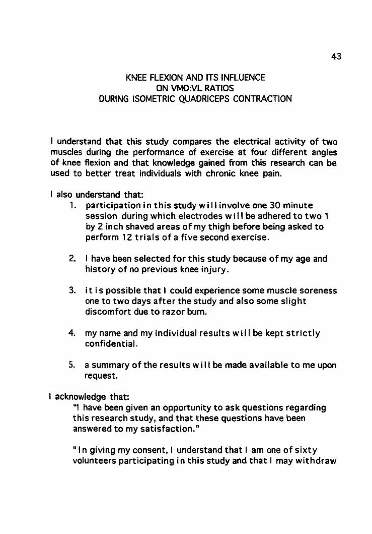
I understand th a t this study com pares th e electrical activity of two muscles during the performance of exercise a t four different angles of knee flexion and th a t knowledge gained from this research can be used to b e tte r tre a t individuals with chronic knee pain.
I also understand tha t:1. partic ipation i n th is study w ill involve one 30 m inute
session during w hich e lectrodes w iII be adhered to tw o 1 by 2 inch shaved a rea s o f my thigh before being asked to perform 12 t r ia l s o f a five second exercise .
2. I have been se lec ted fo r th is study because o f my age and h isto ry o f no previous knee injury.
3. i t is possib le th a t I could experience som e m uscle so ren ess one to tw o days a f te r th e study and also som e s lig h t d iscom fort due to razo r burn.
4. my name and my individual re su lts w i 11 be kept s t r ic t ly confidential.
5. a sum m ary o f th e re su lts w i 11 be made availab le to me upon request.
I acknowledge that:“I have been given an opportunity to ask q uestions regarding th is research study, and th a t th e se questions have been answered to my s a tis fa c tio n .”
" I n giving my consent, I understand th a t I am one o f s ix ty volunteers p artic ip a tin g in th is s tudy and th a t I may w ithd raw

44
a t any tim e during te s tin g .”
“The in v estig a to rs , Bill Allan and J e f f Rendra have my perm ission to perform the above procedures on m e.”
“I hereby au thorize th e in v e s tig a to rs to re le a se th e inform ation obtained in th is study to s c ie n tif ic lite ra tu re . I understand th a t I w ill not be id en tified by nam e.”
“I have th e righ t to con tac t Paul Huizenga, Chair o f Human R esearch Review Com m ittee (8 9 5 -2 4 7 2 ), or Jane Toot (895-3605), a t any tim e i f I have questions.
“I acknowledge th a t I have read and understand th e above inform ation, and th a t I agree to p a r tic ip a te in th e study .”
Witness Signature Participant Signature
Date Date

APPENDIX B.
D ata C ollection S h e e t
45
46
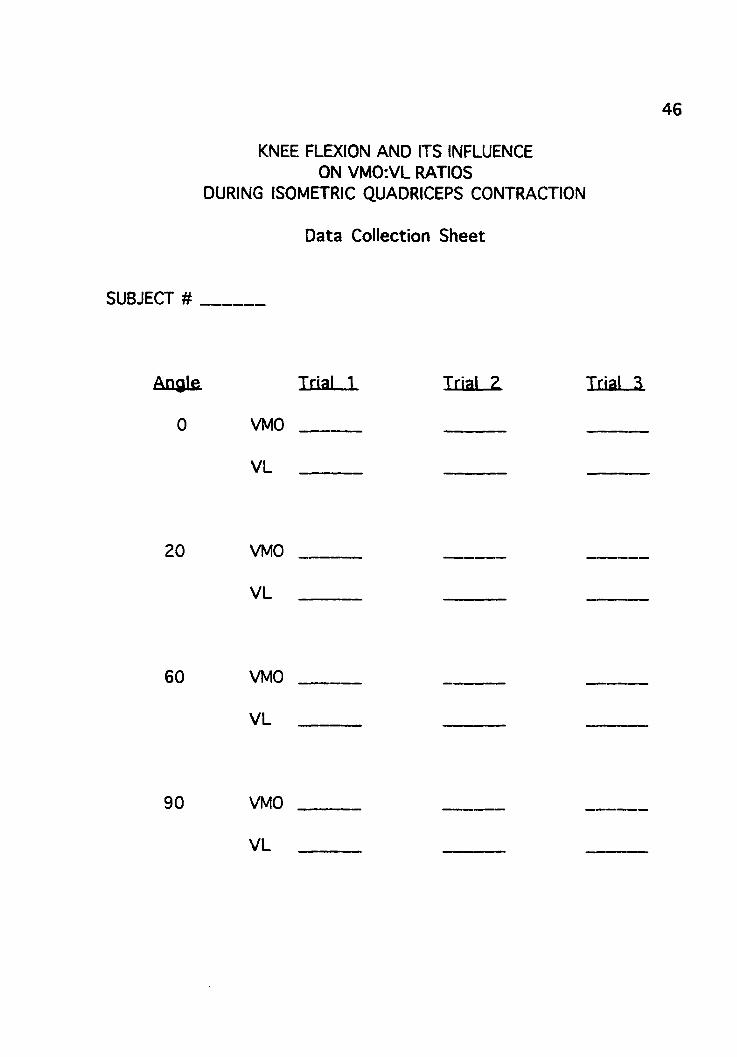
KNEE FLEXION AND ITS INFLUENCE ON VMO:VL RATIOS
DURING ISOMETRIC QUADRICEPS CONTRACTION
Data Collection Sheet
SUBJECT #
Analfi. TüaL.I I n a l-2. Trial 3
0 V M O _________ _______ _______
VL _______ _______ _______
20 VMO
VL
60 VMO
VL
90 VMO
VL

APPENDIX C.
P roposal Summary For Human S u b jec t Review C om m ittee
47
48
P ate llo fem oral Pain Syndrome (PFPS) i s th e number one cause
of knee pain in many clinics and sports medicine cen ters around the
country. The major underlying cause of PFPS is a malalignment of
the patellofemoral Joint. The result of this malalignment is a
laterally tracking patella causing pain for th e patien t.
The vastus m ed ia lis obliquus (VMO) has been id en tified a s th e
only structu re with th e ability to pull th e patella medially.
Theoretically, strengthening th e VMO will co rrec t malalignment thus
relieving pain. Several exercises have been recom m ended by
researchers and therap ists to strengthen th e VMO. However, while
strengthening the VMO, th e se exercises may also strengthen the
vastus lateralis (VL) to th e sam e degree. Even though th e VMO is
strengthened, it may still not be able to pull th e patella medially
because of the sim ultaneous strengthening o f th e VL pulling th e
patella laterally. For an exercise to be efficient in altering
malalignment and to reduce pain it is necessary to streng then the
VMO independent of th e VL or a t least strengthen it to a
significantly greater degree than th e VL.
Several au thors have recommended an iso m e tr ic con trac tion o f
49
th e quadriceps to facilitate strengthening of th e VMO. While some
authors believe VMO strengthening occurs a t terminal knee extension
o thers contend th a t exercise should be performed a t varying degrees
of knee flexion. The problem is th a t th e re is little scientific data
th a t indicates the VMO is strengthened to a g reater degree than the
VL in any of th ese ranges of motion (ROM) during an isom etric
contraction. The purpose of this study is to com pare th e activity of
th e VMO relative to th e activity of th e VL a t various points between
0 and 90 degrees of knee flexion during isometric contraction. Our
hypothesis is th a t isom etric quadriceps contractions betw een 60
and 90 degrees of flexion will result in larger VMO:VL ratios than a t
o ther ranges of motion in healthy college age male and female knees.
T esting wi l l take place i n th e Human Perform ance Lab located
in th e Grand Valley S ta te University Fieldhouse. Subjects for this
study will consist of 60 males and females ranging from 18 to 35
years of age with no history of hip or knee problems. The subjects
will be se lec ted via a sample of convenience using physical therapy
s tu d en ts enrolled a t Grand Valley S ta te University.
S im ultaneous EMG readings w i 11 be taken o f th e VMO and VL
50
during isometric contraction a t angles of 0, 20, 60, and 90 degrees
of knee flexion. The EMG values of th e th ree trials will then be
averaged to establish a mean value for th e VMO and VL. These mean
values will then be used to calculate a VMO:VL ratio for each of th e
designated te s t angles. Finally, a repea ted measures analysis of
variance t e s t will be used to determ ine if a statistically significant
difference exists among the VMOiVL ratios of the four angles.
Consenting su b je c ts w i 11 be te s te d iso m etrica lly a t a rc s o f 0,
20, 60, and 90 degrees of knee flexion. To reduce any effects of
fatigue o f learning, th e order in which th e angles will be te s te d will
be selected randomly.
Once th e order o f angles to be te s te d i s se lec ted , th e s u b je c t 's
leg will be cleaned with an isopropyl alcohol swab and th e bellies of
th e VMO and distal VL will be shaved using a disposable razor. Each
shaved area will be rubbed again with a clean isopropyl alcohol
swab to sanitize th e skin and maximize electrode adherence. While
th e sub ject is seated , th e researcher will place self-adherent
surface electrodes to each of the designated areas. The electrodes
will then be plugged into lead wires connected to a com puter with
51
MYOSOFT EMG softw are for Windows. The sub jec t’s knee will then be
positioned a t the first randomly selected angle. The padded
resistance bar of a CYBEX machine will then be placed a t the
anterior ankle of th e te s t leg for resistance and to maintain th e knee
a t the designated te s t angle. When instructed, th e subject will
perform a maximal isom etric contraction of th e quadriceps femoris
for five seconds against th e padded resistance bar. Subjects will
perform three consecutive trials a t each angle with a one minute
rest between trials. To familiarize te s te rs with both equipment and
procedure, a pilot study with 10 sub jects will be performed. This
will help establish reliability as well as facilitate sub jec t safe ty .
It will be explained to each subject th a t som e muscle soreness will
persist one to two days following participation in this study. It will
also be explained to each subject th a t they have th e right to
withdraw their participation in this experim ent a t any time.