Embed Size (px)
Citation preview
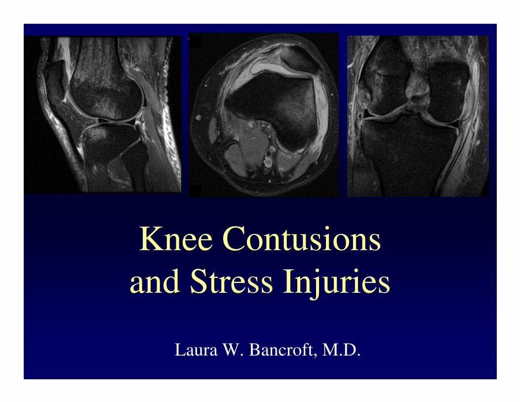
Knee Contusions
and Stress Injuries
Laura W. Bancroft, M.D.

Objectives
• Review 5 types of contusion patterns
– Pivot shift
– Dashboard
– Hyperextension
– Clip
– Lateral patellar dislocation
• Demonstrate various stress injuries,
including patellofemoral stress syndrome
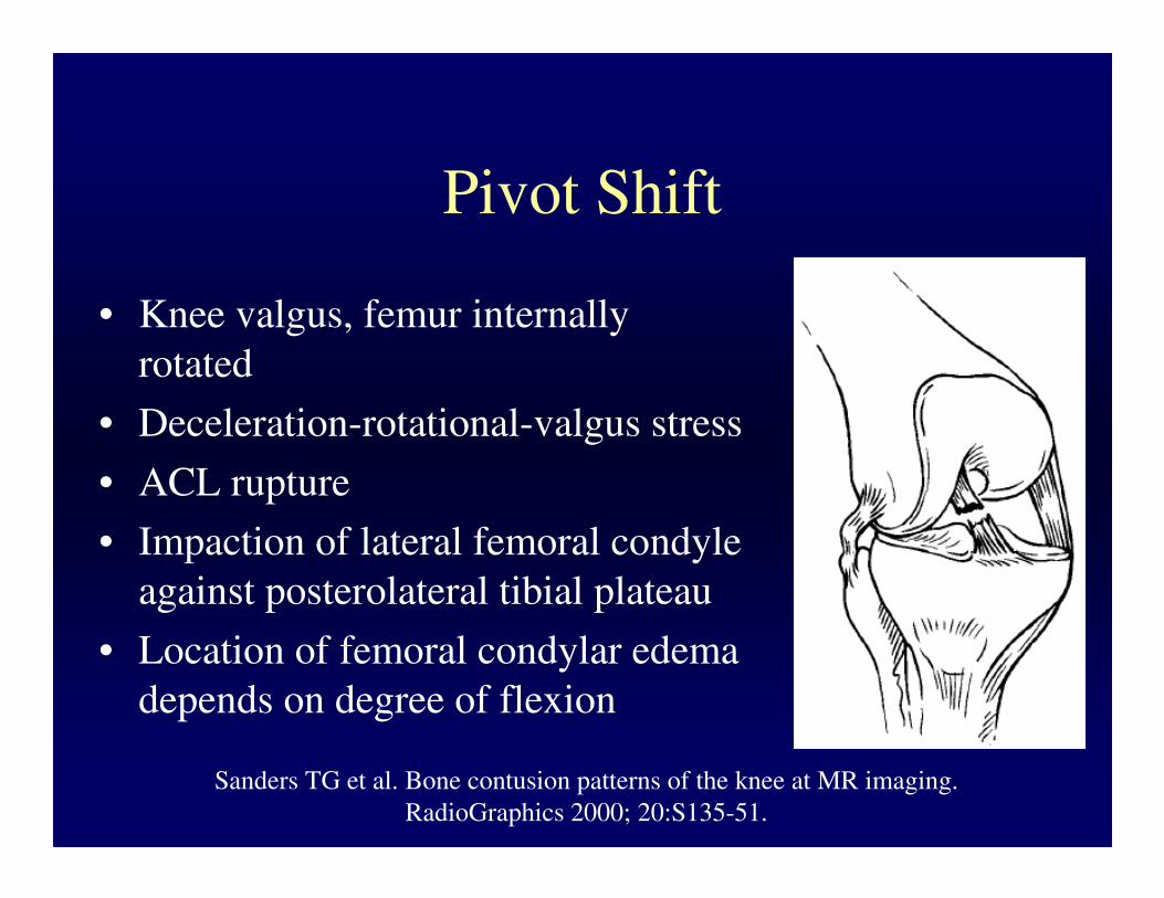
Pivot Shift
• Knee valgus, femur internally
rotated
• Deceleration-rotational-valgus stress
• ACL rupture
• Impaction of lateral femoral condyle
against posterolateral tibial plateau
• Location of femoral condylar edema
depends on degree of flexion
Sanders TG et al. Bone contusion patterns of the knee at MR imaging.
RadioGraphics 2000; 20:S135-51.
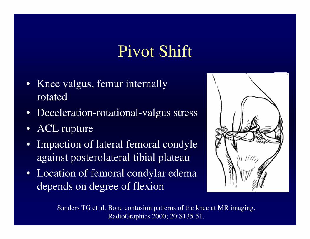
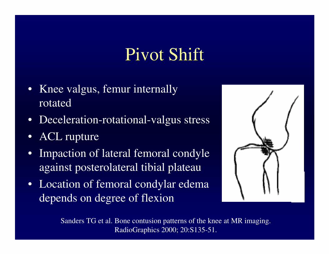
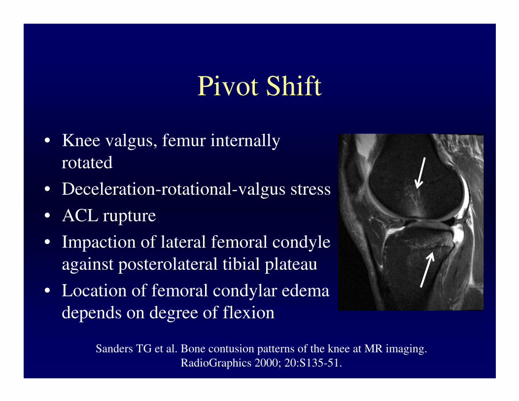

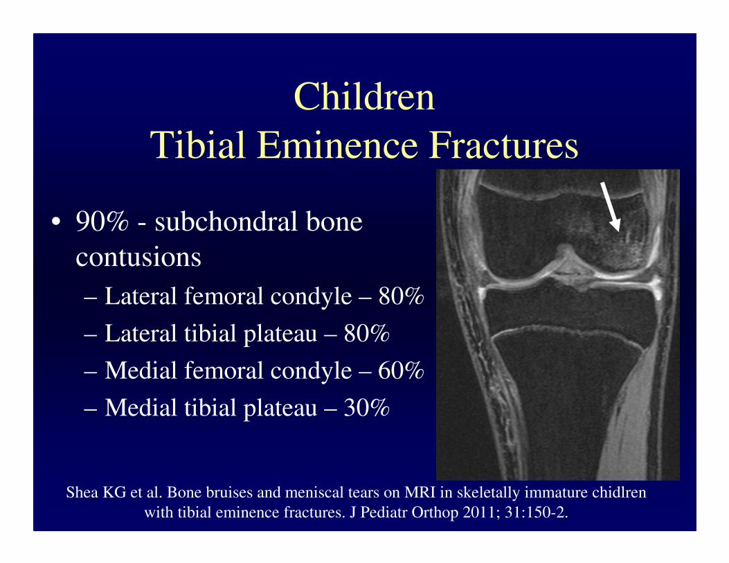
Children
Tibial Eminence Fractures
• 90% - subchondral bone
contusions
– Lateral femoral condyle – 80%
– Lateral tibial plateau – 80%
– Medial femoral condyle – 60%
– Medial tibial plateau – 30%
Shea KG et al. Bone bruises and meniscal tears on MRI in skeletally immature chidlren
with tibial eminence fractures. J Pediatr Orthop 2011; 31:150-2.
Children
• May have intact ACL with typical pivot
shift bone contusions
– 28% of cases
– Ligamentous laxity
Snearly WN et al. Lateral-compartment bone contusions in adolescents with intact
anterior cruciate ligaments. Radiology 1993; 198:205-8.

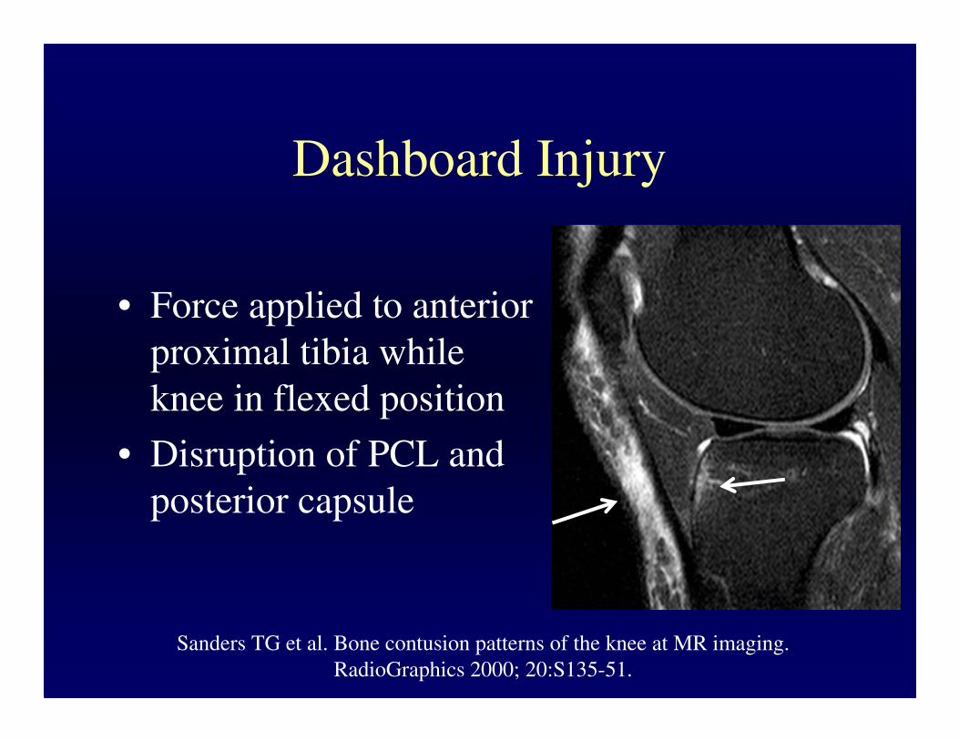
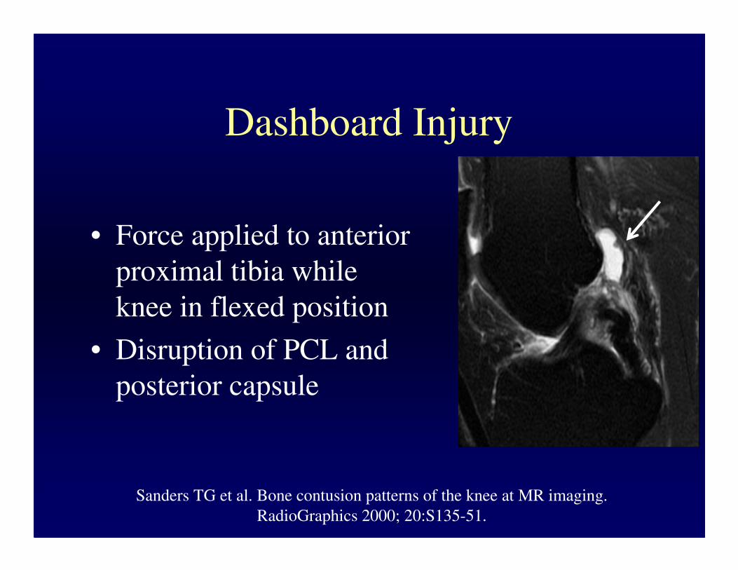
Dashboard Injury
• Force applied to anterior
proximal tibia while
knee in flexed position
• Disruption of PCL and
posterior capsule
Sanders TG et al. Bone contusion patterns of the knee at MR imaging.
RadioGraphics 2000; 20:S135-51.
Dashboard Injury
• Force applied to anterior
proximal tibia while
knee in flexed position
• Disruption of PCL and
posterior capsule
Sanders TG et al. Bone contusion patterns of the knee at MR imaging.
RadioGraphics 2000; 20:S135-51.

Dashboard Injury
• Force applied to anterior
proximal tibia while
knee in flexed position
• Disruption of PCL and
posterior capsule
Sanders TG et al. Bone contusion patterns of the knee at MR imaging.
RadioGraphics 2000; 20:S135-51.

Dashboard Injury
• Force applied to anterior
proximal tibia while
knee in flexed position
• Disruption of PCL and
posterior capsule
Sanders TG et al. Bone contusion patterns of the knee at MR imaging.
RadioGraphics 2000; 20:S135-51.
Dashboard Injury
• Force applied to anterior
proximal tibia while
knee in flexed position
• Disruption of PCL and
posterior capsule
Sanders TG et al. Bone contusion patterns of the knee at MR imaging.
RadioGraphics 2000; 20:S135-51.

SubacuteSubacute Dashboard InjuryDashboard Injury

Hyperextension
• Direct force is applied to anterior
tibia while foot is planted
• Indirect force - forceful kicking
• Direct injury (car bumper hitting
anterior tibia of pedestrian)
Sanders TG et al. Bone contusion patterns of the knee at MR imaging.
RadioGraphics 2000; 20:S135-51.
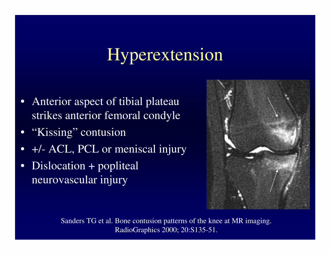
Hyperextension
• Anterior aspect of tibial plateau
strikes anterior femoral condyle
• “Kissing” contusion
• +/- ACL, PCL or meniscal injury
• Dislocation + popliteal
neurovascular injury
Sanders TG et al. Bone contusion patterns of the knee at MR imaging.
RadioGraphics 2000; 20:S135-51.

Hyperextension
• Anterior aspect of tibial plateau
strikes anterior femoral condyle
• “Kissing” contusion
• +/- ACL, PCL or meniscal injury
• Dislocation + popliteal
neurovascular injury
Sanders TG et al. Bone contusion patterns of the knee at MR imaging.
RadioGraphics 2000; 20:S135-51.
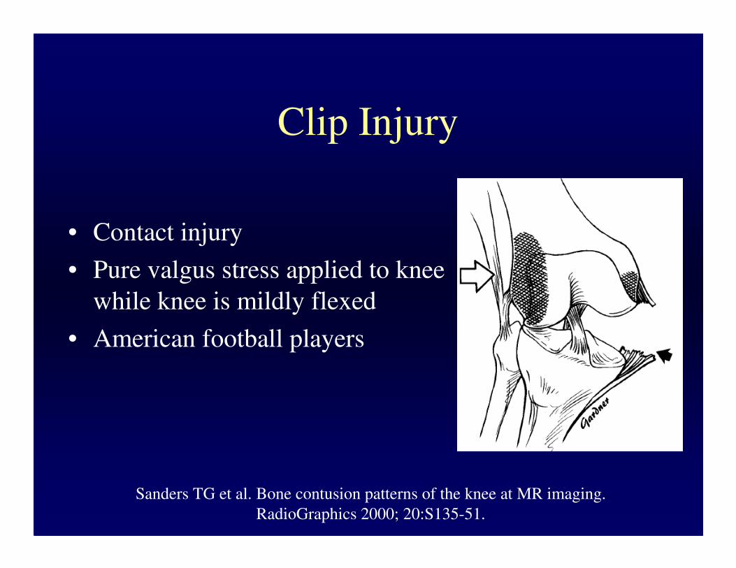
Clip Injury
• Contact injury
• Pure valgus stress applied to knee
while knee is mildly flexed
• American football players
Sanders TG et al. Bone contusion patterns of the knee at MR imaging.
RadioGraphics 2000; 20:S135-51.

Clip Injury
• Bone marrow edema
– Most prominent in lateral
femoral condyle - direct blow
– Second smaller area of edema
in the medial femoral condyle -
avulsive stress to the MCL
Sanders TG et al. Bone contusion patterns of the knee at MR imaging.
RadioGraphics 2000; 20:S135-51.

Clip Injury
• Bone marrow edema
– Most prominent in lateral
femoral condyle - direct blow
– Second smaller area of edema
in the medial femoral condyle -
avulsive stress to the MCL
Sanders TG et al. Bone contusion patterns of the knee at MR imaging.
RadioGraphics 2000; 20:S135-51.
Clip Injury
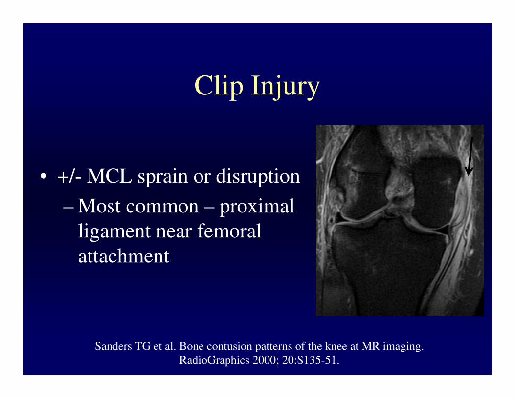
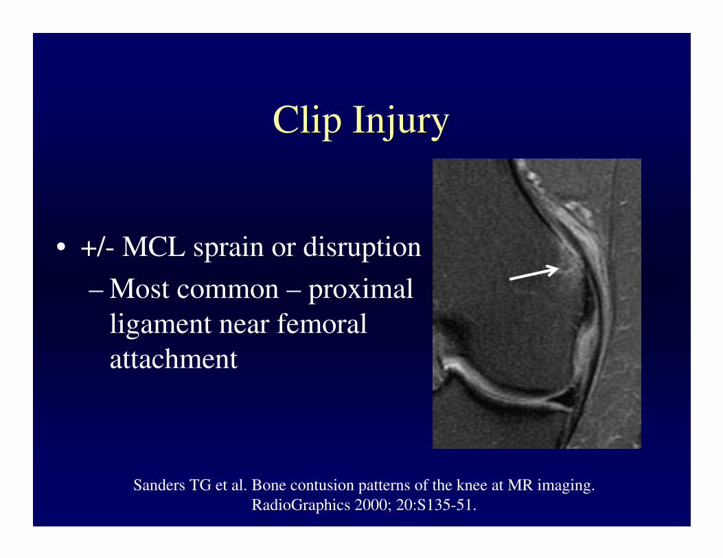
• +/- MCL sprain or disruption
– Most common – proximal
ligament near femoral
attachment
Sanders TG et al. Bone contusion patterns of the knee at MR imaging.
RadioGraphics 2000; 20:S135-51.
Clip Injury
• +/- MCL sprain or disruption
– Most common – proximal
ligament near femoral
attachment
Sanders TG et al. Bone contusion patterns of the knee at MR imaging.
RadioGraphics 2000; 20:S135-51.

Clip Injury
• +/- MCL sprain or disruption
– Most common – proximal
ligament near femoral
attachment
Sanders TG et al. Bone contusion patterns of the knee at MR imaging.
RadioGraphics 2000; 20:S135-51.

• Most common mechanism
of first-time patellar
dislocation
– Flexed, internally rotated
knee
– Planted foot
– Valgus component
Lateral Patellar Dislocation
Sanders TG et al. Bone contusion patterns of the knee at MR imaging.
RadioGraphics 2000; 20:S135-51.

• Most common mechanism
of first-time patellar
dislocation
– Flexed, internally rotated
knee
– Planted foot
– Valgus component
Diederichs G et al. MR imaging of patellar instability. RadioGraphics 2010; 30:961-81.
Lateral Patellar Dislocation
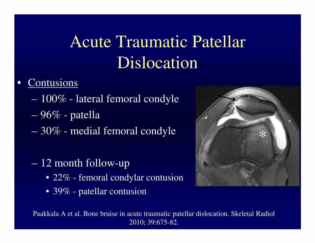
Acute Traumatic Patellar
Dislocation• Contusions
– 100% - lateral femoral condyle
– 96% - patella
– 30% - medial femoral condyle
– 12 month follow-up
• 22% - femoral condylar contusion
• 39% - patellar contusion
Paakkala A et al. Bone bruise in acute traumatic patellar dislocation. Skeletal Radiol
2010; 39:675-82.
*

PDRS – patellofemoral ligament rupture
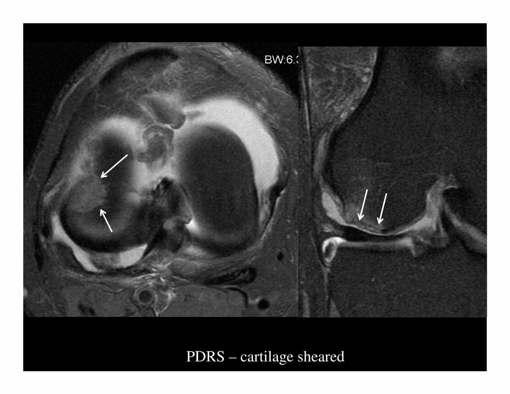
PDRS – cartilage sheared

PDRS – cartilage sheared
*

PDRS – osteochondral fragment

PDRS – osteochondral fragment

PDRS – osteochondral fragment

PDRS – osteochondral fragment

PDRS – osteochondral fragment
Stress Injuries
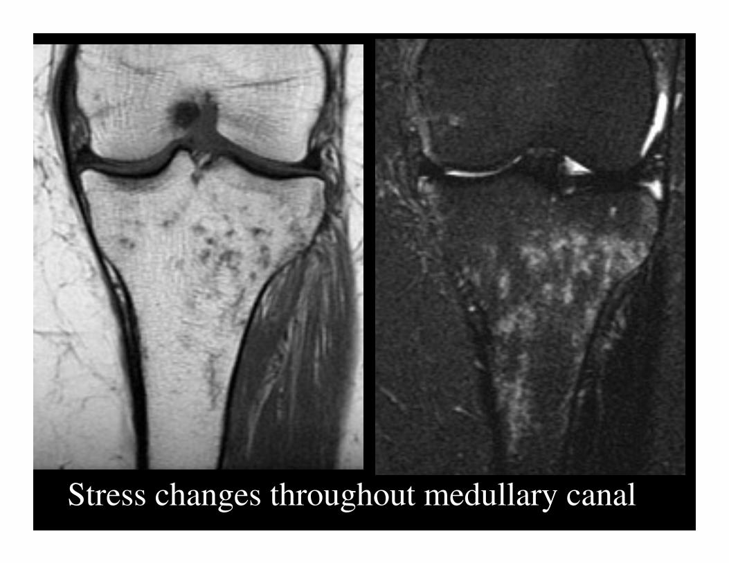
Stress changes throughout medullary canal
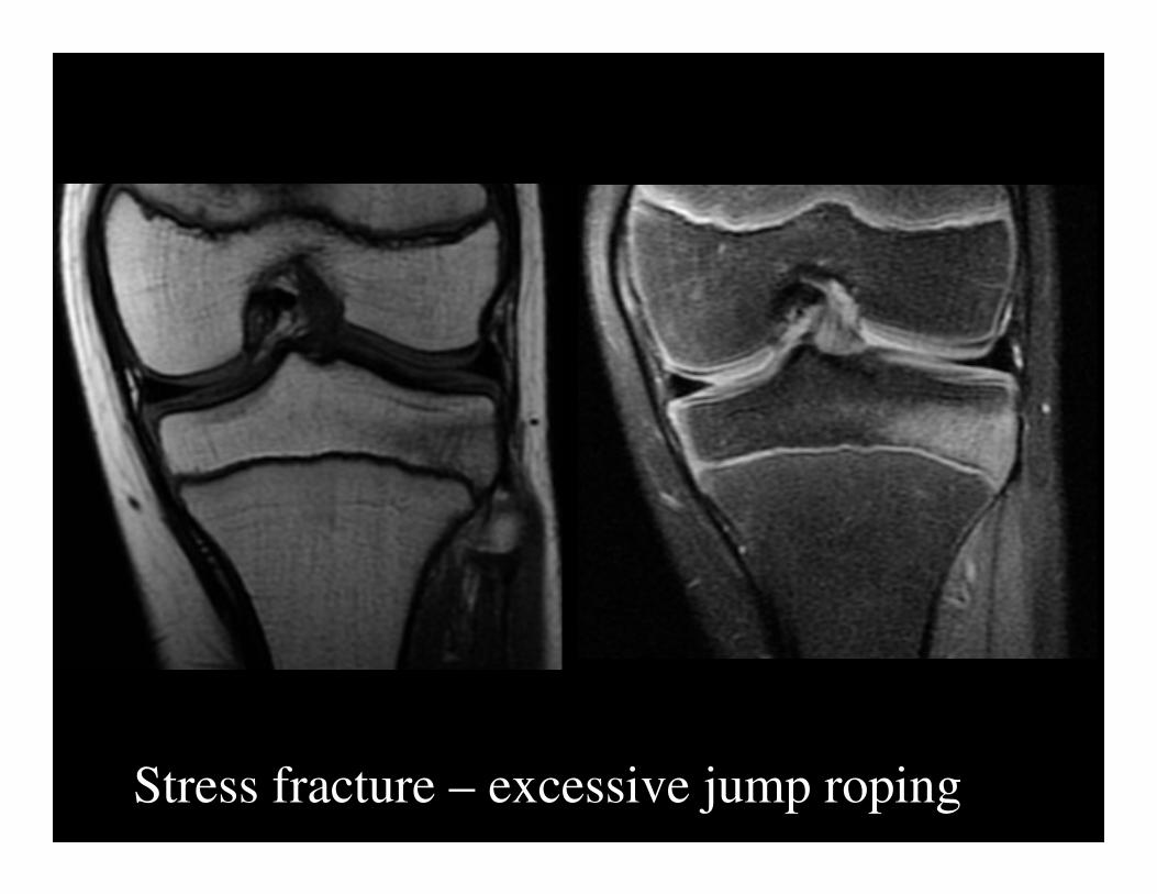
Stress fracture – excessive jump roping
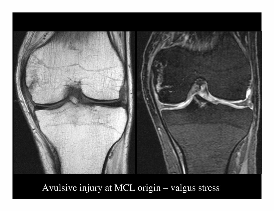
Avulsive injury at MCL origin – valgus stress

Stress fracture - runner

Stress fracture

Stress fracture

Stress fracture

Insufficiency fracture

Insufficiency fracture

Patellofemoral Stress Injuries -
Children
• Sinding-Larsen-Johannson disease
• Osgood-Schlatter
• Patellar sleeve avulsion
• Tibial Tuberosity Avulsion

Sinding-Larsen-Johannson Disease
Davis KW. Imaging pediatric sports injuries. Radiol Clin N Am 2010; 48:1213-35.
• Umbrella term for
syndrome that causes pain
at inferior pole of patella
• Fragmentation or
calcification of inferior
pole

Osgood-Schlatter Disease
Davis KW. Imaging pediatric sports injuries. Radiol Clin N Am 2010; 48:1213-35.
• Traction apophysitis
• Strong forces from quadriceps
mechanism
• Insertion of patellar tendon on
tibial tuberosity

Osgood-Schlatter Disease
Davis KW. Imaging pediatric sports injuries. Radiol Clin N Am 2010; 48:1213-35.
• Common causes of anterior
knee pain
• 12- 15 y/o boys
• 8- 12 y/o girls
• Repeated jumping/squatting
• Local pain, swelling and
tenderness at tuberosity

Osgood-Schlatter Disease
• T2 - High signal within and
surrounding tendon
• Deep infrapatellar bursitis

Osgood-Schlatter Disease
• T2 - High signal within and
surrounding tendon
• Deep infrapatellar bursitis

Patellar Sleeve Avulsion
Davis KW. Imaging pediatric sports injuries. Radiol Clin N Am 2010; 48:1213-35.
• Acute counterpart to
Sinding-Larsen Johannson
• Acute jumping injury
• 8-12 y/o

Patellar Sleeve Avulsion
Davis KW. Imaging pediatric sports injuries. Radiol Clin N Am 2010; 48:1213-35.
• Anterior soft tissue
swelling
• Small fragment of bone
avulsed from inferior
• tip or anterior inferior
patella

Patellar Sleeve Avulsion
Davis KW. Imaging pediatric sports injuries. Radiol Clin N Am 2010; 48:1213-35.
• Osseous fragments - tip of
the iceberg
• Much larger cartilage
fragment unseen on
radiographs
• MR – identify size of
cartilage fragment

Patellar Sleeve Avulsion
Davis KW. Imaging pediatric sports injuries. Radiol Clin N Am 2010; 48:1213-35.
• Adolescent boy jumpers
near skeletal maturity
• Sharp fragments are visible
and elevated on
radiographs

Tibial Tuberosity Avulsion
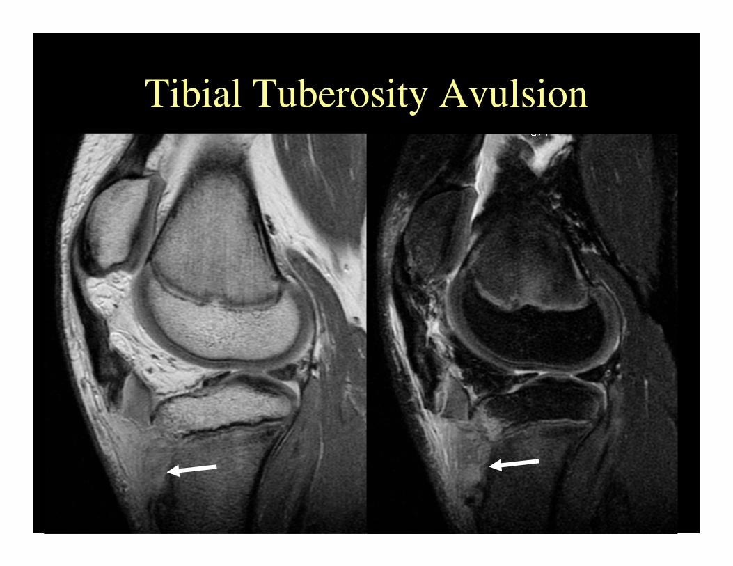
Tibial Tuberosity Avulsion

Conclusion
• Pivot shift injury is caused by deceleration-
rotational-valgus stress, has associated ACL
rupture, and impaction contusions of lateral
femoral condyle and posterolateral tibial plateau
• Dashboard injuries are caused by forces
applied to the anterior proximal tibia while knee
is in flexed position, and leads to disruption of
the PCL and posterior capsule

Conclusion
• Hyperextension injuries are caused by the
anterior tibial plateau striking the anterior
femoral condyle, leading to “kissing”
contusions, with or without ACL, PCL and
meniscal injury
• Clip injuries result from pure valgus stress
applied to mildly flexed knee, resulting in large
lateral femoral condylar contusion and smaller
medial femoral condylar avulsive injury

Conclusion
• Lateral patellar dislocation occurs when
valgus force is applied to the flexed, internally
rotated knee when the foot is planted. Lateral
femoral condylar and medial patellacontusions
occur, often in conjunction with patellofemoral
ligament sprain or tear.
Conclusion
• Linear stress and insufficiency fractures occur
commonly in the proximal tibia, fibula or
subchondral femur.
• Patellofemoral stress syndrome (Sinding-
Larsen-Johannson disease, Osgood-Schlatter
disease, patellar sleeve avulsion and tibial
tuberosity avulsion) is the most common cause
of chronic anterior knee pain in adolescents.