Embed Size (px)
Citation preview
Letter to the Editor
Klinefelter and Trisomy X Syndromes inPatients With Prader-Willi Syndrome andUniparental Maternal Disomy of Chromosome15—A Coincidence?
To the Editor:
We report on two patients (a 7-year-old boy and a12-year-old girl) with Klinefelter syndrome and mosaictrisomy X syndrome, respectively, and Prader-Willisyndrome (PWS) due to uniparental maternal disomyof chromosome 15. Both patients were born to motherswith advanced maternal age. To our knowledge onlyone patient has been reported with PWS and 47,XXXsyndrome [Ferrante et al., 1986], while several pa-tients with PWS and Klinefelter syndrome have beenreported [Dunn, 1968; Bray et al., 1983; Trent et al.,1991; Tu et al., 1992]. Previous studies have not exam-ined whether or not their phenotypes were due to con-certed missegregation of both chromosome 15 and X.Herein, we describe the phenotypes of these two pa-tients and determine the probable etiology of their non-disjunction events.
CLINICAL REPORTSPatient A
Patient A was born to a 36-year-old gravida 1 motherand weighed 2,483 g (15th centile) and was 47 cm (25thcentile) long. Hypotonia was noted immediately afterdelivery with inability to nurse or suck. He was gavagefed in intensive care for 3 weeks. CT scan and MRI of
the head and EEG were normal, as were results ofevaluations for primary neuromuscular or metabolicdisease. He was on home oxygen therapy for the first 3months after discharge. He grew slowly until age 3years, at which time rapid weight gain was noted, al-though there was no reported change in appetite. Hewas evaluated for possible growth hormone deficiencyat age 2 years when he was found to be short and hada bone age of one year. Thyroid and renal function testswere normal. The family history was unremarkable.
Developmental delays were noted. He sat at 11months, crawled at 14 months, and had his first wordsat 19 months and sentences at 2 years. He walked at 2years. He had articulation problems noted at 3 1/2years of age, and he received occupational, physical,and speech therapy. His overall health has been good.Because of development delay and minor anomalies, achromosome study was performed previously at age 2years and a 47,XXY karyotype was found in all periph-eral blood cells analyzed.
A genetic evaluation was requested at 3 years 8months of age at which time his height was 96 cm (5thcentile), weight was 20 kg (95th centile), and head cir-cumference was 51 cm (70th centile). His blood pres-sure was 94/50 mm Hg. He had mild generalized hypo-tonia. He had a midline hair whorl, which was anteri-orly displaced. His bifrontal diameter was narrow. Hehad epicanthal folds and almond-shaped palpebral fis-sures with left esotropia. He had broad alveolar ridgeswith normal saliva. His abdomen was obese. He had asmall penis (stretched length of 2.8 cm) with a small,flat scrotum. The testicles were retractile. His gait wasflat-footed and somewhat broad-based.
Because of findings of Prader-Willi syndrome (seeFig. 1), molecular genetic studies were performed onblood from the mother, father, and child. Maternal uni-parental disomy of chromosome 15 was found usingDNA probe MS620 (D15S586) from the 15q25 region(mother: 5.5 kb, 5.5 kb; father: 4.0 kb, 4.1 kb; and child:
The trisomy X syndrome patient was presented at the 45thannual meeting of the American Society of Human Genetics, Oc-tober 24-28, 1995, Minneapolis, MN.
Peter K. Rogan is now at the Department of Human Genetics,Allegheny University of the Health Sciences, 320 East North Av-enue, Pittsburgh, PA 15212.
*Correspondence to: Dr. Merlin G. Butler, Department of Pe-diatrics, Division of Genetics, Vanderbilt University Medical Cen-ter DD-2205 MCN, Nashville, TN 37232-2578.
Received 19 August 1996; Accepted 17 March 1997
American Journal of Medical Genetics 72:111–114 (1997)
© 1997 Wiley-Liss, Inc.

5.5 kb, 5.5 kb; data not shown) following establishedprotocols [Mascari et al., 1992; Lai et al., 1993]. Addi-tional molecular genetic studies were performed at 7years of age using 15q11q13 microsatellite DNA mark-ers (D15S128, D15S122, GABRB3, GABRA5, andD15S219) and PCR amplification [Malcolm and Don-lon, 1994]. In addition, markers from the X chromo-some (DXS987, DXS991, DXS992, DXS997, DXS1002,and DXS1003) identified alleles in the child that werenot present in his mother. Unfortunately, his fatherwas deceased. These data suggest that X-Y chromo-some non-disjunction occurred during male gameto-genesis. Two different chromosome 15 alleles werefound in the child that were identical to the mother’sand consistent with the previous MS620 probe datashowing uniparental maternal heterodisomy of chro-mosome 15.
Patient B
Patient B is a white female born by repeat C-sectionto a 36-year-old gravida 4, para 2 mother. She weighed2,100 g (30th centile) and was 45.7 cm (40th centile)long. Fetal activity was decreased. Severe generalizedhypotonia was noted at birth with a poor suck reflexand temperature instability. An EEG done at 4 weeksshowed an immature pattern but no evidence of sei-zures. An amino acid screen, cranial CT scan and au-ditory brain stem evoked potential studies were normalduring the neonatal period. Chromosome studies wereobtained from peripheral blood cells and 17 of 30 cells(57%) showed a 47,XXX karyotype, while 13 of 30 cells(43%) demonstrated a normal 46,XX female karyotype,with comparable mosaicism observed in fibroblasts. At
20 months of age, her motor function were at the 6–7month level while social, speech, and language func-tions were at the 9–12 month level. At 22 months ofage, she was 76.8 cm (<5th centile) long, weight was8.44 kg (<5th centile), and her head circumference was47.7 cm (50th centile). She had marked hypotonia withhyperextensible joints and decreased deep tendon re-flexes. She had dolichocephaly with a prominent fore-head, bilateral epicanthal folds, slightly posteriorly ro-tated ears, prominent lateral palentine ridges, mild mi-crognathia, a bland expressionless face with a historyof staring spells, and mild puffiness of the dorsum ofthe feet. She began walking at age 36 months. At 4years 1 month, the family noted rapid weight gain, foodrelated tantrums and sleep disturbances.
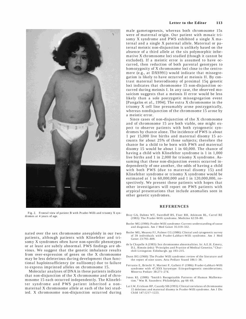
At 5 years, 5 months, her height was 100.3 cm (3rdcentile), weight was 22.7 kg (90th centile) and headcircumference was 51 cm (50th centile). Additionalfindings noted were enamel hypoplasia, sticky saliva,lumbar lordosis, marked truncal obesity, small handsand feet, hypoplastic labia minora, a small clitoris,joint laxity, an increased carrying angle, and skin pick-ing (see Fig. 2). Currently, she is 12 years old with aheight of 142.2 cm (60th centile) and her weight is 70.4kg (>95th centile). She is in the sixth grade and is ap-proximately one year behind academically. Readingskills are her strengths while math is a weakness.
To confirm the diagnosis of PWS, PCR studies wereundertaken using 15q11q13 microsatellite primers aspreviously described for patient A. PCR analysis usingX chromosome markers from both Xq and Xp (DXS987,DXS989, DXS991, DXS992, DXS6789, DXS1001,DXS1105, DXS998, DXS984, DXS6790, DXS988, andAR) was undertaken, and the patient exhibited a singlematernal and a single paternal allele at each of the Xchromosome loci tested, and no third allele was ob-served. The sensitivity of detection for a minor allelepresent in a mosaic cell line is approximately 2% of thepredominant alleles [Pangalos et al., 1994]; thus athird allele could have been detected in our patientwith trisomy X observed in greater than 50% of cellsexamined. PCR analysis with several 15q11q13 mark-ers (D15S128, MN-1, GABRB3, D15S541, D15S165,GABRA5, and D15S122) showed maternal heterodi-somy of chromosome 15 in the patient. Loci studiedfrom other chromosomes were not compatible with non-paternity.
The findings in each patient were consistent withPWS [Butler et al., 1986; Butler, 1990], although thegirl also had epicanthal folds, a flattened nasal bridge,staring spells, and an increased carrying angle, whichcan be seen in patients with trisomy X syndrome. Theboy with Klinefelter syndrome and PWS had findingsconsistent with PWS and few, if any, additional anoma-lies (e.g., tall stature) of Klinefelter syndrome [Jones,1988]. The major manifestations of PWS predominatedin each patient. However, behavior and emotionalproblems, which can occur in both Klinefelter and tri-somy X syndromes, can also overlap in PWS [de laChapelle, 1983]. It is unclear why the clinical presen-tation of an imprinted disorder (i.e., PWS) predomi-
Fig. 1. Frontal view of patient A with Prader-Willi and Klinefelter syn-dromes at 7 years of age.
112 Butler et al.
nated over the sex chromosome aneuploidy in our twopatients, although patients with Klinefelter and tri-somy X syndromes often have non-specific phenotypesor at least are subtly abnormal. PWS findings are ob-vious. We suggest that the genetic imbalance resultsfrom over-expression of genes on the X chromosomemay be less deleterious during development than func-tional haploinsufficiency (or nullisomy) due to failureto express imprinted alleles on chromosome 15.
Molecular analyses of DNA in these patients indicatethat non-disjunction of the X chromosome and of chro-mosome 15 each occurred independently. The Klinefel-ter syndrome and PWS patient inherited a non-maternal X chromosome allele at each of the loci stud-ied. X chromosome non-disjunction occurred during
male gametogenesis, whereas both chromosome 15swere of maternal origin. Our patient with mosaic tri-somy X syndrome and PWS exhibited a single X ma-ternal and a single X paternal allele. Maternal or pa-ternal meiotic non-disjunction is unlikely based on theabsence of a third allele at the six polymorphic infor-mative X chromosome loci studied (though it cannot beexcluded). If a meiotic error is assumed to have oc-curred, then reduction of both parental genotypes tohomozygosity of X chromosome loci close to the centro-mere (e.g., at DXS991) would indicate that missegre-gation is likely to have occurred at meiosis II. By con-trast maternal heterodisomy of proximal 15q geneticloci indicates that chromosome 15 non-disjunction oc-curred during meiosis I. In any case, the observed mo-saicism suggests that a meiosis II error would be lesslikely than a sole postzygotic missegregation event[Pangalos et al., 1994]. The extra X chromosome in thetrisomy X cell line presumably arose postzygotically,whereas nondisjunction of the chromosome 15 arose bya meiotic error.
Since cases of non-disjunction of the X chromosomeand of chromosome 15 are both viable, one might ex-pect to observe patients with both cytogenetic syn-dromes by chance alone. The incidence of PWS is about1 per 15,000 live births and maternal disomy 15 ac-counts for about 25% of those subjects; therefore thechance for a child to be born with PWS and maternaldisomy 15 would be about 1 in 60,000. The chance ofhaving a child with Klinefelter syndrome is 1 in 1,000live births and 1 in 2,000 for trisomy X syndrome. As-suming that these non-disjunction events occurred in-dependently of one another, the odds of having a childwith both PWS (due to maternal disomy 15) andKlinefelter syndrome or trisomy X syndrome would beestimated at 1 in 60,000,000 and 1 in 120,000,000, re-spectively. We present these patients with hopes thatother investigators will report on PWS patients withatypical presentations that include anomalies seen inother genetic syndromes.
REFERENCES
Bray GA, Dahms WT, Swerdloff RS, Fiser RH, Atkinson RL, Carrel RE(1983): The Prader-Willi syndrome. Medicine 62:59–80.
Butler MG (1990): Prader-Willi syndrome: Current understanding of causeand diagnosis. Am J Med Genet 35:319–332.
Butler MG, Meaney FJ, Palmer CG (1986): Clinical and cytogenetic surveyof 39 individuals with Prader-Labhart-Willi syndrome. Am J MedGenet 23:793–809.
de la Chapelle A (1983): Sex chromosome abnormalities. In: A.E.H. Emery,D.L. Rimoin (eds): ‘‘Principles and Practice of Medical Genetics.’’ Chur-chill Livingston: Edinburgh. pp 193–215.
Dunn HG (1968): The Prader Willi syndrome: review of the literature andthe report of nine cases. Acta Pediatr Scand 186:1–38.
Ferrante E, Brinchi V, Marioni P, Galletti F (1986): Prader-Labhart-Willisyndrome with 47,XXX karyotype: Etio-pathogenetic considerations.Minerva Pediatr 38:271–274.
Jones KL (1988): ‘‘Smith’s Recognizable Patterns of Human Malforma-tion.’’ Wm B. Saunders: Philadelphia, pp 66–69.
Lai LW, Erickson RP, Cassidy SB (1993): Clinical correlates of chromosome15 deletions and maternal disomy in Prader-Willi syndrome. Am J DisChild 147:1217–1223.
Fig. 2. Frontal view of patient B with Prader-Willi and trisomy X syn-dromes at 4 years of age.
Letter to the Editor 113
Malcolm S, Donlon TA (1994): Report of the second international workshopon human chromosome 15 mapping. Cytogenet Cell Genet 67:2–14.
Mascari MJ, Gottlieb W, Rogan PK, Butler MG, Waller DA, Armour JAL,Jeffreys A, Ladda RL, Nicholls RD (1992): The frequency of uni-parental disomy in Prader-Willi syndrome. N Engl J Med 326:1599–1607.
Pangalos C, Avramopoulos D, Blouin JL, Raoul O, deBlois MC, Prieur M,Schinzel AA, Gika M, Abazis D, Antonarakis SE (1994): Understandingthe mechanism(s) of mosaic trisomy 21 by using DNA polymorphismanalysis. Am J Hum Gen 54:473–481.
Trent RJ, Volparo F, Smith A, Lindeman R, Wong MK, Warne G, Haan E(1991): Molecular and cytogenetic studies of the Prader-Willi syn-drome. J Med Genet 28:649–654.
Tu JB, Hartridge C, Izawa J (1992): Psychopharmacogenetic aspects ofPrader-Willi syndrome. J Am Acad Child Adol Psychol 31:1137–1140.
Merlin G. Butler*Lora K. HedgesDepartments of Pediatrics, Pathology
and OrthopedicsVanderbilt UniversityNashville, Tennessee
Peter K. RoganJames R. SeipDepartment of PediatricsPennsylvania State UniversityCollege of MedicineHershey, Pennsylvania
Suzanne B. CassidyDepartment of GeneticsCase Western Reserve University and
Center for Human GeneticsUniversity Hospitals of ClevelandCleveland, Ohio
John B. MoeschlerCenter for Genetics and Child DevelopmentDartmouth-Hitchcock Medical CenterLebanon, New Hampshire
114 Butler et al.


![CASE REPORT Open Access Hypomelanosis of Ito with a trisomy 2 mosaicism… · 2017. 8. 27. · exclude the effects of uniparental disomy (UPD) [13]. It is known that cases of HMI](https://img.pdfslide.us/doc/110x75/6113a02ab842ff0515306bcd/case-report-open-access-hypomelanosis-of-ito-with-a-trisomy-2-mosaicism-2017-8.jpg)
![PHYLOGENETIC ANALYSIS OF AVIAN PARENTAL CARE€¦ · types of parental care in bony fishes (i.e. among states of uniparental [male], uniparental [fe- male], biparental, and no parental](https://img.pdfslide.us/doc/110x75/5f0ab0957e708231d42cdb6f/phylogenetic-analysis-of-avian-parental-care-types-of-parental-care-in-bony-fishes.jpg)