Embed Size (px)
Citation preview
JOURNAL OF VIROLOGY,0022-538X/99/$04.0010
May 1999, p. 3893–3903 Vol. 73, No. 5
Copyright © 1999, American Society for Microbiology. All Rights Reserved.
Ikk Mediates NF-kB Activation in Human ImmunodeficiencyVirus-Infected Cells
SUSANA ASIN,1 JULIE A. TAYLOR,1 SERGEY TRUSHIN,1 GARY BREN,1
AND CARLOS V. PAYA1,2*
Department of Immunology1 and Division of Infectious Diseases,2
Mayo Clinic, Rochester, Minnesota 55905
Received 16 September 1998/Accepted 27 January 1999
Human monocytes and macrophages are persistent reservoirs of human immunodeficiency virus (HIV)type-1. Persistent HIV infection of these cells results in increased levels of NF-kB in the nucleus secondary toincreased IkBa, IkBb, and IkB« degradation, a mechanism postulated to regulate viral persistence. Tocharacterize the molecular mechanisms regulating HIV-mediated degradation of IkB, we have sought toidentify the regulatory domains of IkBa targeted by HIV infection. Using monocytic cells stably expressingdifferent transdominant molecules of IkBa, we determined that persistent HIV infection of these cells targetsthe NH2 but not the COOH terminus of IkBa. Further analysis demonstrated that phosphorylation at S32 andS36 is necessary for HIV-dependent IkBa degradation and NF-kB activation. Of the putative N-terminal IkBakinases, we demonstrated that the Ikk complex, but not p90rsk, is activated by HIV infection and mediatesHIV-dependent NF-kB activation. Analysis of viral replication in cells that constitutively express IkBanegative transdominant molecules demonstrated a lack of correlation between virus-induced NF-kB (p65/p50)nuclear translocation and degree of viral persistence in human monocytes.
The Rel family of transcription factors plays an importantrole in the transactivation of several viral genes, includingthose of human immunodeficiency virus (HIV) type 1 (HIV-1)(25, 38). HIV-1 replication is regulated, in part, at the tran-scriptional level through the interplay of viral regulatory pro-teins with cellular transcription factors interacting with theviral long terminal repeat (LTR) (39). Since the identificationand functional characterization of NF-kB cis-acting sequenceswithin the HIV LTR (38), multiple studies have addressed theessential or dispensable role that this transcription factor playsin the reactivation of HIV from a true latent state and in thecontrol of viral persistence (1, 10, 27, 31, 54, 55). Unfortu-nately, these studies have yielded conflicting results as to therole of NF-kB in these two steps of the viral life cycle ininfected host cells. Differences in the type of host cells studied,HIV strain or genetic constructs used, and methodologicalapproaches may explain these conflicting results.
Understanding the potential impact of NF-kB on the regu-lation of HIV latency has again become a priority, as recentstudies suggest that NF-kB controls the reactivation of latentHIV in T cells from HIV-infected patients undergoing highlyactive antiretroviral therapy (19). An additional reservoir ofHIV, separate from that of T cells harboring latent HIV, arecells of the monocyte lineage in which persistent viral replica-tion is observed (36). During all stages of HIV infection, tissuemacrophages provide a unique viral reservoir. In these cells,HIV persistently replicates in the absence of cytopathicity,escapes immune surveillance, and spreads via cell-to-cell con-tact (reviewed in reference 36). The important role of macro-phages in AIDS pathogenesis has prompted the investigationof the molecular mechanisms which regulate HIV-1 persis-tence in these immune cells; one of these mechanisms isthought to be NF-kB dependent. Human macrophages express
a constitutive level of NF-kB in the nuclei in the absence ofexogenous cellular activation (25). This constitutive pool ofnuclear NF-kB may be sufficient to allow for the initiation ofHIV transcription immediately following infection. In addi-tion, NF-kB may be required to further sustain persistent HIVreplication, as multiple studies have demonstrated that persis-tent HIV replication in human macrophages or monocytesfurther upregulates NF-kB activity (2, 34, 40, 43, 48). However,the mechanisms by which HIV infection induces the activationof NF-kB in cells of the monocyte lineage remains unknown.Their identification would greatly enhance the understandingof this process and allow future testing of whether inhibition ofthe virus-induced activation of NF-kB may decrease viral per-sistence in cells of the monocyte lineage, hence eliminating animportant reservoir of HIV replication in infected patients.
NF-kB is a heterodimeric protein composed of differentcombinations of members of the Rel family of transcriptionfactors. A well-characterized form of NF-kB is a heterodimerof p50 and Rel-A (p65) (reviewed in references 3 and 4). In themajority of cells studied, NF-kB is anchored in the cytosol byan inhibitory protein, IkB. An extensively studied IkB mole-cule, IkBa, has previously been shown to physically interactwith NF-kB and to mask the nuclear localization signal of p50and Rel-A (6). Following cell activation by one of an array ofextracellular stimuli, IkBa undergoes a hyperphosphorylationevent that renders the inhibitory molecule susceptible to deg-radation (7, 13, 47). This process results in the release ofNF-kB, which undergoes nuclear translocation and drives genetranscription. Significant advances in the understanding of themolecular mechanisms and the structure-function of the phos-phorylation and degradation of IkBa have recently been made.IkBa is constitutively phosphorylated at its COOH terminus byprotein kinase-casein kinase II (PK-CK2) (5, 33, 35, 45). Whilethe exact function of this phosphorylation is poorly under-stood, it appears that phosphorylation at the COOH terminusmay play a role in the constitutively rapid protein turnover ofIkBa in resting cells, thus potentially favoring a low degree ofcontinuous NF-kB translocation. On the contrary, the N ter-
* Corresponding author. Mailing address: Mayo Clinic, 200 First St.SW, Guggenheim 501, Rochester, MN 55905. Phone: (507) 284-3747.Fax: (507) 284-3757. E-mail: [email protected].
3893
on August 25, 2018 by guest
http://jvi.asm.org/
Dow
nloaded from
minus contains two series (S32 and S36) which are required forstimulus-dependent phosphorylation (8, 9, 11, 15, 46, 49, 50,52) by specific kinases, such as the ones present in the Ikkcomplex (Ikka and Ikkb) or p90rsk (12, 16, 20, 24, 37, 42, 44,53, 57). Phosphorylation at these sites primes IkBa to undergoubiquitination and subsequent degradation by the proteosome.
Our group has previously determined that a mechanism where-by HIV infection results in an increase in the nuclear translo-cation of NF-kB involves modification and enhancement ofIkBa turnover (34). The half-life of IkBa in HIV-infected cellsis reduced by at least 50% compared to that in uninfected cells,and this fact directly correlates with increased levels of thenuclear pool of NF-kB in HIV-infected cells. That IkBa is thetarget of persistent HIV infection in monocytic cells has beenfurther confirmed by other groups (14, 27); one of those groupsfurther demonstrated that inhibition of IkBa degradation withproteosome inhibitors decreases HIV-induced NF-kB activa-tion (27). What remain to be elucidated are the molecular mech-anisms whereby HIV infection targets IkBa. Potential mecha-nisms regulated by HIV infection could target the COOHterminus of IkBa, favoring an enhanced “basal” turnover ofthis inhibitor molecule by activating PK-CK2 or the proteolyticmachinery. Alternatively, HIV infection could result in theactivation of other IkBa kinases that target S32 and S36, thuscontinuously priming IkBa to be degraded via the proteosome.Lastly, HIV could target other regulatory sites of IkBa or evenother molecules, such as Rel-A, that could result in the disso-ciation of NF-kB from IkBa, thus rendering IkBa less stable.
To investigate these possibilities, we have used a cell modelof monocytic cells in which persistent HIV replication resultsin NF-kB activation and a variety of genetically modifiedtagged IkBa molecules can be constitutively overexpressed.Our results indicate that HIV infection targets the NH2 ter-minus of IkBa, specifically S32 and S36, causing the enhanceddegradation of IkBa and hence increased NF-kB nucleartranslocation. The Ikk complex kinase activity is selectivelyactivated and is shown to mediate increased NF-kB activationin HIV-infected cells. In addition, we demonstrate that HIV-mediated NF-kB activation is not necessary to maintain viralpersistence in monocytic cells.
MATERIALS AND METHODS
Reagents and antibodies. Tumor necrosis factor (TNF) was purchased fromGenzyme (Cambridge, Mass.) and stored in aliquots at 270°C. Cycloheximidewas purchased from Sigma (St. Louis, Mo.) and stored at 220°C. Calpaininhibitor I (N-acetyl-Leu-Leu-norleucinal or ALLN) was purchased from Boehr-inger Mannheim Biochemicals (Indianapolis, Ind.), solubilized in ethyl alcohol,and stored in aliquots at 220°C. Bay 11-7082 (41) was purchased from Biomol(Plymouth Meeting, Pa.), solubilized in ethyl alcohol, and stored at 220°C. G418was purchased from Calbiochem-Novabiochem Corporation (La Jolla, Calif.),solubilized in RPMI medium, and stored in aliquots at 220°C.
The expression of the Flag-tagged IkB constructs was monitored with ananti-Flag monoclonal antibody (Kodak, New Haven, Conn.). To control forequal loading of proteins in sodium dodecyl sulfate (SDS)-polyacrylamide gelelectrophoresis (PAGE) analysis, an anti–b-actin polyclonal antibody (Sigma)and an anti-p90rsk antibody (Santa Cruz Biotechnology, Santa Cruz, Calif.) wereused. Polyclonal anti-IkBa serum was generated with a glutathione S-transferase(GST)–MAD3 fusion protein (34). The viral envelope protein gp120 was de-tected with an anti-gp120 polyclonal antibody (Center for Biologics Evaluationand Research, Food and Drug Administration, Bethesda, Md.). The identity ofthe complexes binding DNA in the gel shift assays was determined with poly-clonal antibodies against the different members of the Rel family (Santa CruzBiotechnology). Antibodies against p90rsk, Ikka, Ikkb, Raf-1, and NF-kB werepurchased from Santa Cruz Biotechnology.
DNA constructs. pCMV2-FLAG-IkBa-wt consisted of the full-length “wild-type” sequence of human IkBa (26) cloned into the SmaI-HindIII sites ofpCMV2FLAG (Kodak) to generate N-terminally Flag-tagged IkBa-wt (Flag-IkBa-wt). Flag-IkBa-wt was used as a template for subsequent mutations anddeletions by PCR-based techniques. Flag-IkBa-DN consisted of an N-terminaldeletion lacking the first 37 amino acids. This construct was generated with thesense primer wt-FLAG (59CGGAATTCATGGACTACAAAGACGAT39) and
the antisense primer wt-B (59GGAATTCCTCATAACGTCAGACGCTG39).EcoRI sites were created upstream and downstream of the coding sequence.Flag-IkBa-DC consisted of a C-terminal deletion lacking the last 40 amino acidsand was generated with the sense primer wt-FLAG and the antisense primer DC(59GCGAATTCTCAAAGGTTTTCTAGTGTC39). This construct containedan EcoRI site downstream of the coding sequence. Flag-IkBa-2N consisted ofthe full-length sequence of IkBa-wt in which S32 and S36 were mutated to alanineresidues. To generate these mutations, a sense primer with the sequence 59GACGCAGGCCTGGACGCAATG39 and an antisense primer with the sequence59CATTGCGTCCAGGCCTGCGTC39 were used. Flag-IkBa-4C was created bymutation of S283, S288, S293, and T291 to alanine residues in the PEST sequence(35), cloning into the HindIII-EcoRI site of pCMV2FLAG, and then PCRamplifying with the sense primer wt-FLAG and the antisense primer wt-B.Flag-IkBa-wt, Flag-IkBa-DN, Flag-IkBa-DC, Flag-IkBa-2N, and Flag-IkBa-4Cwere then digested and cloned into the EcoRI site of SFFV-Neo under thetranscriptional regulation of the Friend spleen focus-forming virus (SFFV) 59LTR (22). All of the cloning was verified by DNA sequencing.
Plasmid kB-luc contains three tandem copies of the kB motif of the HIV LTRcloned upstream of the minimal conalbumin-luciferase (con-luc) promoter re-porter gene. Plasmid pBLCAT 2 is a mammalian reporter vector designed for theexpression of chloramphenicol acetyltransferase (CAT) in mammalian cells tran-scribed by the minimal thymidine kinase (TK) promoter (Promega, Madison,Wis.). Plasmids Ikka wt and kinase dead were kind gifts from Alain Israel,Institute Pasteur, Paris, France. Plasmids Ikkb wt and kinase dead were obtainedfrom M. Roth (Tularik, San Jose, Calif.). Ikka kinase dead was generated bymutation of aspartic acid 144 to asparagine. Ikkb kinase dead was created bymutation of lysine 44 to alanine. pcDNA3-Ikk expression vectors were generatedby cloning the cDNA of wild-type Ikka or Ikkb or its respective mutant into thecytomegalovirus expression vector pcDNA3 (Invitrogen).
Gene transfection and generation of cell lines. The U937 promonocytic cellline was purchased from the American Type Culture Collection and grown inRPMI 1640 supplemented with 5% heat-inactivated fetal bovine serum (Inter-gen), 1% glutamine, and 1% penicillin-streptomycin. To generate IkBa-express-ing cell lines, 107 freshly thawed and exponentially growing U937 cells wereresuspended in RPMI 1640 and electroporated with 20 mg of previously linear-ized DNA by use of a BTX cell electroporator at 250 V for 10 ms. U937 cellselectroporated without DNA were used as controls. At 24 h after transfection,cells were resuspended in selection medium containing 5% fetal bovine serumand 700 mg of G418 per ml. After 3 to 4 weeks, upon the incipient growth ofneomycin-resistant bulk cultures, cells were cloned by limiting dilution (30).Stable integration and expression of the transfected genes within each monoclo-nal population were verified by serial passages of the cultures in the absence ofthe selective antibiotic and by immunoblotting with anti-Flag antibodies.
Separate clones expressing equal levels of Flag-IkBa constructs were selected,and their CD4 surface expression was verified by flow cytometry analysis. There-after, three clones expressing each of the Flag-IkBa constructs were pooled, andexponentially growing cells were mock or HIV infected. The level of expressionof each of the tagged IkBa constructs was confirmed before and during theperiod of HIV infection by immunoblotting of cytosolic extracts with anti-Flagantibodies.
Transient transfection of U937 cells was performed as follows. A total of 107
exponentially growing U937 cells were incubated with 4 mg of the con-luc orkB-con-luc reporter construct, 6 mg of the pDNA3-Ikk construct, 4 mg of thepBLCAT2 reporter, and 300 mg of DEAE-dextran (Pharmacia, Piscataway, N.J.)per ml for 90 min at room temperature. Dimethyl sulfoxide (10%) was thenadded for 3 min, followed by extensive washing and plating at 0.5 3 107/cells/ml.Two days later, cells were harvested. Luciferase levels were measured with thePromega luciferase assay system, and CAT activity was measured with a CATenzyme-linked immunosorbent assay kit (Boehringer).
HIV infection and measurement of HIV replication. U937 cells expressingSFFV, Flag-IkBa-wt, Flag-IkBa-DN, Flag-IkBa-DC, Flag-IkBa-2N, and Flag-IkBa-4C were infected with the HIV LAV-Bru strain as previously described (2,34, 40). Briefly, 107 exponentially growing U937 cells were sedimented by low-speed centrifugation and resuspended overnight in 10 ml of infective supernatantcontaining 360 ng of p24 per ml. Mock-infected cells were used as a control.After 24 h, cells were extensively washed and resuspended in culture medium.Cells were passaged twice a week at 0.25 3 106 cells/ml and used from day 30through day 90 postinfection. During this period, cell supernatants were col-lected, precleared by centrifugation at 1,500 rpm for 5 min at 4°C, and stored forfuture analysis of HIV p24 antigen content by an enzyme-linked immunosorbentassay (Coulter-Immunotech Immunology, Westbrook, Maine). At least eightconsecutive infections were used for each of these experiments. All the cell linesstudied maintained HIV persistence and 100% viability during the study period,except for the U937 clones expressing Flag-IkBa-DC, which maintained viabilityand normal growth while uninfected which underwent immediate and massivecytopathicity upon HIV infection in four consecutive attempts. Therefore, aU937 cell line expressing Flag-IkBa-DC could not support a persistent HIVinfection. In addition, in some experiments, HIV gp120 expression was deter-mined by immunoblotting with anti-gp120 antibodies.
Nuclear and cytosolic extracts, electrophoretic mobility shift assays, and im-munoblotting. Nuclear and cytosolic extracts were prepared by a modification ofthe method of Dignam et al. (17). A total of 107 cells were washed with ice-cold
3894 ASIN ET AL. J. VIROL.
on August 25, 2018 by guest
http://jvi.asm.org/
Dow
nloaded from
phosphate-buffered saline and then with buffer A (10 mM HEPES [pH 7.9], 1.5mM MgCl2, 10 mM KCl). Cells were then lysed for 10 min on ice in the samebuffer containing 0.1% Nonidet P-40, 0.5 mM dithiothreitol (DTT), 0.5 mMphenylmethylsulfonyl fluoride (PMSF), 2 mg of aprotinin per ml, 2 mg of leu-peptin per ml, and 2 mg of pepstatin per ml. After centrifugation, cells werewashed twice with buffer A. Nuclei were pelleted by centrifugation, lysed byresuspension in 25 ml of buffer C (20 mM HEPES, 25% glycerol, 0.42 M NaCl,1.5 mM MgCl2, 0.2 mM EDTA, DTT, PMSF, aprotinin, leupeptin, pepstatin)and rotated at 4°C for 30 min. After centrifugation, the supernatants were dilutedin 50 ml of buffer D (20 mM HEPES, 20% glycerol, 0.05 M KCl, 0.2 M EDTA,DTT, PMSF, aprotinin, leupeptin, pepstatin) and stored at 270°C.
For electrophoretic mobility shift assays, 6 mg of nuclear extract was incubatedwith a [g-32P]ATP-labeled double-stranded NF-kB oligonucleotide probe in 15ml of DNA binding buffer for 15 min at room temperature as previously de-scribed (2, 34, 40). Components of the HIV-induced DNA binding proteincomplexes were identified by incubation of the extract with specific polyclonalantibodies against p50 and Rel-A prior to addition of the labeled probe. Theresulting protein-DNA complexes were resolved on a 5% polyacrylamide gel andvisualized by autoradiography.
To characterize the level of expression of the Flag-IkBa constructs in unin-fected and infected cells, 40 mg of cytosolic protein was analyzed by SDS–10%PAGE. Proteins were transferred to Immobilon-P membranes (Millipore) bystandard procedures and blotted with an anti-Flag monoclonal antibody, fol-lowed by incubation with rabbit anti-mouse immunoglobulin G (Pierce) and thenhorseradish peroxidase (Amersham, Buckinghamshire, England). Immunoreac-tive proteins were detected with an ECL Western blotting detection kit (Amer-sham). b-Actin and p90rsk were used as internal controls for equal loading in allexperiments.
Preparation of recombinant IkBa. The IkBa-MAD3 cDNA (26) plasmid wasobtained from Cetus Corporation and was used as a template for subsequentPCR amplification.
The amino-terminal IkBa-MAD3 (positions 1 to 54) sequence was amplifiedwith wild-type primer A (59CGGGATCCATGTTCCAGGCGGCCGAG39) asthe sense primer, creating a BamHI site upstream of the coding sequence, andwild-type primer B (59GGAATTCCTCAGCGGATCTCCTGCAGCT39) as theantisense primer, creating an EcoRI site downstream of the coding sequence. AnS32/36A double mutant was amplified from the full-length cDNA by use of prim-ers to create alanines at S32 and S36. Following digestion with BamHI-EcoRI,these sequences were ligated into pGEX-KG (derived from pGEX-2T, fromPharmacia, Piscataway, N.J.). These constructs were transformed into Esche-richia coli DH5a cells, which were grown exponentially. After 60 min of stimu-lation with isopropylthiogalactopyranoside (Sigma), cells were lysed. Proteinswere isolated by affinity chromatography on glutathione-bonded 4% cross-linkedagarose (Sigma). The purity of GST-IkBa (positions 1 to 54) containing the first54 amino acids of IkBa and GST-IkBa (positions 1 to 54) containing S32/36A wasanalyzed by SDS–10% PAGE and subsequent Coomassie blue staining. Thepurity of both proteins was greater than 90%.
Immunoprecipitation of IkBa kinases and in vitro kinase assays. Whole-cellextracts were prepared for immunoprecipitation and in vitro kinase assays asfollows. Aliquots of 107 exponentially growing U937 cells were washed twice withcold phosphate-buffered saline, resuspended in lysis buffer containing 40 mMTris-HCl (pH 8), 0.3 M NaCl, 0.1% Nonidet P-40, 6 mM EDTA, 6 mM EGTA,10 mM NaF, 10 mM p-nitrophenyl phosphate (PNPP), 10 mM b-glycerolphos-phate, 300 mM sodium orthovanadate, 1 mM DDT, 2 mM PMSF, 10 mg ofaprotinin per ml, 1 mg of leupeptin per ml, and 1 mg of pepstatin per ml, andincubated on ice. Cells were then centrifuged at 12,000 3 g for 15 min at 4°C. Theresultant supernatant contained total cellular proteins, which were quantitatedwith a Bio-Rad protein assay.
For immunoprecipitation of the Ikk complex, p90rsk, or Raf-1, 100 mg of cellextract was incubated with anti-Ikka, anti-Ikkb, anti-p90rsk, or anti–Raf-1 anti-bodies for 1 h at 4°C, after which protein A-agarose beads (Life Technologies,Gaithersburg, Md.) were added for 1 h. The beads were then washed three timeswith 0.5 M NaCl-based lysis buffer, followed by one wash with a buffer containing50 mM Tris-HCl (pH 7.4) and 40 mM NaCl. The washed beads were thenincubated in 15 ml of kinase buffer (20 mM HEPES [pH 7.4], 2 mM MgCl, 2 mMMnCl, 10 mM ATP, 10 mM NaF, 10 mM PNPP, 10 mM b-glycerolphosphate, 300mM sodium orthovanadate, 2 mM PMSF, 10 mg of aprotinin per ml, 1 mg ofleupeptin per ml, 1 mg of pepstatin per ml, 1 mM DTT) with 2 mg of GST-IkBa(positions 1 to 54) or GST-IkBa (positions 1 to 54) containing S32/36A and 0.1mCi of [g-32P]ATP. The kinase reaction was performed for 30 min at 30°C, andsamples were resolved by SDS-PAGE, transferred to Immobilon-P membranes,and exposed to film.
RESULTS
Increased degradation of Flag-IkBa-wt in HIV-infected cells.To confirm the expression and functionality of the Flag-IkBaconstructs, pooled clones expressing equal levels of Flag-IkBconstructs were treated or not treated with TNF, followed bythe analysis of the cytosolic extracts by SDS-PAGE and immu-
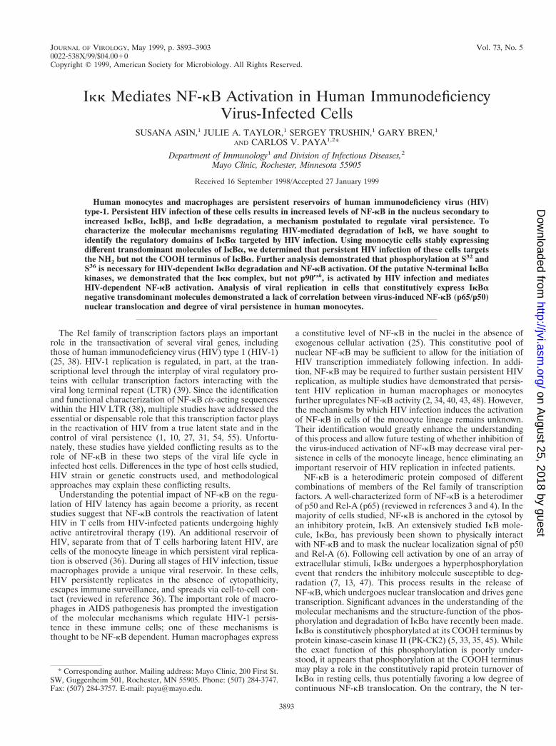
noblotting with anti-Flag antibodies. As shown in Fig. 1A, TNFstimulation led to the rapid hyperphosphorylation and subse-quent degradation of Flag-IkBa-wt. In contrast, Flag-IkBa-DNand Flag-IkBa-2N were refractory to TNF-induced hyperphos-phorylation and subsequent degradation. Flag-IkBa-4C be-haved similarly to Flag-IkBa-wt in that it was susceptible toTNF-mediated hyperphosphorylation and degradation. Theseresults confirm that the constitutively overexpressed Flag-IkBamolecules are regulated as previously described for nativeIkBa and highlight the functional relevance of the N terminuscontaining S32 and S36 in TNF-induced IkBa hyperphosphor-ylation and degradation.
As expected (34), persistent HIV infection of U937 cells re-sulted in decreased cytosolic levels of native IkBa. Moreover,IkBb and IkBε protein levels were also significantly decreasedin HIV-infected cells compared to uninfected cells (Fig. 1B).
Having determined that overexpressed Flag-IkBa constructsfunction similarly to native IkBa upon stimulation with knowninducers of NF-kB and that HIV infection of U937 cells resultsin decreased steady-state levels of endogenous IkB, we nextinvestigated whether Flag-IkBa-wt is also a target of HIV in-fection. Immunoblotting of cytosolic fractions from mock- andHIV-infected cells expressing Flag-IkBa-wt was performedwith anti-Flag antibodies. U937 cells transfected with the pa-rental empty retrovirus vector (SFFV) were also mock or HIVinfected and used as controls. As shown in Fig. 1C, the steady-state protein levels of Flag-IkBa-wt were decreased in thecytosolic fractions of HIV-infected cells compared to mock-infected cells, confirming that HIV infection decreases thecytosolic levels of Flag-IkBa and indicating that tagged IkBaconstructs can be used to study the regulatory domain(s) tar-geted by persistent HIV infection in monocytes.
Having previously demonstrated that the decreased level ofnative IkBa is a result of the enhanced rate of IkBa degrada-tion in persistently HIV-infected monocytes, we investigatedwhether this process also accounted for the decreased level ofFlag-IkBa-wt in infected cells. The half-life of Flag-IkBa-wtwas estimated by immunoblotting Flag-IkBa-wt from cytosolicfractions from mock- and HIV-infected cells treated for dif-ferent time periods with cycloheximide. As shown in Fig. 1D,the turnover of Flag-IkBa-wt was increased in HIV-infectedcells compared to mock-infected cells. The half-lives of Flag-IkBa-wt calculated from Fig. 1D were found to be approxi-mately 60 min in HIV-infected cells and 128 min in uninfectedcells (Fig. 1E).
The NH2 terminus but not the PEST sequence present in theCOOH terminus of IkBa is necessary for IkBa degradation byHIV infection. To characterize which of the regulatory do-mains of IkBa is targeted by HIV infection, we first focused onthe NH2-terminal domain of IkBa. We analyzed the turnoverand half-life of Flag-IkBa-DN. U937 cells stably transfectedwith the empty retrovirus vector (SFFV), Flag-IkBa-wt, orFlag-IkBa-DN were mock or HIV infected. The half-lives ofthese constructs were measured by analyzing the levels of theFlag-IkBa constructs in cytosolic extracts from cell culturestreated for different time periods with cycloheximide (as forFig. 1). As shown in Fig. 2A, Flag-IkBa-DN was very stable notonly in mock-infected but also in HIV-infected U937 cells, withthe resulting half-lives being estimated at greater than 4 h (Fig.2B). The enhanced stability of Flag-IkBa-DN in both mock-and HIV-infected cells contrasts with the more rapid turnoverof Flag-IkBa-wt in mock-infected cells and even more rapidturnover in HIV-infected cells (Fig. 1D and Fig. 2A). Theseresults indicate that the increased degradation of IkBa thatensues in HIV-infected cells appears to be dependent on theNH2-terminal domain of the molecule. In addition, these re-
VOL. 73, 1999 Ikk MEDIATES NF-kB ACTIVATION IN HIV-INFECTED CELLS 3895
on August 25, 2018 by guest
http://jvi.asm.org/
Dow
nloaded from
sults highlight the potential relevance of this IkBa domain inthe regulation of the basal turnover of IkBa in unstimulatedtransformed cells.
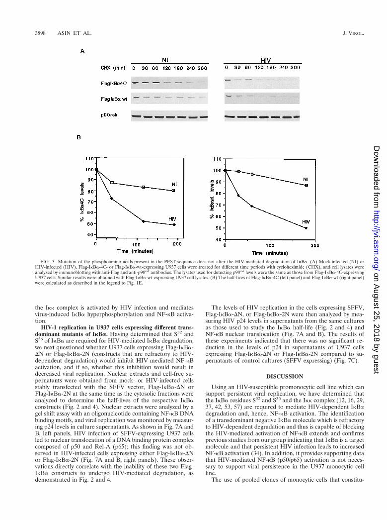
Previous studies have demonstrated that mutation of S283,S288, T291, and S293 to alanines eliminates the constitutivephosphorylation of IkBa mediated by PK-CK2 and may influ-ence the turnover of IkBa (5, 33, 35, 45). Based on this infor-mation, we analyzed the turnover and half-life of Flag-IkBa-4C in mock- or HIV-infected U937 cells and compared them tothose of Flag-IkBa-wt. In mock-infected U937 cells, the basal
turnover of Flag-IkBa-4C was slightly longer than that of Flag-IkBa-wt (Fig. 3), suggesting a potential role of the C-terminalamino acids S283, S288, T291, and S293 in the basal turnover ofIkBa in unstimulated monocytic cells. The half-life of Flag-IkBa-4C was shorter in HIV-infected cells than in mock-in-fected cells but was similar to the half-life of Flag-IkBa-wt inHIV-infected cells (Fig. 3). These results demonstrate that theamino acids present in the PEST sequence of IkBa are notinvolved in the HIV-mediated degradation and turnover ofIkBa.
FIG. 1. Functional characterization of Flag-IkBa molecules in U937 cells. (A) Pooled clones of U937 cells expressing the different Flag-IkBa constructs werestimulated with TNF for different time periods, and the cell lysates were analyzed by immunoblotting with anti-Flag antibodies. The hyperphosphorylated form of IkBais indicated by a small circle. (B) Immunoblotting of cell lysates from mock-infected (NI) or HIV-infected (HIV), SFFV-expressing U937 cells with anti-IkBa,anti-IkBb, anti-IkBε, and antiactin antibodies. The hyperphosphorylated form of IkBε is indicated by a small circle. (C) Immunoblotting of cell lysates frommock-infected (NI) or HIV-infected (HIV), SFFV- or Flag-IkBa-wt-expressing U937 cells with anti-Flag and antiactin antibodies. (D) The half-life of Flag-IkBa-wtwas estimated by immunoblotting of cell lysates from mock-infected (NI) or HIV-infected (HIV), Flag-IkBa-wt-expressing U937 cells treated with cycloheximide(CHX) for different periods of time with anti-Flag antibodies. Equal protein loading was calculated by immunoblotting the same membrane with anti-p90rsk antibody.(E) The half-life of Flag-IkBa-wt was calculated by measuring with a densitometer the disintegrations per minute of Flag-IkBa-wt and normalizing them to those forp90rsk from each experimental time point shown in panel D.
3896 ASIN ET AL. J. VIROL.
on August 25, 2018 by guest
http://jvi.asm.org/
Dow
nloaded from
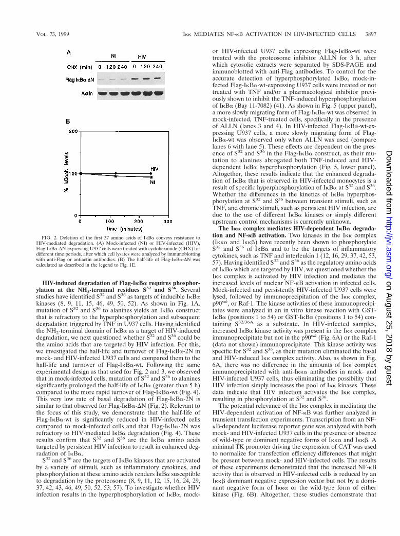
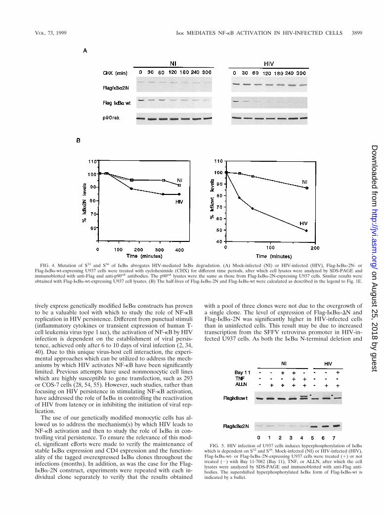
HIV-induced degradation of Flag-IkBa requires phosphor-ylation at the NH2-terminal residues S32 and S36. Severalstudies have identified S32 and S36 as targets of inducible IkBakinases (8, 9, 11, 15, 46, 49, 50, 52). As shown in Fig. 1A,mutation of S32 and S36 to alanines yields an IkBa constructthat is refractory to the hyperphosphorylation and subsequentdegradation triggered by TNF in U937 cells. Having identifiedthe NH2-terminal domain of IkBa as a target of HIV-induceddegradation, we next questioned whether S32 and S36 could bethe amino acids that are targeted by HIV infection. For this,we investigated the half-life and turnover of Flag-IkBa-2N inmock- and HIV-infected U937 cells and compared them to thehalf-life and turnover of Flag-IkBa-wt. Following the sameexperimental design as that used for Fig. 2 and 3, we observedthat in mock-infected cells, mutation of S32 and S36 to alaninessignificantly prolonged the half-life of IkBa (greater than 5 h)compared to the more rapid turnover of Flag-IkBa-wt (Fig. 4).This very low rate of basal degradation of Flag-IkBa-2N issimilar to that observed for Flag-IkBa-DN (Fig. 2). Relevant tothe focus of this study, we demonstrate that the half-life ofFlag-IkBa-wt is significantly reduced in HIV-infected cellscompared to mock-infected cells and that Flag-IkBa-2N wasrefractory to HIV-mediated IkBa degradation (Fig. 4). Theseresults confirm that S32 and S36 are the IkBa amino acidstargeted by persistent HIV infection to result in enhanced deg-radation of IkBa.
S32 and S36 are the targets of IkBa kinases that are activatedby a variety of stimuli, such as inflammatory cytokines, andphosphorylation at these amino acids renders IkBa susceptibleto degradation by the proteosome (8, 9, 11, 12, 15, 16, 24, 29,37, 42, 43, 46, 49, 50, 52, 53, 57). To investigate whether HIVinfection results in the hyperphosphorylation of IkBa, mock-
or HIV-infected U937 cells expressing Flag-IkBa-wt weretreated with the proteosome inhibitor ALLN for 3 h, afterwhich cytosolic extracts were separated by SDS-PAGE andimmunoblotted with anti-Flag antibodies. To control for theaccurate detection of hyperphosphorylated IkBa, mock-in-fected Flag-IkBa-wt-expressing U937 cells were treated or nottreated with TNF and/or a pharmacological inhibitor previ-ously shown to inhibit the TNF-induced hyperphosphorylationof IkBa (Bay 11-7082) (41). As shown in Fig. 5 (upper panel),a more slowly migrating form of Flag-IkBa-wt was observed inmock-infected, TNF-treated cells, specifically in the presenceof ALLN (lanes 3 and 4). In HIV-infected Flag-IkBa-wt-ex-pressing U937 cells, a more slowly migrating form of Flag-IkBa-wt was observed only when ALLN was used (comparelanes 6 with lane 5). These effects are dependent on the pres-ence of S32 and S36 in the Flag-IkBa construct, as their mu-tation to alanines abrogated both TNF-induced and HIV-dependent IkBa hyperphosphorylation (Fig. 5, lower panel).Altogether, these results indicate that the enhanced degrada-tion of IkBa that is observed in HIV-infected monocytes is aresult of specific hyperphosphorylation of IkBa at S32 and S36.Whether the differences in the kinetics of IkBa hyperphos-phorylation at S32 and S36 between transient stimuli, such asTNF, and chronic stimuli, such as persistent HIV infection, aredue to the use of different IkBa kinases or simply differentupstream control mechanisms is currently unknown.
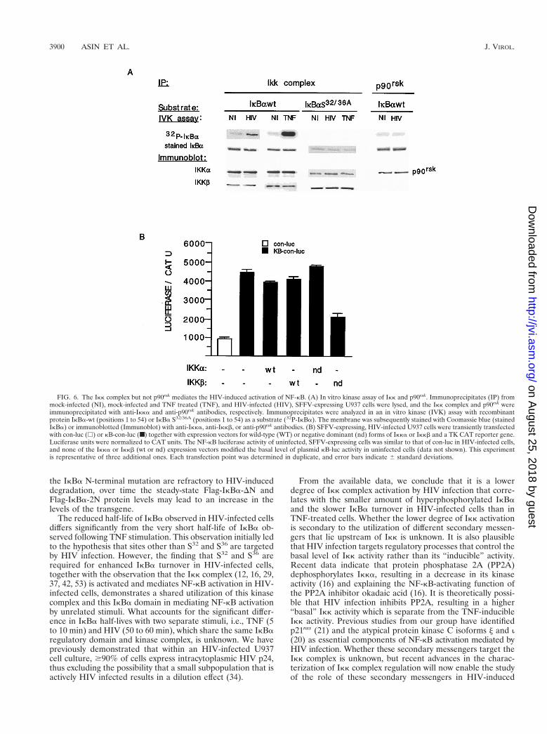
The Ikk complex mediates HIV-dependent IkBa degrada-tion and NF-kB activation. Two kinases in the Ikk complex(Ikka and Ikkb) have recently been shown to phosphorylateS32 and S36 of IkBa and to be the targets of inflammatorycytokines, such as TNF and interleukin 1 (12, 16, 29, 37, 42, 53,57). Having identified S32 and S36 as the regulatory amino acidsof IkBa which are targeted by HIV, we questioned whether theIkk complex is activated by HIV infection and mediates theincreased levels of nuclear NF-kB activation in infected cells.Mock-infected and persistently HIV-infected U937 cells werelysed, followed by immunoprecipitation of the Ikk complex,p90rsk, or Raf-1. The kinase activities of these immunoprecipi-tates were analyzed in an in vitro kinase reaction with GST-IkBa (positions 1 to 54) or GST-IkBa (positions 1 to 54) con-taining S32/36A as a substrate. In HIV-infected samples,increased IkBa kinase activity was present in the Ikk compleximmunoprecipitate but not in the p90rsk (Fig. 6A) or the Raf-1(data not shown) immunoprecipitate. This kinase activity wasspecific for S32 and S36, as their mutation eliminated the basaland HIV-induced Ikk complex activity. Also, as shown in Fig.6A, there was no difference in the amounts of Ikk compleximmunoprecipitated with anti-Ikka antibodies in mock- andHIV-infected U937 cells, thus eliminating the possibility thatHIV infection simply increases the pool of Ikk kinases. Thesedata indicate that HIV infection activates the Ikk complex,resulting in phosphorylation at S32 and S36.
The potential relevance of the Ikk complex in mediating theHIV-dependent activation of NF-kB was further analyzed intransient transfection experiments. Transcription from an NF-kB-dependent luciferase reporter gene was analyzed with bothmock- and HIV-infected U937 cells in the presence or absenceof wild-type or dominant negative forms of Ikka and Ikkb. Aminimal TK promoter driving the expression of CAT was usedto normalize for transfection efficiency differences that mightbe present between mock- and HIV-infected cells. The resultsof these experiments demonstrated that the increased NF-kBactivity that is observed in HIV-infected cells is reduced by anIkkb dominant negative expression vector but not by a domi-nant negative form of Ikka or the wild-type form of eitherkinase (Fig. 6B). Altogether, these studies demonstrate that
FIG. 2. Deletion of the first 37 amino acids of IkBa conveys resistance toHIV-mediated degradation. (A) Mock-infected (NI) or HIV-infected (HIV),Flag-IkBa-DN-expressing U937 cells were treated with cycloheximide (CHX) fordifferent time periods, after which cell lysates were analyzed by immunoblottingwith anti-Flag or antiactin antibodies. (B) The half-life of Flag-IkBa-DN wascalculated as described in the legend to Fig. 1E.
VOL. 73, 1999 Ikk MEDIATES NF-kB ACTIVATION IN HIV-INFECTED CELLS 3897
on August 25, 2018 by guest
http://jvi.asm.org/
Dow
nloaded from
the Ikk complex is activated by HIV infection and mediatesvirus-induced IkBa hyperphosphorylation and NF-kB activa-tion.
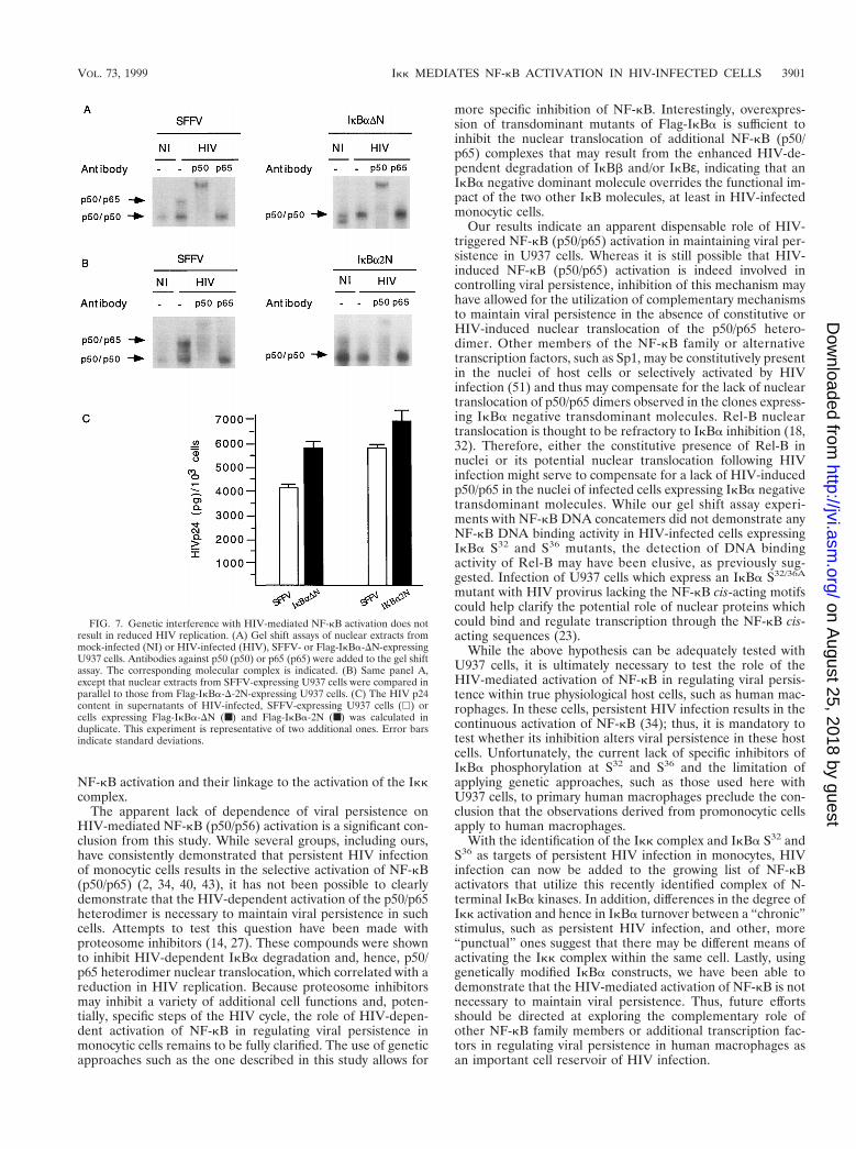
HIV-1 replication in U937 cells expressing different trans-dominant mutants of IkBa. Having determined that S32 andS36 of IkBa are required for HIV-mediated IkBa degradation,we next questioned whether U937 cells expressing Flag-IkBa-DN or Flag-IkBa-2N (constructs that are refractory to HIV-dependent degradation) would inhibit HIV-mediated NF-kBactivation, and if so, whether this inhibition would result indecreased viral replication. Nuclear extracts and cell-free su-pernatants were obtained from mock- or HIV-infected cellsstably transfected with the SFFV vector, Flag-IkBa-DN orFlag-IkBa-2N at the same time as the cytosolic fractions wereanalyzed to determine the half-lives of the respective IkBaconstructs (Fig. 2 and 4). Nuclear extracts were analyzed by agel shift assay with an oligonucleotide containing NF-kB DNAbinding motifs, and viral replication was monitored by measur-ing p24 levels in culture supernatants. As shown in Fig. 7A andB, left panels, HIV infection of SFFV-expressing U937 cellsled to nuclear translocation of a DNA binding protein complexcomposed of p50 and Rel-A (p65); this finding was not ob-served in HIV-infected cells expressing either Flag-IkBa-DNor Flag-IkBa-2N (Fig. 7A and B, right panels). These obser-vations directly correlate with the inability of these two Flag-IkBa constructs to undergo HIV-mediated degradation, asdemonstrated in Fig. 2 and 4.
The levels of HIV replication in the cells expressing SFFV,Flag-IkBa-DN, or Flag-IkBa-2N were then analyzed by mea-suring HIV p24 levels in supernatants from the same culturesas those used to study the IkBa half-life (Fig. 2 and 4) andNF-kB nuclear translocation (Fig. 7A and B). The results ofthese experiments indicated that there was no significant re-duction in the levels of p24 in supernatants of U937 cellsexpressing Flag-IkBa-DN or Flag-IkBa-2N compared to su-pernatants of control cultures (SFFV expressing) (Fig. 7C).
DISCUSSION
Using an HIV-susceptible promonocytic cell line which cansupport persistent viral replication, we have determined thatthe IkBa residues S32 and S36 and the Ikk complex (12, 16, 29,37, 42, 53, 57) are required to mediate HIV-dependent IkBadegradation and, hence, NF-kB activation. The identificationof a transdominant negative IkBa molecule which is refractoryto HIV-dependent degradation and thus is capable of blockingthe HIV-mediated activation of NF-kB extends and confirmsprevious studies from our group indicating that IkBa is a targetmolecule and that persistent HIV infection leads to increasedNF-kB activation (34). In addition, it provides supporting datathat HIV-mediated NF-kB (p50/p65) activation is not neces-sary to support viral persistence in the U937 monocytic cellline.
The use of pooled clones of monocytic cells that constitu-
FIG. 3. Mutation of the phosphoamino acids present in the PEST sequence does not alter the HIV-mediated degradation of IkBa. (A) Mock-infected (NI) orHIV-infected (HIV), Flag-IkBa-4C- or Flag-IkBa-wt-expressing U937 cells were treated for different time periods with cycloheximide (CHX), and cell lysates wereanalyzed by immunoblotting with anti-Flag and anti-p90rsk antibodies. The lysates used for detecting p90rsk levels were the same as those from Flag-IkBa-4C-expressingU937 cells. Similar results were obtained with Flag-IkBa-wt-expressing U937 cell lysates. (B) The half-lives of Flag-IkBa-4C (left panel) and Flag-IkBa-wt (right panel)were calculated as described in the legend to Fig. 1E.
3898 ASIN ET AL. J. VIROL.
on August 25, 2018 by guest
http://jvi.asm.org/
Dow
nloaded from
tively express genetically modified IkBa constructs has provento be a valuable tool with which to study the role of NF-kBreplication in HIV persistence. Different from punctual stimuli(inflammatory cytokines or transient expression of human T-cell leukemia virus type 1 tax), the activation of NF-kB by HIVinfection is dependent on the establishment of viral persis-tence, achieved only after 6 to 10 days of viral infection (2, 34,40). Due to this unique virus-host cell interaction, the experi-mental approaches which can be utilized to address the mech-anisms by which HIV activates NF-kB have been significantlylimited. Previous attempts have used nonmonocytic cell lineswhich are highly susceptible to gene transfection, such as 293or COS-7 cells (28, 54, 55). However, such studies, rather thanfocusing on HIV persistence in stimulating NF-kB activation,have addressed the role of IkBa in controlling the reactivationof HIV from latency or in inhibiting the initiation of viral rep-lication.
The use of our genetically modified monocytic cells has al-lowed us to address the mechanism(s) by which HIV leads toNF-kB activation and then to study the role of IkBa in con-trolling viral persistence. To ensure the relevance of this mod-el, significant efforts were made to verify the maintenance ofstable IkBa expression and CD4 expression and the function-ality of the tagged overexpressed IkBa clones throughout theinfections (months). In addition, as was the case for the Flag-IkBa-2N construct, experiments were repeated with each in-dividual clone separately to verify that the results obtained
with a pool of three clones were not due to the overgrowth ofa single clone. The level of expression of Flag-IkBa-DN andFlag-IkBa-2N was significantly higher in HIV-infected cellsthan in uninfected cells. This result may be due to increasedtranscription from the SFFV retrovirus promoter in HIV-in-fected U937 cells. As both the IkBa N-terminal deletion and
FIG. 4. Mutation of S32 and S36 of IkBa abrogates HIV-mediated IkBa degradation. (A) Mock-infected (NI) or HIV-infected (HIV), Flag-IkBa-2N- orFlag-IkBa-wt-expressing U937 cells were treated with cycloheximide (CHX) for different time periods, after which cell lysates were analyzed by SDS-PAGE andimmunoblotted with anti-Flag and anti-p90rsk antibodies. The p90rsk lysates were the same as those from Flag-IkBa-2N-expressing U937 cells. Similar results wereobtained with Flag-IkBa-wt-expressing U937 cell lysates. (B) The half-lives of Flag-IkBa-2N and Flag-IkBa-wt were calculated as described in the legend to Fig. 1E.
FIG. 5. HIV infection of U937 cells induces hyperphosphorylation of IkBawhich is dependent on S32 and S36. Mock-infected (NI) or HIV-infected (HIV),Flag-IkBa-wt- or Flag-IkBa-2N-expressing U937 cells were treated (1) or nottreated (2) with Bay 11-7082 (Bay 11), TNF, or ALLN, after which the celllysates were analyzed by SDS-PAGE and immunoblotted with anti-Flag anti-bodies. The supershifted hyperphosphorylated IkBa form of Flag-IkBa-wt isindicated by a bullet.
VOL. 73, 1999 Ikk MEDIATES NF-kB ACTIVATION IN HIV-INFECTED CELLS 3899
on August 25, 2018 by guest
http://jvi.asm.org/
Dow
nloaded from
the IkBa N-terminal mutation are refractory to HIV-induceddegradation, over time the steady-state Flag-IkBa-DN andFlag-IkBa-2N protein levels may lead to an increase in thelevels of the transgene.
The reduced half-life of IkBa observed in HIV-infected cellsdiffers significantly from the very short half-life of IkBa ob-served following TNF stimulation. This observation initially ledto the hypothesis that sites other than S32 and S36 are targetedby HIV infection. However, the finding that S32 and S36 arerequired for enhanced IkBa turnover in HIV-infected cells,together with the observation that the Ikk complex (12, 16, 29,37, 42, 53) is activated and mediates NF-kB activation in HIV-infected cells, demonstrates a shared utilization of this kinasecomplex and this IkBa domain in mediating NF-kB activationby unrelated stimuli. What accounts for the significant differ-ence in IkBa half-lives with two separate stimuli, i.e., TNF (5to 10 min) and HIV (50 to 60 min), which share the same IkBaregulatory domain and kinase complex, is unknown. We havepreviously demonstrated that within an HIV-infected U937cell culture, $90% of cells express intracytoplasmic HIV p24,thus excluding the possibility that a small subpopulation that isactively HIV infected results in a dilution effect (34).
From the available data, we conclude that it is a lowerdegree of Ikk complex activation by HIV infection that corre-lates with the smaller amount of hyperphosphorylated IkBaand the slower IkBa turnover in HIV-infected cells than inTNF-treated cells. Whether the lower degree of Ikk activationis secondary to the utilization of different secondary messen-gers that lie upstream of Ikk is unknown. It is also plausiblethat HIV infection targets regulatory processes that control thebasal level of Ikk activity rather than its “inducible” activity.Recent data indicate that protein phosphatase 2A (PP2A)dephosphorylates Ikka, resulting in a decrease in its kinaseactivity (16) and explaining the NF-kB-activating function ofthe PP2A inhibitor okadaic acid (16). It is theoretically possi-ble that HIV infection inhibits PP2A, resulting in a higher“basal” Ikk activity which is separate from the TNF-inducibleIkk activity. Previous studies from our group have identifiedp21ras (21) and the atypical protein kinase C isoforms j and i(20) as essential components of NF-kB activation mediated byHIV infection. Whether these secondary messengers target theIkk complex is unknown, but recent advances in the charac-terization of Ikk complex regulation will now enable the studyof the role of these secondary messengers in HIV-induced
FIG. 6. The Ikk complex but not p90rsk mediates the HIV-induced activation of NF-kB. (A) In vitro kinase assay of Ikk and p90rsk. Immunoprecipitates (IP) frommock-infected (NI), mock-infected and TNF treated (TNF), and HIV-infected (HIV), SFFV-expressing U937 cells were lysed, and the Ikk complex and p90rsk wereimmunoprecipitated with anti-Ikka and anti-p90rsk antibodies, respectively. Immunoprecipitates were analyzed in an in vitro kinase (IVK) assay with recombinantprotein IkBa-wt (positions 1 to 54) or IkBa S32/36A (positions 1 to 54) as a substrate (32P-IkBa). The membrane was subsequently stained with Coomassie blue (stainedIkBa) or immunoblotted (Immunoblot) with anti-Ikka, anti-Ikkb, or anti-p90rsk antibodies. (B) SFFV-expressing, HIV-infected U937 cells were transiently transfectedwith con-luc (h) or kB-con-luc (■) together with expression vectors for wild-type (WT) or negative dominant (nd) forms of Ikka or Ikkb and a TK CAT reporter gene.Luciferase units were normalized to CAT units. The NF-kB luciferase activity of uninfected, SFFV-expressing cells was similar to that of con-luc in HIV-infected cells,and none of the Ikka or Ikkb (wt or nd) expression vectors modified the basal level of plasmid kB-luc activity in uninfected cells (data not shown). This experimentis representative of three additional ones. Each transfection point was determined in duplicate, and error bars indicate 6 standard deviations.
3900 ASIN ET AL. J. VIROL.
on August 25, 2018 by guest
http://jvi.asm.org/
Dow
nloaded from
NF-kB activation and their linkage to the activation of the Ikkcomplex.
The apparent lack of dependence of viral persistence onHIV-mediated NF-kB (p50/p56) activation is a significant con-clusion from this study. While several groups, including ours,have consistently demonstrated that persistent HIV infectionof monocytic cells results in the selective activation of NF-kB(p50/p65) (2, 34, 40, 43), it has not been possible to clearlydemonstrate that the HIV-dependent activation of the p50/p65heterodimer is necessary to maintain viral persistence in suchcells. Attempts to test this question have been made withproteosome inhibitors (14, 27). These compounds were shownto inhibit HIV-dependent IkBa degradation and, hence, p50/p65 heterodimer nuclear translocation, which correlated with areduction in HIV replication. Because proteosome inhibitorsmay inhibit a variety of additional cell functions and, poten-tially, specific steps of the HIV cycle, the role of HIV-depen-dent activation of NF-kB in regulating viral persistence inmonocytic cells remains to be fully clarified. The use of geneticapproaches such as the one described in this study allows for
more specific inhibition of NF-kB. Interestingly, overexpres-sion of transdominant mutants of Flag-IkBa is sufficient toinhibit the nuclear translocation of additional NF-kB (p50/p65) complexes that may result from the enhanced HIV-de-pendent degradation of IkBb and/or IkBε, indicating that anIkBa negative dominant molecule overrides the functional im-pact of the two other IkB molecules, at least in HIV-infectedmonocytic cells.
Our results indicate an apparent dispensable role of HIV-triggered NF-kB (p50/p65) activation in maintaining viral per-sistence in U937 cells. Whereas it is still possible that HIV-induced NF-kB (p50/p65) activation is indeed involved incontrolling viral persistence, inhibition of this mechanism mayhave allowed for the utilization of complementary mechanismsto maintain viral persistence in the absence of constitutive orHIV-induced nuclear translocation of the p50/p65 hetero-dimer. Other members of the NF-kB family or alternativetranscription factors, such as Sp1, may be constitutively presentin the nuclei of host cells or selectively activated by HIVinfection (51) and thus may compensate for the lack of nucleartranslocation of p50/p65 dimers observed in the clones express-ing IkBa negative transdominant molecules. Rel-B nucleartranslocation is thought to be refractory to IkBa inhibition (18,32). Therefore, either the constitutive presence of Rel-B innuclei or its potential nuclear translocation following HIVinfection might serve to compensate for a lack of HIV-inducedp50/p65 in the nuclei of infected cells expressing IkBa negativetransdominant molecules. While our gel shift assay experi-ments with NF-kB DNA concatemers did not demonstrate anyNF-kB DNA binding activity in HIV-infected cells expressingIkBa S32 and S36 mutants, the detection of DNA bindingactivity of Rel-B may have been elusive, as previously sug-gested. Infection of U937 cells which express an IkBa S32/36A
mutant with HIV provirus lacking the NF-kB cis-acting motifscould help clarify the potential role of nuclear proteins whichcould bind and regulate transcription through the NF-kB cis-acting sequences (23).
While the above hypothesis can be adequately tested withU937 cells, it is ultimately necessary to test the role of theHIV-mediated activation of NF-kB in regulating viral persis-tence within true physiological host cells, such as human mac-rophages. In these cells, persistent HIV infection results in thecontinuous activation of NF-kB (34); thus, it is mandatory totest whether its inhibition alters viral persistence in these hostcells. Unfortunately, the current lack of specific inhibitors ofIkBa phosphorylation at S32 and S36 and the limitation ofapplying genetic approaches, such as those used here withU937 cells, to primary human macrophages preclude the con-clusion that the observations derived from promonocytic cellsapply to human macrophages.
With the identification of the Ikk complex and IkBa S32 andS36 as targets of persistent HIV infection in monocytes, HIVinfection can now be added to the growing list of NF-kBactivators that utilize this recently identified complex of N-terminal IkBa kinases. In addition, differences in the degree ofIkk activation and hence in IkBa turnover between a “chronic”stimulus, such as persistent HIV infection, and other, more“punctual” ones suggest that there may be different means ofactivating the Ikk complex within the same cell. Lastly, usinggenetically modified IkBa constructs, we have been able todemonstrate that the HIV-mediated activation of NF-kB is notnecessary to maintain viral persistence. Thus, future effortsshould be directed at exploring the complementary role ofother NF-kB family members or additional transcription fac-tors in regulating viral persistence in human macrophages asan important cell reservoir of HIV infection.
FIG. 7. Genetic interference with HIV-mediated NF-kB activation does notresult in reduced HIV replication. (A) Gel shift assays of nuclear extracts frommock-infected (NI) or HIV-infected (HIV), SFFV- or Flag-IkBa-DN-expressingU937 cells. Antibodies against p50 (p50) or p65 (p65) were added to the gel shiftassay. The corresponding molecular complex is indicated. (B) Same panel A,except that nuclear extracts from SFFV-expressing U937 cells were compared inparallel to those from Flag-IkBa-D-2N-expressing U937 cells. (C) The HIV p24content in supernatants of HIV-infected, SFFV-expressing U937 cells (h) orcells expressing Flag-IkBa-DN (■) and Flag-IkBa-2N (■) was calculated induplicate. This experiment is representative of two additional ones. Error barsindicate standard deviations.
VOL. 73, 1999 Ikk MEDIATES NF-kB ACTIVATION IN HIV-INFECTED CELLS 3901
on August 25, 2018 by guest
http://jvi.asm.org/
Dow
nloaded from
REFERENCES
1. Alcamı, J., T. Laın de Lera, L. Folgueira, M.-A. Pedraza, J.-M. Jacque, F.Bachelerie, A. R. Noriega, R. T. Hay, D. Harrich, R. B. Gaynor, J.-L. Vire-lizier, and F. Arenzana-Seisdedos. 1995. Absolute dependence on kB re-sponsive elements for initiation and TAT-mediated amplification of HIVtranscription in blood CD4 T lymphocytes. EMBO J. 14:1552–1560.
2. Bachelerie, F., J. Alcami, F. Arenzana-Seisdedos, and J.-L. Virelizier. 1991.HIV enhancer activity perpetuated by NF-kB induction on infection ofmonocytes. Nature (London) 350:709–712.
3. Baeuerle, P. A., and D. Baltimore. 1996. NF-kB ten years after. Cell 87:13–20.
4. Baeuerle, P. A., and T. Henkel. 1994. Function and activation of NF-kB inthe immune system. Annu. Rev. Immunol. 12:141–179.
5. Barroga, C. F., J. K. Stevenson, E. M. Schwarz, and I. M. Verma. 1995.Constitutive phosphorylation of IkBa by casein kinase II. Proc. Natl. Acad.Sci. USA 92:7637–7641.
6. Beg, A. A., S. M. Ruben, R. I. Scheinman, S. Haskill, C. A. Rosen, and A. S.Baldwin. 1992. IkB interacts with the nuclear localization sequences of thesubunits of NF-kB: a mechanism for cytoplasmic retention. Genes Dev. 6:1899–1913.
7. Beg, A. A., T. S. Finco, P. V. Nantermet, and A. S. Baldwin. 1993. Tumornecrosis factor and interleukin-1 lead to phosphorylation and loss of IkBa:a mechanism of NF-kB activation. Mol. Cell. Biol. 13:3301–3310.
8. Brockman, J. A., D. C. Scherer, T. A. McKinsey, S. M. Hall, X. Qi, W. Y. Lee,and D. W. Ballard. 1995. Coupling of a signal response domain in IkBa tomultiple pathways for NF-kB activation. Mol. Cell. Biol. 15:2809–2818.
9. Brown, K., S. Gerstberger, L. Carlson, G. Franzoso, and U. Siebenlist. 1995.Control of IkBa proteolysis by site-specific, signal-induced phosphorylation.Science 267:1485–1488.
10. Chen, B. K., M. B. Feinberg, and D. Baltimore. 1997. The kB sites in thehuman immunodeficiency virus type 1 long terminal repeat enhance virusreplication yet are not absolutely required for viral growth. J. Virol. 71:5495–5504.
11. Chen, Z., J. Hagler, V. J. Palombella, F. Melandri, D. Scherer, D. Ballard,and T. Maniatis. 1995. Signal-induced site-specific phosphorylation targetsIkBa to the ubiquitin-proteosome pathway. Genes Dev. 9:1586–1587.
12. Chen, Z. J., L. Parent, and T. Maniatis. 1996. Site-specific phosphorylationof IkBa by a novel ubiquitination-dependent protein kinase activity. Cell 84:853–862.
13. Cordle, S. R., R. Donald, M. A. Reed, and J. Hawiger. 1993. Lipopolysac-charide induces phosphorylation of MAD3 and activation of c-rel and re-lated NF-kB proteins in human monocytic THP-1 cells. J. Biol. Chem. 268:11803–11810.
14. DeLuca, C., A. Roulston, A. Koromilas, M. A. Wainberg, and J. Hiscott.1996. Chronic human immunodeficiency virus type 1 infection of myeloidcells disrupts the autoregulatory control of the NF-kB/Rel pathway via en-hanced IkBa degradation. J. Virol. 70:5183–5193.
15. DiDonato, J., F. Mercurio, C. Rosette, J. Wu-Li, H. Suyang, S. Ghosh, andM. Karin. 1996. Mapping of the inducible IkB phosphorylation sites thatsignal its ubiquitination and degradation. Mol. Cell. Biol. 16:1295–1304.
16. DiDonato, J. A., M. Hayakawa, D. M. Rothwarf, E. Zandi, and M. Karin.1997. A cytokine-responsive IkB kinase that activates the transcription factorNF-kB. Nature 388:548–554.
17. Dignam, J. D., R. M. Lebovitz, and R. G. Roeder. 1983. Accurate transcrip-tion initiation by RNA polymerase II in a soluble extract from isolatedmammalian nuclei. Nucleic Acids Res. 11:1475–1489.
18. Ferreira, V., N. Tarantino, and M. Korner. 1998. Discrimination betweenRelA and RelB transcriptional regulation by a dominant negative mutant ofIkBa. J. Biol. Chem. 273:592–599.
19. Finzl, D., M. Hermankova, T. Pierson, L. M. Carruth, C. Buck, R. E.Chaisson, T. C. Quinn, K. Chadwick, J. Margolick, R. Brookmeyer, J. Gal-lant, M. Markowitz, D. D. Ho, D. D. Richman, and R. F. Siliciano. 1997.Identification of a reservoir for HIV-1 in patients on highly active antiret-roviral therapy. Science 278:1295–1300.
20. Folgueira, L., J. A. McElhinny, G. D. Bren, W. S. MacMorran, M. T. Diaz-Meco, J. Moscat, and C. V. Paya. 1996. Protein kinase C-j mediates NF-kBactivation in human immunodeficiency virus-infected monocytes. J. Virol.70:223–231.
21. Folgueira, L., A. Algeciras, W. S. MacMorran, G. D. Bren, and C. V. Paya.1996. The ras-raf pathway is activated in human immunodeficiency virus-infected monocytes and participates in the activation of NF-kB. J. Virol. 70:2332–2338.
22. Fuhlbrigge, R. C., S. M. Fine, E. R. Unanue, and D. D. Chaplin. 1988.Expression of membrane interleukin 1 by fibroblasts transfected with murinepro-interleukin 1a cDNA. Proc. Natl. Acad. Sci. USA 85:5649–5653.
23. Fuminori, H., H. Tanaka, Y. Hirano, M. Hiramoto, H. Handa, I. Makino,and C. Scheidereit. 1998. Functional interference of Sp1 and NF-kB throughthe same DNA binding site. Mol. Cell. Biol. 18:1266–1274.
24. Ghoda, L., X. Lin, and W. C. Green. 1997. The 90-kDa ribosomal S6 kinase(pp90rsk) phosphorylates the N-terminal regulatory domain of IkBa andstimulates its degradation in vitro. J. Biol. Chem. 272:21281–21288.
25. Griffin, G. E., K. Leung, T. M. Folks, S. Kunkel, and G. J. Nabel. 1989.
Activation of HIV gene expression during monocyte differentiation by in-duction of NF-kB. Nature 339:70–73.
26. Haskill, D., A. A. Beg, S. M. Tompkins, J. S. Morris, A. D. Yurochko, A.Sampson-Johannes, K. Mondal, P. Ralph, and A. S. Baldwin, Jr. 1991.Characterization of an immediate-early gene induced in adherent monocytesthat encodes IkB-like activity. Cell 65:1281–1289.
27. Jacque, J.-M., B. Fernandez, F. Arenzana-Seisdedos, D. Thomas, F. Baleux,J. L. Virelizier, and F. Bachelerie. 1996. Permanent occupancy of the humanimmunodeficiency virus type 1 enhancer by NF-kB is needed for persistentviral replication in monocytes. J. Virol. 70:2930–2938.
28. Kwon, H., N. Pelletier, C. DeLuca, P. Genin, S. Cisternas, R. Lin, M. A.Wainberg, and J. Hiscott. 1998. Inducible expression of IkBa repressormutants interferes with NF-kB activity and HIV-1 replication in Jurkat Tcells. J. Biol. Chem. 273:7431–7440.
29. Lee, F. S., J. Hagler, Z. J. Chen, and T. Maniatis. 1997. Activation of theIkBa kinase complex by MEKK1, a kinase of the JNK pathway. Cell 88:213–222.
30. Lefkovits, I., and H. Waldmann. 1979. Limiting dilution analysis of cells inthe immune system. Cambridge University Press, Cambridge, England.
31. Leonard, J., C. Parrott, A. J. Buckler-White, W. Turner, E. K. Ross, M. A.Martin, and A. B. Rabson. 1989. The NF-kB binding sites in the humanimmunodeficiency virus type 1 long terminal repeat are not required for virusinfectivity. J. Virol. 63:4919–4924.
32. Lernbecher, T., B. Kistler, and T. Wirth. 1994. Two distinct mechanismscontribute to the constitutive activation of relB in lymphoid cells. EMBO J.13:4060–4069.
33. Lin, R., P. Beauparlant, C. Makris, S. Meloche, and J. Hiscott. 1996. Phos-phorylation of IkBa in the C-terminal PEST domain by casein kinase IIaffects intrinsic protein stability. Mol. Cell. Biol. 16:1401–1409.
34. McElhinny, J. A., W. S. MacMorran, G. D. Bren, R. M. Ten, A. Israel, andC. V. Paya. 1995. Regulation of IkBa and p105 in monocytes and macro-phages persistently infected with human immunodeficiency virus. J. Virol.69:1500–1509.
35. McElhinny, J. A., S. A. Trushin, G. D. Bren, N. Chester, and C. V. Paya.1996. Casein kinase II phosphorylates IkBa at S-283, S-288, S-293, and T-291and is required for its degradation. Mol. Cell. Biol. 16:899–906.
36. Meltzer, M. S., D. R. Skillman, D. L. Hoover, B. D. Hanson, J. A. Turpin, D.Chester Kalter, and H. E. Gendelman. 1990. Macrophages and the humanimmunodeficiency virus. Immunol. Today 11:217–223.
37. Mercurio, F., H. Zhu, B. W. Murray, A. Shevchenko, B. L. Bennett, J. Wu Li,D. B. Young, M. Barbosa, M. Mann, A. Manning, and A. Rao. 1997. Ikk-1and Ikk-2: cytokine-activated IkB kinases essential for NFkB activation.Science 278:860–866.
38. Nabel, G., and D. Baltimore. 1987. An inducible transcription factor acti-vates expression of human immunodeficiency virus in T cells. Nature (Lon-don) 326:711–713.
39. Oh, S. H. I., and R. B. Gaynor. 1995. Intracellular factors involved in geneexpression of human retroviruses, p. 97–187. In J. A. Levy (ed.), The Ret-roviridae, 4th ed. Plenum Press, New York, N.Y.
40. Paya, C. V., R. M. Ten, C. Bessia, J. Alcami, and R. T. Hay. 1992. NF-kB-dependent induction of the NF-kB p50 subunit gene promoter underliesself-perpetuation of human immunodeficiency virus transcription in mono-cytic cells. Proc. Natl. Acad. Sci. USA 89:7826–7830.
41. Pierce, J. W., R. Schoenleber, G. Jesmok, J. Best, S. A. Moore, T. Collins,and M. E. Gerritsen. 1997. Novel inhibitors of cytokine-induced IkBa phos-phorylation and endothelial cell adhesion molecule expression show anti-inflammatory effect in vivo. J. Biol. Chem. 272:21096–21103.
42. Regnier, C. H., H. Y. Song, X. Gao, D. V. Goeddel, Z. Cao, and M. Rothe.1997. Identification and characterization of an IkB kinase. Cell 90:373–383.
43. Roulston, A., M. D’Addario, F. Boulerice, S. Caplan, M. A. Wainberg, and J.Hiscott. 1992. Induction of monocyte differentiation and NF-kB-like activi-ties by human immunodeficiency virus 1 infection of myelomonoblastic cells.J. Exp. Med. 175:751–752.
44. Schouten, G. J., A. C. O. Vertegaal, S. T. Whiteside, A. Israel, M. Toebes,J. C. Dorsman, A. J. van der Eb, and A. Zantema. 1997. IkBa is a target forthe mitogen-activated 90 kDa ribosomal S6 kinase. EMBO J. 16:3133–3144.
45. Schwarz, E. M., D. V. Antwerp, and I. M. Verma. 1996. Constitutive phos-phorylation of IkBa by casein kinase II occurs preferentially at serine 293:requirement for degradation of free IkBa. Mol. Cell. Biol. 16:3554–3559.
46. Sun, S.-C., J. Elwood, and W. C. Greene. 1996. Both amino- and carboxyl-terminal sequences within IkBa regulate its inducible degradation. Mol.Cell. Biol. 16:1058–1065.
47. Sun, S.-C., J. Elwood, C. Beraud, and W. C. Greene. 1994. Human T-cellleukemia type 1 tax activation of NF-kB/Rel involves phosphorylation anddegradation of IkBa- and RelA (p65)-mediated induction of the c-rel gene.Mol. Cell. Biol. 14:7377–7384.
48. Suzan, M., D. Salaun, C. Neuveut, B. Spire, I. Hirsch, P. de Bouteiller, G.Querat, and J. Sire. Induction of NF-kB during monocyte differentiation byHIV type 1 infection. J. Immunol. 146:377–383.
49. Traenckner, E. B.-M., H. L. Pahl, T. Henkel, K. N. Schmidt, S. Wilk, andP. A. Baeuerle. 1995. Phosphorylation of human IkBa on serines 32 and 36
3902 ASIN ET AL. J. VIROL.
on August 25, 2018 by guest
http://jvi.asm.org/
Dow
nloaded from
controls IkBa proteolysis and NFkB activation in response to diverse stimuli.EMBO J. 14:2876–2883.
50. Van Antwerp, D. J., and I. M. Verma. 1996. Signal-induced degradation ofIkBa: association with NF-kB and the PEST sequence in IkBa are notrequired. Mol. Cell. Biol. 16:6037–6045.
51. Wang, L., S. Mukherjee, F. Jia, O. Narayan, and L.-J. Zhao. 1995. Interac-tion of virion protein Vpr of human immunodeficiency virus type 1 withcellular transcription factor Sp1 and transactivation of viral long terminalrepeat. J. Biol. Chem. 270:25564–25569.
52. Whiteside, S. T., M. K. Ernst, O. LeBail, C. Laurent-Winter, N. Rice, and A.Israel. 1995. N- and C-terminal sequences control degradation of MAD3/IkBa in response to inducers of NF-kB activity. Mol. Cell. Biol. 15:5339–5345.
53. Woronicz, J. D., X. Gao, Z. Cao, M. Rothe, and D. V. Goeddel. 1997. IkBkinase-b: NFkB activation and complex formation with IkB kinase-a and
NIK. Science 278:866–869.54. Wu, B.-Y., C. Woffendin, C. S. Duckett, T. Ohno, and G. J. Nabel. 1995.
Regulation of human retroviral latency by the NF-kB/IkBa family: inhibitionof human immunodeficiency virus replication by IkB through a Rev-depen-dent mechanism. Proc. Natl. Acad. Sci. USA 92:1480–1484.
55. Wu, B.-Y., C. Woffendin, I. MacLachlan, and G. J. Nabel. 1997. Distinctdomains of IkBa inhibit human immunodeficiency virus type 1 replicationthrough NF-kB and Rev. J. Virol. 71:3161–3167.
56. Yin, M.-J., L. B. Christerson, Y. Yamamoto, Y.-T. Kwak, S. Xu, F. Mercurio,M. Barbosa, M. H. Cobb, and R. B. Gaynor. 1998. HTLV-1 Tax protein bindsto Mekk1 to stimulate IkB kinase activity and NF-kB activation. Cell 93:875–884.
57. Zandi, E., D. M. Torhwarf, M. Delhase, M. Hayakawa, and M. Karin. 1997.The IkB kinase complex (Ikk) contains two kinase subunits, Ikka and Ikkb,necessary for IkB phosphorylation and NF-kB activation. Cell 91:243–252.
VOL. 73, 1999 Ikk MEDIATES NF-kB ACTIVATION IN HIV-INFECTED CELLS 3903
on August 25, 2018 by guest
http://jvi.asm.org/
Dow
nloaded from