Embed Size (px)
Citation preview
Version:2 Last Updated: 18 March 2013
ab110043 –Fumarase Specific Activity Microplate Assay Kit
Instructions for Use
For the quantitative measurement of Fumarase activity and quantity in Human and Bovine samples, activity only in Rat samples only
This product is for research use only and is not intended for diagnostic use.

1
Table of Contents
1. Introduction 3
2. Assay Summary 6
3. Kit Contents 8
4. Storage and Handling 9
5. Additional Materials Required 9
6. Preparation of Buffers and Samples 9
7. Assay Method 12
8. Data Analysis 16
9. Specificity 21
2
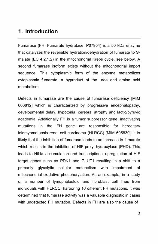
1. Introduction
Fumarase (FH, Fumarate hydratase, P07954) is a 50 kDa enzyme
that catalyzes the reversible hydration/dehydration of fumarate to S-
malate (EC 4.2.1.2) in the mitochondrial Krebs cycle, see below. A
second fumarase isoform exists without the mitochondrial import
sequence. This cytoplasmic form of the enzyme metabolizes
cytoplasmic fumarate, a byproduct of the urea and amino acid
metabolism.
Defects in fumarase are the cause of fumarase deficiency [MIM
606812] which is characterized by progressive encephalopathy,
developmental delay, hypotonia, cerebral atrophy and lactic/pyruvic
academia. Additionally FH is a tumor suppressor gene; inactivating
mutations in the FH gene are responsible for hereditary
leiomyomataosis renal cell carcinoma (HLRCC) [MIM 605839]. It is
likely that the inhibition of fumarase leads to an increase in fumarate
which results in the inhibition of HIF prolyl hydroxylase (PHD). This
leads to Hif1 accumulation and transcriptional upregulation of HIF
target genes such as PDK1 and GLUT1 resulting in a shift to a
primarily glycolytic cellular metabolism with impairment of
mitochondrial oxidative phosphorylation. As an example, in a study
of a number of lymophblastoid and fibroblast cell lines from
individuals with HLRCC, harboring 16 different FH mutations, it was
determined that fumarase activity was a valuable diagnostic in cases
with undetected FH mutation. Defects in FH are also the cause of
3
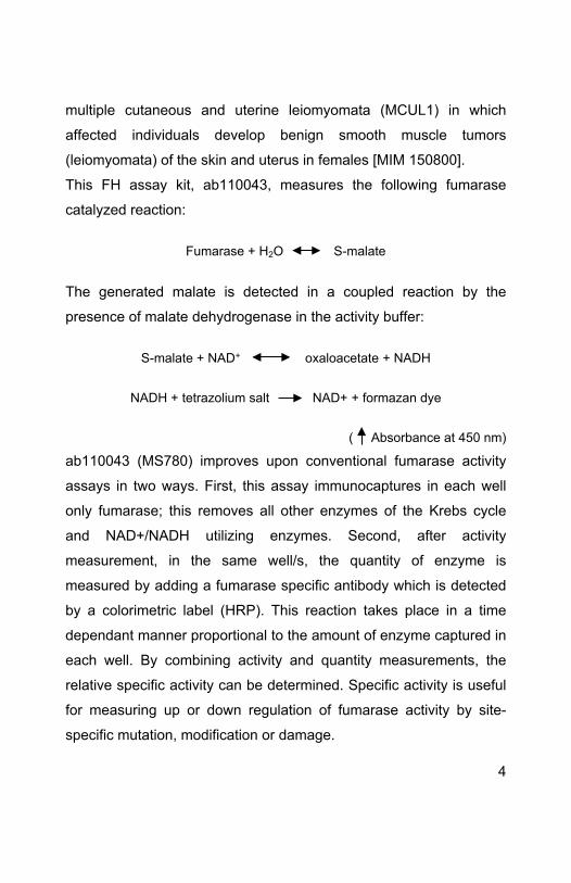
multiple cutaneous and uterine leiomyomata (MCUL1) in which
affected individuals develop benign smooth muscle tumors
(leiomyomata) of the skin and uterus in females [MIM 150800].
This FH assay kit, ab110043, measures the following fumarase
catalyzed reaction:
Fumarase + H2O S-malate
The generated malate is detected in a coupled reaction by the
presence of malate dehydrogenase in the activity buffer:
S-malate + NAD+ oxaloacetate + NADH
NADH + tetrazolium salt NAD+ + formazan dye
( Absorbance at 450 nm)
ab110043 (MS780) improves upon conventional fumarase activity
assays in two ways. First, this assay immunocaptures in each well
only fumarase; this removes all other enzymes of the Krebs cycle
and NAD+/NADH utilizing enzymes. Second, after activity
measurement, in the same well/s, the quantity of enzyme is
measured by adding a fumarase specific antibody which is detected
by a colorimetric label (HRP). This reaction takes place in a time
dependant manner proportional to the amount of enzyme captured in
each well. By combining activity and quantity measurements, the
relative specific activity can be determined. Specific activity is useful
for measuring up or down regulation of fumarase activity by site-
specific mutation, modification or damage.
4
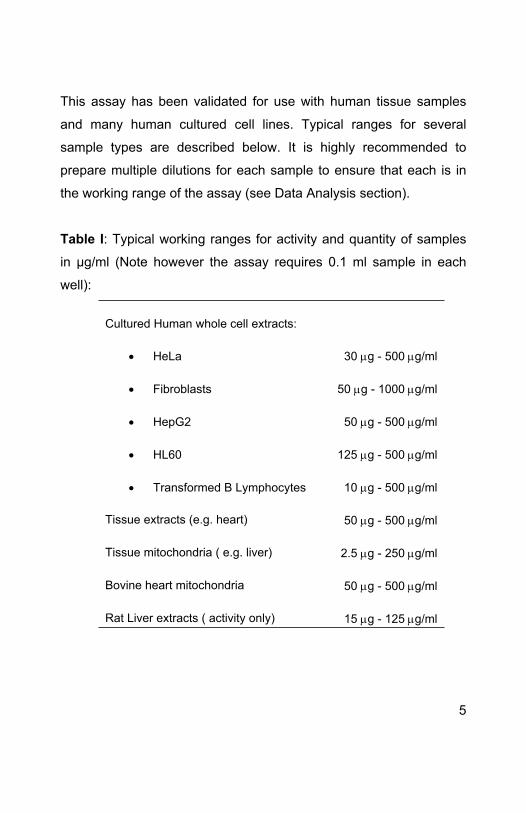
This assay has been validated for use with human tissue samples
and many human cultured cell lines. Typical ranges for several
sample types are described below. It is highly recommended to
prepare multiple dilutions for each sample to ensure that each is in
the working range of the assay (see Data Analysis section).
Table I: Typical working ranges for activity and quantity of samples
in µg/ml (Note however the assay requires 0.1 ml sample in each
well):
Cultured Human whole cell extracts:
HeLa 30 g - 500g/ml
Fibroblasts 50 g - 1000g/ml
HepG2 50 g - 500g/ml
HL60 125 g - 500g/ml
Transformed B Lymphocytes 10 g - 500g/ml
Tissue extracts (e.g. heart) 50 g - 500g/ml
Tissue mitochondria ( e.g. liver) 2.5 g - 250g/ml
Bovine heart mitochondria 50 g - 500g/ml
Rat Liver extracts ( activity only) 15 g - 125g/ml
5
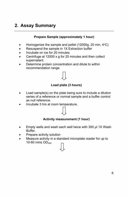
2. Assay Summary
Prepare Sample (approximately 1 hour)
Homogenize the sample and pellet (12000g, 20 min, 4oC) Resuspend the sample in 1X Extraction buffer Incubate on ice for 20 minutes. Centrifuge at 12000 x g for 20 minutes and then collect
supernatant. Determine protein concentration and dilute to within
recommendation range
Load plate (3 hours)
Load sample(s) on the plate being sure to include a dilution series of a reference or normal sample and a buffer control as null reference.
Incubate 3 hrs at room temperature.
Activity measurement (1 hour)
Empty wells and wash each well twice with 300 μl 1X Wash Buffer.
Prepare activity solution Measure activity in a standard microplate reader for up to
10-60 mins OD450
6
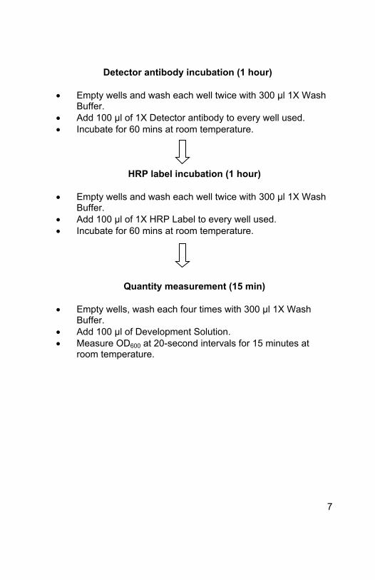
Detector antibody incubation (1 hour)
Empty wells and wash each well twice with 300 μl 1X Wash Buffer.
Add 100 μl of 1X Detector antibody to every well used. Incubate for 60 mins at room temperature.
HRP label incubation (1 hour)
Empty wells and wash each well twice with 300 μl 1X Wash Buffer.
Add 100 μl of 1X HRP Label to every well used. Incubate for 60 mins at room temperature.
Quantity measurement (15 min)
Empty wells, wash each four times with 300 μl 1X Wash Buffer.
Add 100 μl of Development Solution. Measure OD600 at 20-second intervals for 15 minutes at
room temperature.
7
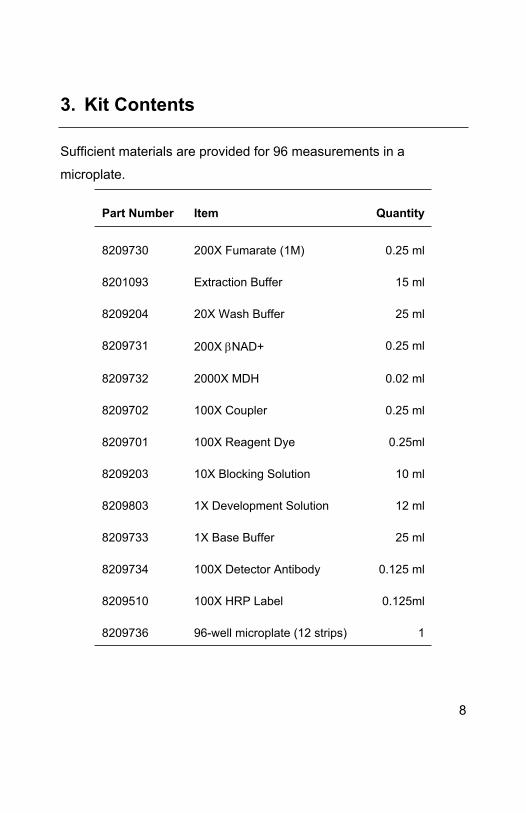
3. Kit Contents
Sufficient materials are provided for 96 measurements in a
microplate.
Part Number Item Quantity
8209730 200X Fumarate (1M) 0.25 ml
8201093 Extraction Buffer 15 ml
8209204 20X Wash Buffer 25 ml
8209731 200X NAD+ 0.25 ml
8209732 2000X MDH 0.02 ml
8209702 100X Coupler 0.25 ml
8209701 100X Reagent Dye 0.25ml
8209203 10X Blocking Solution 10 ml
8209803 1X Development Solution 12 ml
8209733 1X Base Buffer 25 ml
8209734 100X Detector Antibody 0.125 ml
8209510 100X HRP Label 0.125ml
8209736 96-well microplate (12 strips) 1
8
4. Storage and Handling
All components are shipped cold. Upon receipt store 200X
Fumarate, 200X NAD+, 100X Coupler and 100X Reagent Dye at
-20°C or preferably at -80°C for longer stability. Store all other
components at 4°C.
5. Additional Materials Required
Spectrophotometer that measures absorbance:
Activity: (450nm) Quantity: (450, 600 or 650 nm)
Multichannel pipette (50 - 300 μl) and tips
1.5 ml microtubes
A fine needle
Optional 1N HCl
6. Preparation of Buffers and Samples
Note: This protocol contains detailed steps for measuring FH activity in human samples. Be completely familiar with the protocol before beginning the assay. Do not deviate from the specified protocol steps or optimal results may not be obtained.
9
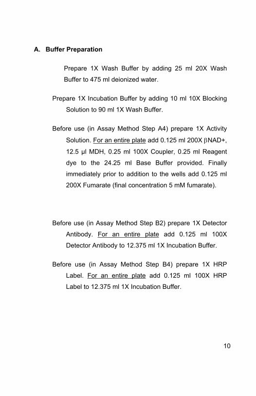
A. Buffer Preparation
Prepare 1X Wash Buffer by adding 25 ml 20X Wash
Buffer to 475 ml deionized water.
Prepare 1X Incubation Buffer by adding 10 ml 10X Blocking
Solution to 90 ml 1X Wash Buffer.
Before use (in Assay Method Step A4) prepare 1X Activity
Solution. For an entire plate add 0.125 ml 200X NAD+,
12.5 µl MDH, 0.25 ml 100X Coupler, 0.25 ml Reagent
dye to the 24.25 ml Base Buffer provided. Finally
immediately prior to addition to the wells add 0.125 ml
200X Fumarate (final concentration 5 mM fumarate).
Before use (in Assay Method Step B2) prepare 1X Detector
Antibody. For an entire plate add 0.125 ml 100X
Detector Antibody to 12.375 ml 1X Incubation Buffer.
Before use (in Assay Method Step B4) prepare 1X HRP
Label. For an entire plate add 0.125 ml 100X HRP
Label to 12.375 ml 1X Incubation Buffer.
10
B Sample preparation
Note: Samples must be detergent extracted by the provided Extraction buffer. To do this cell pellets, tissue homogenate pellets, or mitochondrial pellets must be prepared.
1. Briefly homogenate and mitochondrial preparations are
typically centrifuged at 12000 g for 20 min at 4oC to
pellet. Cells are typically pelleted at 500 g for 10 min at
4oC. Estimate the volume of the pellet and then
resuspend the sample pellet in 9 volumes of Sample
Extraction Buffer.
A total of 15 ml 1X Extraction buffer is provided.
Example - for analyses of multiple samples this is
enough to extract 15 samples at 1 ml, 30 at 0.5 ml, 60 at
0.25 ml etc.
2. Incubate extracts on ice for 20 minutes. Centrifuge
12000 x g, 4°C, for 20 minutes. Save the supernatant
and discard the pellet.
3. Determine the protein concentration of the supernatant
extract (a protein assay method unaffected by the
presence of detergent is critical e.g. BCA assay
recommended or OD280).
11
4. The sample should be diluted to within the working
range of the assay as described in Assay Method Step
A1 and first consulting Table I. Undiluted extract can be
frozen at -80°C.
7. Assay Method
A. Activity Measurement
1. Detergent extracted samples should be diluted in 1X
Incubation Buffer. A dilution series of samples is
recommended to ensure that samples are within the
working range of the activity and the quantity assays.
Also include a buffer control (1X Incubation Buffer only)
as a null or background reference. Add 100 μl of each
diluted sample into individual wells.
2. Cover/seal the plate and incubate for 3 hours at room
temperature with shaking.
3. Wash the plate
a. Empty the wells by turning the plate over a
receptacle and firmly shaking out the well contents
in one rapid downward motion. Dispose of biological
samples appropriately.
12
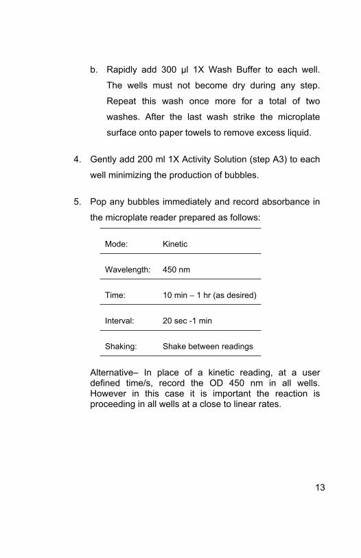
b. Rapidly add 300 μl 1X Wash Buffer to each well.
The wells must not become dry during any step.
Repeat this wash once more for a total of two
washes. After the last wash strike the microplate
surface onto paper towels to remove excess liquid.
4. Gently add 200 ml 1X Activity Solution (step A3) to each
well minimizing the production of bubbles.
5. Pop any bubbles immediately and record absorbance in
the microplate reader prepared as follows:
Mode: Kinetic
Wavelength: 450 nm
Time: 10 min – 1 hr (as desired)
Interval: 20 sec -1 min
Shaking: Shake between readings
Alternative– In place of a kinetic reading, at a user defined time/s, record the OD 450 nm in all wells. However in this case it is important the reaction is proceeding in all wells at a close to linear rates.
13
6. Record the data for analysis and proceed to section B –
Quantity Measurement.
B. Quantity Measurement
1. Repeat the wash procedure in step A3. Collect and
dispose of solutions and any added enzyme inhibitors
appropriately.
2. Add 100 μl of 1X Detector Antibody to each well used.
Cover/seal the plate and incubate for 1 hour at room
temperature with shaking.
3. Repeat the wash procedure in step A3.
4. Add 100 μl of 1X HRP Label to each well used.
Cover/seal the plate and incubate 1 hour at room
temperature with shaking. Meanwhile, prepare the
microplate spectrophotometer using the parameters
described below (Step B6).
5. Repeat the wash procedure in step A3 however
performing a total of four washes.
14
6. Add 100 μl HRP Development Solution to each empty
well, rapidly pop bubbles, and immediately record the
blue color development in the microplate reader
prepared as follows:
Mode: Kinetic
Wavelength: 600 nm
Time: up to 15 min
Interval: 20 sec -1 min
Shaking: Shake between readings
Alternative– In place of a kinetic reading, at a user defined
time, record the endpoint OD data at 600 nm or 450 nm. The
reaction can be stopped by adding 50 μl stop solution (1N
HCl) to each well and record the OD at 450 nm - however an
intense, sub-saturated, blue color could become saturating
by stopping the reaction and measuring at 450nm. However
in this case it is important the reaction is proceeding in all
wells at close at a close to linear rates.
Record the data and proceed to Data Analysis.
15
8. Data Analysis
A. Calculation of activity and Quantity
Determine the relative activity and quantity of an unknown
sample by first creating standard curves for the reference
sample.
The following two-fold dilution series of HeLa cell extract
was prepared in incubation buffer:
0, 8, 16, 32, 64, 125, 250, 500, 1000 µg/ml of HeLa cell
extract
Activity and quantity data were sequentially collected as
described in this protocol using a microplate reader.
Standard curves of these reference sample data were
prepared using the microplate reader software, which is
capable of a 4-parameter data analysis (Figure 1). Activity
and quantity are clearly measurable in the 30-500 µg/ml
range when such a fit was applied (alternatively raw data
can be exported to other graphing software).
16
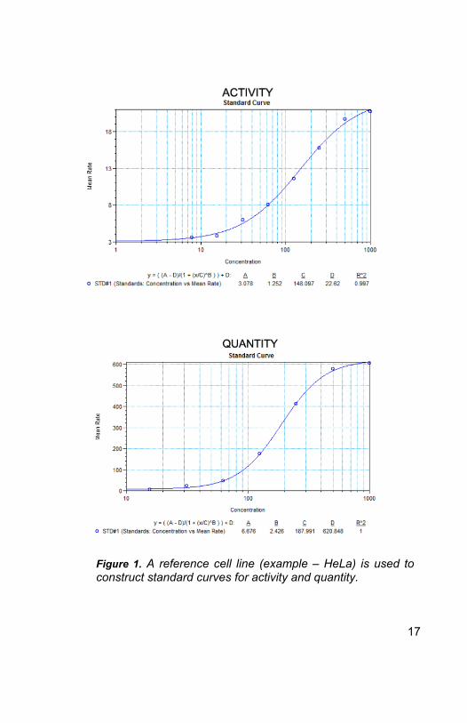
Figure 1. A reference cell line (example – HeLa) is used to construct standard curves for activity and quantity.
17
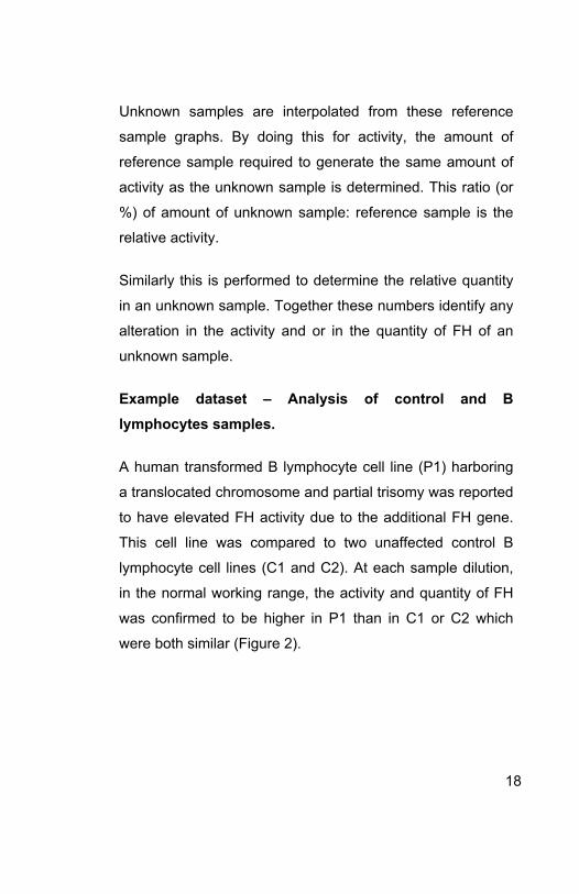
Unknown samples are interpolated from these reference
sample graphs. By doing this for activity, the amount of
reference sample required to generate the same amount of
activity as the unknown sample is determined. This ratio (or
%) of amount of unknown sample: reference sample is the
relative activity.
Similarly this is performed to determine the relative quantity
in an unknown sample. Together these numbers identify any
alteration in the activity and or in the quantity of FH of an
unknown sample.
Example dataset – Analysis of control and B lymphocytes samples.
A human transformed B lymphocyte cell line (P1) harboring
a translocated chromosome and partial trisomy was reported
to have elevated FH activity due to the additional FH gene.
This cell line was compared to two unaffected control B
lymphocyte cell lines (C1 and C2). At each sample dilution,
in the normal working range, the activity and quantity of FH
was confirmed to be higher in P1 than in C1 or C2 which
were both similar (Figure 2).
18
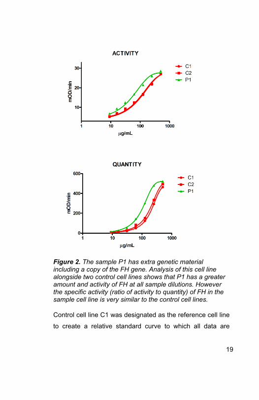
Figure 2. The sample P1 has extra genetic material including a copy of the FH gene. Analysis of this cell line alongside two control cell lines shows that P1 has a greater amount and activity of FH at all sample dilutions. However the specific activity (ratio of activity to quantity) of FH in the sample cell line is very similar to the control cell lines.
Control cell line C1 was designated as the reference cell line
to create a relative standard curve to which all data are
19
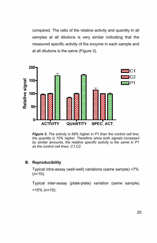
compared. The ratio of the relative activity and quantity in all
samples at all dilutions is very similar indicating that the
measured specific activity of the enzyme in each sample and
at all dilutions is the same (Figure 3).
Figure 3. The activity is 68% higher in P1 than the control cell line; the quantity is 70% higher. Therefore since both signals increased by similar amounts, the relative specific activity is the same in P1 as the control cell lines, C1,C2.
B. Reproducibility Typical intra-assay (well-well) variations (same sample) <7% (n=10).
Typical inter-assay (plate-plate) variation (same sample)
<10% (n=10).
20
9. Specificity
This kit measures activity and amount of fumarase in human and
bovine cells and tissues.
This kit does is not compatible with mouse samples, however the
activity assay is compatible with rat samples.
21

22

UK, EU and ROWEmail:
[email protected]: +44 (0)1223 696000www.abcam.com
US, Canada and Latin AmericaEmail: [email protected]: 888-77-ABCAM (22226)www.abcam.com
China and Asia Pacific Email: [email protected]
Tel: 400 921 0189 / +86 21 2070 0500www.abcam.cn
JapanEmail: [email protected]: +81-(0)3-6231-0940www.abcam.co.jp
23
Copyright © 2012 Abcam, All Rights Reserved. The Abcam logo is a registered trademark.
All information / detail is correct at time of going to print.