Embed Size (px)
Citation preview

Available online at www.sciencedirect.com
Kinome signaling through regulated protein–protein interactionsin normal and cancer cellsTony Pawson1,2 and Michael Kofler1
The flow of molecular information through normal and
oncogenic signaling pathways frequently depends on protein
phosphorylation, mediated by specific kinases, and the
selective binding of the resulting phosphorylation sites to
interaction domains present on downstream targets. This
physical and functional interplay of catalytic and interaction
domains can be clearly seen in cytoplasmic tyrosine kinases
such as Src, Abl, Fes, and ZAP-70. Although the kinase and
SH2 domains of these proteins possess similar intrinsic
properties of phosphorylating tyrosine residues or binding
phosphotyrosine sites, they also undergo intramolecular
interactions when linked together, in a fashion that varies from
protein to protein. These cooperative interactions can have
diverse effects on substrate recognition and kinase activity,
and provide a variety of mechanisms to link the stimulation of
catalytic activity to substrate recognition. Taken together,
these data have suggested how protein kinases, and the
signaling pathways in which they are embedded, can evolve
complex properties through the stepwise linkage of domains
within single polypeptides or multi-protein assemblies.
Addresses1 Samuel Lunenfeld Research Institute, Mt. Sinai Hospital, 600 University
Avenue, Toronto, Ontario M5G 1X5, Canada2 Department of Molecular Genetics, University of Toronto, Toronto,
Ontario M5S 1A8, Canada
Corresponding author: Pawson, Tony ([email protected])
Current Opinion in Cell Biology 2009, 21:147–153
This review comes from a themed issue on
Cell regulation
Edited by Brian Hemmings and Nikolas Tonks
Available online 18th March 2009
0955-0674/$ – see front matter
# 2009 Elsevier Ltd. All rights reserved.
DOI 10.1016/j.ceb.2009.02.005
IntroductionProtein kinases, by definition, exert their primary effects
through the phosphorylation of specific substrates. The
ability of protein kinases to recognize appropriate targets
depends, partly, on the sequence of amino acids that
surround the phosphorylation site, which determines
whether the substrate motif can be accommodated by
the active site [1,2]. However, such short motifs are
common in eukaryotic proteomes, and probably do not
provide sufficient information to ensure that only a given
subset of substrates are phosphorylated by a protein
www.sciencedirect.com
kinase within the cell. To circumvent this problem,
protein kinases have evolved additional mechanisms
for the selective recruitment of substrates, which involve
kinase–substrate interactions at surfaces that are distinct
from the active site of the kinase and the phosphorylation
motif of the substrate. For example, in the case of
cytoplasmic protein-tyrosine kinases, substrate recog-
nition typically involves non-catalytic modules such as
SH2 and SH3 domains that bind to substrates or their
associated scaffolds, and thus position the kinase domain
in the vicinity of relevant substrate sites [3–5] (Figure 1).
Non-catalytic domains may also reduce the complexity of
the potential substrate space by localizing the kinase to a
particular subcellular compartment. This is the case for
the PH domains of Tec family tyrosine kinases and the
Akt/PKB and Pdk1 serine/threonine kinases, which bind
phosphatidylinositol-(3,4,5)-trisphosphate (PIP3), and
thus localize the kinases in which they are embedded
to the plasma membrane upon activation of the PI 30-kinase pathway [6–8]. Although these non-catalytic
domains may have originally been linked to kinase
domains to aid in susbstrate recognition, they have also
acquired an ability to both positively and negatively
regulate kinase activity; the activation of such multi-
domain kinases may therefore be coupled to their ability
to interact with their substrates. This is a convenient
device to promote kinase-substrate specificity, in the
sense that such kinases may only be fully active in the
cell when in the vicinity of their physiological substrates.
The evolution of Src tyrosine kinase regulationA classic example involves the Src tyrosine kinase, which
possesses an N-terminal membrane-localizing myristoyl
group, followed in turn by non-catalytic SH3 and SH2
domains, the kinase domain, and a short C-terminal tail
(Figures 1 and 2). Since Src is constitutively associated
with the membrane, in proximity to its substrates, it must
be held in an inactive state in the absence of an appro-
priate upstream signal. This is achieved through phos-
phorylation of a tyrosine in the C-terminal tail by the
inhibitory tyrosine kinase Csk, which promotes an intra-
molecular interaction between the SH2 domain and the
phosphorylated tail, and consequently an additional
association between the SH3 domain and the SH2-kinase
linker [9–12] (Figures 2 and 3). Although these autoinhi-
bitory interactions involving the Src SH2 and SH3
domains are distant from the kinase active site, they
nonetheless attenuate catalytic activity, partly by clamp-
ing the kinase domain in a rigid state that precludes the
dynamic motions required for substrate phosphorylation
Current Opinion in Cell Biology 2009, 21:147–153

148 Cell regulation
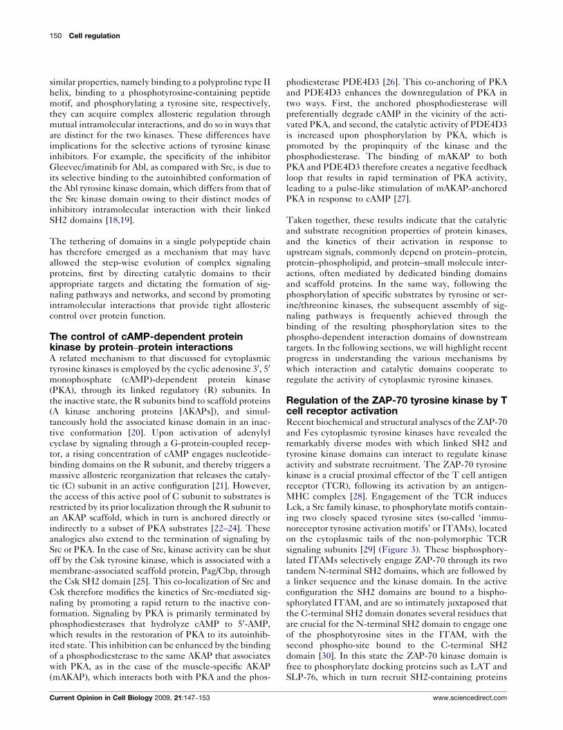
Figure 1
Domain composition of cytoplasmic tyrosine kinases. The SH2-kinase domain combination is conserved in cytoplasmic tyrosine kinases and
represents a functional unit. FABD: F-actin binding domain, F-BAR: FCH-BAR, Myr: myristoyl group, SH2: Src homology 2 domain, SH3: Src homology
3 domain.
[13]. In addition, these interactions suppress adventitious
binding of the interaction domains to other proteins. This
inhibited state can be overcome by tyrosine phosphatases
that dephosphorylate the C-terminal tail, or by external
ligands that engage the SH2 and SH3 domains, and
thereby break the inhibitory grip of the phosphorylated
Figure 2
Interdomain interactions in active and inactive conformations of cytoplasmic
(PDB: 1OPL [42��]), Csk (1K9A [41]), and Fes (3BKB [36��]), and the inactive c
All structures have been aligned at the lower lobe of the kinase domain and a
domains in red, SH3 domains in green, and linkers and C-terminal SH2 bind
Current Opinion in Cell Biology 2009, 21:147–153
tail and linker region [14,15]. In this latter case, target
recognition may be coupled to kinase activation.
Since the SH2 and SH3 domains of Src family kinases
play a dual role, through their ability to repress kinase
activity on the one hand, and to coordinately promote
substrate recognition and kinase activation on the other
tyrosine kinases. The gallery of structures includes the active form of Abl
losed form of Abl (1OPL [18]), Src (2SRC [43]), and ZAP-70 (2OZO [32��]).
re shown in identical orientation. Kinase domains are shown in blue, SH2
ing sites in gray. The aC helix of the kinase domain is depicted in yellow.
www.sciencedirect.com

Kinome signaling through regulated protein–protein interactions in normal and cancer cells Pawson and Kofler 149
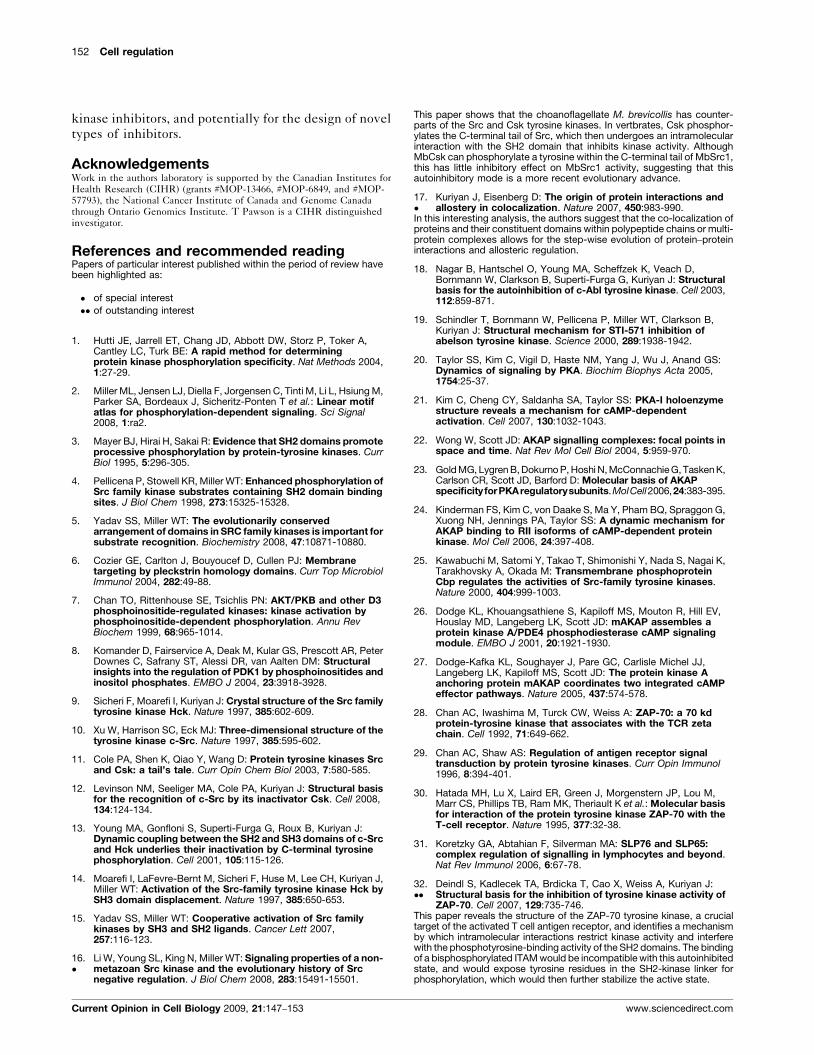
Figure 3
Activation model of cytoplasmic tyrosine kinases. Models for the closed, inactive conformations of Src, Fes, and ZAP-70 tyrosine kinases are shown
on the left, models of the opened, catalytically active states in the presence of ligands for the adaptor domains and kinase domain substrates are
shown on the right. In this active conformation, the aC helix adopts a well-defined orientation and tilts toward the kinase domain C-lobe. Kinase
domains (KD) are depicted in blue, SH2 domains in red, and SH3 domains in green. The aC helix of the kinase domain is depicted in yellow. Black filled
circles labeled with a white ‘P’ and open circles labeled with a black ‘Y’ represent phosphorylated and unphosphorylated tyrosines, respectively, of
SH2 recognition sites and substrate motifs.
(Figure 3), one might wonder which of these activities
came first in the course of evolution. This question has
been addressed by the analysis of a Src family kinase
(MbSrc1) from the unicellular choanoflagellate Monosigabrevicollis, which shares a common ancestry with multi-
cellular animals. Although MbSrc1 has a C-terminal tail
that can be phosphorylated by the M. brevicollis homolog
of Csk (MbCsk), this phosphorylation does not strongly
inhibit MbSrc1 kinase activity [16�]. This observation is
consistent with the notion that the joining of non-catalytic
SH2 and SH3 domains to tyrosine kinase domains
initially served to target kinases to their substrates.
Once covalently joined together, however, intramolecular
interactions between the non-catalytic and kinase
domains could potentially evolve to regulate their
www.sciencedirect.com
respective binding and catalytic properties and to fine-
tune and specify the cellular functions of such enzymes
[17�]. In support of this view, the Abl cytoplasmic tyrosine
kinase has a similar arrangement of SH3, SH2, and kinase
domains to Src, and is also autoinhibited by intramole-
cular interactions of the SH2 and SH3 domains with the
kinase domain (Figure 2). However, the precise mech-
anism by which this is achieved is different for the two
kinases, since the autoinhibted conformation of Abl
involves a phosphotyrosine-independent interaction of
the SH2 domain with the back side of the large lobe of
the kinase domain, which nonetheless inhibits both the
catalytic activity of the kinase domain and phosphotyr-
osine-binding by the SH2 domain, as in the case of Src
[18]. Thus, although the SH3, SH2, and catalytic domains
of the Src and Abl kinases independently have very
Current Opinion in Cell Biology 2009, 21:147–153
150 Cell regulation
similar properties, namely binding to a polyproline type II
helix, binding to a phosphotyrosine-containing peptide
motif, and phosphorylating a tyrosine site, respectively,
they can acquire complex allosteric regulation through
mutual intramolecular interactions, and do so in ways that
are distinct for the two kinases. These differences have
implications for the selective actions of tyrosine kinase
inhibitors. For example, the specificity of the inhibitor
Gleevec/imatinib for Abl, as compared with Src, is due to
its selective binding to the autoinhibted conformation of
the Abl tyrosine kinase domain, which differs from that of
the Src kinase domain owing to their distinct modes of
inhibitory intramolecular interaction with their linked
SH2 domains [18,19].
The tethering of domains in a single polypeptide chain
has therefore emerged as a mechanism that may have
allowed the step-wise evolution of complex signaling
proteins, first by directing catalytic domains to their
appropriate targets and dictating the formation of sig-
naling pathways and networks, and second by promoting
intramolecular interactions that provide tight allosteric
control over protein function.
The control of cAMP-dependent proteinkinase by protein–protein interactionsA related mechanism to that discussed for cytoplasmic
tyrosine kinases is employed by the cyclic adenosine 30, 50
monophosphate (cAMP)-dependent protein kinase
(PKA), through its linked regulatory (R) subunits. In
the inactive state, the R subunits bind to scaffold proteins
(A kinase anchoring proteins [AKAPs]), and simul-
taneously hold the associated kinase domain in an inac-
tive conformation [20]. Upon activation of adenylyl
cyclase by signaling through a G-protein-coupled recep-
tor, a rising concentration of cAMP engages nucleotide-
binding domains on the R subunit, and thereby triggers a
massive allosteric reorganization that releases the cataly-
tic (C) subunit in an active configuration [21]. However,
the access of this active pool of C subunit to substrates is
restricted by its prior localization through the R subunit to
an AKAP scaffold, which in turn is anchored directly or
indirectly to a subset of PKA substrates [22–24]. These
analogies also extend to the termination of signaling by
Src or PKA. In the case of Src, kinase activity can be shut
off by the Csk tyrosine kinase, which is associated with a
membrane-associated scaffold protein, Pag/Cbp, through
the Csk SH2 domain [25]. This co-localization of Src and
Csk therefore modifies the kinetics of Src-mediated sig-
naling by promoting a rapid return to the inactive con-
formation. Signaling by PKA is primarily terminated by
phosphodiesterases that hydrolyze cAMP to 50-AMP,
which results in the restoration of PKA to its autoinhib-
ited state. This inhibition can be enhanced by the binding
of a phosphodiesterase to the same AKAP that associates
with PKA, as in the case of the muscle-specific AKAP
(mAKAP), which interacts both with PKA and the phos-
Current Opinion in Cell Biology 2009, 21:147–153
phodiesterase PDE4D3 [26]. This co-anchoring of PKA
and PDE4D3 enhances the downregulation of PKA in
two ways. First, the anchored phosphodiesterase will
preferentially degrade cAMP in the vicinity of the acti-
vated PKA, and second, the catalytic activity of PDE4D3
is increased upon phosphorylation by PKA, which is
promoted by the propinquity of the kinase and the
phosphodiesterase. The binding of mAKAP to both
PKA and PDE4D3 therefore creates a negative feedback
loop that results in rapid termination of PKA activity,
leading to a pulse-like stimulation of mAKAP-anchored
PKA in response to cAMP [27].
Taken together, these results indicate that the catalytic
and substrate recognition properties of protein kinases,
and the kinetics of their activation in response to
upstream signals, commonly depend on protein–protein,
protein–phospholipid, and protein–small molecule inter-
actions, often mediated by dedicated binding domains
and scaffold proteins. In the same way, following the
phosphorylation of specific substrates by tyrosine or ser-
ine/threonine kinases, the subsequent assembly of sig-
naling pathways is frequently achieved through the
binding of the resulting phosphorylation sites to the
phospho-dependent interaction domains of downstream
targets. In the following sections, we will highlight recent
progress in understanding the various mechanisms by
which interaction and catalytic domains cooperate to
regulate the activity of cytoplasmic tyrosine kinases.
Regulation of the ZAP-70 tyrosine kinase by Tcell receptor activationRecent biochemical and structural analyses of the ZAP-70
and Fes cytoplasmic tyrosine kinases have revealed the
remarkably diverse modes with which linked SH2 and
tyrosine kinase domains can interact to regulate kinase
activity and substrate recruitment. The ZAP-70 tyrosine
kinase is a crucial proximal effector of the T cell antigen
receptor (TCR), following its activation by an antigen-
MHC complex [28]. Engagement of the TCR induces
Lck, a Src family kinase, to phosphorylate motifs contain-
ing two closely spaced tyrosine sites (so-called ‘immu-
noreceptor tyrosine activation motifs’ or ITAMs), located
on the cytoplasmic tails of the non-polymorphic TCR
signaling subunits [29] (Figure 3). These bisphosphory-
lated ITAMs selectively engage ZAP-70 through its two
tandem N-terminal SH2 domains, which are followed by
a linker sequence and the kinase domain. In the active
configuration the SH2 domains are bound to a bispho-
sphorylated ITAM, and are so intimately juxtaposed that
the C-terminal SH2 domain donates several residues that
are crucial for the N-terminal SH2 domain to engage one
of the phosphotyrosine sites in the ITAM, with the
second phospho-site bound to the C-terminal SH2
domain [30]. In this state the ZAP-70 kinase domain is
free to phosphorylate docking proteins such as LAT and
SLP-76, which in turn recruit SH2-containing proteins
www.sciencedirect.com

Kinome signaling through regulated protein–protein interactions in normal and cancer cells Pawson and Kofler 151
such as phospholipase C-g that transmit the TCR signal
[31]. However, recent structural data have shown that in
the inactive state, ZAP-70 adopts an autoinhibited con-
formation that restricts kinase activity [32��]; although the
SH2 domains are exposed for potential ligand-binding,
they are physically separated, which disrupts the phos-
photyrosine-binding pocket of the N-terminal SH2
domain. This configuration is achieved by docking of a
helical sequence located between the two SH2 domains
onto the kinase domain, with the SH2-kinase linker being
captured between this inter-SH2 sequence and the
kinase domain (Figures 2 and 3). Two regulatory tyro-
sines in the SH2-kinase linker, which become phosphory-
lated during TCR signaling, are part of the hydrophobic
environment formed by these interactions. These find-
ings have suggested a model in which binding of a
phosphorylated ITAM to the ZAP-70 SH2 domains
induces a conformational rearrangement that disrupts
the inhibitory interactions between the inter-SH2
sequence, the SH2-kinase linker and the kinase domain.
This reorganization would also expose the tyrosines in the
SH2-kinase linker for phosphorylation, probably by the
TCR-associated Lck tyrosine kinase, which would
antagonize their ability to adopt the autoinhibited con-
formation and stabilize the active state. Thus, a key
feature of ZAP-70 regulation involves the cooperative
binding of the tandem SH2 domains to a phosphorylated
ITAM motif, which stimulates kinase activity.
The SH2 and catalytic domains of the Fescytoplasmic tyrosine kinase form a functionalunitThe Fes tyrosine kinase has an N-terminal F-BAR
domain, an SH2 domain, and a tyrosine kinase domain
[33] (Figure 1). Genetic, biochemical, and functional
analysis of the oncogenic avian Fes homolog (v-Fps)
has suggested that the SH2 domain associates with the
kinase domain, and has a strongly positive effect on
catalytic activity and substrate recognition [34,35]. Struc-
tural analysis of the linked SH2 and kinase domains of the
human Fes protein has recently revealed that the SH2
domain interacts with the aC helix in the small lobe of the
kinase domain, in a fashion that promotes an active kinase
conformation [36��] (Figure 2). This active configuration
is further stabilized by the binding of a ligand to the SH2
domain; in addition, the activation loop of the Fes kinase
domain adopts an active conformation both as a result of
autophosphorylation and binding to a substrate peptide.
Such data have suggested that the Fes SH2-kinase unit is
stabilized in the active state by binding of the SH2
domain to a phosphorylated site on a polypeptide, which
then orients a substrate motif toward the active site for
phosphorylation of a second tyrosine residue (Figure 3).
Genetic and functional evidence suggests that Src family
tyrosine kinases lie upstream of Fes, and might therefore
phosphorylate sites that engage the Fes SH2 domain
[37,38]. The Fes F-BAR sequence belongs to a family
www.sciencedirect.com
of domains that form oligomeric structures and bind to
phospholipids in the context of curved membranes
[39,40]. Interaction of the F-BAR domain with specific
membrane sites may position the Fes tyrosine kinase in
the vicinity of substrates, and indeed during FceR1
signaling in mast cells, the Fes F-BAR appears to act
in concert with the SH2 domain to promote substrate
phosphorylation and degranulation [39]. Fes therefore
represents a kinase in which the SH2 domain not only
appears to target the kinase toward its substrates but also
simultaneously stimulates kinase activity. Similar obser-
vations have been made for the SH2 and SH3 domains of
Csk, which interact with the small lobe of the kinase
domain to stimulate activity [41]. In the case of Src,
binding of the SH2/SH3 interaction domains to external
ligands relieves an autoinhibited state, and by default
releases the kinase domain in an active conformation,
with little detectable interaction between the SH2/SH3
domains and the kinase following activation. By contrast,
in Fes the ligand-bound SH2 domain is required to
promote an active configuration of the kinase domain
through intramolecular interactions.
Complex regulation of the Abl tyrosine kinaseThe Abl cytoplasmic tyrosine kinase may represent an
intermediate between Src and Fes. As noted, in its auto-
inhibited conformation the Abl SH2 and SH3 domains
interact with the kinase domain and SH2-kinase linker to
inhibit kinase activity. Upon activation, however, the
SH2 domain appears to adopt a new interaction with
the kinase domain, involving the small, rather than the
large lobe [42��] (Figure 2). In cells, this novel Abl SH2–kinase interaction appears important for full kinase acti-
vation [36��,42��], although the effect is less pronounced
than for Fes and the mechanistic basis remains unclear.
This apparent coupling of the Abl SH2 and kinase
domains in the active state may be important for the
ability of Abl to mediate processive multi-site phos-
phorylation of substrates such as p130cas.
ConclusionTaken together, these data are consistent with a general
scheme in which the activation of cytoplasmic tyrosine
kinases is coupled to substrate recognition. They
suggest that intramolecular interactions between linked
interaction and catalytic domains have evolved to con-
trol the precise circumstances under which a cyto-
plasmic tyrosine kinase becomes active, and show
that although the domains of the various kinases retain
very similar intrinsic functions in binding or phosphor-
ylating exogenous proteins, they have evolved a wide
range of intramolecular regulatory interactions (Figures
2 and 3). Related mechanisms are used to direct serine/
threonine protein kinases such as PKA to their sub-
strates, and to control the kinetics of kinase activation.
These observations have interesting implications for
understanding the mechanisms of action of existing
Current Opinion in Cell Biology 2009, 21:147–153
152 Cell regulation
kinase inhibitors, and potentially for the design of novel
types of inhibitors.
AcknowledgementsWork in the authors laboratory is supported by the Canadian Institutes forHealth Research (CIHR) (grants #MOP-13466, #MOP-6849, and #MOP-57793), the National Cancer Institute of Canada and Genome Canadathrough Ontario Genomics Institute. T Pawson is a CIHR distinguishedinvestigator.
References and recommended readingPapers of particular interest published within the period of review havebeen highlighted as:
� of special interest
�� of outstanding interest
1. Hutti JE, Jarrell ET, Chang JD, Abbott DW, Storz P, Toker A,Cantley LC, Turk BE: A rapid method for determiningprotein kinase phosphorylation specificity. Nat Methods 2004,1:27-29.
2. Miller ML, Jensen LJ, Diella F, Jorgensen C, Tinti M, Li L, Hsiung M,Parker SA, Bordeaux J, Sicheritz-Ponten T et al.: Linear motifatlas for phosphorylation-dependent signaling. Sci Signal2008, 1:ra2.
3. Mayer BJ, Hirai H, Sakai R: Evidence that SH2 domains promoteprocessive phosphorylation by protein-tyrosine kinases. CurrBiol 1995, 5:296-305.
4. Pellicena P, Stowell KR, Miller WT: Enhanced phosphorylation ofSrc family kinase substrates containing SH2 domain bindingsites. J Biol Chem 1998, 273:15325-15328.
5. Yadav SS, Miller WT: The evolutionarily conservedarrangement of domains in SRC family kinases is important forsubstrate recognition. Biochemistry 2008, 47:10871-10880.
6. Cozier GE, Carlton J, Bouyoucef D, Cullen PJ: Membranetargeting by pleckstrin homology domains. Curr Top MicrobiolImmunol 2004, 282:49-88.
7. Chan TO, Rittenhouse SE, Tsichlis PN: AKT/PKB and other D3phosphoinositide-regulated kinases: kinase activation byphosphoinositide-dependent phosphorylation. Annu RevBiochem 1999, 68:965-1014.
8. Komander D, Fairservice A, Deak M, Kular GS, Prescott AR, PeterDownes C, Safrany ST, Alessi DR, van Aalten DM: Structuralinsights into the regulation of PDK1 by phosphoinositides andinositol phosphates. EMBO J 2004, 23:3918-3928.
9. Sicheri F, Moarefi I, Kuriyan J: Crystal structure of the Src familytyrosine kinase Hck. Nature 1997, 385:602-609.
10. Xu W, Harrison SC, Eck MJ: Three-dimensional structure of thetyrosine kinase c-Src. Nature 1997, 385:595-602.
11. Cole PA, Shen K, Qiao Y, Wang D: Protein tyrosine kinases Srcand Csk: a tail’s tale. Curr Opin Chem Biol 2003, 7:580-585.
12. Levinson NM, Seeliger MA, Cole PA, Kuriyan J: Structural basisfor the recognition of c-Src by its inactivator Csk. Cell 2008,134:124-134.
13. Young MA, Gonfloni S, Superti-Furga G, Roux B, Kuriyan J:Dynamic coupling between the SH2 and SH3 domains of c-Srcand Hck underlies their inactivation by C-terminal tyrosinephosphorylation. Cell 2001, 105:115-126.
14. Moarefi I, LaFevre-Bernt M, Sicheri F, Huse M, Lee CH, Kuriyan J,Miller WT: Activation of the Src-family tyrosine kinase Hck bySH3 domain displacement. Nature 1997, 385:650-653.
15. Yadav SS, Miller WT: Cooperative activation of Src familykinases by SH3 and SH2 ligands. Cancer Lett 2007,257:116-123.
16.�
Li W, Young SL, King N, Miller WT: Signaling properties of a non-metazoan Src kinase and the evolutionary history of Srcnegative regulation. J Biol Chem 2008, 283:15491-15501.
Current Opinion in Cell Biology 2009, 21:147–153
This paper shows that the choanoflagellate M. brevicollis has counter-parts of the Src and Csk tyrosine kinases. In vertbrates, Csk phosphor-ylates the C-terminal tail of Src, which then undergoes an intramolecularinteraction with the SH2 domain that inhibits kinase activity. AlthoughMbCsk can phosphorylate a tyrosine within the C-terminal tail of MbSrc1,this has little inhibitory effect on MbSrc1 activity, suggesting that thisautoinhibitory mode is a more recent evolutionary advance.
17.�
Kuriyan J, Eisenberg D: The origin of protein interactions andallostery in colocalization. Nature 2007, 450:983-990.
In this interesting analysis, the authors suggest that the co-localization ofproteins and their constituent domains within polypeptide chains or multi-protein complexes allows for the step-wise evolution of protein–proteininteractions and allosteric regulation.
18. Nagar B, Hantschel O, Young MA, Scheffzek K, Veach D,Bornmann W, Clarkson B, Superti-Furga G, Kuriyan J: Structuralbasis for the autoinhibition of c-Abl tyrosine kinase. Cell 2003,112:859-871.
19. Schindler T, Bornmann W, Pellicena P, Miller WT, Clarkson B,Kuriyan J: Structural mechanism for STI-571 inhibition ofabelson tyrosine kinase. Science 2000, 289:1938-1942.
20. Taylor SS, Kim C, Vigil D, Haste NM, Yang J, Wu J, Anand GS:Dynamics of signaling by PKA. Biochim Biophys Acta 2005,1754:25-37.
21. Kim C, Cheng CY, Saldanha SA, Taylor SS: PKA-I holoenzymestructure reveals a mechanism for cAMP-dependentactivation. Cell 2007, 130:1032-1043.
22. Wong W, Scott JD: AKAP signalling complexes: focal points inspace and time. Nat Rev Mol Cell Biol 2004, 5:959-970.
23. Gold MG, Lygren B, Dokurno P, Hoshi N, McConnachie G, Tasken K,Carlson CR, Scott JD, Barford D: Molecular basis of AKAPspecificity forPKAregulatorysubunits.MolCell2006,24:383-395.
24. Kinderman FS, Kim C, von Daake S, Ma Y, Pham BQ, Spraggon G,Xuong NH, Jennings PA, Taylor SS: A dynamic mechanism forAKAP binding to RII isoforms of cAMP-dependent proteinkinase. Mol Cell 2006, 24:397-408.
25. Kawabuchi M, Satomi Y, Takao T, Shimonishi Y, Nada S, Nagai K,Tarakhovsky A, Okada M: Transmembrane phosphoproteinCbp regulates the activities of Src-family tyrosine kinases.Nature 2000, 404:999-1003.
26. Dodge KL, Khouangsathiene S, Kapiloff MS, Mouton R, Hill EV,Houslay MD, Langeberg LK, Scott JD: mAKAP assembles aprotein kinase A/PDE4 phosphodiesterase cAMP signalingmodule. EMBO J 2001, 20:1921-1930.
27. Dodge-Kafka KL, Soughayer J, Pare GC, Carlisle Michel JJ,Langeberg LK, Kapiloff MS, Scott JD: The protein kinase Aanchoring protein mAKAP coordinates two integrated cAMPeffector pathways. Nature 2005, 437:574-578.
28. Chan AC, Iwashima M, Turck CW, Weiss A: ZAP-70: a 70 kdprotein-tyrosine kinase that associates with the TCR zetachain. Cell 1992, 71:649-662.
29. Chan AC, Shaw AS: Regulation of antigen receptor signaltransduction by protein tyrosine kinases. Curr Opin Immunol1996, 8:394-401.
30. Hatada MH, Lu X, Laird ER, Green J, Morgenstern JP, Lou M,Marr CS, Phillips TB, Ram MK, Theriault K et al.: Molecular basisfor interaction of the protein tyrosine kinase ZAP-70 with theT-cell receptor. Nature 1995, 377:32-38.
31. Koretzky GA, Abtahian F, Silverman MA: SLP76 and SLP65:complex regulation of signalling in lymphocytes and beyond.Nat Rev Immunol 2006, 6:67-78.
32.��
Deindl S, Kadlecek TA, Brdicka T, Cao X, Weiss A, Kuriyan J:Structural basis for the inhibition of tyrosine kinase activity ofZAP-70. Cell 2007, 129:735-746.
This paper reveals the structure of the ZAP-70 tyrosine kinase, a crucialtarget of the activated T cell antigen receptor, and identifies a mechanismby which intramolecular interactions restrict kinase activity and interferewith the phosphotyrosine-binding activity of the SH2 domains. The bindingof a bisphosphorylated ITAM would be incompatible with this autoinhibitedstate, and would expose tyrosine residues in the SH2-kinase linker forphosphorylation, which would then further stabilize the active state.
www.sciencedirect.com
Kinome signaling through regulated protein–protein interactions in normal and cancer cells Pawson and Kofler 153
33. Greer P: Closing in on the biological functions of Fps/Fes andFer. Nat Rev Mol Cell Biol 2002, 3:278-289.
34. Sadowski I, Stone JC, Pawson T: A noncatalytic domainconserved among cytoplasmic protein-tyrosine kinasesmodifies the kinase function and transforming activity ofFujinami sarcoma virus P130gag-fps. Mol Cell Biol 1986,6:4396-4408.
35. Koch CA, Moran M, Sadowski I, Pawson T: The common srchomology region 2 domain of cytoplasmic signaling proteinsis a positive effector of v-fps tyrosine kinase function. Mol CellBiol 1989, 9:4131-4140.
36.��
Filippakopoulos P, Kofler M, Hantschel O, Gish GD, Grebien F,Salah E, Neudecker P, Kay LE, Turk BE, Superti-Furga G et al.:Structural coupling of SH2-kinase domains links Fes and Ablsubstrate recognition and kinase activation. Cell 2008,134:793-803.
This paper identifies the mechanism by which the SH2 domain of the Festyrosine kinase interacts with the adjacent kinase domain to stimulatecatalytic activity, and indicates that substrate-binding to the SH2 andkinase domains stabilizes the active state of the SH2-kinase unit. Thepaper also indicates that the SH2 domain of the Abl tyrosine kinase has astimulatory effect in the active state in cells.
37. Udell CM, Samayawardhena LA, Kawakami Y, Kawakami T,Craig AW: Fer and Fps/Fes participate in a Lyn-dependentpathway from FcepsilonRI to platelet-endothelial celladhesion molecule 1 to limit mast cell activation. J Biol Chem2006, 281:20949-20957.
www.sciencedirect.com
38. Murray MJ, Davidson CM, Hayward NM, Brand AH: The Fes/Fernon-receptor tyrosine kinase cooperates with Src42A toregulate dorsal closure in Drosophila. Development 2006,133:3063-3073.
39. McPherson VA, Everingham S, Karisch R, Smith JA, Udell CM,Zheng J, Jia Z, Craig AW: Contributions of F-BAR and SH2domains of Fes protein tyrosine kinase for coupling to theFcepsilonRI pathway in mast cells. Mol Cell Biol 2009,29:389-401.
40. Heath RJ, Insall RH: F-BAR domains: multifunctional regulatorsof membrane curvature. J Cell Sci 2008, 121:1951-1954.
41. Ogawa A, Takayama Y, Sakai H, Chong KT, Takeuchi S,Nakagawa A, Nada S, Okada M, Tsukihara T: Structure of thecarboxyl-terminal Src kinase, Csk. J Biol Chem 2002,277:14351-14354.
42.��
Nagar B, Hantschel O, Seeliger M, Davies JM, Weis WI, Superti-Furga G, Kuriyan J: Organization of the SH3-SH2 unit in activeand inactive forms of the c-Abl tyrosine kinase. Mol Cell 2006,21:787-798.
This paper not only shows how the SH3-SH2 unit of the Abl kinase bindsto the back side of the kinase domain to inhibit catalytic activity but alsoidentifies a conformation that probably corresponds to an active state inwhich the domains are in an extended configuration, and in which the SH2domain interacts with the kinase domain.
43. Xu W, Doshi A, Lei M, Eck MJ, Harrison SC: Crystal structures ofc-Src reveal features of its autoinhibitory mechanism. Mol Cell1999, 5:629-638.
Current Opinion in Cell Biology 2009, 21:147–153