Embed Size (px)
Citation preview

Kinetics of Desmosome Assembly in Madin-Darby Canine Kidney Epithelial Cells: Temporal and Spatial Regulation of Desmoplakin Organization and Stabilization upon Cell-Cell Contact. I. Biochemical Analysis Mani jeh Pasdar and W. J ames Nelson
Institute for Cancer Research, Philadelphia, Pennsylvania 19Ill
Abstract. The functional interaction of cells in the formation of tissues requires the establishment and maintenance of cell-cell contact by the junctional complex. However, little is known biochemically about the mechanism(s) that regulates junctional complex as- sembly. To address this problem, we have initiated a study of the regulation of assembly of one component of the junctional complex, the desmosome, during in- duction of cell-cell contact in cultures of Madin-Darby canine kidney epithelial cells. Here we have analyzed two major protein components of the desmosomal plaque, desmoplakins I (Mr of 250,000) and II (Mr of 215,000). Analysis of protein levels of desmoplakins I and II by immunoprecipitation with an antiserum that reacts specifically with an epitope com- mon to both proteins revealed that desmoplakins I and II are synthesized and accumulate at steady state in a ratio of 3-4:1 (in the absence or presence of cell-cell contact). The kinetics of desmoplakins I and II stabili- zation and assembly were analyzed after partitioning of newly synthesized proteins into a soluble and in-
soluble protein fraction by extraction of whole cells in a Triton X-IO0 high salt buffer. In the absence of cell- cell contact, both the soluble and insoluble pools of desmoplakins I and II are unstable and are degraded rapidly (t,/: ,x, 8 h). Upon induction of cell-cell con- tact, the capacity of the insoluble pool increases ,'~three- fold as a proportion of the soluble pool of newly syn- thesized desmoplakins I and II is titrated into the insoluble pool. The insoluble pool becomes relatively stable (t,/, > 72 h), whereas proteins remaining in the soluble pool (m25-40% of the total) are degraded rap- idly (t,/~ ~ 8 h). Furthermore, we show that desmo- plakins I and II can be recruited from this unstable soluble pool of protein to the stable insoluble pool upon induction of cell-cell contact 4 h after synthesis; significantly, the stabilization of this population of newly synthesized desmoplakins I and II is blocked by the addition of cycloheximide at the time of cell-cell contact, indicating that the coordinate synthesis of an- other protein(s) is required for protein stabilization.
haracteristic feature of tissue organization is the close structural and functional interaction of constituent cells. During development, this interaction is estab-
lished by a junctional complex that forms rapidly between adjacent cells upon induction of cell-cell contact. Subse- quently, the junctional complex appears to be involved in the maintenance of cell-cell adhesion and the functional interac- tions between adjacent cells.
The junctional complex exhibits cell and tissue specificity in the expression of different junction components. In simple epithelia, the complex may comprise up to five components (14): zonula occludens (50, 51), zonula adherens (21, 54), a cell adhesion molecule (L-CAM or uvomorulin) (12), gap junctions, and desmosomes (1, 4, 11, 14, 18, 19, 27, 38, 47). Of these components, the desmosome has been extensively analyzed in terms of its tissue distribution, morphology, and molecular anatomy (1-10, 14, 16, 19, 20, 22, 24, 27, 28, 39,
41, 42, 44, 46, 52). The desmosome can be isolated intact and is resistant to solubilization by nonionic detergents and buffers of high and low ionic strength (11, 35, 48, 49). It is composed of 7-12 proteins that are segregated into two asso- ciated structures, a cytoplasmic plaque and a membrane core (7, I1, 17, 23, 29, 35, 48, 49). The cytoplasmic plaque consists of nonglycosylated proteins (Mrs of 250,000, 215,000 [the des- moplakins], 83,000 and 75,000) and is an attachment site for cytokeratin intermediate filaments. The plaque appears to be attached to the plasma membrane core of the desmosome. The core consists of glycoproteins (Mrs of 150,000, 110,000, 97,000, and 22,000), some of which span the lipid bilayer and some of which are located extracellularly (3, 6, 8, 9, 16, 23, 35). The assembly of this complex structure is thought to confer a high degree of structural integrity on the epithelial cell monolayer (t).
Early morphological studies of junctional complex forma-
�9 The Rockefeller University Press, 0021-9525/88/03/677/9 $2.00 The Journal of Cell Biology, Volume 106, March 1988 677-685 677
on April 10, 2019jcb.rupress.org Downloaded from http://doi.org/10.1083/jcb.106.3.677Published Online: 1 March, 1988 | Supp Info:

tion between epithelial cells demonstrated that an initial event upon cell-cell contact was the symmetrical appearance of a desmosomal half-plaque on the plasma membrane of each adjacent cell (40, 42-44). The plaque appeared to form rapidly from proteins that were present in the cytoplasm of the cell before induction of cell-cell contact (25, 56). These results suggest that there is a close temporal and spatial coor- dination between cell-cell contact and desmosome assem- bly. However, little or nothing is known biochemically about the regulation of assembly of this multisubunit complex on the plasma membrane upon induction of cell-cell contact.
To address this question, we have initiated a study of des- mosome assembly in Madin-Darby canine kidney (MDCK) epithelial cells (reference 33). MDCK cells express a junc- tional complex characteristic of simple epithelia that in- cludes desmosomes (33, 55). Desmosome formation can be analyzed conveniently in MDCK cells since the degree of cell-cell contact can be modulated by adjusting the Ca ++ concentration of the growth media (33, 34, 37). Confluent monolayers of MDCK cells established in growth media con- raining 5 I.tM Ca ++ exhibit little or no cell-cell contact and no evidence of junctional complex formation. However, sub- sequent incubation in complete growth medium containing 1.8 mM Ca ++ results in the rapid and synchronous induction of cell-cell contact throughout the monolayer and the assem- bly of the junctional complex (37).
The initial set of experiments reported here focus on the synthesis and assembly of the two major protein components of the cytoplasmic plaque, desmoplakins I (250,000 Mr) and II (215,000 Mr) (35). Our results demonstrate biochemically that induction of cell-cell contact correlates with a dramatic increase in the stability and insolubility of newly synthesized desmoplakins I and II. Furthermore, our results indicate that the assembly of newly synthesized desmoplakins I and II is limited in MDCK cells and is regulated at three discrete stages.
Materials and Methods
Cells
The morphology, growth characteristics, and culture conditions of the MDCK cell clone (No. 8) used in this study have been described previously (32, 36). The degree of cell-cell contact in confluent cultures of cells was modulated by adjusting the concentration of Ca ++ in the growth medium (for details, see reference 37).
Preparation of a Desmosome-enriched Fraction from Bovine Muzzle Epidermis Desmosomes were purifed from bovine muzzle epidermis as described pre- viously (23, 35, 49). Fig. 1, lane 2 shows the protein profile of a typical desmosome preparation, which comprises a characteristic group of des- mosome-specific proteins including desmoplakins I (250,000 M,) and II (215,000 M,) (cf. references 11, 23, 35, 49).
Preparation of an Antiserum against Desmoplakins I and H The desmosome-enriched fraction was separated on preparative SDS 5 % polyacrylamide gels (30). The protein bands corresponding to the 250,000- (desmoplakin I) and 215,000-Mr (desmoplakin II) components of the des- mosome were excised from the gel and electroeluted (sample concentrator; Isco, Inc., Lincoln, NE) and used to immunize New Zealand white rabbits (36). The serum was tested for antibody production using an ELISA assay (13) and immunoblotting (see Fig. 1). The IgG fraction of the antiserum was precipitated with (NH4)2SO4 at 50% saturation.
Immunoblotting Proteins were transferred electrophoretically from SDS polyacrylamide gels to nitrocellulose filters (53) as described previously (36). Filters were in- cubated with l:l,000-5,000 dilution of antiserum and then ~25I-protein A (10 ~tCi/~tg) as described previously (36). Filters were dried and exposed at -80~ to XAR-5 x-ray film using two intensifying screens (DuPont Co., Wilmington, DE).
Metabolic Labeling and Cell Fractionation
Confluent monolayers of MDCK cells were grown in low Ca ++ (5 laM) medium (LC medium) I or high Ca ++ (1.8 mM) medium (HC medium) on collagen-coated 35 mm petri dishes. Cells were labeled metabolically with either [35S]methionine (1,200 Ci/mmol; New England Nuclear, Boston, MA) or [3H]leucine 046.7 Ci/mmol; New England Nuclear) for either 15 min or 2 h in LC or HC medium, and then chased in a >10,000-fold excess of unlabeled methionine in the appropriate growth medium (37). At the end of the period of incubation, cells were transferred to 4~ rinsed twice with ice-cold Tris-saline containing 1 mM phenylmethylsulfonyl fluoride (PMSF), and then extracted in situ with 1 ml of a buffer containing 50 mM NaCI, 300 mM sucrose, 10 mM Pipes (pH 6.8), 3 mM MgClz, 0.5% vol/ vol Triton X-100, 1.2 mM PMSF, 0.1 mg/ml DNase, and 0.1 mg/ml RNase (CSK buffer, see reference 15) for 20 min on a rocker platform at 4~ A solution of 2.5 M (NH4)2SO4 was added to a final concentration of 250 mM and incubation was continued for another 5 min. Cells were scraped from the petri dish with a rubber policeman and centrifuged at 48,000 g for 10 min to yield a supernatant (soluble fraction) and pellet (insoluble frac- tion). The samples were processed for quantitative immunoprecipitation and fluorography (36, 31) as described in detail previously (36). The relative amount of radioactivity in bands corresponding to desmoplakins I and II was determined from the resulting fluorograms by scanning densitometry using a spectrophotometer (Du-7; Beckman Instruments, Inc., Palo Alto, CA) equipped for automatic integrations. All experiments were performed at least three times with similar results, and the data from one typical experi- ment is presented. In all cases, desmoplakins I and II exhibited identical trends in stability and distribution between the soluble and insoluble pools (see Figs. 2 and 3). However, only the data for desmoplakin I is presented.
Analysis of Soluble Desmoplakins I and II by Sucrose Gradient Centrifugation
Confluent monolayers of MDCK cells were metabolically labeled with [35S]methionine and extracted with CSK buffer as described above. The soluble fraction (200 ~tl) was loaded onto a 3.8-ml linear 5-20% wt/wt su- crose gradient in 10 mM Tris-HCl, pH 7.5, 120 mM NaCl, 2 mM EDTA, 0.1 mM dithiothreitol (DTT). The gradient was centrifuged at 60,000 rpm in the SW6OTi rotor of the L8-70M ultracentrifuge (Beckman Instruments, Inc.) for 3-6 h at 4~ The gradient was fractionated from the bottom to the top (200 I.tl per fraction) and individual fractions were processed for im- munoprecipitation with the desmoplakin II antiserum, followed by SDS 5 % polyacrylamide gel electrophoresis and fluorography as described above.
Results
Characterization of the Antibody to Desmoplakins I and H
Immunoblotting of total proteins of purified bovine desmo- somes revealed that the antiserum reacted with desmoplakins I (250,000 Mr) and II (215,000 Mr) (Fig. 1, lane 3). Immu- noblotting (Fig. l, lane 4) or high stringency immunoprecip- itation (Fig. l, lane 5) of MDCK proteins insoluble in CSK buffer also revealed that the antiserum reacted with two pro- teins with apparent molecular masses similar to those of des- moplakins I and II, respectively. Antibodies affinity purified against either purified desmoplakins I or II (from bovine epidermis) reacted with both proteins in MDCK cells (Fig.
1. Abbreviations used in this paper: CSK buffer, cytoskeleton extraction buffer; HC medium, high Ca ++ (1.8 mM)-containing medium; LC medi- um, low Ca ++ (5 I.tM)-containing medium.
The Journal of Cell Biology, Volume 106, 1988 678

Figure 1. Characterization of an antiserum raised against des- moplakins I and II. Lane 1, relative molecular mass markers: (a) myosin (205,000 Mr), (b) 13-galactosidase (116,000 Mr), (c) phos- phorylase b (97,000 Mr), (d) BSA (68,000 M0, (e) ovalbumin (45,000 Mr). Lane 2, Coomassie Blue-stained SDS/5-12.5% lin- ear gradient polyacrylamide gel of purified desmosomes from bo- vine muzzle epidermis. The protein comprising the 215,000 Mr band (desmoplakin II) was purified and used as an antigen to im- munize a New Zealand white rabbit. Lane 3, autoradiogram of total bovine epidermal desmosomal proteins (lane 2) after immunoblot- ting with the antiserum raised against desmoplakin II. Lane 4, au- toradiogram of CSK buffer-extracted cell residue of MDCK cells immunoblotted with the desmoplakin II antiserum. Lane 5, silver- stained SDS/5-12.5% polyacrylamide gel of the CSK buffer-ex- tracted cell residue of MDCK cells after immunoprecipitation with the desmoplakin II antiserum. Lanes 6 and 7, autoradiograms of CSK buffer-extracted cell residues of MDCK cells immunoblotted with antibodies affinity purified against bovine epidermal desmo- some desmoplakin I (lane 6) or desmoplakin II (lane 7).
1, lanes 6, and 7, respectively), indicating that the antiserum recognizes an epitope common to both proteins. These re- sults confirm a previous finding that desmoplakins I and II are present in MDCK cells (33); however, several other studies have been unable to positively identify desmoplakin II in nonstratified epithelia (5, 24, 45).
Unequal Synthesis and Accumulation of desmoplakins I and H in M D C K Cells
Analysis of steady-state protein levels of desmoplakins I and II in MDCK cells by immunoblotting (Fig. 1, lanes 4, 5, and 7) and immunoprecipitation (Fig. 1, lane 5) revealed that these proteins are present in a ratio of 3:1-4:1. To determine whether this ratio was a reflection of an unequal rate of syn- thesis of desmoplakins I and II, replicate 35-mm petri dishes of confluent MDCK cells were labeled metabolically for short periods of time with [35S]methionine or [3H]leucine, extracted in situ with CSK buffer, and the resulting soluble (Fig. 2 B) and insoluble fractions (Fig. 2 C) were subjected to immunoprecipitation with the desmoplakin I and II antise- rums. The results demonstrate that in MDCK cells, des- moplakin I is synthesized in an approximately three to four- fold excess of desmoplakin II, and that this ratio was manifested in both the soluble and insoluble fractions at all periods of synthesis (Fig. 2, B and C).
Soluble Desmoplakins I and H Cosediment on Sucrose Density Gradients
We sought to determine the nature of the soluble pool of these newly synthesized proteins by sucrose density gradient cen- trifugation. [35S]methionine-labeled proteins were solubi- lized with CSK buffer and fractionated on linear 5-20% su- crose density gradients (Fig. 3). The distribution of newly synthesized desmoplakins I and II on the gradient was deter- mined by immunoprecipitation and fluorography (Fig. 3 A). The results demonstrate that desmoplakins I and II have identical distributions on the sucrose gradient. The peak dis- tribution was in fractions 11 and 12, which corresponds to an apparent sedimentation coefficient of "~7.3 S compared with that of protein standards (Fig. 3, A and B). Significantly, desmoplakins I and II were present in a ratio of '~3-4:1, similar to that of the proteins in the insoluble pool (compare with Fig. 2, A and B). When the centrifugation was per- formed for different times (3, 5, and 6 h) desmoplakins I and II invariably cosedimented, indicating that they were behav- ing as a complex (data not shown).
Figure 2. Unequal synthesis of desmoplakins I and II in MDCK cells. (A) MDCK cells were metabol- ically labeled for 2 h with either [35S]methionine (lanes 1 and 2) or [3H]leucine (lanes 3 and 4). Cells were extracted with CSK buffer and the solu- ble (lanes 1 and 3) and insoluble (lanes 2 and 4) fractions were immunoprecipitated with des- moplakin II antiserum and analyzed by SDS/5 % polyacrylamide gel electrophoresis and fluorogra- phy (7). (B) Rate of synthesis of desmoplakins I (solid line) and II (broken line) in confluent monolayers of MDCK cells grown in LC medium. Cells were metabolically labeled with [35S]methi- onine for different periods (15-120 min). Subse- quently, cells were extracted with CSK buffer and the soluble (open circles, open squares) and in- soluble (solid circles, solid squares) fractions were analyzed by immunoprecipitation with des- moplakin II antiserum followed by fluorography and scanning densitometry (37).
Pasdar and Nelson Desmosome Assembly in Epithelial Cells 1 679
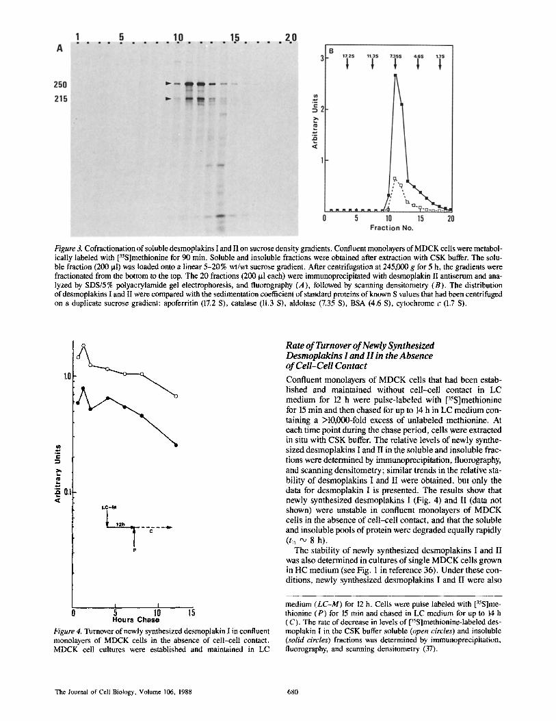
Figure 3. Cofractionation of soluble desmoplakins I and II on sucrose density gradients. Confluent monolayers of MDCK cells were metabol- ically labeled with [35S]methionine for 90 min. Soluble and insoluble fractions were obtained after extraction with CSK buffer. The solu- ble fraction (200 ~tl) was loaded onto a linear 5-20% wt/wt sucrose gradient. After centrifugation at 245,000 g for 5 h, the gradients were fractionated from the bottom to the top. The 20 fractions (200 t~1 each) were immunoprecipitated with desmoplakin II antiserum and ana- lyzed by SDS/5% polyacrylamide gel electrophoresis, and fluorography (A), followed by scanning densitometry (B). The distribution of desmoplakins I and II were compared with the sedimentation coefficient of standard proteins of known S values that had been centrifuged on a duplicate sucrose gradient: apoferritin (17.2 S), catalase (11.3 S), aldolase (7.35 S), BSA (4.6 S), cytochrome c (1.7 S).
1.0
W
C
o.1
LC-M
p
| I ...........
s lo Hours Chase
Figure 4. Turnover of newly synthesized desmoplakin I in confluent monolayers of MDCK cells in the absence of cetl-ceU contact. MDCK cell cultures were established and maintained in LC
Rate of Turnover of Newly Synthesized Desmoplakins I and H in the Absence of Cell-Cell Contact
Confluent monolayers of MDCK cells that had been estab- lished and maintained without cell-cell contact in LC medium for 12 h were pulse-labeled with [35S]methionine for 15 min and then chased for up to 14 h in LC medium con- taining a >10,000-fold excess of unlabeled methionine. At each time point during the chase period, cells were extracted in situ with CSK buffer. The relative levels of newly synthe- sized desmoplakins I and II in the soluble and insoluble frac- tions were determined by immunoprecipitation, fluorography, and scanning densitometry; similar trends in the relative sta- bility of desmoplakins I and II were obtained, but only the data for desmoplakin I is presented. The results show that newly synthesized desmoplakins I (Fig. 4) and II (data not shown) were unstable in confluent monolayers of MDCK cells in the absence of cell-cell contact, and that the soluble and insoluble pools of protein were degraded equally rapidly (t~ ~ 8 h).
The stability of newly synthesized desmoplakins I and II was also determined in cultures of single MDCK cells grown in HC medium (see Fig. 1 in reference 36). Under these con- ditions, newly synthesized desmoplakins I and II were also
medium (LC-M) for 12 h. Cells were pulse labeled with [35S]me- thionine (P) for 15 min and chased in LC medium for up to 14 h (C). The rate of decrease in levels of [35S]methionine-labeled des- moplakin I in the CSK buffer soluble (open circles) and insoluble (solid circles) fractions was determined by immunoprecipitation, fluorography, and scanning densitometry (37).
The Journal of Cell Biology, Volume 106, 1988 680

degraded rapidly (t,~ ~ 9 h; data not shown). Together these results indicate that the instability of newly synthesized desmoplakins I and II in MDCK cells grown in LC medium is not due to the low levels of calcium in the medium per se, but to the absence of cell-cell contact.
Differential Stabilization of Soluble and Insoluble Pools of Newly Synthesized Desmoplakins I and H upon Induction of Cell-Cell Contact
Analysis of the fate of newly synthesized desmoplakins I and II 3 h after synchronous induction of cell-cell contact re- vealed dramatic changes in the rates of turnover of the solu- ble and insoluble pools of protein (Fig. 5). For the first 5-6 h of the chase period, the soluble pool of desmoplakin I (and desmoplakin II, data not shown) was depleted rapidly of newly synthesized protein (t,~ ",, 2 h). Concomitantly, there was a proportional increase in the amount of newly synthe- sized protein in the insoluble pool. Subsequently, the fates of these two pools of newly synthesized proteins were dra- matically different. Desmoplakins I and II remaining in the soluble pool (,~,25-40% of the total) were degraded rapidly
0.1 <
,.~ t
LC-M HC-M
12h 13h
P
I I I I 0 ..... 5 10 15 20 25
Hours Chase Figure 5. Rate of turnover ofdesmoplakin I in confluent monolayers of MDCK cells 3 h after the induction of cell-cell contact. MDCK cell cultures were established in LC medium (LC-M) for 12 h. Cell-cell contact was induced by exchanging the LC medium with HC medium (HC-M). 3 h later cells were pulse labeled with [35S]methionine (P) for 15 min and then chased in HC medium for up to 24 (C). Soluble (open circles) and insoluble (solid circles) fractions were obtained by extraction in CSK buffer. The rate of de- crease in levels of [35S]methionine-labeled desmoplakin I was de- termined by immunoprecipitation, flurorography, and scanning densitometry (37).
with a half-life similar to that of the soluble proteins in cells maintained in LC medium (t,~ "~ 8-10 h). In contrast, in the insoluble pool desmoplakins I and II were relatively stable and exhibited little or no decay during the 24-h period of chase (t~ > 72 h).
Temporal Coordination of CeU-Cell Contact and Stabilization of the Insoluble Pool of Newly Synthesized Desmoplakins I and H
To determine how closely coordinated the induction of cell- cell contact and increased stability of desmoplakins I and II are, we analyzed the rates of turnover of newly synthesized protein at the time of induction of cell-cell contact, 30 min and 3 h after induction (Fig. 6). The results showed that at all time points examined a substantial proportion of newly synthesized desmoplakin I (and desmoplakin II, data not shown) was rapidly titrated from the soluble pool (Fig. 6 A) to the insoluble pool of protein (Fig. 6 B). In all cases, this titration occurred over a period of 3-5 h after the short period of labeling (15 min). The protein remaining in the soluble pool was degraded relatively rapidly (r/2 '~ 4-8 h), whereas the protein in the insoluble pool was relatively sta- ble (t,/2 > 72 h).
Recruitment of Unstable Newly Synthesized Desmoplakins I and H into a Stable, Insoluble Protein Pool upon Induction of Cell-Cell Contact
The experiments described above were designed to deter- mine the fate of desmoplakins I and II synthesized in the ab- sence or presence of cell-cell contact. Next, we wanted to analyze the fate of these proteins synthesized in MDCK cells that did not initially have cell-cell contact but which were later induced to form cell-cell contact. Since the insoluble pool of newly synthesized desmoplakins I and II is unstable in cells without cell-cell contact (t,~ ~ 8 h; see Fig. 4), we sought to determine whether this pool of protein could be sta- bilized upon cell-cell contact.
As shown earlier (Fig. 4), in the absence of ceil-cell con- tact both the soluble (Fig. 7 A) and insoluble (Fig. 7 B) pools of newly synthesized desmoplakin I (and desmoplakin II, data not shown) were degraded relatively rapidly (t~ ~ 8 h). However, if cell-cell contact was induced 2 h (line B) or 4 h (line C) after synthesis, the insoluble pool of desmoplakins I and II increased in size at the expense of the soluble pool; soluble protein titrated into the insoluble pool for a period of up to 2-3 h after induction of cell-cell contact. Subse- quently, the insoluble pool became stabilized (t,~ > 72 h). On the other hand, newly synthesized desmoplakins I and II that remained in the soluble pool were degraded rapidly with a rate of turnover similar to that of the soluble proteins in cells without cell-cell contact (r~ ,~ 8 h).
Is Protein Synthesis Required for the Stabilization of Newly Synthesized Desmoplakins I and H upon Induction of Cell-CeU Contact? To determine whether protein synthesis is required for stabilization of newly synthesized desmoplakins I and II, we analyzed the fate of newly synthesized desmoplakins I and II upon induction of cell-cell contact in the presence of cy- cloheximide (Fig. 8). The results showed that the rate of turnover of protein in both the soluble pool and the insoluble
Pasdar and Nelson Desmosome Assembly in Epithelial Cells I 681

1.0
C
m
.Q
~ o.J
A .~,
"-o.',~.:.. A
LC-M HC-M
L ~ _ _ _ ~ _ _ . . ~ .
A:Omin B: 3Omin C:180min
I 10
Hours Chase
B " ; . . . . . �9 . . . . . . . . . " - . . B
i i ," �9 11- . . . . . . 41 . . . . A t
z/:m. J" vt"
I I 5 10 15
Figure 6. Fate of the newly synthesized desmoplakin I upon induction of cell-cell contact in confluent monolayers of MDCK cells�9 MDCK cell cultures were established in LC medium (LC-M) for 12 h. Cells were metabolically labeled with [35S]methionine (P) for 15 min in HC medium (HC-M) at the time of cell-cell con- tact (line A), 30 rain after induction of cell-cell contact (line B), or 180 rain after induction of cell-cell contact (line C), and then chased for up to 14 h in HC medium (c). Cells were extracted with CSK buffer and the rate of decrease in levels of [35S]methionine-labeled des- moplakin I in the soluble (A) and insoluble (B) fractions was determined by immunoprecipitation, fluorography, and scanning densitometry (37).
1.0
c
I,,.
o.1 ,<
"-'~c ~zx B
LC-M
A: I 12h
LC-M
B: I 12h
LC-M
C: I 12h
I 0 5
T . . . . ~ . . . . P HC-M
P 4h H -MC -~
P
I 10
Hours C h a s e
B
/A__ ; ) -..-.: . . : . i "B / ,;" " C / ,"
~ A
I I 5 10
Figure 7. Recruitment of the newly synthesized unstable desmoplakin I into a stable insoluble pool upon induction of cell-cell contact. MDCK cell cultures were estab- lished in LC medium (LC-M) for 12 h. Cells were pulse labeled with [35S]methionine for 15 min (P) and chased (C) continuously in LC medium (line A), or induced to form cell-cell contact after 2 (line B) or 4 h (line C) in LC medium by replacing the media with HC medium. The decrease in levels of [35S]methionine-labeled des- moplakin I in the CSK buffer soluble (A) and insoluble (B) fractions was determined by immunoprecipitation, fluorography, and scanning densitometry (37).
The Journal of Cell Biology, Volume 106, 1988 682

L, .
�9 .~ 0.1
L C - M HC-M+CHX
p
1.0
I I
0 5 10 15 Hours Chase
Figure 8. Degradation of newly synthesized desmoplakin I in confluent monolayers of MDCK cells induced to form cell-cell contact in the presence of cycloheximide. MDCK cell cultures were established in LC medium (LC-M) for 12 h. Cells were pulse la- beled with [35S]methionine (P) for 15 min and then incubated in HC medium (HC-M) containing 4 ~tg/ml cycloheximide (CHX) (C). Cells were extracted with CSK buffer and the rate of decline in the levels of [35S]methionine-labeled desmoplakin I in the soluble (open circles) and insoluble (solid circles) fractions was determined by immunoprecipitation, fluorography and scanning densitometry (37).
pool was rapid (t,~ '~ 5 h). In addition, we did not detect a titration of newly synthesized protein from the soluble into the insoluble pool, as had been shown previously to be the case upon induction of cell-cell contact in the presence of protein synthesis (Figs. 5-7).
Discuss ion
The experiments performed in this study have sought to ana- lyze biochemically and kinetically the regulation of assem- bly of the desmosome upon cell-cell contact in MDCK epi- thelial cells. Particular emphasis has been placed on the two major cytoplasmic plaque proteins, desmoplakins I and II. These proteins were identified using a polyclonal antiserum that recognized an epitope common to both proteins. To study the asseembly of desmosomes, monolayers of MDCK cells were extracted in situ in order to preserve the native subcellular distribution and conformation of proteins with respect to cell-cell and cell-substratum contact (cf. refer- ence 15). The extraction of MDCK cells with CSK buffer resulted in a soluble and insoluble protein fraction of newly
synthesized desmoplakins I and II (see below). Significantly, the fates of these two pools of protein were different upon in- duction of cell-cell contact, indicating that they are not de- rived artificially from a single pool of protein (see below).
Temporal Coordination between Cell-Cell Contact and the Assembly of Desmoplakins I and H
The fate of protein pools of newly synthesized desmoplakins I and II in the presence and absence of cell-cell contact was determined by modulating the degree of cell-cell contact in confluent monolayers of MDCK epithelial cells by adjusting the Ca ++ concentration of the growth medium (37). In the absence of cell-cell contact we detected both a soluble and insoluble pool of newly synthesized desmoplakins I and II, both of which were unstable and were degraded equally rap- idly (t,~ "~ 8 h). The fact that a proportion (~35%) of des- moplakins I and II was insoluble under extraction conditions that did not solubilize desmosomes (11, 15, 23, 25, 33, 35, 48, 49) indicates that they were in a conformation, perhaps with other desmosomal proteins (see below), similar to that in fully assembled desmosomes, even though desmosomes had not formed at the plasma membrane (see accompanying paper).
When the rate of degradation of newly synthesized des- moplakins I and II was analyzed at the time of induction of cell-cell contact or 3 h after induction, we found that there was a significant difference in the fate of the insoluble and soluble pools of protein. While the soluble pool of protein was degraded as rapidly as in cells without cell-cell contact (t,~ "~ 8 h), the insoluble pool was stabilized (t,~ > 72 h). Thus, there appears to be a close temporal coordination be- tween the induction of cell-cell contact and the stabilization of this insoluble pool of newly synthesized desmoplakins I and II.
In addition to the change in the stability of the insoluble pool of newly synthesized desmoplakins I and II upon cell-cell contact, we observed that there was a change in its relative size (Figs. 5-7). In the absence of cell-cell contact, after 1 h of chase, the relative sizes of the insoluble and solu- ble pools of newly synthesized protein remained constant and were subsequently reduced at the same rate by degrada- tion (tw ~ 8 h). However, after induction of cell-cell con- tact, there was a gradual titration of newly synthesized pro- tein from the soluble to the insoluble pool (Fig. 5). That the soluble pool was being depleted of newly synthesized protein was reflected in the very rapid loss of protein from this pool (tv2 '~ 2 h). Concomitantly, the insoluble pool increased in size by approximately three to fourfold over a period of 3-5 h, and then the level of protein remained relatively constant. At this time, the rate of loss of protein from the soluble pool slowed to a rate similar to that in cells without cell-cell con- tact (t,~ '~ 8 h), at which time loss of protein was presum- ably due to proteolytic degradation. Significantly, we found that when cell-cell contact was induced 2 h or 4 h after syn- thesis of a discrete population of desmoplakins I and II, there was a similar rapid titration of these proteins from the unsta- ble soluble pool to the stable insoluble pool. Hence, the ca- pacity of the insoluble pool of desmoplakins I and II in- creases upon cell-cell contact as newly synthesized proteins are recruited from an unstable soluble pool of protein. Therefore, while the formation of the insoluble pool of des-
Pasdar and Nelson Desmosome Assembly in Epithelial Cells I 683

moplakins I and II is independent of cell-cell contact (see above), its capacity and stability are strictly dependent upon cell-cell contact.
That newly synthesized desmoplakins I and II can be rap- idly recruited from the soluble to insoluble pool upon cell- cell contact indicates that the soluble pool of protein might represent a precursor step in the assembly of desmoplakins I and II into the insoluble pool. Significantly, analysis of the soluble pool by sucrose density gradient centrifugation re- vealed that desmoplakins I and II cosediment as a complex in the same molar ratio as that of the newly synthesized pro- teins in the insoluble pool. This is consistent with the possi- bility that the soluble desmoplakins I and II are a precursor pool of protein that can be rapidly titrated into the insoluble pool.
Regulation of Assembly of Desmoplakins I and H Even under conditions of extensive cell-cell contact, not all of the soluble newly synthesized desmoplakins I and II are titrated into the insoluble pool of protein. This result indi- cates that the capacity of the insoluble pool of newly synthe- sized desmoplakins I and II is limited. This is supported by our analysis of the effect(s) of inhibition of protein synthesis on cell-cell contact. In this experiment, we analyzed the fate of desmoplakins I and II synthesized in a narrow window of time (0-15 min) before the induction of cell-cell contact and the addition of cycloheximide. Under these conditions, the preformed soluble and insoluble pools of newly synthesized desmoplakins I and II were degraded equally rapidly %/2 ~ 5 h). While there is clearly a dramatic effect of inhibition of protein synthesis on the stability of preformed soluble and in- soluble pools of newly synthesized desmoplakins I and II, there appears to be little or no temporal or spatial effect of this inhibition on the assembly of desmoplakins I and II at steady state into desmosomes upon cell-cell contact in MDCK cells (see accompanying paper). Thus, these results indicate that the assembly of newly synthesized desmoplak- ins I and II requires the coordinate synthesis of a protein(s) that regulates the capacity and stability of the insoluble pool of this population of newly synthesized desmoplakins I and II. However, desmoplakins I and II that had accumulated at steady state together with this limiting protein(s) before the induction of cell-cell contact are able to assemble into des- mosomes in the absence of additional protein synthesis. This protein(s) appears not to be involved in the formation of the insoluble pool of desmoplakins I and II per se, since the ratio of proteins in the soluble and insoluble pools was the same in the presence of cycloheximide as in the absence of cell- cell contact. However, since the increase in capacity of the insoluble pool upon cell-cell contact coincides temporarily with the formation of desmosomes on the plasma membrane (see accompanying paper), one of the membrane core sub- units located on the plasma membrane may play a role in stabilizing newly synthesized desmoplakins I and II.
This study of the stabilization and organization of des- moplakins I and H raises several questions concerning the regulation of protein transition between these assembly stages, and the interconnection of the soluble and insoluble pools of protein. Analysis of the organization and stability of other desmosomal proteins, particularly with respect to the soluble and insoluble pools of desmoplakins I and II, may
provide answers to some of these questions. In the accom- panying paper, we have reevaluated the coordination of cell- cell contact and assembly of desmoplakins I and II by im- munofluorescence microscopy in the light of these new ob- servations, and have sought to determine the spatial nature of the soluble and insoluble pools of these proteins.
We thank Secretarial Services for typing the manuscript. This work was supported in part by grants to W. J. Nelson from the Na-
tional Institutes of Health (GM-35527) and the National Science Foundation (DCB-8609091); from the National Institutes of Health to the Institute for Cancer Research (CA-06927, RR-05539); and by an appropriation from the Commonwealth of Pennsylvania. M. Pasdar was also supported by a post- doctoral fellowship from the National Institutes of Health (CA-09035).
Received for publication 7 August 1987, and in revised form 3 November 1987.
References
1. Arnn, J., and L. A. Staehelin. 1981. The structure and function of spot des- mosomes. Int. J. Dermatol. 20:330-339.
2. Campbell, R. D., and J. H. Campbell. 1971. Origin and continuity of des- mosomes. In Origin and Continuity of Cell Organelles, J. Reinert and H. Ursprung, editors. Springer-Verlag, Berlin. 261-298.
3. Cohen, S. M., G. Gorbsky, and M. S. Steinberg. 1983. Immunochemical characterization of related families of glycoproteins in desmosomes. J. BioL Chem. 258(Suppl. 4):2621-2627.
4. Cowin, P., W. W. Franke, C. Grund, H. P. Kapperell, and J. Kartenbeck. 1985. The desmosome-intermediate filament complex. In The Celt in Contact: Adhesion and Junctions as Morphogenetic Determinants. G. Edelman and J. P. Thiery, editors. John Wiley and Sons, Inc., New York. 427-460.
5. Cowin, P., H. P. Kapprell, and W. W. Franke. 1985. The complement of desmosomal plaque proteins in different cell types, J. Cell Biol. 101: 1442-1454.
6. Cowin, P., H. P. Kapprell, W. W. Franke, J. Tamkun, and R. O. Hynes. I986. Plakoglobin: a protein common to different kinds of intercellular adhering junctions. Cell. 46:1063-1073.
7. Cowin, P., and D. R. Garrod. 1983. Antibodies to epithelial desmosomes show wide tissue and species cross reactivity. Nature (Lond.). 302: 148-150.
8. Cowin, P., D. L. Mattey, and D. R. Garrod. 1984. Distribution of des- mosomal components in the tissues of vertebrates studied by fluorescent antibody staining. J. Cell Sci. 66:119-132.
9. Cowin, P., D. L. Mattey, and D. R. Garrod. 1984. Identification of des- mosomal surface components (desmocollins) and inhibition of desmo- some formation by specific Fab. J. Cell Sci. 70:41-60.
10. Docherty, R. J., J. G. Edwards, D. R. Garrod, and D~ L. Mattey. 1984. Chick embryonic pigmented retina is one of the group of epitheloid tis- sues that lack cytokeratins and desmosomes and have intermediate fila- ments composed of vimentin. J. Cell Sci. 71:61-74.
l 1. Drochmans, P., C. Freudenstein, J. C. Wanson, L. Lewrent, T. W. Kee- nan, J. Stadler, R. Leloup, and W. W. Franke. 1978. Structure and bio- chemical composition of desmosome and tonofilaments isolated from calf muzzle epidermis. J. Cell Biol. 79:427-443.
12. Edelman, G. M. 1985. Cell adhesion and the molecular processes of mor- phogenesis. Annu. Rev. Biochem. 54:135-169.
13. Engvall, E., and P. Perlman. 1971. Enzyme-linked immunosorbent assay (ELISA). Quantitative assay of immunoglobulin-G, tmmunochemistry. 8(Suppl. 9):871-874.
14. Farquhar, M. G., and G. E. Palade. 1963. Junctional complexes in various epithelia. J. Cell Biol. 17:375-412.
15. Fey, E. G., K. M. Wan, and S. Penman. 1984. Epithelial cytoskeletal framework and nuclear matrix-intermediate filament scaffold: three- dimensional organization and protein composition. J. Cell Biol. 98: 1973-1984.
16. Franke, W. W., H. Mueller, S. Mittnacht, H. P. Kapprell, and J. L. Jor- cano. 1983. Significance of two desmosome plaque-associated polypep- tides of molecular weights 75,000 and 83,000. EMBO (Eur. Mot. Biol. Organ.) J. 2:2211-2215.
17. Franke, W. W., E. Schmid, C. Grund, H. Muller, H. Engelbrecht, R. Moll, J. Stadler, and E. D. Jarasch. 1981. Antibodies to high molecular weight polypeptides of desmosome: specific localization of a class of junctional proteins in cells and tissues. Differentiation. 20:217-241.
18. Garrod, D. R. 1986. Formation of desmosomes in polarized and non- polarized epithelial cells: implications for epithelia morphogenesis. Bio- chem. Soc. Trans. 14:172-175.
19. Garrod, D. R. 1986. Desmosomes, cell adhesion molecules, and the adhe- sive properties of tissue cells. J. Cell Sci. Suppl. 4:221-237.
The Journal of Cell Biology, Volume 106, 1988 684

20. Garrod, D. R., and P. Cowin. 1986. Desmosomes structure and function. In Receptors in Tumor Biology. C. M. Chadwick, editor. Cambridge University Press, Cambridge. 95-130,
21. Greiger, B., Z. Avnur, T. Volberg and T. Volk. 1985. Molecular domains of adherens junctions. In The Cell in Contact. G. M. Edelman and J. P. Thiery, editors. John Wiley and Sons, Inc., New York. 461-489.
22. Geiger, B., E. Schmid, and W. W. Franke. 1983. Spatial distribution of proteins specific for desmosomes and adherens junctions in epithelial cells demonstrated by double immunofluorescence microscopy. Differen- tiation. 23:189-205.
23. Gorbsky, G., and M. S. Steinberg. 1981. Isolation of intercellular glyco- proteins of desmosomes. J. Cell Biol. 90:243-248.
24. Giudice, G. L, S. M. Cohen, N. H. Patel, and M. S. Steinberg. 1984. Im- munological comparison of desmosomal components from several bovine tissues. J. Cell. Biochem. 26:35-45.
25. Jones, J. C, R., and R. D. Goldman. 1985. Intermediate filaments and initi- ation of desmosome assmebly. 3". Cell Biol. 101:506-517.
26. Jones, J. C. R., A. E. Goldman, P. M. Steinert, S. Yuspa, and R. D. Gold- man. 1982. Dynamic aspects of the supramolecular organization of inter- mediate filaments network in cultured epidermal cells. Cell Motil. 2: 197-213.
27. Kelly, D. E. 1966. Fine structure of desmosomes, hemi-desmosomes and an adepidermal globular layer in developing newt epidermis. J. Celt BioL 28:51-72.
28. Kelly, D. E., and F. L. Shienvold. 1976. The desmosome: line structure studies with freeze-fracture replication and tannic acid staining of sec- tioned epidermis. Cell Tissue Res. 272:309-323.
29. Kapprell, H. P., P. Cowin, and W. W. Franke. 1985. Biochemical charac- terization of desmosomal proteins isolated from bovine muzzle epider- mis: amino acid and carbohydrate composition. Eur. J. Cell Biol. 36: 217-229.
30. Lamelli, U. K. 1970. Cleavage of structural proteins during the assembly of the head of bacteriophage T4. Nature (Lond.). 227:680-685.
31. Laskey, R. A., and D. Mills. 1975. Quantitative detection of 3H and ~4C in polyacrylamide gels by fluorography. Eur. J. Biochem. 56:335-341.
32. Madin, S. J., and N. B. Darby. 1979. American Type Culture Collection Catalogue of Strains II. American Type Culture Collection, Rockville, MD. 30.
33. Mattey, D. L., and D. R. Garrod. 1986. Calcium-induced desmosome for- mation in cultured kidney epithelial cells. J. Cell Sci. 85:95-111.
34. Mattey, D. L., and D. R. Garrod. 1986. Splitting and internalization of the desmosomes of cultured kidney epithelial cells by reduction in calcium concentration. J. Cell Sci. 85:113-124.
35. Mueller, H., and W. W. Franke. 1983~ Biochemical and immunological characterization of Desmoplakins I and lI, the major polypeptides of the desmosomal plaque. J. MoL Biol. 163:647-671.
36. Nelson, W. J., and P. J. Veshnock. 1986. Dynamics of mambrane-skeleton (fodrin) organization during development of polarity in Madin-Darby kidney epithelial cells. J. Cell Biol. 103:1751-1765.
37. Nelson, W. J., and P. J. Veshnock. 1987. Modulation of membrane- skeleton (fodrin) stability by cell-cell contact in Madin-Darby canine kidney epithelial cells. J. Cell Biol. 104:1527-1537.
38~ Overton, J. 1962. Desmosome development in normal and reassociating cells of early chick blastoderm. Dev. Biol. 4:532-548.
39. Overton, J. 1968. The fate of desmosomes in trypsinized tissue. J. F,~. Zool. 168:203-214.
40. Overton, J. 1973, Experimental manipulation of desmosome formation. J. Cell Biol. 56:636-646.
41. Overton, J. 1974. Cell junctions and their development. Prog. Surf. Membr. Sci. 8:161-208.
42. Overton, J. 1974. Selective formation of desmosomes in chick cell reag- gregates. Dev. Biol. 39:210-225.
43. Overton, J. 1975. Experiments with junctions of the adherens types. Curt. Top. Dev. Biol. 10:1-34.
44~ Overton, J., and R. DeSalle. 1980. Control ofdesmosome formation in ag- gregating embryonic chick cells. Dev. BioL 75:168-t76.
45. Penn, E. J., C. Hobson, D. A. Rees, and A. I. Magee. 1987. Structure and assembly of desmosome junctions: biosynthesis, processing, and trans- port of the major protein and glycoprotein components in cultured epithe- lial cells. J. Cell Biol. 105:57-68.
46. Pirbazari, M., and D. E. Kelly. 1985. Analysis of desmosomal intramem- brahe particle populations and cytoskeletal elements: detergent extraction and freeze-fracture. Cell Tissue Res. 241: 341-351.
47. Skerrow, C. J. 1985. Desmosomal proteins. In Biology of the Integrument. Vertebrates Vol. 2. J. Bereiter-Hahn, A. G. Matoltsy, and K. S. Richards, editors. Springer-Verlag, Berlin. 763-787.
48. Skerrow, C. J., I. Hunter, and D. Skerrow. 1987. Dissection of the bovine epidermal desmosome into cytoplasmic protein and membrane glycopro- tein. J. Cell Sei. 87:4111--421.
49. Skerrow, C. J., and A. G. Matoltsy. 1974. Isolation of epidermal desmo- somes. J. Cell Biol. 63:515-523.
50. Stevenson, B. R., and D. A. Goodenough. 1984. Zonulae occludentes in junctional complex-enriched fractions from mouse liver. Preliminary morphological and biochemical characterization. J. Cell BioL 98:1209- 1221.
5l. Stevenson, B. R., J. D. Siliciano, M. S. Mooseker, and D. A. Goodenough. 1986. Identification of ZO-I: a high molecular weight polypeptide as- sociated with the tight junction (zonula occludens) in a variety of epithe- lia. J. Cell Biol. 103:755-766.
52. Suhrbier, A., and D, Garrod. 1986. An investigation of the molecular com- ponents of desmosomes in epithelial cells of five vertebrates. J. Cell Sci. 81:223-242.
53. Towbin, H., T. Staehlin, and J. Gordon. 1979. Electrophoretic transfer of proteins from polyacrylamide gels to nitrocellulose sheets: procedures and some applications. Proc. Natl. Acad. Sci. USA. 76:4350-4354.
54, Volk, T., and B. Geiger. 1984. A 135-kd membrane protein of intercellular adherens junctions. EMBO (Eur. Mol. Biol. Organ.) J. 3(10):2249- 2260.
55. Warren, S. L., and W. J. Nelson. 1987. Nonmitogenic morphoregulatory action of pp60 ' ' ~ on multicellular epithelial structures. Mol. Celt. Biol. 7 (Suppl. 4): 1326-1337.
56. Watt, F. M., D. L. Mattey, and D. R. Garrod. 1984. Calcium-induced re- organization of desmosomal components in cultured keratinocytes. J, Cell Biol. 99:2211-2215,
Pasdar and Nelson Desmosome Assembly in Epithelial Cells I 685