Embed Size (px)
Citation preview
Biophysical Journal Volume 98 May 2010 1911–1920 1911
Kinetics and Reaction Coordinates of the Reassembly of ProteinFragments Via Forward Flux Sampling
Ernesto E. Borrero, Lydia M. Contreras Martı́nez, Matthew P. DeLisa, and Fernando A. Escobedo*School of Chemical and Biomolecular Engineering, Cornell University, Ithaca, New York
ABSTRACT We studied the mechanism of the reassembly and folding process of two fragments of a split lattice protein byusing forward flux sampling (FFS). Our results confirmed previous thermodynamics and kinetics analyses that suggested thatthe disruption of the critical core (of an unsplit protein that folds by a nucleation mechanism) plays a key role in the reassemblymechanism of the split system. For several split systems derived from a parent 48-mer model, we estimated the reaction coor-dinates in terms of collective variables by using the FFS least-square estimation method and found that the reassembly transitionis best described by a combination of the total number of native contacts, the number of interchain native contacts, and the totalconformational energy of the split system. We also analyzed the transition path ensemble obtained from FFS simulations usingthe estimated reaction coordinates as order parameters to identify the microscopic features that differentiate the reassembly ofthe different split systems studied. We found that in the fastest folding split system, a balanced distribution of the original-coreamino acids (of the unsplit system) between protein fragments propitiates interchain interactions at early stages of the foldingprocess. Only this system exhibits a different reassembly mechanism from that of the unsplit protein, involving the formationof a different folding nucleus. In the slowest folding system, the concentration of the folding nucleus in one fragment causesits early prefolding, whereas the second fragment tends to remain as a detached random coil. We also show that the reassemblyrate can be either increased or decreased by tuning interchain cooperativeness via the introduction of a single point mutation thateither strengthens or weakens one of the native interchain contacts (prevalent in the transition state ensemble).
INTRODUCTION
Protein fragment complementation assays (PCAs) have been
powerful experimental tools for assaying highly specific
interactions involving cellular proteins (1–5). This approach
is based on splitting a reporter protein into two individual
fragments that by themselves remain inactive but on reas-
sembly, yield the original properly folded and active protein
structure. Examples of these systems include split green
fluorescent protein (GFP) and its spectral variants YFP and
CFP (2,6), ubiquitin (7), murine dihydrofolate reductase
(mDHFR) (8), b-lactamase (9,10), and firefly luciferase (11).
The reconstituted activity of all these proteins can be conve-
niently detected by fluorescence or well-established enzy-
matic assays. The application of PCAs in living cells has
become an invaluable tool for mapping protein-protein and
protein-nucleic acid interaction networks.
A major factor limiting the usefulness of split proteins
is the slow folding kinetics and formation of misfolded
aggregates that is associated with the reassembly process
of multiple fragments (2,12). This inefficiency hinders the
effective application of PCAs on biologically relevant time-
scales. For instance, although GFP fluorescence can be
detected in minutes, the two fragments that result when the
protein is dissected in the middle of the sequence fail to asso-
ciate and reassemble when expressed in bacteria (2). Even
when the fragments are each fused to strongly interacting
Submitted August 14, 2009, and accepted for publication December 15,2009.
*Correspondence: [email protected]
Editor: Costas D. Maranas.
� 2010 by the Biophysical Society
0006-3495/10/05/1911/10 $2.00
leucine zippers (KD ~1–2 mM), folding and fluorescence
activity of the reconstituted protein is not observed until after
1–2 days (13). Given that similar drawbacks have been
observed for different split reporters, significant effort has
been focused on strategies to accelerate the formation of
a reassembled protein. Some attempts include: i), identifica-
tion of multiple permissive split sites along the protein,
typically away from the catalytic site, using circular permu-
tation (14,15); ii), structure-guided design, using bioinfor-
matic and theoretical analysis (9,16,17); iii), optimization
of target sequence using directed evolution for more efficient
folding/reassembly (18,19); and, most recently, iv), the addi-
tion of hybridizing molecules (20). Despite these efforts, our
understanding of how parameters such as the split site posi-
tion in the primary sequence and size of resulting fragments
contribute to the efficiency of protein reassembly remains
limited.
Proteins can fold by diverse pathways including nucle-
ation-condensation, framework (hierarchical) model, and
hydrophobic collapse models (21). The nucleation-conden-
sation mechanism describes the overall features of folding
of most domains by uniting features of the other two folding
models and invoking the formation of hydrophobic and long
range interactions in the transition state (TS) to stabilize
weak secondary structures. Given that in a nucleation folding
mechanism a few key residues known as the folding nucleus
provide a significant driving force in the formation of the TS
leading to the folding of many proteins (22–24), a clear
understanding of how these amino acids are distributed
between fragments could be key to our progress in designing
doi: 10.1016/j.bpj.2009.12.4329
1912 Borrero et al.
efficient split protein systems. Intrigued by this notion, we
previously used brute-force Monte Carlo simulations to
analyze how the thermodynamics and kinetics of the reas-
sembly process for two split protein model systems were
affected by the location of the split site (25). In that study,
we rationalized thermodynamically why reassembly of a
split fragment system is significantly slower than the folding
of an unsplit protein. We showed that strategic splitting
of the folding nucleus, where the nucleus is more equally
shared between the two fragments, drastically accelerated
reassembly by: i), preventing the permanent preassembly
of an individual fragment that would otherwise lead to a
slower two-step assembly process where chain preassembly
precedes interchain contacts; and ii), driving the formation of
interchain native contacts that promoted a cooperative and
productive folding. Interestingly, reassembly of split ubiqui-
tin is observed experimentally when the protein is frag-
mented such that the amino acid residues that make up the
compact hydrophobic core (26,27) have a 60–40% distribu-
tion between fragments (7). In Contreras Martı́nez et al. (25),
however, we did not provide a precise characterization of the
folding mechanism or of the TS.
Several transition path sampling methods have been
developed to study the kinetics of biomolecular rare events
(28,29) by enhancing the sampling of the transition region.
These methods allow the estimation of rate constants and
the collection of pathways (i.e., the transition path ensemble
(TPE) to establish the transition mechanism, which would
be impractical via conventional brute-force simulations.
The technique of choice for this study is the forward flux
sampling (FFS) method because of its simplicity and effi-
ciency. Two of us (E. Borrero and F. Escobedo) used FFS
previously to evaluate the kinetics of the transition pathways
for the folding mechanism of a single chain (unsplit) 48-mer
lattice protein (30). We showed that the initial formation of
a critical core of amino acids (24,31) is the most important
step during folding, a result that is relevant for proteins after
a nucleation folding mechanism.
In this study, we further investigate the mechanism of
reassembly of the same model 48-mer system and split sys-
tems, taking advantage of optimized FFS-type approaches
(32,33) to analyze mechanistic details of their folding path-
ways (TPE) and estimate a suitable reaction-coordinate
(RxC). The RxC essentially corresponds to the committor
probability (pB) surface, which gives the probability of any
particular configuration to reach the final folded state. After
the RxC is parameterized in terms of physically meaningful
properties that describe the system’s dynamics, the mecha-
nistic details of the transition can be extracted by screening
the microscopic properties of the ensemble of configurations
belonging to different pB isosurfaces (e.g., for pB ¼ 0.5,
which corresponds to the TS). Our results provide insight
to two interesting questions that arose from our previous
analysis: i), whether the split fragments exhibited the same
folding nucleus and nucleation-driving folding mechanism
Biophysical Journal 98(9) 1911–1920
as the parent protein; and ii), whether the same folding mech-
anism for the parent protein (or a highly similar one) would
be optimal for fragment reassembly.
SIMULATION DETAILS
In FFS, interfaces are used to partition the phase space along
an order parameter l connecting the initial and final regions
of interest. On each point at each interface, multiple trial runs
(ki) are carried out to promote successful partial paths
between interfaces. In the Supporting Material, a short
description is given of the branched growth (BG) method
adopted that can optimize the selection of l as RxC, the
spacing of l, and the number of trial trajectories per point ki.
Unsplit system
The 48-mer protein model adopted here exhibits a fast and
stable proteinlike folding into a unique native structure via
a two-state (unfolded-folded) process whose transition path-
ways are known to follow a nucleation-driven folding mech-
anism (30). The formation of a critical core of amino acids
mediates the folding of the single-chain (unsplit) protein
(23,24). This critical core is formed at an early stage of the
process by those residues that have a higher chance of being
in contact in the TS (30) and was composed of several
(mostly hydrophobic) amino acid residues, that have >80%
probability of forming native contacts (i.e., residues: 13, 16,
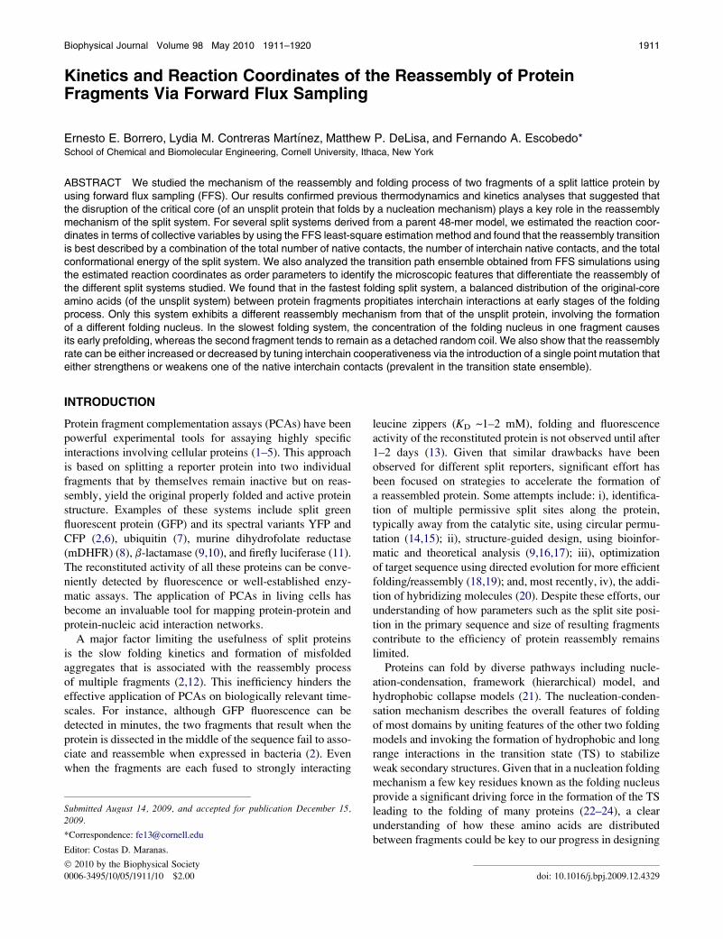
17, 19–24, 26–31, and 34–37). Fig. 1 shows the contact map
density for all the native contacts belonging to the ensemble
of configurations at isocommittor pB ¼ 0.2, 0.5, and 0.8 sur-
faces. The configurations belonging to different regions of
the isocommittor surface were collected by calculating the
pB value for each TPE interfacial configuration via the
following equation:
pBðNC;EÞ ¼ �0:404 þ 0:017ðNCÞ � 0:029ðEÞ: (1)
This RxC model estimates the probability of any TPE
interfacial point to commit to the folded state from the NCand E values of that point (30). Fig. 1, center, shows that
the 15 critical core (CC) native contacts with higher proba-
bility to belong to the TS (pB¼ 0.5) are formed by those resi-
dues forming the folding nucleus. Fig. 1, top, shows that
these CC native contacts start to form the nucleus at early
stages of the folding process (pB ¼ 0.2). Fig. 1, bottom,
shows that at late stages of the folding process (pB ¼ 0.8),
the protein acquires its native structure by forming contacts
around the folding nucleus.
Split system preparation
The split lattice model proteins were generated by dissecting
the 48-mer at one of three possible positions: between resi-
dues 16 and 17 (N-split), 24 and 25 (M-split), and 32 and
33 (C-split). In all these cases the folded state is identical
to the one reached by the single 48-mer chain and
FIGURE 1 Contact density map for the unsplit 48-mer system for ensem-
bles of configurations belonging to isocommittor surfaces: pB ¼ 0.2 (top),
pB ¼ 0.5 (center), and pB ¼ 0.8 (bottom). The x and y axis represent the
amino acid (aa) position in the 48-mer sequence. The ensembles were
collected by estimating pB values for all the interfacial points in the TPE
from the RxC model in Eq. 1. The lower triangle (below the diagonal
line) shows the probability of a native contact to belong to the ensemble;
the color code is given by the vertical bar. The lower triangle at the isocom-
mittor surface pB ¼ 0.8 (bottom) shows the 57 pair contacts for the native
structure. The upper triangle shows those native contacts with at least
80% probability to belong to the corresponding pB ensemble. Encircled
symbols represent native contacts that form the critical folding nucleus.
Snapshots depicting typical configurations observed for each ensemble are
also shown where red/dark gray indicates native contacts and white indicates
native contacts that form the critical folding nuclei.
Kinetics of Protein Reassembly 1913
characterized by NNC¼58 native contacts. Table 1 gives the
main characteristics for all the systems (i.e., the split and the
unsplit systems), including the number of interchain CC
contacts (InterC) that involve interacting residues from
both chains and intrachain CC contacts (IntraC) that involve
interacting residues within the same chain. These InterC and
IntraC are formed by the original 15 critical core native
contacts (identified in Fig. 1) on protein fragmentation.
Note that in the C- and Mid-split cases, the folding core resi-
dues are well distributed between fragments and give a signif-
icant number of InterC. In contrast, for the N-split system
most of the folding core residues are concentrated in chain
B and are not involved in InterC (25). Moreover, the N-split
and the C-split systems are symmetrical, with each system
having one 16-mer fragment and one 32-mer fragment; this
allows a comparison in the absence of chain length dispar-
ities. Further details on the model unsplit and split systems,
including their structure and thermodynamics are given in
Contreras Martı́nez et al. (25).
Conformational sampling
Conformational local sampling was carried out through a set
of MC moves based on the Verdier-Stockmayer algorithm
(34). Relative to these local moves, whole-fragment diffu-
sional translation of a randomly selected chain was also
attempted after each MC step with a priori probability
(%10�4) (25). For simulating the folding kinetics, the
temperature was fixed at T ¼ 0.25, a value close to the fold-
ing transition temperature of the unsplit system. The system
was confined inside a relatively large 3-D cubic box of side
length (L) 12 s (where s is the lattice size ¼ size of a protein
residue) corresponding to a protein volume fraction of ~3%
(25). Because of this dilution, it is assumed that spatial
restriction affects the translational entropy of symmetrically
and asymmetrically split systems in a commensurate way,
and has negligible effect on conformational entropy (25,30).
In comparing different split protein systems, the analysis in
our previous work (25) indicated that differences in thermo-
dynamic and kinetic behavior were not determined by diffu-
sion limitations of the fragments trying to find each other.
The spatial constriction also mimics a moderately crowded
environment relative to open space, ensuring a timely asso-
ciation of the different fragments.
Candidate collective variables
In the simulations, the following macroscopic properties
were calculated for all the state points collected at the l
interfaces in the TPE trajectories: total number of native
contacts (NC), number native contacts in chain A (NNA),
number native contacts in chain B (NNB), number of con-
tacts between fragments (IC), number of native contacts
between fragments (INC), conformational energy (E), and
the number of critical core contacts (CC) (the latter as iden-
tified for the unsplit system). These collective variables were
used for the RxC analysis via the FFS-LSE method.
RESULTS
A first preliminary BG simulation was carried out using the
number of native contacts as initial guess of the order param-
eter (i.e., l ¼ NC) with the purpose of optimizing the posi-
tion (l values) and sampling (k values) of 12 interface
ensembles. Details of this calculation and its results are given
in the Supporting Material. These optimized parameters were
then used to obtain the pB history data via BG simulations
with the FFS-LSE method (see Supporting Material). These
pB data were then used to screen a set of candidate collective
properties (see above) for an optimized order parameter
model l, as described in the Supporting Material. Thereafter,
Biophysical Journal 98(9) 1911–1920
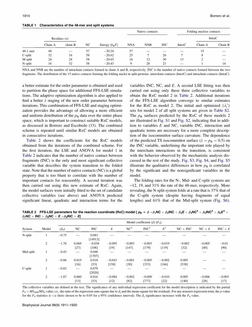
TABLE 1 Characteristics of the 48-mer and split systems
Residues (n)
NC Energy [kBT]
Native contacts Folding nucleus contacts
NNA NNB INC InterC
IntraC
Chain A chain B Chain A Chain B
48-1 mer 48 — 57 �20.24 57 — — — 15 —
C-split 32 16 58 �20.62 25 7 26 7 8 —
M-split 24 24 58 �20.65 16 12 30 13 2 —
N-split 16 32 58 �20.43 9 28 21 — 1 14
NNA and NNB are the number of intrachain contacts formed in chain A and B, respectively. INC is the number of native contacts formed between the two
fragments. The distribution of the 15 native contacts forming the folding nuclei in split-proteins: interchain contacts (InterC) and intrachain contacts (IntraC).
1914 Borrero et al.
a better estimate for the order parameter is obtained and used
to partition the phase space for additional FFS-LSE simula-
tions. The adaptive optimization algorithm is also applied to
find a better l staging of the new order parameter between
iterations. This combination of FFS-LSE and staging optimi-
zation provides the advantage of allowing a more efficient
and uniform distribution of the pB data over the entire phase
space, which is important to construct suitable RxC models,
as discussed in Borrero and Escobedo (33). The combined
scheme is repeated until similar RxC models are obtained
in consecutive iterations.
Table 2 shows the coefficients for the RxC models
obtained from the iterations of the combined scheme. For
the first iteration, the LSE and ANOVA for model 1 in
Table 2 indicates that the number of native contact between
fragments (INC) is the only and most significant collective
variable that describes the system transition to the folded
state. Note that the number of native contacts (NC) is a global
property that is too blunt to correlate with the number of
important contacts for reassembly. A second iteration was
then carried out using this new estimate of RxC. Again,
the model surfaces were initially fitted to the set of candidate
collective variables (see above) and ANOVA predicted
significant linear, quadratic and interaction terms for the
TABLE 2 FFS-LSE parameters for the reaction coordinate (RxC) mo
b7NC � INC þ b8NC � E þb9INC � E]
System Model
M
(b0) NC INC E NC
N-split 1 �0.75 — 0.083
[14913]
— —
2 �1.78 0.069
[27]
�0.038
[108]
�0.095
[19]
�0.
[14
Mid-split 1 �0.43 — 0.048
[1707]
— —
2 �0.86 0.019
[16]
0.018
[53]
�0.043
[158]
�0.
[3
C-split 1 �0.82 — 0.079
[2020]
— —
2 �1.87 0.060
[13]
0.016
[53]
�0.084
[12]
�0.
[8
The collective variables are defined in the text. The significance of any individu
F0¼MSSSR/MSE value; i.e., the ratio of the regression sum-square due to bj and th
for the F0 statistics is <a (here chosen to be to 0.05 for a 95% confidence inter
Biophysical Journal 98(9) 1911–1920
variables INC, NC, and E. A second LSE fitting was then
carried out using only these three collective variables to
obtain the RxC model 2 in Table 2. Additional iterations
of the FFS-LSE algorithm converge to similar estimates
for the RxC as model 2. The initial and optimized {li0}
sets for model 2 of all split systems are given in Table S2.
The pB surfaces predicted by the RxC of these models 2
are illustrated in Fig. S1 and Fig. S2, indicating that in addi-
tion to variables E and NC, variable INC, interaction and
quadratic terms are necessary for a more complete descrip-
tion of the isocommittor surface curvature. The dependence
of the predicted TS isocommittor surface (l ¼ pB ¼ 0.5) on
the INC variable, underlining the important role played by
the interchain interactions in the transition, is consistent
with the behavior observed by the mechanistic analysis dis-
cussed in the rest of the study. Fig. S3, Fig. S4, and Fig. S5
illustrate the qualitative differences in how pB is correlated
by the significant and the nonsignificant variables in the
model.
The folding rates for the N-, Mid- and C-split systems are
~12, 19, and 31% the rate of the 48-mer, respectively. More
revealing, the N-split system folds at a rate that is 37% that of
the C-split system (despite having fragments of equal
lengths) and 61% that of the Mid-split system (Fig. S6).
del [ pB z b þb1NC þ b2INC þ b3E þ b4NC2 þ b5INC2 þ b6E 2 þ
odel coefficient (b) [F0]
2 INC2 E2 NC � INC NC � E INC � E
— — — — —
003
7]
�0.003
[179]
�0.019
[119]
�0.002
[32]
�0.005
[40]
�0.01
[90]
— — — — —
001
0]
�0.005
[333]
�0.002
[166]
0.005
[538]
— —
— — — — —
002
2]
�0.009
[777]
�0.010
[22]
0.005
[140]
�0.006
[28]
�0.003
[17]
al regression coefficient for the model description is indicated by the partial
e mean-square for the residuals. For any nonzero regression term, the p-value
val). The bj significance increases with the F0 value.
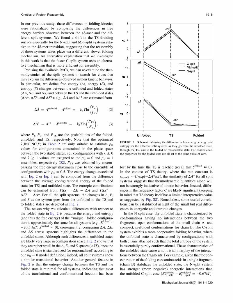
FIGURE 2 Schematic showing the difference in free energy, energy, and
entropy for the different split systems as they go from the unfolded state,
through the TS, and to the folded or reassembled state. For convenience,
the properties for the folded state are all set to the same value of zero.
Kinetics of Protein Reassembly 1915
In our previous study, these differences in folding kinetics
were rationalized by comparing the differences in free
energy barriers observed between the 48-mer and the dif-
ferent split systems. We found a shift in the TS dividing
surface especially for the N-split and Mid-split systems rela-
tive to the 48-mer transition, suggesting that the reassembly
of these systems takes place via a different, slower folding
mechanism. An alternative explanation that we investigate
in this work is that the faster C-split system uses an alterna-
tive mechanism that is more efficient for assembly.
Perusing the available RxCs, we can re-examine the ther-
modynamics of the split systems to search for clues that
may explain the differences observed in their kinetic behavior.
In particular, we define free energy (A), energy (E), and
entropy (S) changes between the unfolded and folded states
(DA, DE, and DS) and between the TS and the unfolded states
(DA*, DE*, and DS*); e.g., DA and DA* are estimated from
DA ¼ Aunfolded � Afolded ¼ �kBTln
�Pu
Pf
�; (2)
DA� ¼ ATS � Aunfolded ¼ �kBTln
�PTS
Pu
�; (3)
where Pf, Pu, and PTS are the probabilities of the folded,
unfolded, and TS, respectively. Note that the optimized
l(INC,NC,E) in Table 2 are only suitable to estimate pB
values for configurations constrained in the phase space
between the two stable states, i.e., configurations with l % 0
and l R 1 values are assigned to the pB ¼ 0 and pB ¼ 1
ensembles, respectively (32). PTS was obtained by encom-
passing the free energy maximum close to the ensemble of
configurations with pB ¼ 0.5. The energy change associated
with Eq. 2 or Eq. 3 can be computed from the difference
between the average configurational energy of the folded
state (or TS) and unfolded state. The entropic contributions
can be estimated from TDS ¼ DE � DA and TDS* ¼DE* � DA*. For all the split systems, the changes in A, E,
and S as the system goes from the unfolded to the TS and
to folded states are depicted in Fig. 2.
The reason why we calculate differences with respect to
the folded state in Eq. 2 is because the energy and entropy
(and thus the free energy) of the ‘‘unique’’ folded configura-
tion is approximately the same for all systems (e.g., Efolded ~
�20.5 kBT, Sfolded z 0); consequently, comparing DA, DE,
and DS across systems highlights the differences in the
unfolded states. Although such differences in unfolded states
are likely very large in configuration space, Fig. 2 shows that
they are rather small in the A, E, and S spaces (<kT), once the
unfolded state is standardized (or renormalized) according to
our pB ¼ 0 model definition; indeed, all split systems show
a similar transitional behavior. Another general feature in
Fig. 2 is that the entropy change between the TS and the
folded state is minimal for all systems, indicating that most
of the translational and conformational freedom has been
lost by the time the TS is reached (recall that Sfolded z 0).
In the context of TS theory, where the rate constant is
kA/B z C exp(�DA*/kT), the similarity of DA* for all split
systems suggests that thermodynamic quantities alone will
not be strongly indicative of kinetic behavior. Instead, differ-
ences in the frequency factor C are likely significant (keeping
in mind that TS theory itself has a limited interpretative value
as suggested by Fig. S2). Nonetheless, some useful correla-
tions can be established in light of the small but real differ-
ences in energetic and entropic changes.
In the N-split case, the unfolded state is characterized by
conformations having no interactions between the two
fragments, open conformations of the small chain A, and
compact, prefolded conformations for chain B. The C-split
system exhibits a more cooperative folding behavior, where
the unfolded state is characterized by configurations with
both chains attached such that the total entropy of the system
is essentially purely conformational. These characteristics of
the unfolded state cause a nontrivial interplay of the interac-
tions between the fragments. For example, given that the con-
centration of the folding core amino acids in a single fragment
(chain B) stabilizes the unfolded state, the N-split system
has stronger (more negative) energetic interactions than
the unfolded C-split case ðEunfoldedN�split � Eunfolded
C�split ¼ �0:67kTÞ;
Biophysical Journal 98(9) 1911–1920
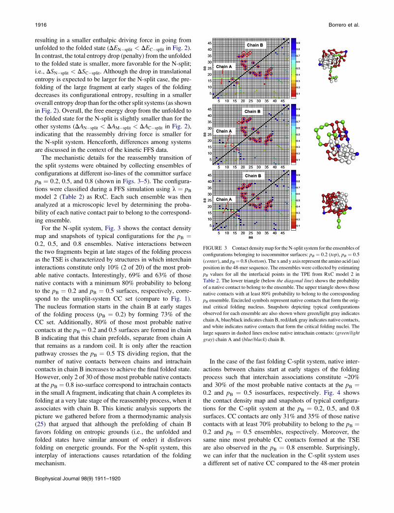
FIGURE 3 Contact density map for the N-split system for the ensembles of
configurations belonging to isocommittor surfaces: pB ¼ 0.2 (top), pB ¼ 0.5
(center), and pB¼ 0.8 (bottom). The x and y axis represent the amino acid (aa)
position in the 48-mer sequence. The ensembles were collected by estimating
pB values for all the interfacial points in the TPE from RxC model 2 in
Table 2. The lower triangle (below the diagonal line) shows the probability
of a native contact to belong to the ensemble. The upper triangle shows those
native contacts with at least 80% probability to belong to the corresponding
pB ensemble. Encircled symbols represent native contacts that form the orig-
inal critical folding nucleus. Snapshots depicting typical configurations
observed for each ensemble are also shown where green/light gray indicates
chain A, blue/black indicates chain B, red/dark gray indicates native contacts,
and white indicates native contacts that form the critical folding nuclei. The
large squares in dashed lines enclose native intrachain contacts: (green/light
gray) chain A and (blue/black) chain B.
1916 Borrero et al.
resulting in a smaller enthalpic driving force in going from
unfolded to the folded state (DEN�split < DEC�split in Fig. 2).
In contrast, the total entropy drop (penalty) from the unfolded
to the folded state is smaller, more favorable for the N-split;
i.e., DSN�split < DSC�split. Although the drop in translational
entropy is expected to be larger for the N-split case, the pre-
folding of the large fragment at early stages of the folding
decreases its configurational entropy, resulting in a smaller
overall entropy drop than for the other split systems (as shown
in Fig. 2). Overall, the free energy drop from the unfolded to
the folded state for the N-split is slightly smaller than for the
other systems (DAN�split < DAM�split < DAC�split in Fig. 2),
indicating that the reassembly driving force is smaller for
the N-split system. Henceforth, differences among systems
are discussed in the context of the kinetic FFS data.
The mechanistic details for the reassembly transition of
the split systems were obtained by collecting ensembles of
configurations at different iso-lines of the committor surface
pB ¼ 0.2, 0.5, and 0.8 (shown in Figs. 3–5). The configura-
tions were classified during a FFS simulation using l ¼ pB
model 2 (Table 2) as RxC. Each such ensemble was then
analyzed at a microscopic level by determining the proba-
bility of each native contact pair to belong to the correspond-
ing ensemble.
For the N-split system, Fig. 3 shows the contact density
map and snapshots of typical configurations for the pB ¼0.2, 0.5, and 0.8 ensembles. Native interactions between
the two fragments begin at late stages of the folding process
as the TSE is characterized by structures in which interchain
interactions constitute only 10% (2 of 20) of the most prob-
able native contacts. Interestingly, 69% and 63% of those
native contacts with a minimum 80% probability to belong
to the pB ¼ 0.2 and pB ¼ 0.5 surfaces, respectively, corre-
spond to the unsplit-system CC set (compare to Fig. 1).
The nucleus formation starts in the chain B at early stages
of the folding process (pB ¼ 0.2) by forming 73% of the
CC set. Additionally, 80% of those most probable native
contacts at the pB ¼ 0.2 and 0.5 surfaces are formed in chain
B indicating that this chain prefolds, separate from chain A
that remains as a random coil. It is only after the reaction
pathway crosses the pB ¼ 0.5 TS dividing region, that the
number of native contacts between chains and intrachain
contacts in chain B increases to achieve the final folded state.
However, only 2 of 30 of those most probable native contacts
at the pB ¼ 0.8 iso-surface correspond to intrachain contacts
in the small A fragment, indicating that chain A completes its
folding at a very late stage of the reassembly process, when it
associates with chain B. This kinetic analysis supports the
picture we gathered before from a thermodynamic analysis
(25) that argued that although the prefolding of chain B
favors folding on entropic grounds (i.e., the unfolded and
folded states have similar amount of order) it disfavors
folding on energetic grounds. For the N-split system, this
interplay of interactions causes retardation of the folding
mechanism.
Biophysical Journal 98(9) 1911–1920
In the case of the fast folding C-split system, native inter-
actions between chains start at early stages of the folding
process such that interchain associations constitute ~20%
and 30% of the most probable native contacts at the pB ¼0.2 and pB ¼ 0.5 isosurfaces, respectively. Fig. 4 shows
the contact density map and snapshots of typical configura-
tions for the C-split system at the pB ¼ 0.2, 0.5, and 0.8
surfaces. CC contacts are only 31% and 35% of those native
contacts with at least 70% probability to belong to the pB ¼0.2 and pB ¼ 0.5 ensembles, respectively. Moreover, the
same nine most probable CC contacts formed at the TSE
are also observed in the pB ¼ 0.8 ensemble. Surprisingly,
we can infer that the nucleation in the C-split system uses
a different set of native CC compared to the 48-mer protein
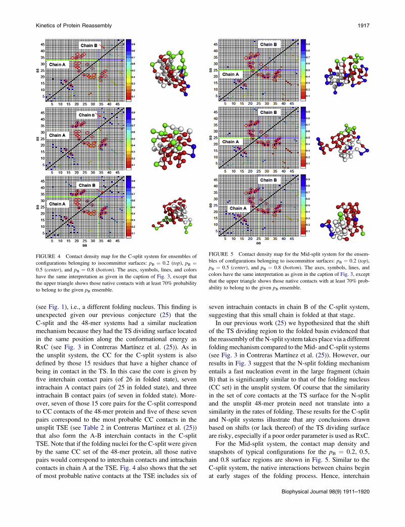
FIGURE 4 Contact density map for the C-split system for ensembles of
configurations belonging to isocommittor surfaces: pB ¼ 0.2 (top), pB ¼0.5 (center), and pB ¼ 0.8 (bottom). The axes, symbols, lines, and colors
have the same interpretation as given in the caption of Fig. 3, except that
the upper triangle shows those native contacts with at least 70% probability
to belong to the given pB ensemble.
FIGURE 5 Contact density map for the Mid-split system for the ensem-
bles of configurations belonging to isocommittor surfaces: pB ¼ 0.2 (top),
pB ¼ 0.5 (center), and pB ¼ 0.8 (bottom). The axes, symbols, lines, and
colors have the same interpretation as given in the caption of Fig. 3, except
that the upper triangle shows those native contacts with at least 70% prob-
ability to belong to the given pB ensemble.
Kinetics of Protein Reassembly 1917
(see Fig. 1), i.e., a different folding nucleus. This finding is
unexpected given our previous conjecture (25) that the
C-split and the 48-mer systems had a similar nucleation
mechanism because they had the TS dividing surface located
in the same position along the conformational energy as
RxC (see Fig. 3 in Contreras Martı́nez et al. (25)). As in
the unsplit system, the CC for the C-split system is also
defined by those 15 residues that have a higher chance of
being in contact in the TS. In this case the core is given by
five interchain contact pairs (of 26 in folded state), seven
intrachain A contact pairs (of 25 in folded state), and three
intrachain B contact pairs (of seven in folded state). More-
over, seven of those 15 core pairs for the C-split correspond
to CC contacts of the 48-mer protein and five of these seven
pairs correspond to the most probable CC contacts in the
unsplit TSE (see Table 2 in Contreras Martı́nez et al. (25))
that also form the A-B interchain contacts in the C-split
TSE. Note that if the folding nuclei for the C-split were given
by the same CC set of the 48-mer protein, all those native
pairs would correspond to interchain contacts and intrachain
contacts in chain A at the TSE. Fig. 4 also shows that the set
of most probable native contacts at the TSE includes six of
seven intrachain contacts in chain B of the C-split system,
suggesting that this small chain is folded at that stage.
In our previous work (25) we hypothesized that the shift
of the TS dividing region to the folded basin evidenced that
the reassembly of the N-split system takes place via a different
folding mechanism compared to the Mid- and C-split systems
(see Fig. 3 in Contreras Martı́nez et al. (25)). However, our
results in Fig. 3 suggest that the N-split folding mechanism
entails a fast nucleation event in the large fragment (chain
B) that is significantly similar to that of the folding nucleus
(CC set) in the unsplit system. Of course that the similarity
in the set of core contacts at the TS surface for the N-split
and the unsplit 48-mer protein need not translate into a
similarity in the rates of folding. These results for the C-split
and N-split systems illustrate that any conclusions drawn
based on shifts (or lack thereof) of the TS dividing surface
are risky, especially if a poor order parameter is used as RxC.
For the Mid-split system, the contact map density and
snapshots of typical configurations for the pB ¼ 0.2, 0.5,
and 0.8 surface regions are shown in Fig. 5. Similar to the
C-split system, the native interactions between chains begin
at early stages of the folding process. Hence, interchain
Biophysical Journal 98(9) 1911–1920
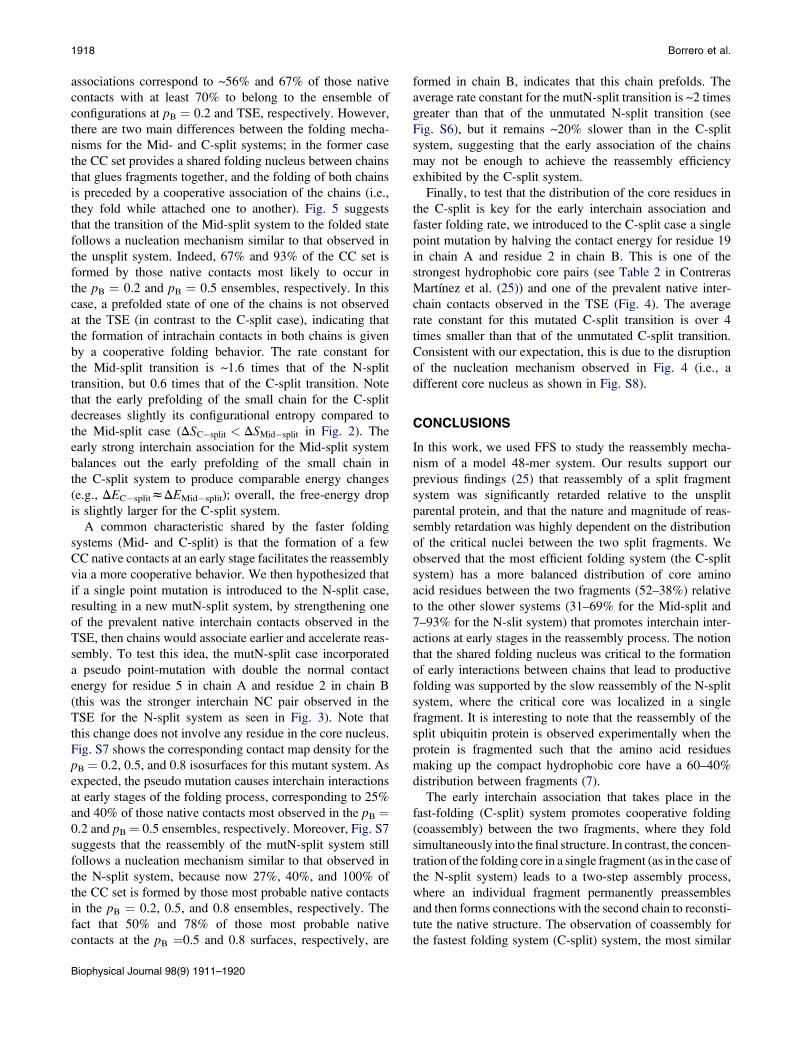
1918 Borrero et al.
associations correspond to ~56% and 67% of those native
contacts with at least 70% to belong to the ensemble of
configurations at pB ¼ 0.2 and TSE, respectively. However,
there are two main differences between the folding mecha-
nisms for the Mid- and C-split systems; in the former case
the CC set provides a shared folding nucleus between chains
that glues fragments together, and the folding of both chains
is preceded by a cooperative association of the chains (i.e.,
they fold while attached one to another). Fig. 5 suggests
that the transition of the Mid-split system to the folded state
follows a nucleation mechanism similar to that observed in
the unsplit system. Indeed, 67% and 93% of the CC set is
formed by those native contacts most likely to occur in
the pB ¼ 0.2 and pB ¼ 0.5 ensembles, respectively. In this
case, a prefolded state of one of the chains is not observed
at the TSE (in contrast to the C-split case), indicating that
the formation of intrachain contacts in both chains is given
by a cooperative folding behavior. The rate constant for
the Mid-split transition is ~1.6 times that of the N-split
transition, but 0.6 times that of the C-split transition. Note
that the early prefolding of the small chain for the C-split
decreases slightly its configurational entropy compared to
the Mid-split case (DSC�split < DSMid�split in Fig. 2). The
early strong interchain association for the Mid-split system
balances out the early prefolding of the small chain in
the C-split system to produce comparable energy changes
(e.g., DEC�splitzDEMid�split); overall, the free-energy drop
is slightly larger for the C-split system.
A common characteristic shared by the faster folding
systems (Mid- and C-split) is that the formation of a few
CC native contacts at an early stage facilitates the reassembly
via a more cooperative behavior. We then hypothesized that
if a single point mutation is introduced to the N-split case,
resulting in a new mutN-split system, by strengthening one
of the prevalent native interchain contacts observed in the
TSE, then chains would associate earlier and accelerate reas-
sembly. To test this idea, the mutN-split case incorporated
a pseudo point-mutation with double the normal contact
energy for residue 5 in chain A and residue 2 in chain B
(this was the stronger interchain NC pair observed in the
TSE for the N-split system as seen in Fig. 3). Note that
this change does not involve any residue in the core nucleus.
Fig. S7 shows the corresponding contact map density for the
pB ¼ 0.2, 0.5, and 0.8 isosurfaces for this mutant system. As
expected, the pseudo mutation causes interchain interactions
at early stages of the folding process, corresponding to 25%
and 40% of those native contacts most observed in the pB ¼0.2 and pB¼ 0.5 ensembles, respectively. Moreover, Fig. S7
suggests that the reassembly of the mutN-split system still
follows a nucleation mechanism similar to that observed in
the N-split system, because now 27%, 40%, and 100% of
the CC set is formed by those most probable native contacts
in the pB ¼ 0.2, 0.5, and 0.8 ensembles, respectively. The
fact that 50% and 78% of those most probable native
contacts at the pB ¼0.5 and 0.8 surfaces, respectively, are
Biophysical Journal 98(9) 1911–1920
formed in chain B, indicates that this chain prefolds. The
average rate constant for the mutN-split transition is ~2 times
greater than that of the unmutated N-split transition (see
Fig. S6), but it remains ~20% slower than in the C-split
system, suggesting that the early association of the chains
may not be enough to achieve the reassembly efficiency
exhibited by the C-split system.
Finally, to test that the distribution of the core residues in
the C-split is key for the early interchain association and
faster folding rate, we introduced to the C-split case a single
point mutation by halving the contact energy for residue 19
in chain A and residue 2 in chain B. This is one of the
strongest hydrophobic core pairs (see Table 2 in Contreras
Martı́nez et al. (25)) and one of the prevalent native inter-
chain contacts observed in the TSE (Fig. 4). The average
rate constant for this mutated C-split transition is over 4
times smaller than that of the unmutated C-split transition.
Consistent with our expectation, this is due to the disruption
of the nucleation mechanism observed in Fig. 4 (i.e., a
different core nucleus as shown in Fig. S8).
CONCLUSIONS
In this work, we used FFS to study the reassembly mecha-
nism of a model 48-mer system. Our results support our
previous findings (25) that reassembly of a split fragment
system was significantly retarded relative to the unsplit
parental protein, and that the nature and magnitude of reas-
sembly retardation was highly dependent on the distribution
of the critical nuclei between the two split fragments. We
observed that the most efficient folding system (the C-split
system) has a more balanced distribution of core amino
acid residues between the two fragments (52–38%) relative
to the other slower systems (31–69% for the Mid-split and
7–93% for the N-slit system) that promotes interchain inter-
actions at early stages in the reassembly process. The notion
that the shared folding nucleus was critical to the formation
of early interactions between chains that lead to productive
folding was supported by the slow reassembly of the N-split
system, where the critical core was localized in a single
fragment. It is interesting to note that the reassembly of the
split ubiquitin protein is observed experimentally when the
protein is fragmented such that the amino acid residues
making up the compact hydrophobic core have a 60–40%
distribution between fragments (7).
The early interchain association that takes place in the
fast-folding (C-split) system promotes cooperative folding
(coassembly) between the two fragments, where they fold
simultaneously into the final structure. In contrast, the concen-
tration of the folding core in a single fragment (as in the case of
the N-split system) leads to a two-step assembly process,
where an individual fragment permanently preassembles
and then forms connections with the second chain to reconsti-
tute the native structure. The observation of coassembly for
the fastest folding system (C-split) system, the most similar
Kinetics of Protein Reassembly 1919
to the parent protein in terms of folding rate, and a two-step
folding for the slowest folding (N-split) protein led us to
hypothesize that the concentration of core native contacts in
a single fragment changed the cooperative folding mechanism
observed in the parent protein (where all amino acids are
linked) to a coassembly process. Intuitively, it seemed that
sharing the core between two fragments preserved the overall
folding mechanism exhibited by the parent protein so that the
process was still productive when the protein is fragmented.
However, a precise characterization of the folding mechanism
and the TS for the split systems revealed the surprising
result that the folding mechanism of the unsplit protein was
unchanged in the slower folding (N-split and Mid-split)
systems but significantly changed in the case of the fast
folding (C-split) system, where a different TS was observed.
Given the higher degree of freedom of split protein
systems (as compared to a single protein chain) to search
for multiple folding pathways, it is significant that a split
protein system can reassemble via the same folding mecha-
nism as its parent structure. Yet, it is possible that the early
commitment to the formation of the same parental TS and
folding pathway could be a suboptimal strategy to reassemble
the split system (as in the N-split case), because the system
might not fully explore alternative, faster pathways. We there-
fore speculate that the utilization of a folding pathway
different from the one used by the unsplit parent protein, leads
to the efficient reassembly of the C-split fragment system.
It remains unclear, however, whether optimal reassembly
via a novel folding pathway is a general phenomenon or a
highly system-specific occurrence in our split protein sys-
tems. Also, we cannot at present rule out the existence of a split
point yielding a system that folds as fast as the C-split system
and follows the mechanism of the parent protein.
One of the experimental strategies to control the folding or
assembly mechanism of a protein system involves changes to
amino acids and protein domains; e.g., the addition of
leucine zipper domains has been experimentally observed
to enhance fragment interactions and promote fragment reas-
sembly for several split protein systems such as GFP, DHFR
and ubiquitin (2,6–12). In this study, the rational introduc-
tion of a single point mutation to the N-split system was
found to promote fragment association and speed up the
reassembly kinetics. This mutation, however, did not change
the overall folding mechanism nor did it match the folding
efficiency of the C-split system, suggesting that additional
factors to early interchain interactions contributed to the effi-
cient folding of the latter. Although our results for a mini-
malist lattice model are not directly applicable to real split
proteins, they suggest that reassembly of split protein frag-
ments could be optimized by designing strategic fragmenta-
tion patterns that lead to different, more efficient folding
mechanisms or by altering the sequence itself in a manner
that promotes the interaction between fragments without
necessarily affecting the overall folding process. The use
of such key mutations presents an alternative to the introduc-
tion of additional protein domains (i.e., leucine zippers) that
have been reported to be important to the natural activity and
mode of action of several well-characterized proteins (35).
With respect to experiments, we plan to test some of these
predictions in the split ubiquitin system model by intro-
ducing various mutations that affect its well-characterized
compact hydrophobic core. With respect to simulations,
current work is directed to the reassembly mechanism of split
lattice protein models exhibiting two domains (correspond-
ing to independently secondary motifs) and following a hier-
archical folding mechanism; future work will also aim at
studying the kinetics of small (computationally tractable)
atomistic split-protein models.
SUPPORTING MATERIAL
Two tables and eight figures are available at http://www.biophysj.org/
biophysj/supplemental/S0006-3495(10)00133-5.
This work was supported by the National Science Foundation (grant
0756248).
REFERENCES
1. Hu, C. D., and T. K. Kerppola. 2003. Simultaneous visualization ofmultiple protein interactions in living cells using multicolor fluores-cence complementation analysis. Nat. Biotechnol. 21:539–545.
2. Magliery, T. J., C. G. M. Wilson, ., L. Regan. 2005. Detecting protein-protein interactions with a green fluorescent protein fragment reassem-bly trap: scope and mechanism. J. Am. Chem. Soc. 127:146–157.
3. Ozawa, T., Y. Sako, ., Y. Umezawa. 2003. A genetic approach toidentifying mitochondrial proteins. Nat. Biotechnol. 21:287–293.
4. Remy, I., and S. W. Michnick. 2004. A cDNA library functionalscreening strategy based on fluorescent protein complementationassays to identify novel components of signaling pathways. Methods.32:381–388.
5. Stains, C. I., J. R. Porter, ., I. Ghosh. 2005. DNA sequence-enabledreassembly of the green fluorescent protein. J. Am. Chem. Soc. 127:10782–10783.
6. Ghosh, I., A. D. Hamilton, and L. Regan. 2000. Antiparallel leucinezipper-directed protein reassembly: application to the green fluorescentprotein. J. Am. Chem. Soc. 122:5658–5659.
7. Johnsson, N., and A. Varshavsky. 1994. Split ubiquitin as a sensor ofprotein interactions in vivo. Proc. Natl. Acad. Sci. USA. 91:10340–10344.
8. Pelletier, J. N., F. X. Campbell-Valois, and S. W. Michnick. 1998. Olig-omerization domain-directed reassembly of active dihydrofolate reduc-tase from rationally designed fragments. Proc. Natl. Acad. Sci. USA.95:12141–12146.
9. Galarneau, A., M. Primeau, ., S. W. Michnick. 2002. Beta-lactamaseprotein fragment complementation assays as in vivo and in vitro sensorsof protein protein interactions. Nat. Biotechnol. 20:619–622.
10. Wehrman, T., B. Kleaveland, ., H. M. Blau. 2002. Protein-proteininteractions monitored in mammalian cells via complementation ofbeta -lactamase enzyme fragments. Proc. Natl. Acad. Sci. USA. 99:3469–3474.
11. Paulmurugan, R., and S. S. Gambhir. 2003. Monitoring protein-proteininteractions using split synthetic Renilla luciferase protein-fragment-as-sisted complementation. Anal. Chem. 75:1584–1589.
12. Deo, S. K. 2004. Exploring bioanalytical applications of assisted proteinreassembly. Anal. Bioanal. Chem. 379:383–390.
Biophysical Journal 98(9) 1911–1920
1920 Borrero et al.
13. Wilson, C. G., T. J. Magliery, and L. Regan. 2004. Detecting protein-protein interactions with GFP-fragment reassembly. Nat. Methods.1:255–262.
14. Hennecke, J., P. Sebbel, and R. Glockshuber. 1999. Random circularpermutation of DsbA reveals segments that are essential for proteinfolding and stability. J. Mol. Biol. 286:1197–1215.
15. Iwakura, M., T. Nakamura, ., K. Maki. 2000. Systematic circularpermutation of an entire protein reveals essential folding elements.Nat. Struct. Biol. 7:580–585.
16. Betton, J. M., and M. Hofnung. 1994. In vivo assembly of activemaltose binding protein from independently exported protein fragments.EMBO J. 13:1226–1234.
17. Paszkiewicz, K. H., M. J. E. Sternberg, and M. Lappe. 2006. Predictionof viable circular permutants using a graph theoretic approach. Bioinfor-matics. 22:1353–1358.
18. Cabantous, S., T. C. Terwilliger, and G. S. Waldo. 2005. Proteintagging and detection with engineered self-assembling fragments ofgreen fluorescent protein. Nat. Biotechnol. 23:102–107.
19. Paulmurugan, R., and S. S. Gambhir. 2007. Combinatorial libraryscreening for developing an improved split-firefly luciferase fragment-assisted complementation system for studying protein-protein interac-tions. Anal. Chem. 79:2346–2353.
20. Demidov, V. V., N. V. Dokholyan, ., N. E. Broude. 2006. Fastcomplementation of split fluorescent protein triggered by DNA hybrid-ization. Proc. Natl. Acad. Sci. USA. 103:2052–2056.
21. Daggett, V., and A. R. Fersht. 2003. Is there a unifying mechanism forprotein folding? Trends Biochem. Sci. 28:18–25.
22. Abkevich, V. I., A. M. Gutin, and E. I. Shakhnovich. 1994. Specificnucleus as the transition state for protein folding: evidence from thelattice model. Biochemistry. 33:10026–10036.
23. Fersht, A. R. 1995. Optimization of rates of protein folding: the nucle-ation-condensation mechanism and its implications. Proc. Natl. Acad.Sci. USA. 92:10869–10873.
24. Fersht, A. R. 1997. Nucleation mechanisms in protein folding. Curr.Opin. Struct. Biol. 7:3–9.
Biophysical Journal 98(9) 1911–1920
25. Contreras Martı́nez, L. M., E. E. Borrero Quintana, ., M. P. DeLisa.
2008. In silico protein fragmentation reveals the importance of critical
nuclei on domain reassembly. Biophys. J. 94:1575–1588.
26. Krantz, B. A., R. S. Dothager, and T. R. Sosnick. 2004. Discerning the
structure and energy of multiple transition states in protein folding using
psi-analysis. J. Mol. Biol. 337:463–475.
27. Levy, Y., P. G. Wolynes, and J. N. Onuchic. 2004. Protein topology
determines binding mechanism. Proc. Natl. Acad. Sci. USA. 101:
511–516.
28. Dellago, C., and P. G. Bolhuis. 2007. Transition path sampling simula-
tions of biological systems. In Atomistic Approaches in Modern
Biology: from Quantum Chemistry to Molecular Simulations., Vol.268 Springer-Verlag Berlin, Berlin, pp. 291–317.
29. Dellago, C., P. G. Bolhuis, and P. L. Geissler. 2002. Transition path
sampling. Adv. Chem. Phys. 123:1–78.
30. Borrero, E. E., and F. A. Escobedo. 2006. Folding kinetics of a lattice
protein via a forward flux sampling approach. J. Chem. Phys.125:164904–164914.
31. Vendruscolo, M., E. Paci, ., M. Karplus. 2001. Three key residues
form a critical contact network in a protein folding transition state.
Nature. 409:641–645.
32. Borrero, E. E., and F. A. Escobedo. 2007. Reaction coordinates and tran-
sition pathways of rare events via forward flux sampling. J. Chem. Phys.127:164101–164117.
33. Borrero, E. E., and F. A. Escobedo. 2008. Optimizing the sampling and
staging for simulations of rare events via forward flux sampling
schemes. J. Chem. Phys. 129:024115–024116.
34. Frenkel, D., and B. Smit. 2002. Understanding Molecular Simulation:
From Algorithms to Applications,2nd ed . Academic, Boston, MA.
35. Zhu, W. L., Y. M. Song, ., S. Y. Shin. 2007. Substitution of the
leucine zipper sequence in melittin with peptoid residues affects self-
association, cell selectivity, and mode of action. Biochim. Biophys.Acta. 1768:1506–1517.


![Terracotta Reassembly from Fragments Based on Surface ...diglib.eg.org/bitstream/handle/10.2312/egp20161049/025-026.pdf · reassembly method described in [MRS10] may work, it is semi-automatic](https://img.pdfslide.us/doc/110x75/607b744d973cd76d7a5559e6/terracotta-reassembly-from-fragments-based-on-surface-reassembly-method-described.jpg)