Embed Size (px)
Citation preview
Bridgewater State UniversityVirtual Commons - Bridgewater State University
Honors Program Theses and Projects Undergraduate Honors Program
5-7-2018
Kinetic studies on the aqueous chemistry of theglutathione-methyleneoxindole conjugate to modelits inhibition of glyoxalase IAriane Borges
Follow this and additional works at: http://vc.bridgew.edu/honors_proj
Part of the Chemistry Commons
This item is available as part of Virtual Commons, the open-access institutional repository of Bridgewater State University, Bridgewater, Massachusetts.
Recommended CitationBorges, Ariane. (2018). Kinetic studies on the aqueous chemistry of the glutathione-methyleneoxindole conjugate to model itsinhibition of glyoxalase I. In BSU Honors Program Theses and Projects. Item 300. Available at: http://vc.bridgew.edu/honors_proj/300Copyright © 2018 Ariane Borges
Kinetic studies on the aqueous chemistry of the glutathione-methyleneoxindole conjugate
to model its inhibition of glyoxalase I
Ariane Borges
Submitted in Partial Completion of the
Requirements for Departmental Honors in Chemistry
Bridgewater State University
May 7, 2018
Dr. Edward Brush, Thesis Advisor Dr. Saritha Nellutla, Committee Member
Dr. Samer Lone, Committee Member
2
Kinetic studies on the aqueous chemistry of the glutathione-methyleneoxindole conjugate to model its inhibition of
glyoxalase I
Ariane Borges Undergraduate Honors Thesis
Mentor:
Dr. Edward J. Brush
Bridgewater State University Department of Chemical Sciences
Bridgewater, MA 02325

3
This page intentionally left blank.
4
Abstract
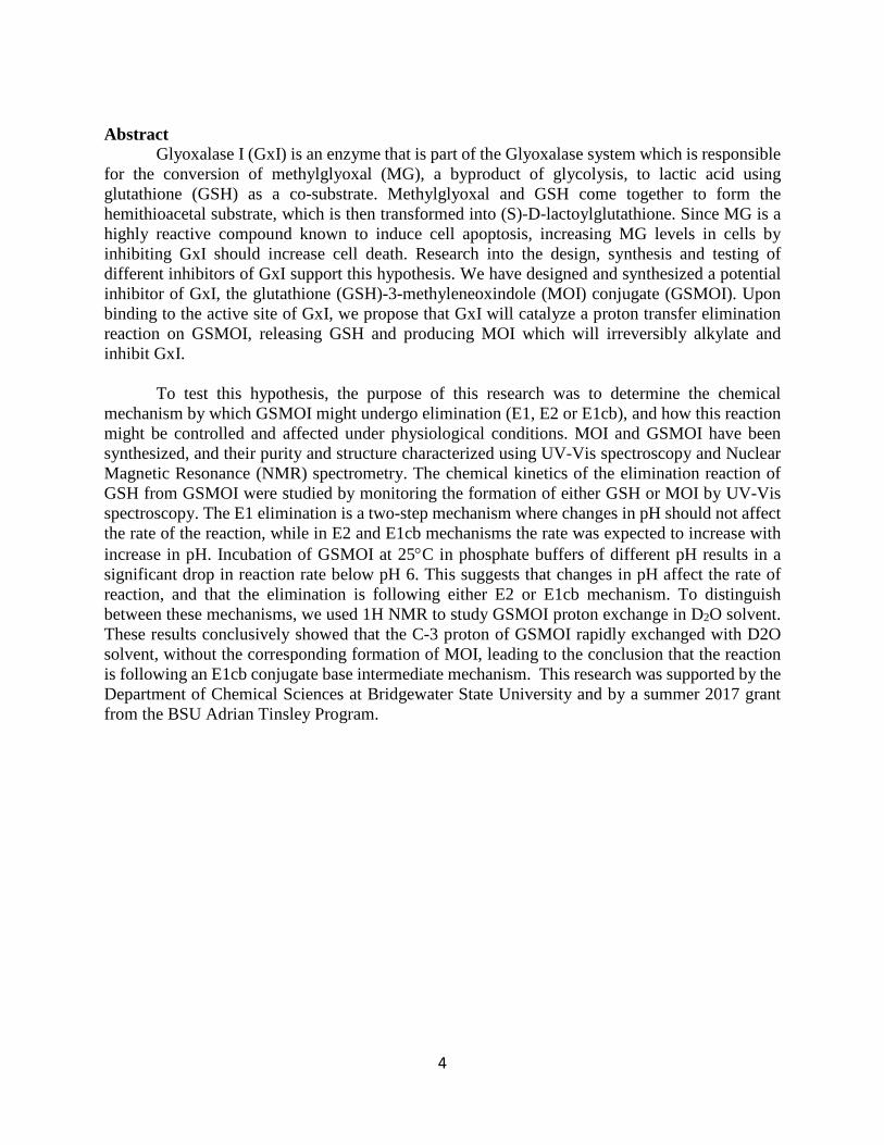
Glyoxalase I (GxI) is an enzyme that is part of the Glyoxalase system which is responsible for the conversion of methylglyoxal (MG), a byproduct of glycolysis, to lactic acid using glutathione (GSH) as a co-substrate. Methylglyoxal and GSH come together to form the hemithioacetal substrate, which is then transformed into (S)-D-lactoylglutathione. Since MG is a highly reactive compound known to induce cell apoptosis, increasing MG levels in cells by inhibiting GxI should increase cell death. Research into the design, synthesis and testing of different inhibitors of GxI support this hypothesis. We have designed and synthesized a potential inhibitor of GxI, the glutathione (GSH)-3-methyleneoxindole (MOI) conjugate (GSMOI). Upon binding to the active site of GxI, we propose that GxI will catalyze a proton transfer elimination reaction on GSMOI, releasing GSH and producing MOI which will irreversibly alkylate and inhibit GxI.
To test this hypothesis, the purpose of this research was to determine the chemical
mechanism by which GSMOI might undergo elimination (E1, E2 or E1cb), and how this reaction might be controlled and affected under physiological conditions. MOI and GSMOI have been synthesized, and their purity and structure characterized using UV-Vis spectroscopy and Nuclear Magnetic Resonance (NMR) spectrometry. The chemical kinetics of the elimination reaction of GSH from GSMOI were studied by monitoring the formation of either GSH or MOI by UV-Vis spectroscopy. The E1 elimination is a two-step mechanism where changes in pH should not affect the rate of the reaction, while in E2 and E1cb mechanisms the rate was expected to increase with increase in pH. Incubation of GSMOI at 25°C in phosphate buffers of different pH results in a significant drop in reaction rate below pH 6. This suggests that changes in pH affect the rate of reaction, and that the elimination is following either E2 or E1cb mechanism. To distinguish between these mechanisms, we used 1H NMR to study GSMOI proton exchange in D2O solvent. These results conclusively showed that the C-3 proton of GSMOI rapidly exchanged with D2O solvent, without the corresponding formation of MOI, leading to the conclusion that the reaction is following an E1cb conjugate base intermediate mechanism. This research was supported by the Department of Chemical Sciences at Bridgewater State University and by a summer 2017 grant from the BSU Adrian Tinsley Program.
5
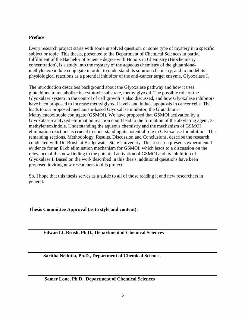
Preface Every research project starts with some unsolved question, or some type of mystery in a specific subject or topic. This thesis, presented to the Department of Chemical Sciences in partial fulfillment of the Bachelor of Science degree with Honors in Chemistry (Biochemistry concentration), is a study into the mystery of the aqueous chemistry of the glutathione-methyleneoxindole conjugate in order to understand its solution chemistry, and to model its physiological reactions as a potential inhibitor of the anti-cancer target enzyme, Glyoxalase I.
The introduction describes background about the Glyoxalase pathway and how it uses glutathione to metabolize its cytotoxic substrate, methylglyoxal. The possible role of the Glyoxalase system in the control of cell growth is also discussed, and how Glyoxalase inhibitors have been proposed to increase methylglyoxal levels and induce apoptosis in cancer cells. That leads to our proposed mechanism-based Glyoxalase inhibitor, the Glutathione-Methyleneoxindole conjugate (GSMOI). We have proposed that GSMOI activation by a Glyoxalase-catalyzed elimination reaction could lead to the formation of the alkylating agent, 3-methyleneoxindole. Understanding the aqueous chemistry and the mechanism of GSMOI elimination reactions is crucial to understanding its potential role in Glyoxalase I inhibition. The remaining sections, Methodology, Results, Discussion and Conclusions, describe the research conducted with Dr. Brush at Bridgewater State University. This research presents experimental evidence for an E1cb elimination mechanism for GSMOI, which leads to a discussion on the relevance of this new finding to the potential activation of GSMOI and its inhibition of Glyoxalase I. Based on the work described in this thesis, additional questions have been proposed inviting new researchers to this project.
So, I hope that this thesis serves as a guide to all of those reading it and new researchers in general.
Thesis Committee Approval (as to style and content): Edward J. Brush, Ph.D., Department of Chemical Sciences Saritha Nellutla, Ph.D., Department of Chemical Sciences Samer Lone, Ph.D., Department of Chemical Sciences
6
Acknowledgements First, I would like to thank my mentor, Dr. Edward J. Brush for all his guidance and support throughout my research, and for mentoring me. I would like to thank him for making me aware of great opportunities and for always being patient and kind with me. Without him I wouldn’t have discovered my passion for chemistry or research and I wouldn’t have written this thesis. I am eternally grateful for everything he has done for me. I would also like to thank my adviser for my Biology degree Dr. Jeffery Bowen, for always being helpful and guiding me through the burden of picking the right classes at the right times. I would like to thank all the professors that made a difference in my undergraduate journey both in the humanities and in the sciences. Each one of them pushed me to become a better version of myself and a better student. I would like to thank them all for their constructive criticism, life lessons and for always being there for me. I would like to thank my colleagues in the Biology department who have graduated, for being the best upper classmates anyone could wish for. I have a profound thanks to my chemistry colleagues for always being supportive and helpful. I am very glad I got to meet each and every one of you and my college journey wouldn’t have been the same without you. Lastly, I would like to thank all my family and friends, especially my parents for always being very supportive, I wouldn’t be able to accomplish this if it weren’t for your unconditional support and guidance. The following research was supported by the BSU Adrian Tinsley Program. The JEOL ECX-400 MHz NMR was obtained through NSF-MRI grant 0421081. Thesis is being submitted in partial fulfillment of a Bachelor of Science degree with Departmental Honors in Chemistry (Biochemistry concentration).
7
Table of Contents Abstract…………………………………………………………………………………..………. 3
1. Introduction
1.1. Glyoxalase System……………………………………………………………………7-11 1.2. Inhibition of Glyoxalase I…………………………………………………………….....12
1.3. Glutathione Methyleneoxindole conjugation (GSMOI)…………………………......13-17 1.4. Significance of this Work……………………………………………………………….17
2. Materials and Methods 2.1. Synthesis of 3-Bromooxindol-3-acetic acid…………………………………………18-19 2.2. Synthesis of 3-methylyeoxindole (MOI) from 3-Bromooxindole-3-acetic acid
(BOAA)……………………………………………………………………………...19-20 2.3. Synthesis of Glutathione-3-Methyleneoxindole Conjugate, GSMOI from 3-
methylyoxindole……………………………………………………………………..20-21 2.4. Characterization using NMR……………………………………………………………21 2.5. UV-Vis Kinetic Studies…………………………………………………………...…21-22 2.6. Determination of reaction order……………………………………………..………..…22 2.7. Effect of pH on the rate of GSH elimination …………….……………………...……...23 2.8. Effect of potential catalyst ……………………………………………………...........…23 2.9. Effect of buffer concentration on rate constant K……………………………………...23 2.10. GSMOI C-3 Proton exchange kinetics………………………………………………...24
3. Results and discussion……………………………………………………………………25-36 4. Conclusion………………………………………………………………………………..37-41 5. Future Work………………………………………………………………………………….42 6. Bibliography……………………………………………………………………………...43-44
8
1. Introduction
1.1 Glyoxalase System
1.1.1 Discovery and first insight
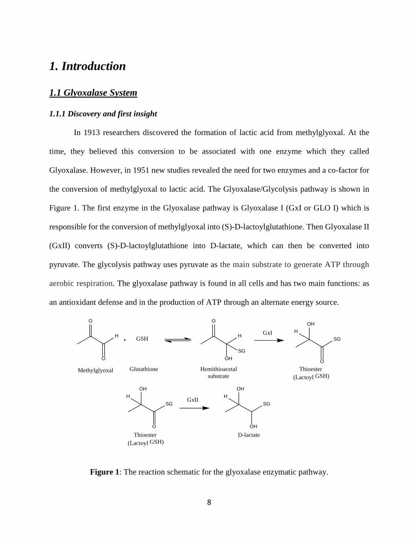
In 1913 researchers discovered the formation of lactic acid from methylglyoxal. At the
time, they believed this conversion to be associated with one enzyme which they called
Glyoxalase. However, in 1951 new studies revealed the need for two enzymes and a co-factor for
the conversion of methylglyoxal to lactic acid. The Glyoxalase/Glycolysis pathway is shown in
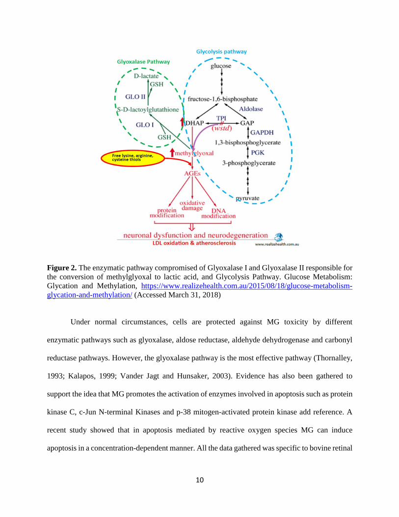
Figure 1. The first enzyme in the Glyoxalase pathway is Glyoxalase I (GxI or GLO I) which is
responsible for the conversion of methylglyoxal into (S)-D-lactoylglutathione. Then Glyoxalase II
(GxII) converts (S)-D-lactoylglutathione into D-lactate, which can then be converted into
pyruvate. The glycolysis pathway uses pyruvate as the main substrate to generate ATP through
aerobic respiration. The glyoxalase pathway is found in all cells and has two main functions: as
an antioxidant defense and in the production of ATP through an alternate energy source.
Figure 1: The reaction schematic for the glyoxalase enzymatic pathway.
H
O
O
GSH
Methylglyoxal Glutathione
H
OHSG
O
Hemithioacetal substrate
GxISG
O
OHH
Thioester
(Lactoyl GSH)
SG
O
OHH GxII
SG
OH
OHH
Thioester
(Lactoyl GSH)D-lactate
9
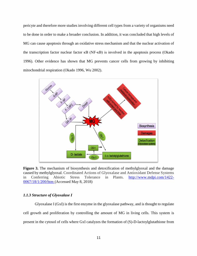
1.1.2 Methylglyoxal: Biosynthesis, damage and detoxification
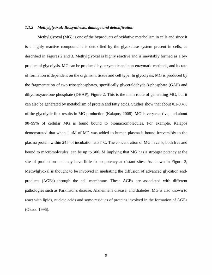
Methylglyoxal (MG) is one of the byproducts of oxidative metabolism in cells and since it
is a highly reactive compound it is detoxified by the glyoxalase system present in cells, as
described in Figures 2 and 3. Methylglyoxal is highly reactive and is inevitably formed as a by-
product of glycolysis. MG can be produced by enzymatic and non-enzymatic methods, and its rate
of formation is dependent on the organism, tissue and cell type. In glycolysis, MG is produced by
the fragmentation of two triosephosphates, specifically glyceraldehyde-3-phosphate (GAP) and
dihydroxyacetone phosphate (DHAP), Figure 2. This is the main route of generating MG, but it
can also be generated by metabolism of protein and fatty acids. Studies show that about 0.1-0.4%
of the glycolytic flux results in MG production (Kalapos, 2008). MG is very reactive, and about
90–99% of cellular MG is found bound to biomacromolecules. For example, Kalapos
demonstrated that when 1 μM of MG was added to human plasma it bound irreversibly to the
plasma protein within 24 h of incubation at 37°C. The concentration of MG in cells, both free and
bound to macromolecules, can be up to 300µΜ implying that MG has a stronger potency at the
site of production and may have little to no potency at distant sites. As shown in Figure 3,
Methylglyoxal is thought to be involved in mediating the diffusion of advanced glycation end-
products (AGEs) through the cell membrane. These AGEs are associated with different
pathologies such as Parkinson's disease, Alzheimer's disease, and diabetes. MG is also known to
react with lipids, nucleic acids and some residues of proteins involved in the formation of AGEs
(Okado 1996).
10
Figure 2. The enzymatic pathway compromised of Glyoxalase I and Glyoxalase II responsible for the conversion of methylglyoxal to lactic acid, and Glycolysis Pathway. Glucose Metabolism: Glycation and Methylation, https://www.realizehealth.com.au/2015/08/18/glucose-metabolism-glycation-and-methylation/ (Accessed March 31, 2018)
Under normal circumstances, cells are protected against MG toxicity by different
enzymatic pathways such as glyoxalase, aldose reductase, aldehyde dehydrogenase and carbonyl
reductase pathways. However, the glyoxalase pathway is the most effective pathway (Thornalley,
1993; Kalapos, 1999; Vander Jagt and Hunsaker, 2003). Evidence has also been gathered to
support the idea that MG promotes the activation of enzymes involved in apoptosis such as protein
kinase C, c-Jun N-terminal Kinases and p-38 mitogen-activated protein kinase add reference. A
recent study showed that in apoptosis mediated by reactive oxygen species MG can induce
apoptosis in a concentration-dependent manner. All the data gathered was specific to bovine retinal
11
pericyte and therefore more studies involving different cell types from a variety of organisms need
to be done in order to make a broader conclusion. In addition, it was concluded that high levels of
MG can cause apoptosis through an oxidative stress mechanism and that the nuclear activation of
the transcription factor nuclear factor κB (NF-κB) is involved in the apoptosis process (Okado
1996). Other evidence has shown that MG prevents cancer cells from growing by inhibiting
mitochondrial respiration (Okado 1996, Wu 2002).
Figure 3. The mechanism of biosynthesis and detoxification of methylglyoxal and the damage caused by methylglyoxal. Coordinated Actions of Glyoxalase and Antioxidant Defense Systems in Conferring Abiotic Stress Tolerance in Plants. http://www.mdpi.com/1422-0067/18/1/200/htm (Accessed May 8, 2018)
1.1.3 Structure of Glyoxalase I
Glyoxalase I (GxI) is the first enzyme in the glyoxalase pathway, and is thought to regulate
cell growth and proliferation by controlling the amount of MG in living cells. This system is
present in the cytosol of cells where GxI catalyzes the formation of (S)-D-lactoylglutathione from
12
the hemithioacetal which is formed in a non-enzymatic reaction between MG and GSH (see Figure
2). Then, GxII converts (S)-D-lactoylglutathione to lactic acid which is relatively non-toxic and
regenerates the co-substrate, GSH. The glyoxalase pathway is highly conserved throughout
biological life and is present also in cell organelles and mitochondria. This system is active from
embryogenesis until cell death, and plays an important role in cell growth and division.
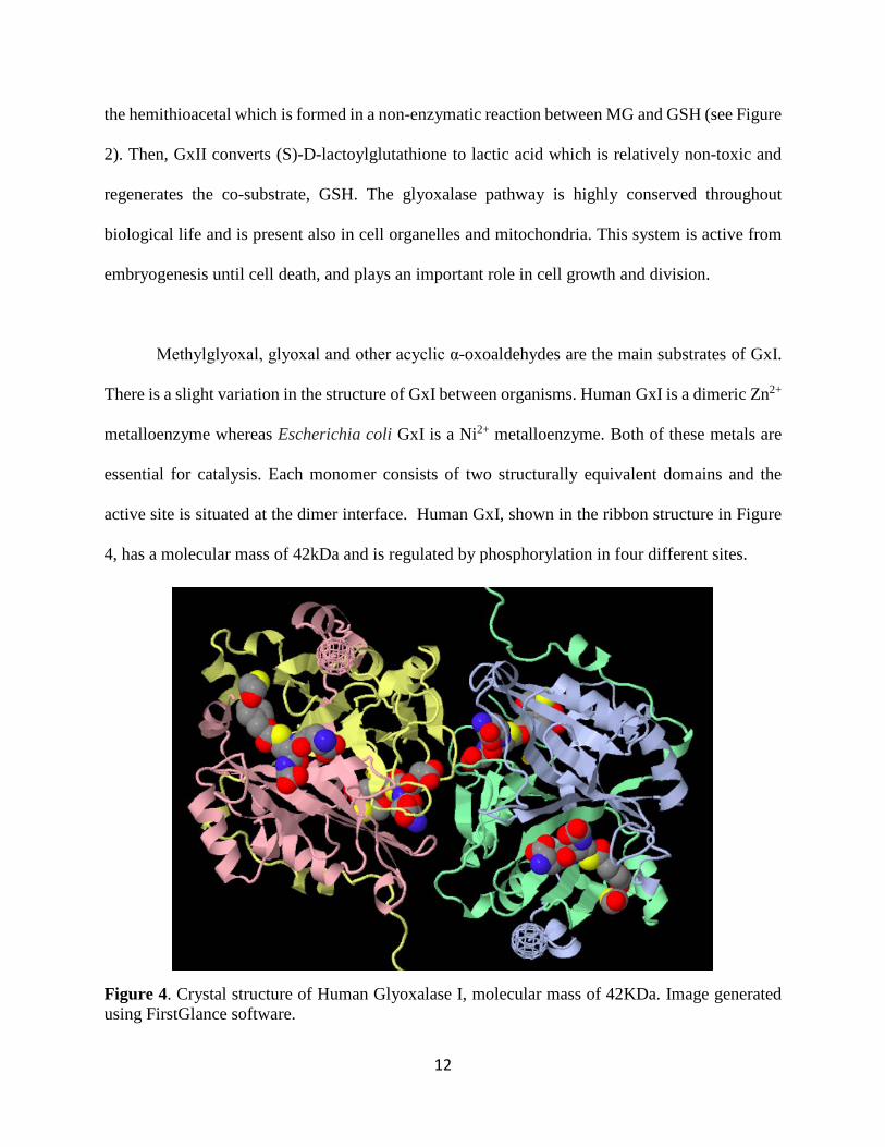
Methylglyoxal, glyoxal and other acyclic α-oxoaldehydes are the main substrates of GxI.
There is a slight variation in the structure of GxI between organisms. Human GxI is a dimeric Zn2+
metalloenzyme whereas Escherichia coli GxI is a Ni2+ metalloenzyme. Both of these metals are
essential for catalysis. Each monomer consists of two structurally equivalent domains and the
active site is situated at the dimer interface. Human GxI, shown in the ribbon structure in Figure
4, has a molecular mass of 42kDa and is regulated by phosphorylation in four different sites.
Figure 4. Crystal structure of Human Glyoxalase I, molecular mass of 42KDa. Image generated using FirstGlance software.
13
1.2 Inhibition of Glyoxalase I
1.2.1 Hallmarks of Apoptosis
GxI has been extensively studied in the past years because its inhibitors are shown to be
potential cancer therapeutic agents. By inhibiting GxI, MG levels should increase, leading to an
increase in cell death by apoptosis or necrosis. The cells of a multicellular organism form an
organized community, and this community is extremely regulated. The regulation involves
controlling the rates of cell division and cell death (White, 2004). When a cell is no longer viable
and is not contributing to the overall well-being of the community, this cell undergoes apoptosis,
which is a process of programed cell death that occurs under normal physiological conditions. This
form of cell death is more organized when compared to necrosis, which occurs when the cell
encounters an extreme variance in physiological conditions. Both processes of cell death are part
of the normal growth and development of an individual, and many pathologies can arise if these
processes are inhibited (White 2004, Osborne 1996). Hence, if GxI is inhibited, the level of MG
will increase, and therefore the rate of cell death by apoptosis will increase. Different competitive
GxI inhibitors, designed based on the active site structure, have been synthesized to test this
hypothesis. These inhibitors have been found to have anti-proliferative activity and suggest that
the increased levels of MG do indeed prevent tumor growth. The best competitive inhibitors of
GxI were found to be GSH derivatives of small, hydrophobic compounds.
14
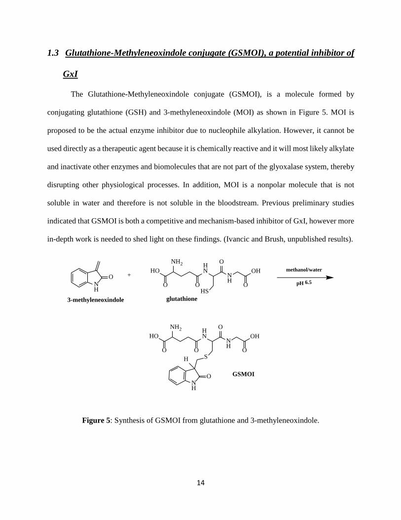
1.3 Glutathione-Methyleneoxindole conjugate (GSMOI), a potential inhibitor of
GxI
The Glutathione-Methyleneoxindole conjugate (GSMOI), is a molecule formed by
conjugating glutathione (GSH) and 3-methyleneoxindole (MOI) as shown in Figure 5. MOI is
proposed to be the actual enzyme inhibitor due to nucleophile alkylation. However, it cannot be
used directly as a therapeutic agent because it is chemically reactive and it will most likely alkylate
and inactivate other enzymes and biomolecules that are not part of the glyoxalase system, thereby
disrupting other physiological processes. In addition, MOI is a nonpolar molecule that is not
soluble in water and therefore is not soluble in the bloodstream. Previous preliminary studies
indicated that GSMOI is both a competitive and mechanism-based inhibitor of GxI, however more
in-depth work is needed to shed light on these findings. (Ivancic and Brush, unpublished results).
Figure 5: Synthesis of GSMOI from glutathione and 3-methyleneoxindole.
HONH2
O
HN
OS
NH
OOH
O
HONH2
O
HN
OHS
NH
OOH
O
NH
O
H
NH
O
glutathione
GSMOI
+
3-methyleneoxindole
methanol/water
pH 6.5
15
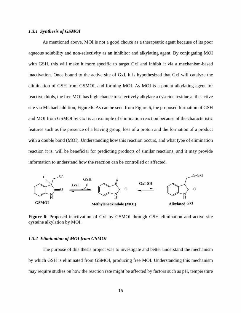
1.3.1 Synthesis of GSMOI
As mentioned above, MOI is not a good choice as a therapeutic agent because of its poor
aqueous solubility and non-selectivity as an inhibitor and alkylating agent. By conjugating MOI
with GSH, this will make it more specific to target GxI and inhibit it via a mechanism-based
inactivation. Once bound to the active site of GxI, it is hypothesized that GxI will catalyze the
elimination of GSH from GSMOI, and forming MOI. As MOI is a potent alkylating agent for
reactive thiols, the free MOI has high chance to selectively alkylate a cysteine residue at the active
site via Michael addition, Figure 6. As can be seen from Figure 6, the proposed formation of GSH
and MOI from GSMOI by GxI is an example of elimination reaction because of the characteristic
features such as the presence of a leaving group, loss of a proton and the formation of a product
with a double bond (MOI). Understanding how this reaction occurs, and what type of elimination
reaction it is, will be beneficial for predicting products of similar reactions, and it may provide
information to understand how the reaction can be controlled or affected.
Figure 6: Proposed inactivation of GxI by GSMOI through GSH elimination and active site cysteine alkylation by MOI.
1.3.2 Elimination of MOI from GSMOI
The purpose of this thesis project was to investigate and better understand the mechanism
by which GSH is eliminated from GSMOI, producing free MOI. Understanding this mechanism
may require studies on how the reaction rate might be affected by factors such as pH, temperature
NH
O
SG
GSMOI
H
NH
O
Methyleneoxindole (MOI)
GxI-SH
NH
O
Alkylated GxI
S-GxI
GxIGSH
16
and potential acid or base catalysts. This work depended on the ability to develop an assay to
quantitatively study the rate of the elimination reaction, under conditions that would allow the
study of factors that affect the rate limiting step. This turned out to be a challenge that was solved.
The interpretation of rate data required understanding of the chemical basis behind the three most
common biological elimination mechanisms that could explain the elimination of GSH and
formation of MOI: E1, E2 or E1cb (cf. Figure 7). (Smith, 2001).
The E1 reaction is a minimum two-step mechanism where the first step is the rate limiting
step that involves loss of GSH from GSMOI to form a carbocation intermediate. In the second step
a base removes the C-3 proton forming the double bond. The E2 reaction is a one-step concerted
mechanism in which removal of the C-3 proton by a base and loss of GSH occur simultaneously.
The E1 mechanism is highly unlikely due to the formation of an unstable primary carbocation
intermediate, but the E1 and E2 mechanisms can be easily distinguished by studying the effect of
pH on the reaction. (Smith, 2001).
The E1cb mechanism is also a two-step mechanism, where we have “fast” removal of the
C-3 proton forming a stable conjugate base (from now onwards referred as cb) or carbanion
intermediate. This is followed by a slow elimination step, forming GSH and MOI. The rate of an
E1 mechanism is not expected to change with changes in pH as abstraction of the C-3 proton is
not expected to be rate-limiting, while the rate of the E2 mechanism is predicted to have a
proportional relationship with pH.
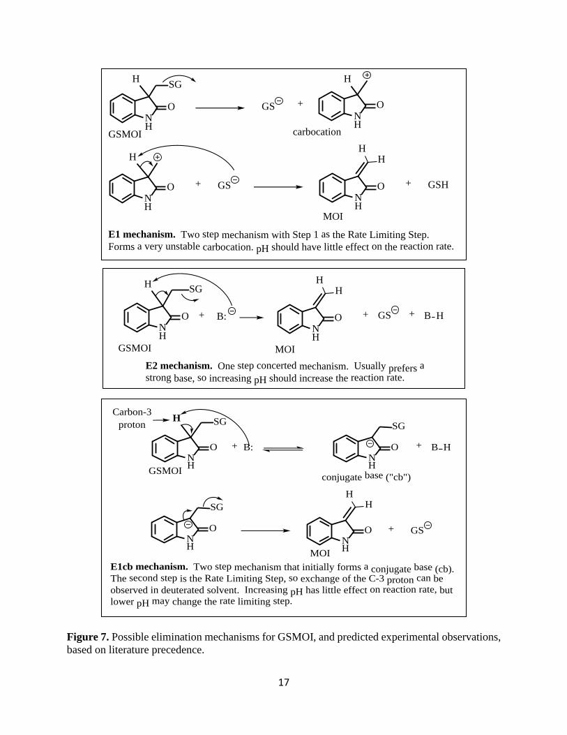
17
Figure 7. Possible elimination mechanisms for GSMOI, and predicted experimental observations, based on literature precedence.
+
SG
NH
O
H
GSNH
O
H
NH
O
H
+ GSNH
O + GSH
HH
MOI
GSMOI carbocation
E1 mechanism. Two step mechanism with Step 1 as the Rate Limiting Step.
Forms a very unstable carbocation. pH should have little effect on the reaction rate.
SG
NH
O
H
GSNH
O +
HH
+ B: + B H
GSMOI MOIE2 mechanism. One step concerted mechanism.
Usually prefers a
strong base, so increasing pH should increase the reaction rate.
SG
NH
O
H
+ GSNH
O
HH
+ B: + B H
SG
NH
O
SG
NH
O
GSMOI conjugate base ("cb")
MOI
Carbon-3 proton
E1cb mechanism. Two step mechanism that initially forms a conjugate base (cb).
The second step is the Rate Limiting Step, so exchange of the C-3 proton can be observed in deuterated solvent.
Increasing pH has little effect on reaction rate, but
lower pH may change the rate limiting step.
18
The rates of both the E2 and E1cb reactions are expected to be pH dependent. However,
the E1cb mechanism can be distinguished by the formation of the intermediate conjugate base.
Formation of the cb can be observed by running the reaction in deuterated water (D2O) and
analyzing kinetic data of proton exchange obtained from 1H NMR spectrometry. For the two step
E1cb mechanism, it is expected that fast proton exchange with D2O solvent will be observed by
the disappearance of the C-3 proton 1H NMR signal, but no (or much slower) GSH elimination
and formation of MOI. For the one step E2 mechanism, the loss of the C-3 proton signal will occur
at the same rate as the development of NMR signals for MOI.
1.4 Significance of this work
The significance of this work can be directly related to the development of cancer
therapeutic agents, an important area of study given that cancer is the cause of 1 in every 8 deaths
worldwide, and research for new and different drugs is a top concern in drug development labs.
The purpose of this research was to discover more relevant information about the inhibition of GxI
by GSMOI that can be used in the future for drug development. If GSMOI is a mechanism-based
inhibitor of GxI, then understanding how GSMOI undergoes elimination under physiological
conditions is critical. By understanding this mechanism, predictions on how other enzymes might
be affected by this inhibitor can be proposed. Overall this research will lead to a better
understanding of the interactions of GSH, MOI, and GxI, and how these interactions can teach
alternate approaches to one day curing cancer.
19
2. Materials and Methods
All reagents were purchased from Sigma-Aldrich or Fisher Scientific and used without further
purification. Methanol was purchased at 99.9% purity. All glassware was washed with Micro-90
cleaner, and then rinsed with deionized water and acetone before oven drying at 80oC. Dry ice was
purchased from Dry Ice Corp., Hingham, MA.
To study and characterize the elimination of GSH from GSMOI, kinetic assays were
performed, in triplicate, in 50 mM potassium phosphate buffer at 25oC, using UV/Vis spectroscopy
on a PerkinElmer Lambda 25 UV/vis spectrometer equipped with a PE PCB 1500 Water Peltier
System for temperature control, and the UV WinLab software. To confirm the structure of the
products, Nuclear Magnetic Resonance (1H NMR) was performed using a JEOL ECX-400M
spectrometer. All NMR samples were prepared in 50 mM potassium phosphate buffer at a
specified pH that had been exchanged into D2O solvent (pD = pH + 0.4).
2.1 Synthesis of 3-Bromooxindole-3-acetic acid (BOAA)
To prepare BOAA, 3.00 g of indole acetic acid, IAA, (17.1 mmoles) were added to a 3-neck
round bottom flask (RBF). The RBF was previously dried in the oven and covered with aluminum
foil. To the same flask, 75ml of warm tert-butanol was added along with a magnetic stir bar. The
IAA was stirred until dissolved, then 6.30 g of N-Bromosuccinimide (35.4 mmoles) were added
over a 60 minute time period. The color of the mixture changed from orange to reddish, and the
mixture was stirred for an additional 10 minutes. The reaction was stored in the -80oC freezer
overnight, and the next day was allowed to warm to room temperature before getting separated
into different portions in a pear-shaped RBF. Each portion was put on the high vac in order to
20
evaporate the tert-butanol. After 30 minutes, all the tert-butanol was evaporated and the resulting
syrup solution had a brown color. This was then dissolved in 50 mL of dry ethyl ether and
magnesium sulfate was added as the drying agent. The mixture was stirred for 15 minutes and was
filtrated to remove succinimide and the drying agent. The mixture was then placed on a rotavap
for 20 minutes to evaporate the ether and dissolved again in 25 mL of ether. After 1 hour at room
temperature, the mixture was filtrated again and concentrated on the rotavap. Residual ether
entrapped in the brown syrup was evaporated on the high vac for approximately 10 minutes. The
viscous syrup was dissolved in 25 mL of chloroform and stored for two hours at -20oC to allow
for the precipitation of BOAA. The product was isolated by vacuum filtration with a Hirsch funnel.
The product was scrapped onto a watch glass and put in a vacuum desiccator to dry overnight and
then characterized by melting point and thin layer chromatography.
2.2 Synthesis of 3-methylyeoxindole (MOI) from 3-Bromooxindole-3-acetic acid
(BOAA)
To prepare MOI, 199 mg (0.736 mmoles) of BOAA, were dissolved in 1.5ml of 100%
ethanol. This solution was then pipet filtrated (with cotton) to remove any insoluble material. The
solution was transferred to a 10mL glass vial and stirred while cooling in an ice bath. While
stirring, a 7mL ice cold solution containing 124 mg (1.48 mmoles) of sodium bicarbonate was
added to the reaction mixture dropwise over a period of 3 minutes. The solution was allowed to
stir for a couple of minutes until a yellow color was observed, and pH paper was used to verify
that the solution was weakly acidic. The precipitate was filtrated using a Hirsch funnel, then
quickly washed with 20mL of ice-cold deionized water to remove all traces of the sodium
bicarbonate. The product was then transferred to a pre-weighed watch glass and placed in a vacuum
21
desiccator to dry overnight in the dark. The dried product was weighed, the percent yield was
calculated, and the purity was determined using UV-Vis spectroscopy and 1H NMR spectrometry.
2.3 Synthesis of Glutathione-3-Methyleneoxindole Conjugate (GSMOI) from 3-
Methylyoxindole and Glutathione.
To make GSMOI, 140 ml of 25% methanol in deionized H2O was added to a 250 ml
Erlenmeyer flask. The solution was stirred under an atmosphere of argon for 15 minutes, then
70mg (0.228 mmole) of reduced glutathione (GSH) were added to the flask and stirred for an
additional 5 minutes. The pH of the solution was adjusted using 1M potassium hydroxide to pH 6.
Then, to the stirring solution, 39.7 mg (0.274 mmole) of 3-methyleneoxindole (MOI) were added
in three portions over a period of 10 minutes. The solution was stirred for an additional one hour
until all the yellow color from MOI dissipated. The reaction mixture was gravity filtrated into a
clean flask to remove any white precipitate due to MOI polymerization, and the filtrate was cooled
in ice. The filtrate was concentrated to about 20ml on the high vac at 30˚C to remove methanol,
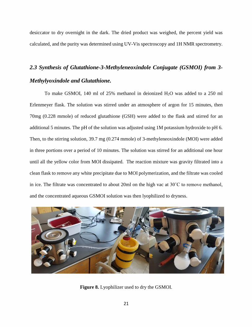
and the concentrated aqueous GSMOI solution was then lyophilized to dryness.
Figure 8. Lyophilizer used to dry the GSMOI.
22
The product was obtained as a fluffy, white powder which was then washed with anhydrous
ether two times to extract excess MOI. The ether was removed with a pipet and the white solid
dried under a gentle stream of dry argon for 5 minutes. The product was transferred to a pre-
weighed watch glass and dried thoroughly in a vacuum desiccator overnight in the dark. Then
GSMOI was stored under dry conditions at -20oC. The yield of the dry product was determined,
and the structure verified by UV-Vis spectroscopy and 1H NMR spectrometry.
2.4 Characterization using NMR
1H NMR was used to determine the purity of both MOI and GSMOI. For MOI 6 mg of
solid sample was dissolved in Acetone d6. GSMOI NMR samples were prepared in two ways.
Method I: 7-10 mg of GSMOI were dissolved in 1ml of D2O and lyophilized to dryness. This
process was repeated two more time with fresh D2O to exchange acidic H for D. Then the dry,
exchanged sample was dissolved in 1 ml of Deuterated Potassium Phosphate, (D)KP buffer at a
specified pD. Method II: 7-10 mg of GSMOI were dissolved directly in 1ml of (D)KP buffer at a
specified pD. The (D)KP was prepared by exchanging 10 mL of (H)KP buffer at a specified pH
with D2O on the high vac. Samples were flushed with dry argon to limit exposure to air. The pD
correction factor is +0.4 units, so for a pH 7 buffer, the pD is 7.4.
2.5 Development of UV-VIS Spectroscopic Methods for Elimination Kinetics
Studies
A stock solution of GSMOI was prepared in deionized (DI) water (4.86 mM). The stock
solution was kept on ice during use, and stored at -20oC. Under these conditions this solution was
23
stable for at least 4 weeks. A 5 mM stock solution of 5,5-dithio-bis-(2-nitrobenzoic acid) (DTNB,
Ellman’s reagent) was prepared in KP buffer, kept on ice during use, and stored at 4oC.
Method I. 50 mM KP buffer in a quartz cuvette (1.0 mL) was pre-incubated for 10 minutes
at 25˚C. To study the rate of GSH elimination, the reaction was initiated by adding an aliquot of
the GSMOI stock solution to a final concentration of 50 µM. Repeat wavelength scans (200-500
nm) were done at specified time intervals for 3 hours. To determine the rate of formation of MOI
the change in absorbance at 248 nm was analyzed. Due to the low extinction coefficient of MOI
at 248 nm this method did not provide the sensitivity for kinetic analysis. We switched to Elman’s
Reagent, DTNB (5,5-dithio-bis-(2-nitrobenzoic acid) as described below.
Method II. A 5 mM solution of DTNB was pre-incubated with 50 mM KP buffer in a quartz
cuvette at 25 ºC. After equilibration for 5 minutes, reaction was initiated by the addition of various
concentrations of the GSMOI stock solution. Initial rate slopes from absorbance (412 nm) vs time
curves were determined over the initial 10-15% of reaction. The extinction coefficient of DTNB
at 412 nm is (14150 m-1 cm-1).
2.6 Determination of Reaction Order
To determine the order of the reaction, a 5 mM solution of DTNB was incubated with 50
mM potassium phosphate buffer in a quartz cuvette at 25oC. After temperature equilibration for at
least 5 minutes, the reaction was initiated by the addition of different aliquots of the GSMOI stock
solution (10, 20, 40 and 80 µL). Absorbance vs time curves at 412 nm were recorded and initial
rate slopes from the first 10-15% of the reaction were used to calculate reaction rates.

24
2.7 Effect of pH on the rate of GSH elimination
To determine the effect of pH on the reaction, a 5mM solution of DTNB was incubated
with 50 mM potassium phosphate buffer (at pH 6.50, 7.00 and 7.50) in a quartz cuvette at 25oC.
After equilibration for 5 minutes, the reaction was initiated by the addition of different aliquots of
GSMOI stock solution to the cuvette. The change in absorbance at 412 nm was recorded and initial
10-15% of the reaction was used in order to collect the initial slopes from absorbance vs time
curves.
2.8 Effect of potential catalysts
The DTNB assay used in section 2.6, was used again in order to determine the effect of
different bio-molecules as potential catalysts for the elimination reaction. In addition to DTNB, a
250 mM solution of the potential catalyst was incubated with 50 mM potassium phosphate buffer
in a quartz cuvette at 25ºC. After equilibration for 5 minutes, the reaction was initiated by the
addition of various concentrations of GSMOI. The absorbance at 412 nm was recorded and initial
10-15% of the reaction was used in order to collect the initial slopes from absorbance vs time
curves.
2.9 Effect of buffer concentration on rate constant k
A 5 mM solution of DTNB was incubated with 10, 50 and 250 mM potassium phosphate
buffer in a quartz cuvette at 25 ºC (pH 7). After equilibration for 5 minutes, reaction was initiated
by the addition of various concentrations of GSMOI to the cuvette. Initial rate slopes from
absorbance vs time curves were determined over the initial 10-15% of reaction.
25
2.10 GSMOI C-3 Proton exchange kinetics
All glassware was dried in an oven at 100oC, and stored in a desiccator. Dry argon was
used to flush all samples and protect from air. 7-10 mg of GSMOI were dissolved in 1ml of (D)KP
buffer at pD 6.69 and 7.34. A stopwatch was started upon addition of D-KP buffer to the GSMOI
samples, and these solutions were immediately transferred to an NMR tube and scans started
within 2-3 minutes. The scans were collected every 8 minutes for approximately one hour, and the
C-3-H and cys-α-H signals were integrated.
26
3. Results and Discussion
3.1 Synthesis of 3-Bromooxindole-3-acetic acid (BOAA) and 3-methylyeoxindole
(MOI)
Because access to an NMR was restricted at the time, BOAA was only characterized by
observing the melting point range of 153-158oC. This compares very well with the literature mp
of 158oC.
After drying in a vacuum desiccator overnight, the MOI sample looked bright yellow, and
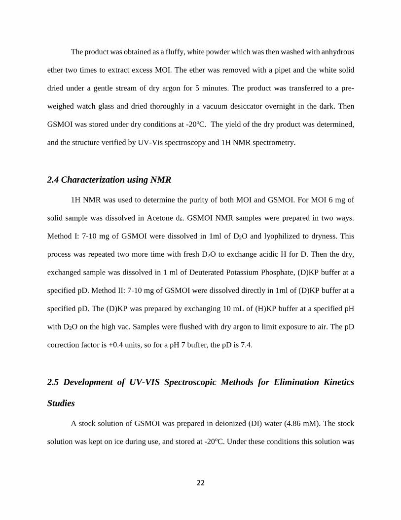
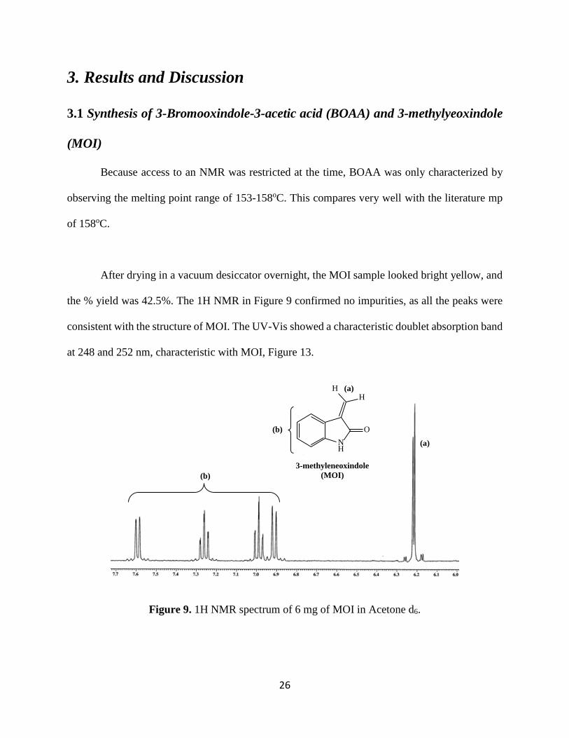
the % yield was 42.5%. The 1H NMR in Figure 9 confirmed no impurities, as all the peaks were
consistent with the structure of MOI. The UV-Vis showed a characteristic doublet absorption band
at 248 and 252 nm, characteristic with MOI, Figure 13.
Figure 9. 1H NMR spectrum of 6 mg of MOI in Acetone d6.
NH
O
3-methyleneoxindole (MOI)
HH
(a)
(b)
(a)
(b)
27
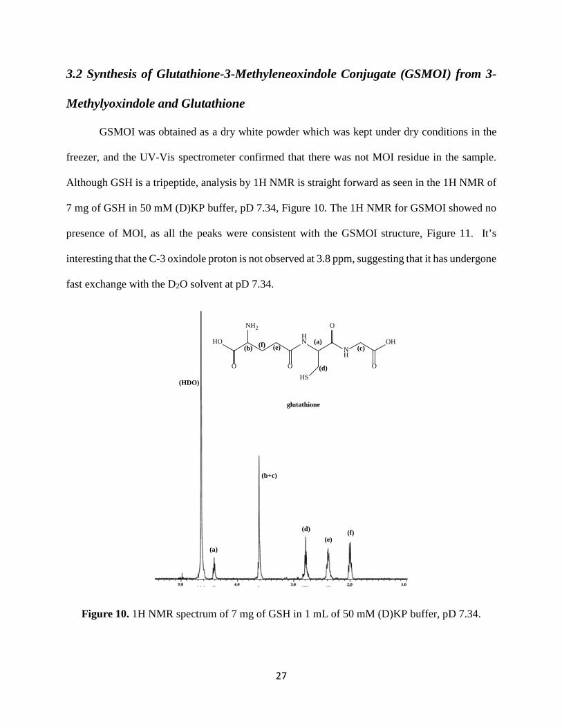
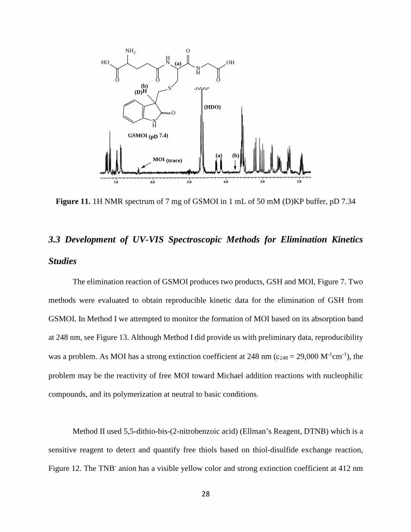
3.2 Synthesis of Glutathione-3-Methyleneoxindole Conjugate (GSMOI) from 3-
Methylyoxindole and Glutathione
GSMOI was obtained as a dry white powder which was kept under dry conditions in the
freezer, and the UV-Vis spectrometer confirmed that there was not MOI residue in the sample.
Although GSH is a tripeptide, analysis by 1H NMR is straight forward as seen in the 1H NMR of
7 mg of GSH in 50 mM (D)KP buffer, pD 7.34, Figure 10. The 1H NMR for GSMOI showed no
presence of MOI, as all the peaks were consistent with the GSMOI structure, Figure 11. It’s
interesting that the C-3 oxindole proton is not observed at 3.8 ppm, suggesting that it has undergone
fast exchange with the D2O solvent at pD 7.34.
Figure 10. 1H NMR spectrum of 7 mg of GSH in 1 mL of 50 mM (D)KP buffer, pD 7.34.
HO
NH2
O
HN
OHS
NH
O
OH
O
glutathione
(HDO)
(a)
(a)
(b)
(b+c)
(c)
(d)(e)
(f)
(d)
(e)(f)
28
Figure 11. 1H NMR spectrum of 7 mg of GSMOI in 1 mL of 50 mM (D)KP buffer, pD 7.34
3.3 Development of UV-VIS Spectroscopic Methods for Elimination Kinetics
Studies
The elimination reaction of GSMOI produces two products, GSH and MOI, Figure 7. Two
methods were evaluated to obtain reproducible kinetic data for the elimination of GSH from
GSMOI. In Method I we attempted to monitor the formation of MOI based on its absorption band
at 248 nm, see Figure 13. Although Method I did provide us with preliminary data, reproducibility
was a problem. As MOI has a strong extinction coefficient at 248 nm (ε248 = 29,000 M-1cm-1), the
problem may be the reactivity of free MOI toward Michael addition reactions with nucleophilic
compounds, and its polymerization at neutral to basic conditions.
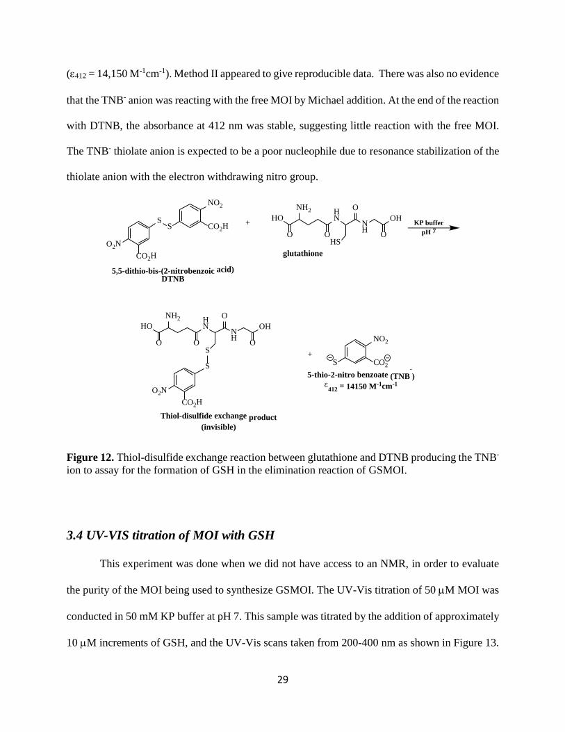
Method II used 5,5-dithio-bis-(2-nitrobenzoic acid) (Ellman’s Reagent, DTNB) which is a
sensitive reagent to detect and quantify free thiols based on thiol-disulfide exchange reaction,
Figure 12. The TNB- anion has a visible yellow color and strong extinction coefficient at 412 nm
HO
NH2
O
HN
OS
NH
O
OH
O
NH
O
H
GSMOI (pD 7.4)
(D)
(a)
(b)
(a)
(b)
(HDO)
MOI (trace)
29
(ε412 = 14,150 M-1cm-1). Method II appeared to give reproducible data. There was also no evidence
that the TNB- anion was reacting with the free MOI by Michael addition. At the end of the reaction
with DTNB, the absorbance at 412 nm was stable, suggesting little reaction with the free MOI.
The TNB- thiolate anion is expected to be a poor nucleophile due to resonance stabilization of the
thiolate anion with the electron withdrawing nitro group.
Figure 12. Thiol-disulfide exchange reaction between glutathione and DTNB producing the TNB- ion to assay for the formation of GSH in the elimination reaction of GSMOI.
3.4 UV-VIS titration of MOI with GSH
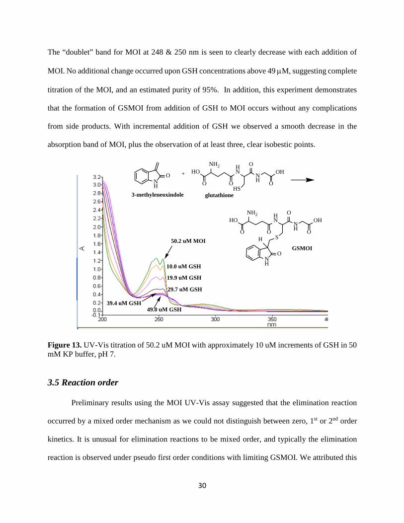
This experiment was done when we did not have access to an NMR, in order to evaluate
the purity of the MOI being used to synthesize GSMOI. The UV-Vis titration of 50 µM MOI was
conducted in 50 mM KP buffer at pH 7. This sample was titrated by the addition of approximately
10 µM increments of GSH, and the UV-Vis scans taken from 200-400 nm as shown in Figure 13.
HONH2
O
HN
OHS
NH
OOH
O
glutathione
+ KP bufferpH 7
CO2H
S
NO2
S
O2N
CO2H
5,5-dithio-bis-(2-nitrobenzoic acid) DTNB
HONH2
O
HN
OS
NH
OOH
O
5-thio-2-nitro benzoate (TNB-)
ε412
= 14150 M-1cm-1
+
CO2H
S
O2N
Thiol-disulfide exchange product (invisible)
S
NO2
CO2
30
The “doublet” band for MOI at 248 & 250 nm is seen to clearly decrease with each addition of
MOI. No additional change occurred upon GSH concentrations above 49 µM, suggesting complete
titration of the MOI, and an estimated purity of 95%. In addition, this experiment demonstrates
that the formation of GSMOI from addition of GSH to MOI occurs without any complications
from side products. With incremental addition of GSH we observed a smooth decrease in the
absorption band of MOI, plus the observation of at least three, clear isobestic points.
Figure 13. UV-Vis titration of 50.2 uM MOI with approximately 10 uM increments of GSH in 50 mM KP buffer, pH 7.
3.5 Reaction order
Preliminary results using the MOI UV-Vis assay suggested that the elimination reaction
occurred by a mixed order mechanism as we could not distinguish between zero, 1st or 2nd order
kinetics. It is unusual for elimination reactions to be mixed order, and typically the elimination
reaction is observed under pseudo first order conditions with limiting GSMOI. We attributed this
50.2 uM MOI
10.0 uM GSH
19.9 uM GSH
29.7 uM GSH
39.4 uM GSH49.0 uM GSH
HONH2
O
HN
OS
NH
OOH
O
HONH2
O
HN
OHS
NH
OOH
O
NH
O
H
NH
O
glutathione
GSMOI
+
3-methyleneoxindole
31
preliminary result to problems with the UV-Vis assay where we were observing the decrease in
the MOI absorbance band at 248 nm.
Using the DTNB UV-Vis assay to monitor the elimination of GSH produced more
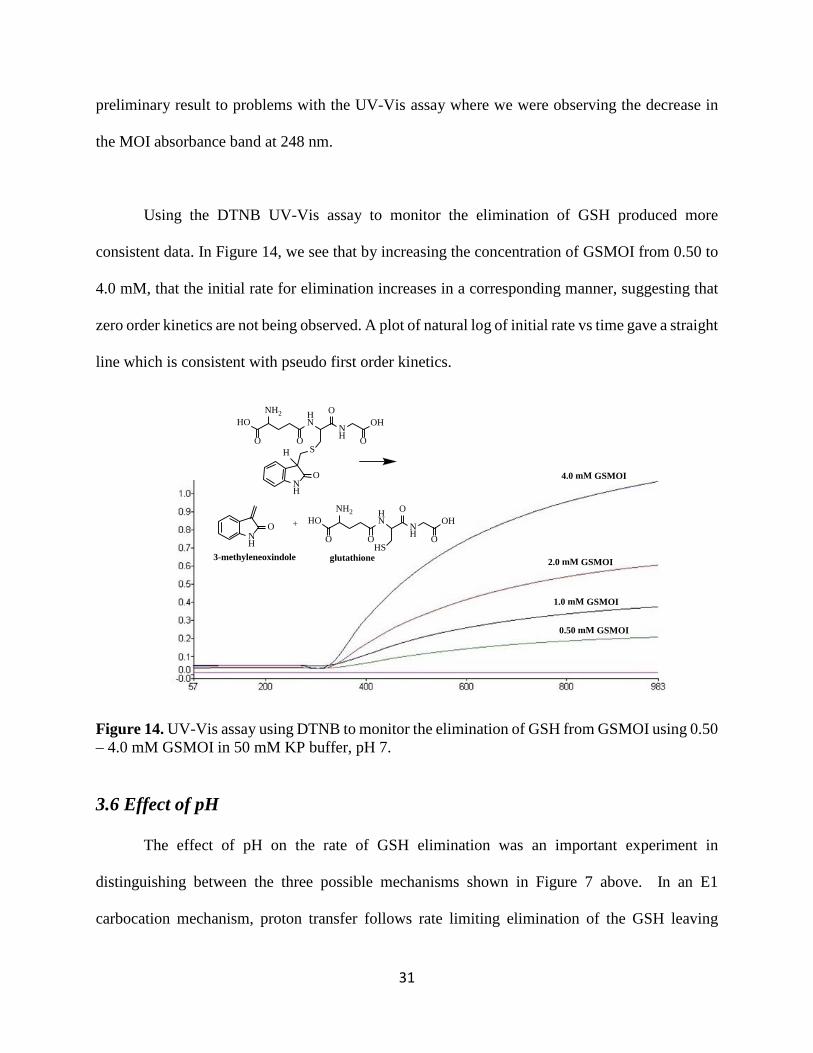
consistent data. In Figure 14, we see that by increasing the concentration of GSMOI from 0.50 to
4.0 mM, that the initial rate for elimination increases in a corresponding manner, suggesting that
zero order kinetics are not being observed. A plot of natural log of initial rate vs time gave a straight
line which is consistent with pseudo first order kinetics.
Figure 14. UV-Vis assay using DTNB to monitor the elimination of GSH from GSMOI using 0.50 – 4.0 mM GSMOI in 50 mM KP buffer, pH 7.
3.6 Effect of pH
The effect of pH on the rate of GSH elimination was an important experiment in
distinguishing between the three possible mechanisms shown in Figure 7 above. In an E1
carbocation mechanism, proton transfer follows rate limiting elimination of the GSH leaving
4.0 mM GSMOI
2.0 mM GSMOI
HONH2
O
HN
OS
NH
OOH
O
HONH2
O
HN
OHS
NH
OOH
O
NH
O
H
NH
O
glutathione
+
3-methyleneoxindole
1.0 mM GSMOI
0.50 mM GSMOI
32
group, so pH should have little effect on rate. The concerted E2 mechanism requires proton transfer
at the same time as GSH elimination, so the reaction rate would be expected to increase with
increasing pH. The rate of an E1cb reaction is also expected to increase with increasing pH as the
fast proton transfer step is followed by slow GSH elimination.
A 5mM solution of DTNB was incubated in 50 mM potassium phosphate buffer (at pH
6.50, 7.00 and 7.50) in quartz cuvettes at 25oC. The reaction was initiated by the addition of various
concentrations of GSMOI to the cuvette. The change in absorbance at 412 nm was recorded and
the initial 10-15% of the reaction was used in order to collect the initial slopes from absorbance vs
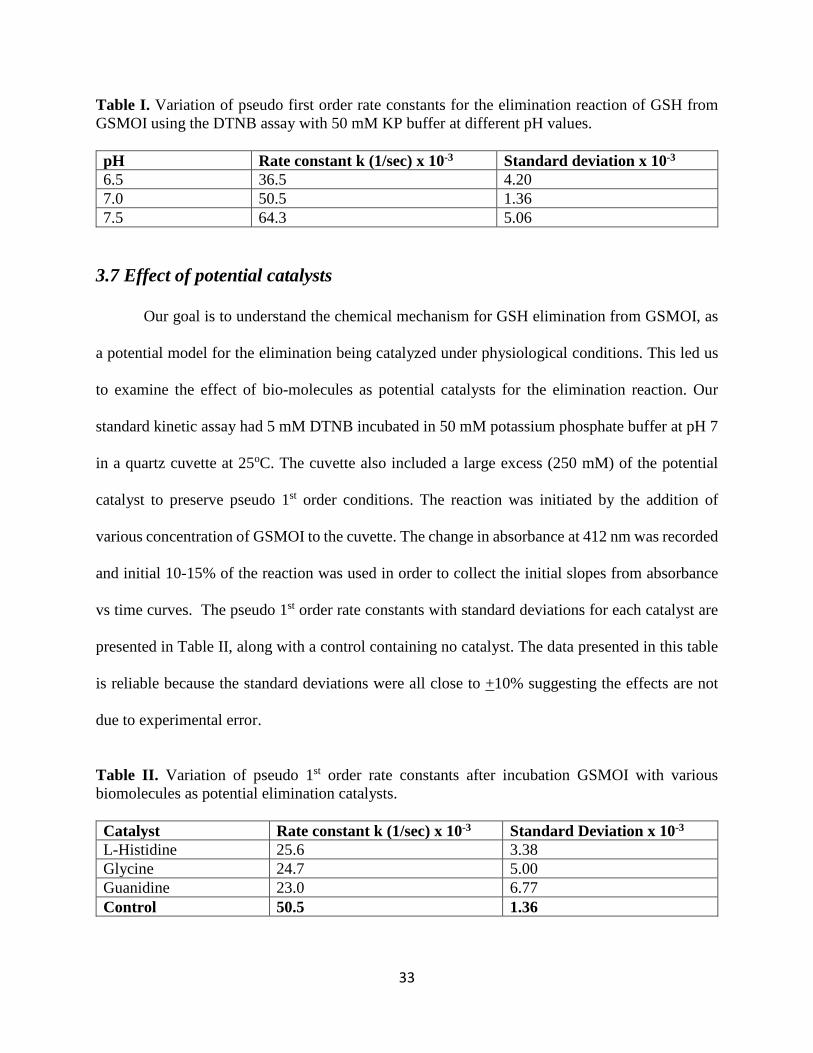
time curves. The pseudo 1st order rate constants with standard deviations at each pH are presented
in Table I, and indicate that the rate constants increase with increasing pH. This suggests that
changes in pH affect the rate of reaction, and that elimination is following either an E2 or E1cb
mechanism. In our preliminary experiments using the less sensitive MOI assay at 248 nm, we
observed little change in elimination reaction rate between pH 6.50-7.50, but a significant decrease
in the reaction rate at pH 6.00. We believe that this may suggest a change in the rate limiting step
consistent with an E1cb elimination mechanism because the “fast” proton transfer step is now
competing with GSH elimination. Although the DNTB assay is more reproducible, and the data
follow a similar trend, the decrease in rate with decreasing pH was not as pronounced. We believe
that this discrepancy results from the DTNB assay being less sensitive below pH 7, as protonation
of the TNB- thiolate ion inhibits conjugation and decreases production of the yellow color at 412
nm. The data presented in this table are reliable because the standard deviations were all close to
+10% suggesting the rate effects are not due to experimental error.
33
Table I. Variation of pseudo first order rate constants for the elimination reaction of GSH from GSMOI using the DTNB assay with 50 mM KP buffer at different pH values. pH Rate constant k (1/sec) x 10-3 Standard deviation x 10-3 6.5 36.5 4.20 7.0 50.5 1.36 7.5 64.3 5.06
3.7 Effect of potential catalysts
Our goal is to understand the chemical mechanism for GSH elimination from GSMOI, as
a potential model for the elimination being catalyzed under physiological conditions. This led us
to examine the effect of bio-molecules as potential catalysts for the elimination reaction. Our
standard kinetic assay had 5 mM DTNB incubated in 50 mM potassium phosphate buffer at pH 7
in a quartz cuvette at 25oC. The cuvette also included a large excess (250 mM) of the potential
catalyst to preserve pseudo 1st order conditions. The reaction was initiated by the addition of
various concentration of GSMOI to the cuvette. The change in absorbance at 412 nm was recorded
and initial 10-15% of the reaction was used in order to collect the initial slopes from absorbance
vs time curves. The pseudo 1st order rate constants with standard deviations for each catalyst are
presented in Table II, along with a control containing no catalyst. The data presented in this table
is reliable because the standard deviations were all close to +10% suggesting the effects are not
due to experimental error.
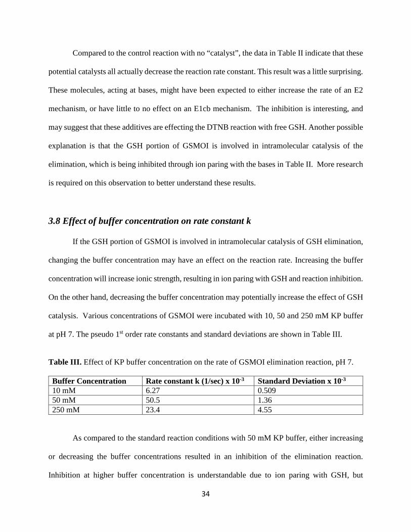
Table II. Variation of pseudo 1st order rate constants after incubation GSMOI with various biomolecules as potential elimination catalysts. Catalyst Rate constant k (1/sec) x 10-3 Standard Deviation x 10-3 L-Histidine 25.6 3.38 Glycine 24.7 5.00 Guanidine 23.0 6.77 Control 50.5 1.36
34
Compared to the control reaction with no “catalyst”, the data in Table II indicate that these
potential catalysts all actually decrease the reaction rate constant. This result was a little surprising.
These molecules, acting at bases, might have been expected to either increase the rate of an E2
mechanism, or have little to no effect on an E1cb mechanism. The inhibition is interesting, and
may suggest that these additives are effecting the DTNB reaction with free GSH. Another possible
explanation is that the GSH portion of GSMOI is involved in intramolecular catalysis of the
elimination, which is being inhibited through ion paring with the bases in Table II. More research
is required on this observation to better understand these results.
3.8 Effect of buffer concentration on rate constant k
If the GSH portion of GSMOI is involved in intramolecular catalysis of GSH elimination,
changing the buffer concentration may have an effect on the reaction rate. Increasing the buffer
concentration will increase ionic strength, resulting in ion paring with GSH and reaction inhibition.
On the other hand, decreasing the buffer concentration may potentially increase the effect of GSH
catalysis. Various concentrations of GSMOI were incubated with 10, 50 and 250 mM KP buffer
at pH 7. The pseudo 1st order rate constants and standard deviations are shown in Table III.
Table III. Effect of KP buffer concentration on the rate of GSMOI elimination reaction, pH 7. Buffer Concentration Rate constant k (1/sec) x 10-3 Standard Deviation x 10-3 10 mM 6.27 0.509 50 mM 50.5 1.36 250 mM 23.4 4.55
As compared to the standard reaction conditions with 50 mM KP buffer, either increasing
or decreasing the buffer concentrations resulted in an inhibition of the elimination reaction.
Inhibition at higher buffer concentration is understandable due to ion paring with GSH, but
35
inhibition at lower buffer concentration is confusing. KP buffers are not known to act as catalysts,
and it is unlikely that changing the buffer concentrations are effecting the DTNB assay. Maybe at
low concentrations of buffer, there is intermolecular ion-pairing of the GSH parts of GSMOI pairs,
and this might be expected to inhibit GSH catalysis. Again, this is an interesting result, but
additional experimentation work is needed to understand these data. The data presented in this
table are reliable because the standard deviations were all close to +10% suggesting the effects are
not due to experimental error.
3.9 GSMOI C-3 Proton exchange kinetics
The most direct way to distinguish between the E2 and E1cb mechanisms was to attempt
to observe removal or exchange, respectively, of the C-3 GSMOI oxindole proton using 1H NMR
in (D)KP buffer. For an E2 mechanism, as the C-3 proton is removed and its 1H NMR signal
decreases, there should be a corresponding increase in the vinyl protons of MOI at 6.2 ppm. On
the other hand, an E1cb mechanism will result in a reversible, fast exchange of the C-3 proton with
deuterium, its 1H NMR signal will quickly decrease, but with very little MOI being produced.
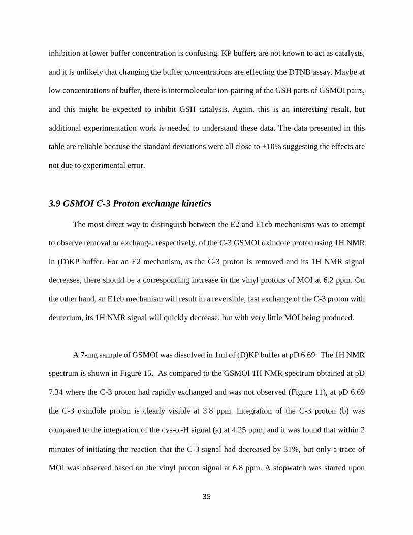
A 7-mg sample of GSMOI was dissolved in 1ml of (D)KP buffer at pD 6.69. The 1H NMR
spectrum is shown in Figure 15. As compared to the GSMOI 1H NMR spectrum obtained at pD
7.34 where the C-3 proton had rapidly exchanged and was not observed (Figure 11), at pD 6.69
the C-3 oxindole proton is clearly visible at 3.8 ppm. Integration of the C-3 proton (b) was
compared to the integration of the cys-α-H signal (a) at 4.25 ppm, and it was found that within 2
minutes of initiating the reaction that the C-3 signal had decreased by 31%, but only a trace of
MOI was observed based on the vinyl proton signal at 6.8 ppm. A stopwatch was started upon
36
addition of D-KP buffer to the GSMOI sample, and NMR scans started within 2-3 minutes. Scans
were collected every 8 minutes for approximately one hour, and the C-3-H and cys-α-H signals
were observed.
Figure 15. 1H NMR spectrum of 7 mg of GSMOI in 1 mL of 50 mM (D)KP buffer, pD 6.69.
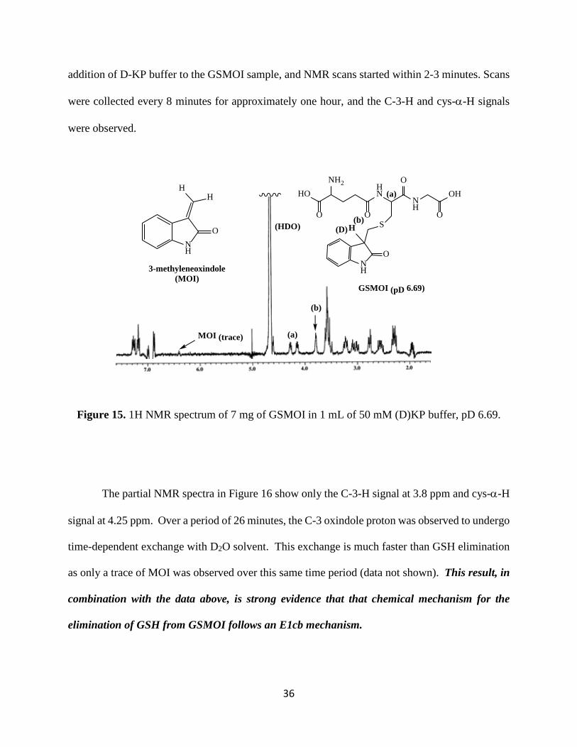
The partial NMR spectra in Figure 16 show only the C-3-H signal at 3.8 ppm and cys-α-H
signal at 4.25 ppm. Over a period of 26 minutes, the C-3 oxindole proton was observed to undergo
time-dependent exchange with D2O solvent. This exchange is much faster than GSH elimination
as only a trace of MOI was observed over this same time period (data not shown). This result, in
combination with the data above, is strong evidence that that chemical mechanism for the
elimination of GSH from GSMOI follows an E1cb mechanism.
HONH2
O
HN
OS
NH
OOH
O
NH
O
H
GSMOI (pD 6.69)
(D)(b)
(a)
(HDO)
NH
O
3-methyleneoxindole (MOI)
HH
(a)
(b)
MOI (trace)
37
Figure 16. 1H NMR spectrum of 7 mg of GSMOI in 1 mL of 50 mM (D)KP buffer, pD 6.69 undergoing time dependent exchange of the oxindole C-3 proton.
HO
NH2
O
HN
OS
NH
O
OH
O
NH
O
H
GSMOI (pD 6.7)
(D)
(a)
(b)
(a)
(b)
t = 129 sec
t = 519 sec
t = 801 sec
t = 1260 sec
t = 1587 sec
38
4. Conclusions
The significance of this work can be directly related to the development of cancer
therapeutic agents, an important area of study given that cancer is the cause of 1 in every 8 deaths
worldwide, and research for new and different drugs is a top concern in drug development labs.
The purpose of this research is to discover more relevant information about the inhibition of GxI
by GSMOI that can be used in the future for drug development.
Previous students in Dr. Brush’s research group found preliminary evidence that GSMOI
is both a competitive and mechanism-based inhibitor of GxI, however more study is needed to
better understand those findings. MOI is not a good choice as a therapeutic agent because of its
poor aqueous solubility, and non-selectivity as an alkylating agent. By conjugating MOI with GSH
to form GSMOI, we believe this compound will specifically target GxI and inhibit by mechanism-
based inactivation.
Once bound to the active site, we think that GxI will catalyze the elimination of GSH from
GSMOI, releasing MOI. As MOI is an alkylating agent for cysteine, we believe that the MOI will
selectively alkylate an active site cysteine residue by Michael addition, Figure 6 (above). The
release of MOI and GSH from GSMOI is an example of an elimination reaction. There is a leaving
group (GSH), loss of a proton and the formation of a product with a double bond (MOI). If GSMOI
is a mechanism-based inhibitor of GxI, we needed to understand how GSMOI undergoes
elimination under physiological conditions.
39
The main outcome of this thesis project is that we now have a better understanding of the
mechanism by which GSH is eliminated from GSMOI. We developed an assay to quantitatively
study the rate of the elimination reaction, under conditions that would allow us to study factors
that affect the rate limiting step. We have studied how the reaction rate was affected by pH and
potential catalysts. Our rate data allowed us to distinguish between the three most common
biological elimination mechanisms that could explain the elimination of GSH and formation of
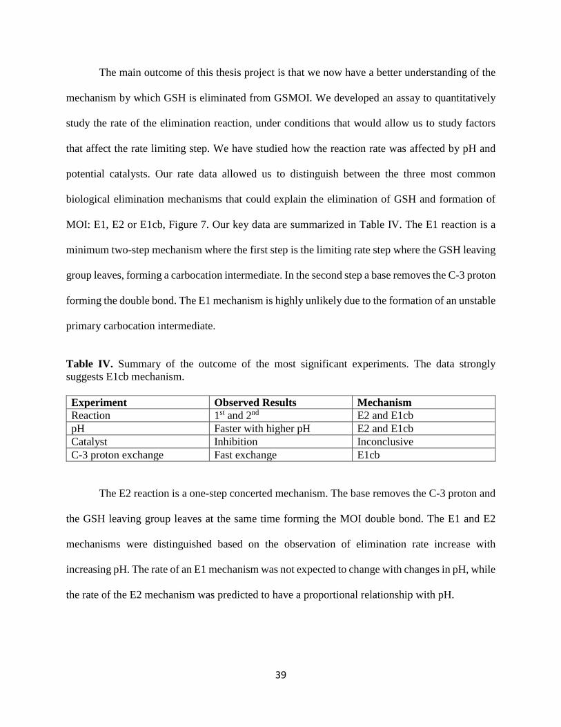
MOI: E1, E2 or E1cb, Figure 7. Our key data are summarized in Table IV. The E1 reaction is a
minimum two-step mechanism where the first step is the limiting rate step where the GSH leaving
group leaves, forming a carbocation intermediate. In the second step a base removes the C-3 proton
forming the double bond. The E1 mechanism is highly unlikely due to the formation of an unstable
primary carbocation intermediate.
Table IV. Summary of the outcome of the most significant experiments. The data strongly suggests E1cb mechanism.
Experiment Observed Results Mechanism Reaction 1st and 2nd E2 and E1cb pH Faster with higher pH E2 and E1cb Catalyst Inhibition Inconclusive C-3 proton exchange Fast exchange E1cb
The E2 reaction is a one-step concerted mechanism. The base removes the C-3 proton and
the GSH leaving group leaves at the same time forming the MOI double bond. The E1 and E2
mechanisms were distinguished based on the observation of elimination rate increase with
increasing pH. The rate of an E1 mechanism was not expected to change with changes in pH, while
the rate of the E2 mechanism was predicted to have a proportional relationship with pH.
40
The E1cb mechanism is also a two-step mechanism, where we have fast removal of the C-
3 proton forming a stable conjugate base “cb” intermediate. This is followed by a slow elimination
step, forming GSH and MOI. The rates of both the E2 and E1cb reactions were expected to be pH
dependent. However, the E1cb mechanism was identified based on data suggesting formation of
the “cb” intermediate based on the observation of fast proton exchange in D2O solvent by 1H
NMR. We observed the disappearance of the C-3 proton 1H NMR signal, and much slower
formation of MOI, Figure 16. This experimental result suggests against the one step E2
mechanism, where the loss of the C-3 proton signal should have occurred at the same rate as the
formation of MOI.
The goals of this thesis project were achieved as the data suggest that the elimination
mechanism is E1cb. What do the results of this research suggest for GSMOI as a mechanism based
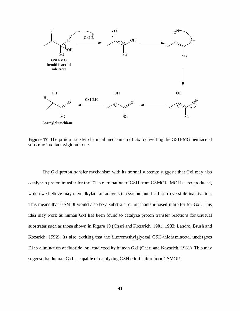
inhibitor of GxI? In its “normal” reaction GxI catalyzes the conversion of the thiohemiacetal of
GSH and MG into (S)-D-lactoylglutathione, Figure 2 (above). The chemical mechanism involves
a proton transfer with an intermediate carbanion, as shown in Figure 17 (Chari and Kozarich,
1981).
41
Figure 17. The proton transfer chemical mechanism of GxI converting the GSH-MG hemiacetal substrate into lactoylglutathione.
The GxI proton transfer mechanism with its normal substrate suggests that GxI may also
catalyze a proton transfer for the E1cb elimination of GSH from GSMOI. MOI is also produced,
which we believe may then alkylate an active site cysteine and lead to irreversible inactivation.
This means that GSMOI would also be a substrate, or mechanism-based inhibitor for GxI. This
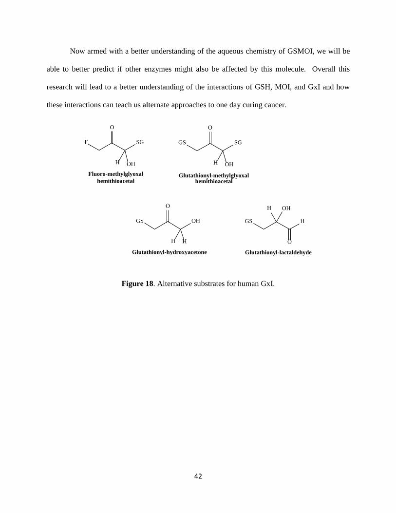
idea may work as human GxI has been found to catalyze proton transfer reactions for unusual
substrates such as those shown in Figure 18 (Chari and Kozarich, 1981, 1983; Landro, Brush and
Kozarich, 1992). Its also exciting that the fluoromethylglyoxal GSH-thiohemiacetal undergoes
E1cb elimination of fluoride ion, catalyzed by human GxI (Chari and Kozarich, 1981). This may
suggest that human GxI is capable of catalyzing GSH elimination from GSMOI!
H
SGOH
O
GSH-MG hemithioacetal
substrate
GxI-B
SG
O
OH
SG
O
OH
SG
OH
O
SG
OH
OGxI-BH
SG
OH
OH
Lactoylglutathione
42
Now armed with a better understanding of the aqueous chemistry of GSMOI, we will be
able to better predict if other enzymes might also be affected by this molecule. Overall this
research will lead to a better understanding of the interactions of GSH, MOI, and GxI and how
these interactions can teach us alternate approaches to one day curing cancer.
Figure 18. Alternative substrates for human GxI.
H
SG
OH
O
Fluoro-methylglyoxal hemithioacetal
F
H
SG
OH
O
Glutathionyl-methylglyoxal hemithioacetal
GS
H
OH
H
O
Glutathionyl-hydroxyacetone
GS
O
H
OH
Glutathionyl-lactaldehyde
GS
H
43
5. Future Work
As to any research project, there are still a few things that need to be done in the future.
The effect of buffer concentration changes were surprising, with various effect on the rate of
reaction. At lower buffer concentration, the reaction rate is very small, and it increases as the buffer
strength is increased. However, at a much higher concentration, this increasing pattern is lost.
Therefore, more studies need to be done in order to determine if the buffer concentration is
affecting the DTNB reaction rather than the elimination mechanism.
Another assumption made while conducting this research was that TNB anion does not
react with MOI, and this needs to be verified experimentally.
Also, the addition of catalyst to the reaction was expected to have little to no effect on the
reaction rate since it is an E1cb reaction. The first step, or removal of proton is very fast while the
second step, elimination of leaving group is very slow. The addition of catalyst appears to be
inhibiting the reaction rate, and therefore more work needs to be done in order to understand the
reasons behind it. A future experiment that can be done to complete this study is analyze the role
of GSH backbone in elimination mechanism. This would involve the synthesis of a smaller MOI
conjugates, such as N-acetyl-cysteine-MOI and N-acetyl-cysteamine-MOI.
44
6. Bibliography Bair III, Warner B., Christopher M. Cabello, Koji Uchida, Alexandra S. Bause, and Georg T. Wandrak. "GLO1 Overexpression in Human Malignant Melanoma." Melanoma Research 20.2 (2010): 85-96. NCBI. Web. 27 Feb. 2017. <https://www.ncbi.nlm.nih.gov/pmc/articles/PMC2891514/pdf/nihms208136.pdf>. Brush, E., Goldberg J., Petrounia I. (1994). “Transient Inactivation of Almond Mandelonitrile Lyase by 3-Methyleneoxindole.” Biochemistry. 33: 2891-99 Chauhan, Swati C., and Rentala Madhubala. "Glyoxalase I Gene Deletion Mutants of Leishmania Donovani Exhibit Reduced Methylglyoxal Detoxification." PLos One (2009): n. pag. NCBI. Web. 27 Feb. 2017. <https://www.ncbi.nlm.nih.gov/pmc/articles/PMC2728510/pdf/pone.0006805.pdf>. Guo, Yi, Yuning Zhang, Xunjun Yang, Panpan Lu, Xijuan Yan, Fanglan Xiao, Huaibin Zhou, Chaowei Wen, Mengru Shi, Jianxin Lu, and Qing H. Meng. "Effects of Methylglyoxal and Glyoxalase I Inhibition on Breast Cancer Cells Proliferation, Invasion, and Apoptosis through Modulation of MAPKs, MMP9, and Bcl-2." Cancer Biology & Therapy 17.2 (2016): 169-80. NCBI. Web. 27 Feb. 2017. <https://www.ncbi.nlm.nih.gov/pmc/articles/PMC4848000/pdf/kcbt-17-02-1121346.pdf>. Igor Allaman, Mireille Bélanger and Pierre J. Magistretti (2015), “Methylglyoxal, the dark side of glycolysis”, Front. Neurosci. 9 (23): 1-12. Web. 25 April 2018. https://www.ncbi.nlm.nih.gov/pmc/articles/PMC4321437/pdf/fnins-09-00023.pdf Landro, JA, Brush, EJ, and Kozarich, JW (1992), “Isomerization of (R)- and (S)-Glutathiolactaldehydes by Glyoxalase I: The Case for Dichotomous Stereochemical Behavior in a Single Active Site?”, Biochemistry,131, 6069-6077. Jaetaek Kim, Jang-Won Son, [...], and Soon-Hyun Shinn. “Methylglyoxal Induces Apoptosis Mediated by Reactive Oxygen Species in Bovine Retinal Pericytes.”(2001) Okado A, Kawasaki Y, Hasuike Y, Takahashi M, Teshima T, Fujii J, Taniguchi N.” Induction of apoptotic cell death by methylglyoxal and 3-deoxyglucosone in macrophage-derived cell lines.” Biochem Biophys Res Commun. 1996;225:219–224. [PubMed] Osborne BA. Apoptosis and the maintenance of homoeostasis in the immune system. Curr Opin Immunol. 1996;8:245–54. [PubMed] Ravi V. J. Chari and John W. Kozarich (1981), “Deuterium Isotope Effects on the Product Partitioning of Fluoromethylglyoxal by Glyoxalase I”, J. Biol. Chem., 256 (19): 9785-9788.
45
Ravi V. J. Chari and John W. Kozarich (1983), “Glutathiohydroxyacetone: 1H NMR Determination of the Stereochemistry of Proton Exchange by Glyoxalase I. Evidence for a cis Enediol Intermediate Based on Mirror-Image Catalysis”, J. Amer. Chem. Soc., 105, 7169-7171. Smith, B. Michael, March Jerry, “Advanced Organic Chemistry, Reactions, Mechanisms and Structures.” John Wiley & Sons, Inc 2001. Thornalley, Paul J. "The Glyoxalase System: New Developments towards Functional Characterization of a Metabolic Pathway Fundamental to Biological Life*." Biochemistry Journal 269 (1990): 1-11. NCBI. Web. 27 Feb. 2017. <https://www.ncbi.nlm.nih.gov/pmc/articles/PMC1131522/pdf/biochemj00180-0011.pdf>. Vander Jagt DL, Hunsaker LA, Vander Jagt TJ, Gomez MS, Gonzales DM, Deck LM, Royer RE. “Inactivation of glutathione reductase by 4-hydroxynonenal and other endogenous aldehydes”. Biochem Pharmacol. 1997;53:1133–1140. [PubMed] White MK, Cinti C. A morphologic approach to detect apoptosis based on electron microscopy. Methods Mol Biol. 2004; 285:105–11. [PubMed] Wu L, Juurlink BH.” Increased methylglyoxal and oxidative stress in hypertensive rat vascular smooth muscle cells.” Hypertension. 2002;39:809–814. [PubMed]