Embed Size (px)
Citation preview
Kinetic and Structural Evidences on Human ProlidasePathological Mutants Suggest Strategies for EnzymeFunctional RescueRoberta Besio1, Roberta Gioia1, Federica Cossu2, Enrico Monzani3, Stefania Nicolis3, Lucia Cucca3,
Antonella Profumo3, Luigi Casella3, Ruggero Tenni1, Martino Bolognesi2, Antonio Rossi1,
Antonella Forlino1*
1 Department of Molecular Medicine, Biochemistry Unit, University of Pavia, Pavia, Italy, 2 Department of BioSciences, CNR-IBF and CIMAINA, University of Milano, Milano,
Italy, 3 Department of Chemistry, University of Pavia, Pavia, Italy
Abstract
Prolidase is the only human enzyme responsible for the digestion of iminodipeptides containing proline or hydroxyprolineat their C-terminal end, being a key player in extracellular matrix remodeling. Prolidase deficiency (PD) is an intractable lossof function disease, characterized by mutations in the prolidase gene. The exact causes of activity impairment in mutantprolidase are still unknown. We generated three recombinant prolidase forms, hRecProl-231delY, hRecProl-E412K andhRecProl-G448R, reproducing three mutations identified in homozygous PD patients. The enzymes showed very lowcatalytic efficiency, thermal instability and changes in protein conformation. No variation of Mn(II) cofactor affinity wasdetected for hRecProl-E412K; a compromised ability to bind the cofactor was found in hRecProl-231delY and Mn(II) wastotally absent in hRecProl-G448R. Furthermore, local structure perturbations for all three mutants were predicted by in silicoanalysis. Our biochemical investigation of the three causative alleles identified in perturbed folding/instability, and inconsequent partial prolidase degradation, the main reasons for enzyme inactivity. Based on the above considerations wewere able to rescue part of the prolidase activity in patients’ fibroblasts through the induction of Heath Shock Proteinsexpression, hinting at new promising avenues for PD treatment.
Citation: Besio R, Gioia R, Cossu F, Monzani E, Nicolis S, et al. (2013) Kinetic and Structural Evidences on Human Prolidase Pathological Mutants Suggest Strategiesfor Enzyme Functional Rescue. PLoS ONE 8(3): e58792. doi:10.1371/journal.pone.0058792
Editor: Paul Taylor, University of Edinburgh, United Kingdom
Received November 21, 2012; Accepted February 6, 2013; Published March 13, 2013
Copyright: � 2013 Besio et al. This is an open-access article distributed under the terms of the Creative Commons Attribution License, which permitsunrestricted use, distribution, and reproduction in any medium, provided the original author and source are credited.
Funding: This work was supported by PRIN 2008 (2008XA48SC) and by Progetto Regione Lombardia (cod. SAL/45) ‘‘Dalla scienza dei materiali alla medicinamolecolare’’ to AF, by Cariplo 201-0270 to RT and by PRIN 2009 (20094C2H2M) to AR. The funders had no role in study design, data collection and analysis,decision to publish, or preparation of the manuscript.
Competing Interests: The authors have declared that no competing interests exist.
* E-mail: [email protected]
Introduction
Missense mutations are genetic alterations, resulting in the
production of a protein with a single amino acid substitution, that
are a common cause of a variety of heritable diseases [1]. The
identification of the molecular defect is indeed a useful diagnostic
tool, but alone it does not allow either deep understanding of the
disease nor the development of appropriate therapies. To
understand the cellular effects of a mutation, and to find the
proper target for clinical intervention, a biochemical investigation
is in fact always needed. Prolidase deficiency (OMIM 170100) is a
loss of function disorder caused by missense mutations for about
half of the characterized cases, and for which no resolutive therapy
is available [2]. It is a severe autosomal recessive connective tissue
disorder linked to mutations in the prolidase gene (PEPD,19cen-
q13.11), which encodes for prolidase (peptidase D, EC 3.4.13.9),
the only human enzyme catalyzing the hydrolysis of dipeptides
containing proline or hydroxyproline residues at their C-terminal
end. In humans, prolidase is a cytosolic Mn(II)-dependent
homodimer of 123 kDa (49362 amino acids), each subunit
hosting a dinuclear Mn(II)-Mn(II) center [3]. A detailed descrip-
tion of the structural environment around the two active sites is
available, whereby both enzyme subunits contribute conserved
residues as ligands to the Mn(II) cofactor [4]. A partial Mn/Zn
substitution has been previously reported [4].
Prolidase is widely distributed among organs and tissues, being
involved in several functions, such as protein catabolism, especially
collagen turnover, and regulation of collagen biosynthesis [5]. It is
also involved in matrix remodeling and cell growth and, through
regulation of growth and transcription factors, it plays roles in
physiological and pathological processes, such as wound healing,
inflammation, angiogenesis, proliferation and carcinogenesis [6,7].
Prolidase is thought to participate in the regulation of nitric oxide
biosynthesis; thus, the strong connection between prolidase and
the pathways regulated by NO expands its roles in many biological
processes [8]. PD patients show a reduced or absent prolidase
activity in erythrocytes, leukocytes and cultured fibroblasts, with
ensuing accumulation of undigested dipeptides in urine [9].
Nowadays twenty-four mutant alleles have been reported, in
homozygosis or compound heterozygosis, as causative of the
disease, but the bases of the clinical outcome and the genotype /
phenotype relationship are still unclear [2]. The phenotypic
spectrum of PD patients is broad: the clinical outcomes include
dermatological manifestations with chronic, slowly healing ulcer-
PLOS ONE | www.plosone.org 1 March 2013 | Volume 8 | Issue 3 | e58792

ations, recurrent infections of the respiratory tract, facial
dysmorphism and different levels of mental retardation [9].
Patients die by infections as a result of severe ulcers.
During the past two decades, different approaches have been
introduced for the treatment of loss of function diseases. Among
them, enzyme replacement therapy (ERT) represented a major
advancement that was successfully exploited in the treatment of
some of these disorders [10]. However, ERT has limitations such
as insufficient bio-distribution of recombinant enzymes and high
costs. Emerging strategies for the treatment of this class of
pathologies focus on molecular, pharmacological and chemical
chaperone therapies, based on the administration of chaperone
species that assist folding of mutated enzymes and improve their
stability and/or correct localization [11]. After proof-of-concept
studies, the chaperone therapy is now being translated into clinical
applications for Fabry [12], Gaucher [13] and Pompe diseases
[14]. Furthermore, a chemical chaperone has been recently
demonstrated to stabilize cystic fibrosis transmembrane conduc-
tance regulator protein [15,16,17], and the use of monoclonal
antibodies with chaperon-like activity has been proposed as
treatment for Alzheimer disease and related foldopathies [18]. In
the present communication we characterized the molecular bases
of prolidase activity loss in three PD patients carrying in
homozygosis mutations differentially located within the enzyme,
namely 231delY, E412K or G448R. In particular residue Y231
falls in a solvent accessible area, within an a-helix at the dimer
interface; E412 is one of the metal ligands in the active site and is
poorly solvent accessible; and G448 is located in an extended b-
strand, in a solvent inaccessible region close to the metal binding
site. Furthermore we identified chaperone-assisted protein fold
recovery as a promising approach for the treatment of prolidase
deficiency.
Materials and Methods
Ethics StatementAll the experiments on human samples were approved by the
Ethics Committee of the University of Pavia, Department of
Internal Medicine and Medical Therapy on 02/08/2005,prot 22/
CE, n1/2005. The Ethics Committee local members were: Prof.
E. Ascari, Prof. R. Fogari, Prof. G. Frigo, Prof G.R. Corazza, Prof.
E. Perucca, Prof. S.B. Solerte and Dr F. Crema; the external
Ethics Committee members were: Dr G. Buniva, Dr. G. Criscuoli,
Prof P. Danesino, Mr G. Caronni, Mr A. Montanari, Prof. A.
Marinoni, Prof. L. Vergine, Mrs A.M. Grugnetti.
Preparation of Cultured Skin FibroblastsDermal fibroblasts, from patients affected by prolidase amino
acid substitutions 231delY [19], G448R [20] and E412K [20], and
controls (n = 2), were established from skin punch biopsies after
written informed consent. Cells were plated at 46104 cells/cm2 in
T75 Flask in Dulbecco’s modified Eagle’s medium (Sigma
Aldrich), supplemented with 10% fetal bovine serum (FBS,
Euroclone), 50 U/ml penicillin (Euroclone), 50 mg/ml streptomy-
cin (Euroclone) at 37uC in a 5% CO2 incubator. At confluence,
fibroblasts were lysed by freezing and thawing in 1 ml of 50 mM
Tris-HCl pH 7.8 and stored at 220uC. Proteins quantitation was
determined by the RC DC Protein Assay (Bio-Rad).
Human Recombinant Prolidase (hRecProl) FormsExpression and Purification
Mutant human recombinant prolidase forms (hRecPro-
231delY, hRecProl-E412K and hRecProl-G448R) were obtained
in prokaryotic host (E. coli) as previously described for the wild type
protein (hRecProl-WT) [21]. Several protein preparations were
needed to obtain a sufficient amount of hRecProl-G448R, due to a
marked precipitation trend of this mutant. The purified proteins
were extensively dialyzed against 10 mM Tris-HCl pH 7.8,
0.57 mM dithiothreitol (DTT) and 300 mM NaCl, aliquoted
and stored at 280uC.
Prolidase Activity AssayProlidase activity was determined at 50uC following an
incubation step performed at the same temperature, as described
in Besio et al., at concentrations fully compatible with maintenance
of the dimeric structure of the proteins [22]. The activity of the
human recombinant enzyme was expressed as mmol of proline
released per min per mg of protein; the activity measured in
fibroblasts lysates was expressed as mmol of proline released per
min per mg of total proteins. All measurements were performed in
triplicate using a Jasco V-550 UV/VIS spectrophotometer.
Proline in 5 mM HCl was used for quantitation.
Kinetic Analysis and Protein Dependence on ManganeseIons
The peptide bond cleavage rate was determined incubating the
recombinant enzymes with different concentrations of the Gly-Pro
or Phe-Pro substrates from 0 to 0.1 M, as previously described
[22]. The reaction was stopped after 10 and 30 min. The first
10 min allowed the reaction mixture to reach equal temperature
and homogeneity. The amount of proline released in 20 min was
calculated as difference between the proline released at 30 and
10 min, respectively. The reaction rate was calculated as the ratio
between the amount of proline released and the time of reaction,
normalized to the amount of protein. The kinetic parameters Vmax
(mmol Pro min21 mg21), kcat (s21), KM (mM) and kcat/KM
(M21 s21) were determined with the Enzyme Kinetic Module 1.1
(Sigma Plot).
The binding constant for the Mn(II) cofactor was determined
from the rate dependence of prolidase activity on MnCl2concentration, at saturating levels of the substrate Gly-Pro, as
previously reported for the wild type enzyme [22].
Metal Content AnalysisThe recombinant protein samples (0.85 mM–1.8 mM), after
48 h of extensive dialysis against 50 mM Tris-HCl pH 7.8,
300 mM NaCl, 10 mM EDTA Chelex-100-treated at 4uC, were
analyzed by ICP-MS (Inductively Coupled Plasma Mass Spec-
trometry). The measurements were performed on three protein
preparations for each recombinant form on a Perkin Elmer Mod
ELAN DRC-e instrument, following the standard procedures
suggested by the manufacturer.
Thermal Stability AnalysisWild type and mutant proteins, as obtained from the
purification, or after a 20 min incubation with 1 mM MnCl2and 4 mM b-mercaptoethanol, were mixed with the fluorescent
dye SYPRO Orange (Sigma Aldrich) in a Thermo-Fast 96-well
PCR plate (VWR International), resulting in a final protein
concentrations of 5 mM (final volume 20 ml). The plate was heated
at a rate of 1uC/min, from 25 to 95uC, and fluorescence was
measured in 1uC increments. The reactions were performed using
the real time PCR Mx3000P apparatus (Stratagene). The
fluorescence signal was filtered through custom interference
excitation (492 nm) and emission (568 nm) filters. The primary
data (relative fluorescence intensity versus temperature) were fitted
Prolidase Inactivity: From Causes to Rescue
PLOS ONE | www.plosone.org 2 March 2013 | Volume 8 | Issue 3 | e58792
to standard equations describing protein thermal stability, as
previously described [23].
Dynamic Light ScatteringDynamic Light Scattering (DLS) measurements were performed
at 20uC in a DynaPro instrument (ProteinSolutions, Charlottes-
ville, USA) after centrifuging all the protein samples at 20uC for 10
minutes at 160006g. For each protein sample two independent
experiments were performed, collecting 20 acquisitions every 30
seconds. Data were recorded with the software Dynamic V5
(DYNAMICS version 5.26.60 � Protein Solutions Inc.) which
allowed the calculation of the molecular weights from the
hydrodynamic radiuses experimentally observed.
Analysis of the Dimerization ProcessBased on the observation that prolidase is active only in the
dimeric state, the recombinant prolidase activity was measured at
substrate saturation (100 mM Gly-Pro), as described in Prolidase
activity assay, at various protein concentrations ranging from
0.1 nM to 80 nM. The ratio between prolidase activity and
protein concentration was plotted against protein concentration.
Circular Dichroism (CD)Far-UV (190–260 nm) CD measurements were performed at
20uC in 0.1 cm pathlength quartz cuvette. CD spectra were
recorded on a Jasco J-720 spectropolarimeter at a scan rate of
50 nm/min with a 1 nm spectral band width, and collecting points
every 0.2 nm. All the spectroscopic measurements were performed
in 50 mM Tris-HCl pH 7.8. Measurements were performed on
protein preparations concentrated at 0.2 mg/ml. Spectra were
recorded also in the presence of 0.57 mM DTT, and after a
20 min incubation at 50uC with 1 mM MnCl2 and 0.57 mM
DTT. Twenty scans were averaged for each spectrum. The results
were expressed as the mean residue ellipticity. The secondary
structure content was estimated from the CD spectra using the
deconvolution algorithms CONTIN [24], CDSSTR [25] and
SELCON3 [26] with the data set 4 at the DICHROWEB server
[27,28].
Fluorescence SpectroscopyFluorescence spectra were measured with a Jasco FP-6500
spectrofluorimeter, using a 0.2 cm quartz cell at room tempera-
ture. Measurements were performed on 0.2 mg/ml protein
preparation. Emission spectra were recorded in the range 290–
400 nm at the excitation wavelengths 270, 280 and 295 nm, with
a scanning rate of 500 nm/min.
Limited ProteolysisRecombinant proteins (10 mg) were digested in a reaction
volume of 100 ml with 250 mg/ml a-chymotrypsin (Cooper
Biomedical) in 50 mM Tris-HCl, pH 7.8, at 37uC; with
0.064 U/ml papain (Sigma Aldrich) in 0.1 M sodium acetate,
pH 5.6, 5 mM cysteine, 5 mM EDTA at 4uC; with 120 mg/ml
trypsin (Sigma Aldrich) in 1 mM HCl at 37uC. After 0, 5, 30,
60 min the reactions were stopped by adding Laemmli sample
buffer (60 mM Tris-Cl, pH 6.8, 2% SDS, 10% glycerol, 0.1 M
DTT, 0.01% bromophenol blue) and immediately heated at 90uC.
The proteolytic patterns were analyzed by 10% SDS-PAGE under
reducing conditions. Coomassie blue was used for staining.
In silico AnalysisTo analyze the prolidase dimerization interface the software
PISA [29] was used, based on the crystal structure of human
prolidase deposited in the Protein Data Bank (ID 2OKN) [30].
The online software http://www.predictprotein.org/was used for
secondary structure predictions. All the structure images were
drawn with Pymol [31].
Heat Shock Proteins StimulationDermal fibroblasts (6.66103 cells/cm2) were plated in 35 mm
Petri dishes to 90% confluence, as described previously. DMEM
without L-glutamine was heated until it equilibrated to 48uC. The
cells were rinsed with PBS to prevent cell damage caused by the
degradation of L-glutamine at high temperatures. PBS was
removed and flasks were filled with 10 ml of the pre warmed
medium and incubated at 48uC in a 5% CO2 incubator from 0 to
20 min. Subsequent to the thermal stimulation, the medium was
immediately removed and cells were washed with PBS. Culture
medium containing 10% FBS was then replenished and the dishes
were returned to 37uC to allow heat shock proteins expression
[32]. 16 h post heating, the time corresponding to maximum Hsps
expression, as previously reported [32,33], cells were collected and
lysed. Total proteins were extracted with 10 mM Tris-HCl
pH 7.6, 5 mM EDTA pH 8.0, 140 mM NaCl, 0.5% Nonidet-
P40 added with protease inhibitors (130 mM benzamidine, 2 mM
NEM, 5 mM EDTA, 1 mM PMSF) and 1.6 mM Na3VO4. The
fibroblasts lysates were used to evaluate both prolidase expression
and activity, and Hsp70/90 protein level. Experiments were
performed at least in triplicate.
Prolidase and Hsp70/90 ExpressionProteins from fibroblast lysates (20 mg) were separated on 10%
SDS-PAGE under reducing conditions and were electro-trans-
ferred to a PVDF membrane (Hybond-P, Amersham Biosciences)
at 100 V for 2 h. After washing with TBS-T solution (20 mM Tris,
500 mM NaCl, pH 7.5, 0.1% Tween) membranes were incubated
for 1 hour at room temperature with 5% dried milk in the same
buffer. Primary antibodies against human prolidase (provided by
Dr. J.M. Phang, Laboratory of Comparative Carcinogenesis, NCI-
Frederick MD, USA) and Hsp70 (Abcam) were diluted 1:3,000
and 1:2,500, respectively, in TBS-T containing 5% milk, while
Hsp90 (Abcam) was diluted 1:500 in TBS-T containing 3% BSA.
The primary antibody incubation was performed overnight at 4uCwhereas the secondary antibody conjugated with horseradish
peroxidase (ECL anti-mouse Peroxidase Labelled, Amersham, GE
Healthcare; donkey anti-rabbit IgG-HRP, Santa Cruz) was
incubated at room temperature for 1 h. The signal was detected
with ECL Western Blotting Detection Reagents (Amersham, GE
Healthcare). Films were acquired by VersaDoc 3000 (BioRad),
and band intensity evaluated by QuantityOne software (BioRad).
a-tubulin was used for protein load normalization.
Statistical AnalysesThe values of the kinetic parameters were expressed as mean 6
SD of at least three different measurements obtained from three
independent protein preparations.
Statistical comparisons were performed by t test analysis. A p-
value less than 0.05 was considered statistically significant (two-
side). The analyses were performed using SigmaPlot Statistics
11.0.
Results
In order to better clarify PD etiology, we selected three naturally
occurring mutations, previously characterized in homozygous
conditions in our patients cohort, 691delTAC (231delY),
1234G.A (E412K) and 1342G.A (G448R) [19,20]
Prolidase Inactivity: From Causes to Rescue
PLOS ONE | www.plosone.org 3 March 2013 | Volume 8 | Issue 3 | e58792
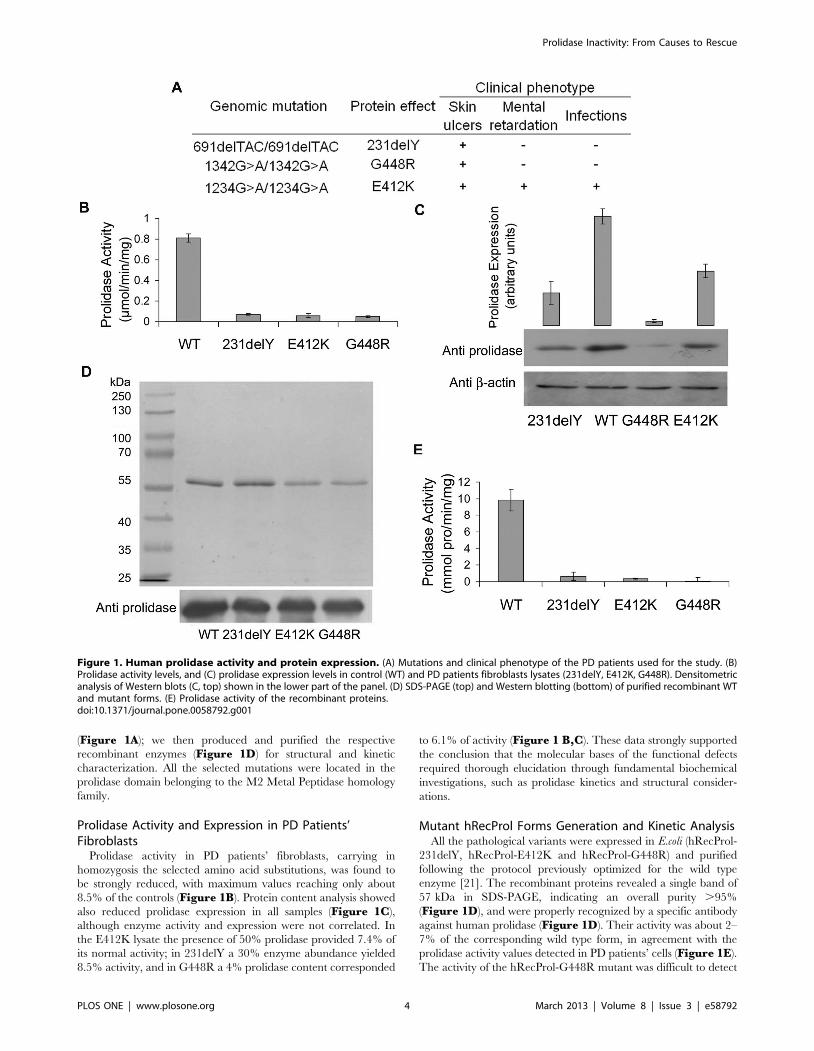
(Figure 1A); we then produced and purified the respective
recombinant enzymes (Figure 1D) for structural and kinetic
characterization. All the selected mutations were located in the
prolidase domain belonging to the M2 Metal Peptidase homology
family.
Prolidase Activity and Expression in PD Patients’Fibroblasts
Prolidase activity in PD patients’ fibroblasts, carrying in
homozygosis the selected amino acid substitutions, was found to
be strongly reduced, with maximum values reaching only about
8.5% of the controls (Figure 1B). Protein content analysis showed
also reduced prolidase expression in all samples (Figure 1C),
although enzyme activity and expression were not correlated. In
the E412K lysate the presence of 50% prolidase provided 7.4% of
its normal activity; in 231delY a 30% enzyme abundance yielded
8.5% activity, and in G448R a 4% prolidase content corresponded
to 6.1% of activity (Figure 1 B,C). These data strongly supported
the conclusion that the molecular bases of the functional defects
required thorough elucidation through fundamental biochemical
investigations, such as prolidase kinetics and structural consider-
ations.
Mutant hRecProl Forms Generation and Kinetic AnalysisAll the pathological variants were expressed in E.coli (hRecProl-
231delY, hRecProl-E412K and hRecProl-G448R) and purified
following the protocol previously optimized for the wild type
enzyme [21]. The recombinant proteins revealed a single band of
57 kDa in SDS-PAGE, indicating an overall purity .95%
(Figure 1D), and were properly recognized by a specific antibody
against human prolidase (Figure 1D). Their activity was about 2–
7% of the corresponding wild type form, in agreement with the
prolidase activity values detected in PD patients’ cells (Figure 1E).
The activity of the hRecProl-G448R mutant was difficult to detect
Figure 1. Human prolidase activity and protein expression. (A) Mutations and clinical phenotype of the PD patients used for the study. (B)Prolidase activity levels, and (C) prolidase expression levels in control (WT) and PD patients fibroblasts lysates (231delY, E412K, G448R). Densitometricanalysis of Western blots (C, top) shown in the lower part of the panel. (D) SDS-PAGE (top) and Western blotting (bottom) of purified recombinant WTand mutant forms. (E) Prolidase activity of the recombinant proteins.doi:10.1371/journal.pone.0058792.g001
Prolidase Inactivity: From Causes to Rescue
PLOS ONE | www.plosone.org 4 March 2013 | Volume 8 | Issue 3 | e58792
due to protein instability, which strongly reduced protein recovery,
reminiscent of what had been observed for the endogenous mutant
form.
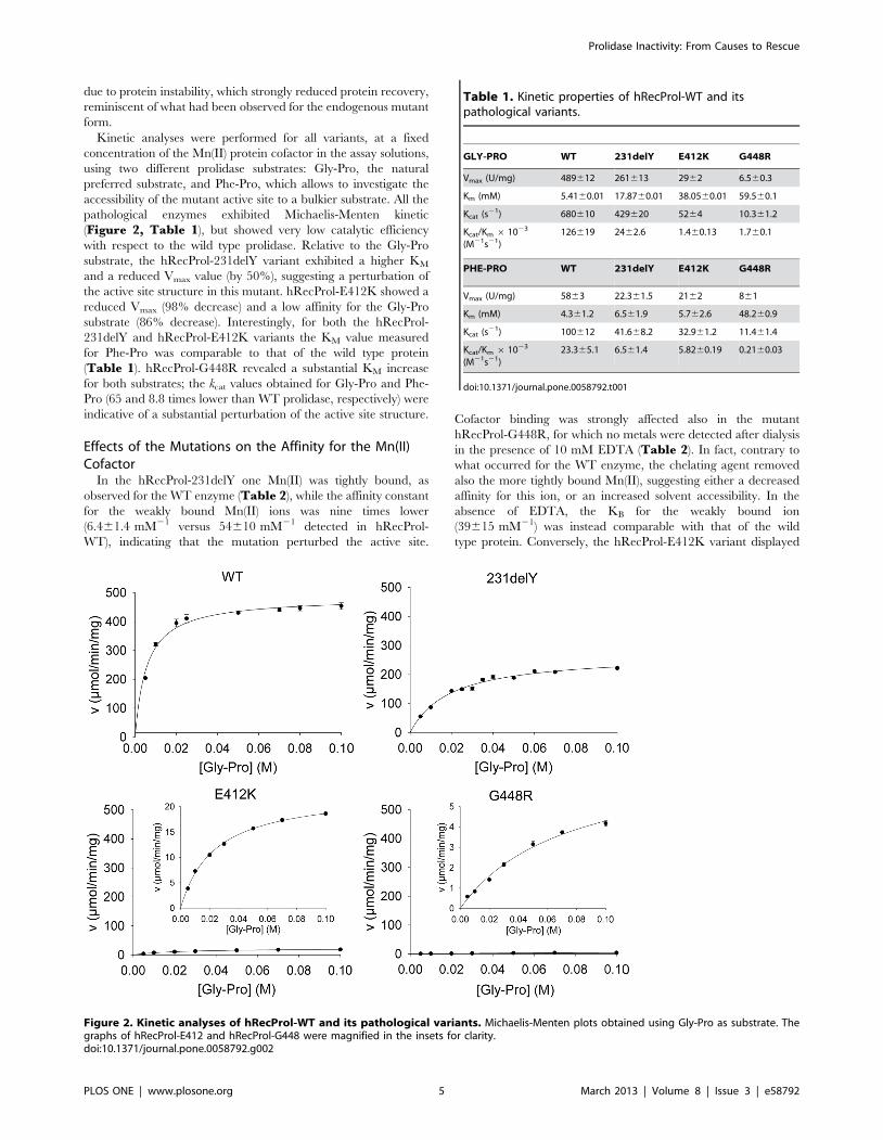
Kinetic analyses were performed for all variants, at a fixed
concentration of the Mn(II) protein cofactor in the assay solutions,
using two different prolidase substrates: Gly-Pro, the natural
preferred substrate, and Phe-Pro, which allows to investigate the
accessibility of the mutant active site to a bulkier substrate. All the
pathological enzymes exhibited Michaelis-Menten kinetic
(Figure 2, Table 1), but showed very low catalytic efficiency
with respect to the wild type prolidase. Relative to the Gly-Pro
substrate, the hRecProl-231delY variant exhibited a higher KM
and a reduced Vmax value (by 50%), suggesting a perturbation of
the active site structure in this mutant. hRecProl-E412K showed a
reduced Vmax (98% decrease) and a low affinity for the Gly-Pro
substrate (86% decrease). Interestingly, for both the hRecProl-
231delY and hRecProl-E412K variants the KM value measured
for Phe-Pro was comparable to that of the wild type protein
(Table 1). hRecProl-G448R revealed a substantial KM increase
for both substrates; the kcat values obtained for Gly-Pro and Phe-
Pro (65 and 8.8 times lower than WT prolidase, respectively) were
indicative of a substantial perturbation of the active site structure.
Effects of the Mutations on the Affinity for the Mn(II)Cofactor
In the hRecProl-231delY one Mn(II) was tightly bound, as
observed for the WT enzyme (Table 2), while the affinity constant
for the weakly bound Mn(II) ions was nine times lower
(6.461.4 mM21 versus 54610 mM21 detected in hRecProl-
WT), indicating that the mutation perturbed the active site.
Cofactor binding was strongly affected also in the mutant
hRecProl-G448R, for which no metals were detected after dialysis
in the presence of 10 mM EDTA (Table 2). In fact, contrary to
what occurred for the WT enzyme, the chelating agent removed
also the more tightly bound Mn(II), suggesting either a decreased
affinity for this ion, or an increased solvent accessibility. In the
absence of EDTA, the KB for the weakly bound ion
(39615 mM21) was instead comparable with that of the wild
type protein. Conversely, the hRecProl-E412K variant displayed
Figure 2. Kinetic analyses of hRecProl-WT and its pathological variants. Michaelis-Menten plots obtained using Gly-Pro as substrate. Thegraphs of hRecProl-E412 and hRecProl-G448 were magnified in the insets for clarity.doi:10.1371/journal.pone.0058792.g002
Table 1. Kinetic properties of hRecProl-WT and itspathological variants.
GLY-PRO WT 231delY E412K G448R
Vmax (U/mg) 489612 261613 2962 6.560.3
Km (mM) 5.4160.01 17.8760.01 38.0560.01 59.560.1
Kcat (s21) 680610 429620 5264 10.361.2
Kcat/Km 6 1023
(M21s21)126619 2462.6 1.460.13 1.760.1
PHE-PRO WT 231delY E412K G448R
Vmax (U/mg) 5863 22.361.5 2162 861
Km (mM) 4.361.2 6.561.9 5.762.6 48.260.9
Kcat (s21) 100612 41.668.2 32.961.2 11.461.4
Kcat/Km 6 1023
(M21s21)23.365.1 6.561.4 5.8260.19 0.2160.03
doi:10.1371/journal.pone.0058792.t001
Prolidase Inactivity: From Causes to Rescue
PLOS ONE | www.plosone.org 5 March 2013 | Volume 8 | Issue 3 | e58792

two tightly bound Mn(II) ions per dimer (Table 2), and a slightly
increased metal binding affinity (103642 mM21), in keeping with
minor effects of the E412K mutation on the cofactor coordination
shell.
Thermal StabilityThe WT and mutant prolidase forms, following incubation with
Mn(II), were heat-denatured in a one-state process. hRecProl-
231delY and hRecProl-G448R showed a reduced TM (55uC and
54uC respectively) compared to WT prolidase (60uC), while
hRecProl-E412K had a TM of 64uC (Figure 3A). The protein
stability in absence of the cofactor was also evaluated, revealing
reduced TM values for all the mutants (hRecProl-231delY
TM = 55uC, hRecProl-E412K TM = 51uC, hRecProl-G448R
TM = 49uC) with respect to hRecProl-WT (TM = 57uC), which
was also reduced of 3uC (Figure S1). hRecProl-G448R showed
the lowest TM, in agreement with the low recombinant protein
purification yield, and with the low protein levels detected in PD
patients fibroblasts.
Prolidase Dimerization in the Mutant SpeciesDLS data on Mn(II)-activated WT and mutant prolidase forms (at
concentrationofca.5 mM)showedthat theprotein samplesdisplayed
hydrodynamic radiuses indicative of a dimeric prolidase (hRecProl-
WT, 5.760.1 nm; hRecProl-231delY, 5.060.2 nm; hRecProl-
E412K, 5.2560.05 nm; hRecProl-G448R, 5.060.3 nm, Table S1)
with a polydispersion of less than 30%.
In order to assess the enzyme quaternary assembly at lower
concentrations, where the DLS analysis fails, the dependence of
prolidase activity on protein concentration was monitored. For
WT prolidase the specific activity did not change indicating that,
the enzyme was present in the dimeric state even at low
nanomolar concentrations (Figure 3B). On the contrary, the
activity of all the mutant enzymes was markedly concentration
dependent (Figure 3B).
Structural Analysis of Prolidase VariantsThe secondary structures of wild type and mutant prolidases
were analyzed by circular dichroism (CD). Far-UV CD spectra of
all proteins showed two minima, at 205–210 and at 215–220 nm
respectively (Figure 4A). Deconvolution of the spectra using three
independent algorithms indicated for the WT enzyme 23% a-
helix, 28% b-sheet and 23% coil relative contents, while the
spectra of hRecProl-E412K and hRecProl-231delY showed an
increased contribution of the random coil signal, reflected by a
reduction of the peak at 220 nm (Table 3), in keeping with an
effect of the mutated residues on proper enzyme folding.
CD spectra were also recorded in the presence of the reducing
agent DTT, and after a 20 min incubation at 50uC in the presence
of Mn(II) and DTT [22]. Both incubation conditions did not affect
the CD spectra, indicating that the metal cofactor was not
responsible for secondary structure variations. On the other hand,
the fine structural properties of the mutated proteins, as evaluated
by fluorescence spectroscopy, indicated the presence of some
conformational changes, since aromatic residues in hRecProl-
E412K and hRecProl-231delY appeared more exposed to solvent
than in the WT enzyme (Figure 4B). For the mutant hRecProl-
G448R it was not possible to perform CD and fluorescence
spectroscopy experiments due to the low protein amount obtained
from the purification.
To investigate the hRecProl-G448R stability, and to further
support the conformational changes revealed in hRecProl-
231delY and hRecProl-E412K, distinct limited proteolysis exper-
iments were performed using a-chymotrypsin, papain and trypsin.
Different proteolytic patterns were identified in all mutated forms,
indicating their different sensitivity to proteolytic cleavage
(Figure 4C). During chymotrypsin digestion, full length hRec-
Prol-231delY was strongly degraded after just 5 minutes, with the
appearance, after 30 min, of a 25 kDa fragment undetected in the
WT prolidase. After trypsin digestion, hRecProl-231delY also
revealed faster proteolysis, characterized by the absence at 30 min
of the ,27 and 32 kDa bands present in the WT protein.
Interestingly, hRecProl-E412K was more resistant than the WT
enzyme to both chymotrypsin and trypsin proteolysis. Although
enough hRecProl-G448R mutant was available for limited
proteolysis analysis, this variant proved very unstable and was
completely degraded by all the tested enzymes after just 5 min
incubation (Figure 4C).
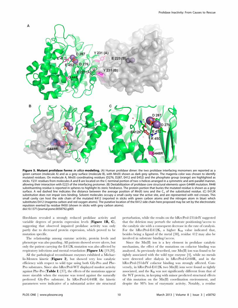
In silico Analysis of Mutants: Structural PredictionThe dimeric assembly of human prolidase (molecules A and B),
as reported by the enzyme crystal structure (PDB ID 2OKN), is
stabilized by different structural motifs; these include a-helices and
coil regions, producing a total interface area of 2861 A2,
characterized by a calculated solvation free energy (DiG) of
225.7 kcal/mol. Residue Y231 is located in one of the two a-
helices involved in association, and represents ,3.1% of the total
interface, with a buried surface area of 88 A2. The Y231 hydroxyl
group, in both protein subunits (A and B), is hydrogen bonded to
the side chain of E223 (Y231 A/B {OH} – E223 B/A
{OE1} = 2.52 A) from the facing protein subunit, contributing to
the stabilization of the associated dimer (Figure 5A).
The calculated residue solvation free energy (DiG) is slightly
positive (0.8 kcal/mol), indicating that Y231 prefers hydrophobic
environment and its location in a buried area is energetically
favored.
Since modeling of the Y231 deletion on the WT prolidase
structure may lead to inaccurate predictions, on a purely indicative
way we considered only the effects of Y231 substitution on subunit
association. In fact, the Y231A substitution in both protein
Table 2. Prolidase metal content.
Protein (nmol) Mn(II) (nmol) Zn(II) (nmol) Mol dimer:Mol Mn(II) Mol dimer: Mol Zn(II)
WT 32 2863 9069 ,1:1 ,1:3
E412K 17 4462 3361 ,1:2.5 ,1:2
231delY 24 1769 4469 ,1:0.7 ,1:1.8
G448R 36 361 33613 ,1:0 ,1:1
Mn(II) and Zn(II) content was analyzed by ICP-MS after 48 h dialysis against metal free buffer based on their presence in the enzyme active site as previouslydemonstrated [4].doi:10.1371/journal.pone.0058792.t002
Prolidase Inactivity: From Causes to Rescue
PLOS ONE | www.plosone.org 6 March 2013 | Volume 8 | Issue 3 | e58792

Figure 3. Prolidase thermal denaturation and dimerization follow up. (A) Protein melting temperatures in the presence of the cofactor asrevealed by Thermofluor Technology. The solvatochromic dye SYPRO orange was used as an indicator of protein unfolding (fluorescence excitationl= 492 nm; fluorescence emission l= 568 nm). (B) The ratio between prolidase activity and protein concentration was plotted against proteinconcentration.doi:10.1371/journal.pone.0058792.g003
Prolidase Inactivity: From Causes to Rescue
PLOS ONE | www.plosone.org 7 March 2013 | Volume 8 | Issue 3 | e58792

Prolidase Inactivity: From Causes to Rescue
PLOS ONE | www.plosone.org 8 March 2013 | Volume 8 | Issue 3 | e58792

monomers produced a decrease in the total interface area
(AY231A = 2784 A2) and an increase of the solvation energy
(DiG = 224,1 kcal/mol) indicative of a slightly less favorable
stabilization process relative to the wild type protein. The effects of
a full amino acid deletion at site 231 were expected to be more
dramatic.
Residue G448 is buried in a protein region, not accessible to
solvent. The residue lies at about 14.5 A from the active site and is
not directly involved in Mn(II) binding. G448 belongs to a b-
strand anti-parallel to a short neighboring strand composed of
G414, I415, Y416, F417 (Figure 5B). The G448R substitution
forces the insertion of a bulky arginine side chain (Figure 5B),
which is not compatible with pairing of the two anti-parallel b-
strands and with the correct assembly of the b-sheet. Furthermore,
the G448R mutation falls only four amino acids before residue
E452, that coordinates one of the Mn(II) cofactor ions; therefore, a
perturbation of the ligand coordination sphere upon G448R
substitution cannot be excluded. Such an observation is in keeping
with the absence of metal ions experimentally detected for this
mutant by ICP-MS.
In WT prolidase the Mn(II) ions are coordinated by the
negatively charged amino acids D276, D287, E412, E452, and by
a phosphate group. Thus the E412K mutation decreases by two
units the negative charge in the coordination sphere; nevertheless,
our in vitro data showed that the mutation does not affect the
binding of the Mn(II) cofactor. Modeling considerations suggested
that K412 side chain, due to electrostatic repulsion by R450, can
relocate into a solvent area adjacent to the active site, and such
accommodation would not impair the Mn(II) coordination
surroundings (Figure 5C). Since the three-dimensional structure
of the enzyme in the presence of a substrate analogue is not
available, any structural prediction on K412-dependent impair-
ment of substrate binding would be largely hypothetic.
Mutant Proteins Stabilization through Heat ShockProteins Induction
Our findings led us to consider enzyme stabilization as a means
for rescuing prolidase activity in vitro, with the aim to evaluate it
in vivo. To test the feasibility of this approach, we chose to
stimulate expression of the endogenous heat shock proteins (Hsps).
Hsps are molecular chaperones that in the presence of adverse
environmental conditions, assist refolding of misfolded proteins
and facilitate the synthesis of new proteins to enhance cell survival
[34]. In vitro Hsps stimulation was performed by means of heat
increase: fibroblasts from patients homozygous for 231delY,
G448R and E412K, and from controls, were heated at 48uC,
for periods ranging from 0 to 20 minutes and, sixteen hours post-
heating, prolidase activity and protein expression level were
evaluated at selected time points. Although the prolidase activity
was expectedly low in all the mutants, with respect to the controls,
and although a partial loss of the protein was caused by the
heating procedure as evidenced by Western blot analysis, following
15 min of heat shock in the G448R lysate the prolidase activity
was increased by 22% relative to the same cells not stimulated. An
even better result was obtained for the E412K mutant that showed
a prolidase activity increase of 40% (Figure 6A, B). No increase
in activity was instead detected in control lysates (Figure 6A, B).
Since the two major known heat inducible cytosolic chaperones
involved in protein folding are Hsp70 and Hsp90 [35], an analysis
of their protein expression levels was performed. A significant
increase of both proteins was detected after stimulation in all the
analyzed cell lines, with the highest level following 20 min heat
shock (Figure 6C, E). Both constitutive and inducible forms of
Hsp70 were detectable. The latter one started to be synthesized
after 10 min heating and reached its maximum after 20 min
(Figure 6C, E). Although the Hsps expression increased by
prolonging the heat shock up to 20 min, prolidase activity reached
its maximum following 15 min and then decreased (Figure 6A,B). Also in control cells the incubation at 48uC for 20 min
produced a slight reduction of prolidase activity, likely indicating
that, under such stressful conditions, even if the chaperone levels
increase, irreversible cellular damages occurred (Figure 6A, B)
[36]. As expected, in mutant cells, Hsp70 was expressed at steady
state at higher concentration relative to the control (Figure 6D).
Unfortunately, an increase in activity following heat stimulation
was not evident for 231delY variant (data not shown), revealing
that the efficacy of the Hsp-mediated refolding process may be
mutation specific.
Discussion
Twenty-four mutant alleles have been reported to date as
responsible for PD, however, the identification of the causative/
pathologic genetic defects in the prolidase gene does not
immediately translate into an understanding of the molecular
basis of the disease. How specific mutations affect the enzyme
activity is still an open question in the field. Although the residues
mutated in the pathologic variants are strictly conserved in
prolidase from different organisms (Figure S2), their contribu-
tions to the hydrolytic reaction and/or to the enzyme structure
have not been thoroughly analyzed yet.
For the biochemical investigation of the PD pathophysiology we
selected three mutations previously characterized in our patients
cohort, namely 691delTAC (231delY), 1234G.A (E412K) and
1342G.A (G448R) [19,20]. The three variants were selected
based on their occurrence frequency, and in order to map
different, but highly conserved regions within the prolidase
structure (Figure S2). The enzyme variants from PD patient
Figure 4. Prolidase spectroscopy analysis. (A) CD spectra of hRecProl-WT, hRecProl-231delY and hRecProl-E412K in the far-UV spectrum region(__). Spectra were recorded also in the presence of 0.75 mM DTT (?????) and of both 0.75 mM DTT and 1 mM MnCl2 (---). The reduction in signalintensity was due to a slight loss of the protein in solution. (B) Fluorecence spectra of hRecProl-WT (__) and its variants hRecProl231delY (---) andhRecProl-E412K (????). The tryptophan excitation wavelength was set at 295 nm, the monitoring emission from 305 to 400 nm. The other spectrarecorded at the wavelengths specific for phenylalanine and tyrosine were similar and thus not reported. (C) Limited proteolysis analyses of mutantprolidase. Digestions were performed with a-chymotrypsin, papain and trypsin. The fragmentation patterns were analyzed by SDS-PAGE underreducing conditions and stained with Comassie blue. In lines 1, *and {only chymotrypsin, papain and trypsin were loaded, respectively.doi:10.1371/journal.pone.0058792.g004
Table 3. Secondary structure composition as estimated bythe CD spectra of the recombinant prolidase variants.
WT 231delY E412K
a-Helix 0.2360.01 0.1660.05 0.1860.10
b-Sheet 0.2860.07 0.2860.04 0.3360.07
Turns 0.2360.03 0.2460.01 0.1960.12
Random coil 0.2660.03 0.3260.01 0.3060.11
doi:10.1371/journal.pone.0058792.t003
Prolidase Inactivity: From Causes to Rescue
PLOS ONE | www.plosone.org 9 March 2013 | Volume 8 | Issue 3 | e58792
fibroblasts revealed a strongly reduced prolidase activity and
variable degrees of protein expression levels (Figure 1B, C),
suggesting that observed impaired prolidase activity was only
partly due to decreased protein expression, which proved to be
mutation specific.
The relationship among enzyme activity, protein levels and
phenotype was also puzzling. All patients showed severe ulcers, but
only the patient carrying the E412K mutation was also affected by
respiratory infections and mental retardation (Figure 1A) [19,20].
All the pathological recombinant enzymes exhibited a Michae-
lis-Menten kinetic (Figure 2), but showed very low catalytic
efficiency with respect to wild type using both Gly-Pro and Phe-
Pro substrates. Since also hRecProl-WT displayed modest activity
against Phe-Pro (Table 1) [37], the effects of the mutations appear
more sizeable when the enzyme was tested against the naturally
preferred Gly-Pro substrate. In hRecProl-G448R the kinetic
parameters were indicative of a substantial active site structural
perturbation, while the results on the hRecProl-231delY suggested
that the deletion may perturb the substrate positioning/access to
the catalytic site with a consequent decrease in the rate of catalysis.
For the hRecProl-E412K, a higher KM value indicated that,
besides being a ligand of the metal [38], residue 412 may also be
involved in substrate binding/access.
Since the Mn(II) ion is a key element in prolidase catalytic
mechanism, the effect of the mutations on cofactor binding was
analyzed. As previously described, one Mn(II) ion was found to be
tightly associated with the wild type enzyme [4], while no metals
were detected after dialysis in hRecProl-G448R, and in the
hRecProl-231delY cofactor binding was strongly affected. Con-
versely, in hRecProl-E412K two Mn(II) ions were found as tightly
associated, and the KB was not significantly different from that of
the WT protein, in keeping with minor predicted structural effects
of this mutation on the Mn(II) coordination environment, and
despite the 98% loss of enzymatic activity. Notably, a residue
Figure 5. Mutant prolidase forms in silico modeling. (A) Human prolidase dimer: the two prolidase interfacing monomers are reported as agreen cartoon (molecule A) and as a grey surface (molecule B), with Mn(II) shown as dark grey spheres. The magenta color was chosen to identifymutated residues. On molecule A, Mn(II) coordinating residues (D276, D287, E412 and E452) and the phosphate group (orange) are highlighted assticks. Y231 residues from molecules A and B are located on the C-terminal portion of two a-helices arranged in a symmetric and anti-parallel manner,allowing their interaction with E223 of the interfacing protomer. (B) Destabilization of prolidase core structural elements upon G448R mutation. R448substituenting residue is reported in spheres to highlight its steric hindrance. The protein portion that buries the mutated residue is shown as a greysurface. A red dashed line indicates the distance between the average position of Mn(II) ions and the Ca of the substituted residue. (C) E412Ksubstitution does not impair ions binding. Solvent molecules occupy a small cavity near the active site, and are represented with red crosses. Thissmall cavity can host the side chain of the mutated K412 (reported in sticks with green carbon atoms and the nitrogen atom in blue) whichsubstitutes E412 (magenta carbon and red oxygen atoms). The putative location of the K412 side chain here proposed may be set by the electrostaticrepulsion exerted by residue R450 (shown in sticks with grey carbon atoms).doi:10.1371/journal.pone.0058792.g005
Prolidase Inactivity: From Causes to Rescue
PLOS ONE | www.plosone.org 10 March 2013 | Volume 8 | Issue 3 | e58792

Prolidase Inactivity: From Causes to Rescue
PLOS ONE | www.plosone.org 11 March 2013 | Volume 8 | Issue 3 | e58792

structurally homologous to E412, recognized in aminopeptidase P
(APPro, EC 3.4.11.9, E383), in methionine aminopeptidase
(MetAP, EC 3.4.11.1a, E204) and in prolidase from Pyrococcus
furiosus (PfProl, E313), was shown to be essential for catalysis:
mutation of E383 reduced porcine APPro, E.coli APPro and E. coli
MetAP prolidase activities by at least two orders of magnitude
[39,40]. Similarly, the E313L mutation in PfProl produced an
inactive highly misfolded protein, recalcitrant to purification [41],
and in the APProE383A mutant, despite the impaired activity,
both Mn(II) ions were still present in the active site [42]. Thus, our
data, together with findings from the homologous enzymes,
suggest that residue E412 contributes to a general structural
maintenance of the active site region [43,44]. The prolidase
catalytic mechanism is still largely uncharacterized but, consider-
ing that the enzyme belongs to the ‘‘pita-bread’’ family, Lowther &
Matthews proposed a common reaction mechanism [44] whereby
the metal ion would coordinate the substrate, polarizing its
carbonyl group, and the glutamate-activated bridging water
molecule would provide the nucleophile hydroxyl group, setting
conditions for hydrolysis of the bound peptide [45,46,47]. Our
kinetic data on the hRecProl-E412K mutant (E383 in APPro) are
in keeping with such water molecule-activator role for residue
E412.
A reduced thermal stability was detected in all three mutant
prolidase forms. Interestingly, a stabilizing effect of the Mn(II)
incubation was detected in hRecProl-WT (DTM = +3uC), in
hRecProl-G448R (DTM = +5uC) and in hRecProl-E412K
(DTM = +13uC), whereas no effect was detected in hRecProl-
231delY. The higher stabilization of hRecProl-E412K, and the
absence of stabilization in hRecProl-231delY, are in keeping with
the Mn(II) cofactor binding properties observed for these mutants.
The monitoring of the ratio between prolidase activity and
protein concentration plotted against protein concentration
revealed that while WT prolidase was active in solution as a
dimer, in the mutant enzymes the activity was concentration
dependent. At very low concentrations (,4 nM), in keeping with
the loss of enzymatic activity, the two subunits appeared
dissociated in all variants; at slightly higher concentrations the
enzymes were presumably present as a mixed population of active
dimers and inactive monomers, while at higher protein concen-
trations (48 nM for hRecProl-231delY, 120 nM for hRecProl-
E412K and 43 nM for hRecProl-G448R) recovery of the
prolidase activity indicated the presence of the dimeric species
(Figure 3B). For multimeric proteins, subunit aggregation is often
a key process in enzyme activation, and the failure or the
decreased association efficiency can translate into reduced activity.
Although our activity tests were performed on the recombinant
prolidase forms, and although the concentration of active prolidase
in the cell is not known, dimerization impairment in all the mutant
enzyme species helps explain their compromised activity.
CD spectra revealed a decrease in a-helix content in the
hRecProl-231delY and hRecProl-E412K variants, indicating that
both mutations have an impact on prolidase secondary structure
(Figure 4A); tryptophan emission fluorescence spectra, together
with proteolytic digestion experiments, confirmed that the
pathological proteins host partly altered conformations
(Figure 4B, C); local structure perturbations for all the mutants
were also predicted by our in silico analyses. Based on structural
considerations, Y231 was indicated as a key residue for
stabilization of the prolidase dimer; deletion of this amino acid,
coupled to loss of its bulky side chain that is involved in interface
interactions, may destabilize one a-helix at the dimer association
interface and affect the DiG for assembly of the dimeric species.
In silico investigation suggested that in hRecProl, the preserva-
tion of the G448 residue may be crucial for the overall enzyme
architecture, since G448 lies in a b-sheet buried in the prolidase
core region, and because bulkier residues would not be compatible
with the compactly packed surroundings. Conversely, due to lack
of a three-dimensional structure of the enzyme in the presence of a
substrate analogue, any structural prediction on K412-dependent
impairment of substrate binding would be largely hypothetic,
leaving a discussion on the molecular basis of reduced activity for
this mutant purely speculative.
All the analyzed mutations introduced significant changes in the
protein that turned into evident effects on the enzyme kinetics:
even structural alterations outside the active site, as in the cases of
G448R and 231delY, were translated to the catalytic center with
impact on the enzyme activity. Thus, based on the data here
reported, we considered that, as for many loss-of-function diseases,
PD pathogenicity is strictly linked to occurrence of a perturbed
protein fold, resulting in faster protein degradation that would
trigger system failure. Ensuring accuracy in protein folding is a
crucial aspect for proper maintenance of cellular functionality and
indeed, in the cell, molecular chaperones recognize protein
substrates in non-native states and assist their proper re-folding
and assembly. We therefore chose to stimulate heat shock proteins
(Hsps), a class of molecular chaperones expressed under sublethal
stressful stimuli, that have previously been identified by several
studies as defense components enhancing cell survival under stress
conditions [32,48,49,50].
As a result of heat stimulation, fibroblasts bearing either the
G448R or the E412K mutations revealed a decrease in prolidase
content, but a statistically significant increase in prolidase activity
(relative to fibroblasts carrying the same mutation, but not heat
stimulated). No beneficial effects were instead detected in heat
stimulated fibroblasts bearing the 231delY mutation, suggesting
that the effects achievable through Hsp-based recovery are
mutation specific. The Y231 deletion, impairing prolidase
dimerization, may be one of the reasons explaining the failure of
Hsp stimulation approach. Interestingly, it was noted that in the
absence of heat stimulation the level of the Hsp70 expression was
higher in G448R and E412K mutant fibroblasts than in the
controls, suggesting that cells specifically activate the Hsp
endogenous response to help folding the mutant prolidase forms,
or more generally to respond to the ensuing cellular stress [51].
Thus, pharmacological stimulation of the Hsp expression appears
as a therapeutic approach in line with the natural response of the
cell.
Altogether, our data suggest that a moderate temperature
increase lead to enhance degradation of the poorly structured
prolidase mutants, minimally affecting the WT form. Improved
stability of the mutant enzymes was found when higher chaperone
Figure 6. Mutant prolidases stabilization though heat shock proteins induction. (A) Prolidase activity (white square) and proteinexpression level (black square) of WT, G448R, E412K fibroblasts lysed 16 h after the heat shock (at 48uC).*, p,.05. (B) Representative western blotperformed to evaluate the prolidase expression in WT, G448R, E412K fibroblasts 16 h after the heat shock. (C) Hsp70/90 expression in WT, G448R,E412K fibroblasts 16 h after the heat shock. (D) Comparison of Hsp70/90 expression in WT, G448R and E412K at time zero, and after 20 min heatshock. The same exposure time was used in order to emphasize the different expression levels. (E) Densitometric analysis of the Hsp70/90 expressionas presented in (C). The proteins expression was quantified by using Quantity One software.doi:10.1371/journal.pone.0058792.g006
Prolidase Inactivity: From Causes to Rescue
PLOS ONE | www.plosone.org 12 March 2013 | Volume 8 | Issue 3 | e58792
levels were present. Such stabilization translated into an increase
in mutant prolidase activity and, even if the prolidase activity after
Hsp stimulation remained lower than that of the WT enzyme,
these findings suggest that drugs effecting the intracellular
chaperone environment, some of which are already FDA
approved [11], could be tested for PD treatment.
ConclusionsOur study on three pathological mutant human prolidase forms
showed that a substantial loss of enzymatic activity can be linked to
enzyme instability and local structural perturbations, partly
affecting the Mn(II) cofactor site; partial degradation of the
mutated enzyme is an additional factor contributing to the general
loss of enzyme activity in PD patients. On these bases, the
possibility to correct functional prolidase defects by promoting
enzyme structure stabilization/recovery was considered. In order
to rescue prolidase activity in vitro we focused on supporting its
folding process by inducing expression of intracellular chaperones.
Such an approach brought to 20–40% recovery of the prolidase
enzymatic activity in fibroblasts bearing the mutated enzymes; the
rescue was mutation-dependent. These results are an exciting
proof-of-principle that bears implications for PD therapy. The
administration of existing drugs stimulating the endogenous
chaperones, or of chaperone-like molecules, might in fact be
considered as a means for rescuing the PD phenotype.
Supporting Information
Figure S1 Protein melting temperatures in the absenceof the cofactor as revealed by Thermofluor Technology.The solvatochromic dye SYPRO orange was used as an indicator
of protein unfolding (fluorescence excitation l= 492 nm; fluores-
cence emission l= 568 nm).
(TIF)
Figure S2 Alignment of different prolidase amino acidsequences from the region involved in cofactor binding.Metal binding residues are indicated by arrows. GenBankHIdentifier (gi) numbers of the displayed sequences are as follow:
189842 (H. sapiens), 296477856 (B. Taurus), 9795244 (M. musculus),
160774330 (D. rerio), 18977119, (P. furiosus), 14590977 (P. horikoshii
OT3).
(TIF)
Table S1 Hydrodynamic radiuses of the recombinantprolidase variants observed with Dynamic Light Scat-tering. Rh values and the percentages of polydispersion are
reported as the averages of two independent experiments for each
protein sample.
(DOC)
Author Contributions
Conceived and designed the experiments: AF. Performed the experiments:
RB RG FC LC. Analyzed the data: AF RB EM AP RT AR SN LC FC
MB. Contributed reagents/materials/analysis tools: AF AP RT AR MB.
Wrote the paper: AF RB MB.
References
1. Amberger J, Bocchini CA, Scott AF, Hamosh A (2009) McKusick’s Online
Mendelian Inheritance in Man (OMIM). Nucleic Acids Research 37: D793–
D796.
2. Lupi A, Tenni R, Rossi A, Cetta G, Forlino A (2008) Human prolidase and
prolidase deficiency: an overview on the characterization of the enzyme involved
in proline recycling and on the effects of its mutations. Amino Acids 35: 739–
752.
3. Besio R, Baratto MC, Gioia R, Monzani E, Nicolis S, et al. (2012) A Mn(II)-
Mn(II) center in human prolidase. Biochim Biophys Acta doi: 10.1016/
j.bbapap.2012.09.008.
4. Besio R, Alleva S, Forlino A, Lupi A, Meneghini C, et al. (2010) Identifying the
structure of the active sites of human recombinant prolidase. Eur Biophys J 39:
935–945.
5. Hu CA, Phang JM, Valle D (2008) Proline metabolism in health and disease.
Preface. Amino Acids 35: 651–652.
6. Palka JA, Phang JM (1997) Prolidase activity in fibroblasts is regulated by
interaction of extracellular matrix with cell surface integrin receptors. J Cell
Biochem 67: 166–175.
7. Rao VH, Royce PM, Steinmann B (1993) Normal production, nature, and
extent of intracellular degradation of newly synthesized collagen in fibroblasts
from a patient with prolidase deficiency. Connect Tissue Res 29: 23–30.
8. Surazynski A, Liu Y, Miltyk W, Phang JM (2005) Nitric oxide regulates prolidase
activity by serine/threonine phosphorylation. J Cell Biochem 96: 1086–1094.
9. Royce PM, Steinmann B, editors (2002) Prolidase deficiency. New-York: Wiley-
Liss. 727–743 p.
10. Brady RO, Schiffmann R (2004) Enzyme-replacement therapy for metabolic
storage disorders. Lancet Neurol 3: 752–756.
11. Cohen FE, Kelly JW (2003) Therapeutic approaches to protein-misfolding
diseases. Nature 426: 905–909.
12. Fan JQ, Ishii S, Asano N, Suzuki Y (1999) Accelerated transport and maturation
of lysosomal alpha-galactosidase A in Fabry lymphoblasts by an enzyme
inhibitor. Nat Med 5: 112–115.
13. Yu Z, Sawkar AR, Kelly JW (2007) Pharmacologic chaperoning as a strategy to
treat Gaucher disease. FEBS J 274: 4944–4950.
14. Okumiya T, Kroos MA, Vliet LV, Takeuchi H, Van der Ploeg AT, et al. (2007)
Chemical chaperones improve transport and enhance stability of mutant alpha-
glucosidases in glycogen storage disease type II. Mol Genet Metab 90: 49–57.
15. Lim M, McKenzie K, Floyd AD, Kwon E, Zeitlin PL (2004) Modulation of
deltaF508 cystic fibrosis transmembrane regulator trafficking and function with
4-phenylbutyrate and flavonoids. Am J Respir Cell Mol Biol 31: 351–357.
16. Kerem E (2005) Pharmacological induction of CFTR function in patients with
cystic fibrosis: mutation-specific therapy. Pediatr Pulmonol 40: 183–196.
17. Wang X, Venable J, LaPointe P, Hutt DM, Koulov AV, et al. (2006) Hsp90
cochaperone Aha1 downregulation rescues misfolding of CFTR in cystic fibrosis.
Cell 127: 803–815.
18. Zilka N, Kontsekova E, Novak M (2008) Chaperone-like antibodies targeting
misfolded tau protein: new vistas in the immunotherapy of neurodegenerative
foldopathies. J Alzheimers Dis 15: 169–179.
19. Lupi A, De Riso A, Torre SD, Rossi A, Campari E, et al. (2004)
Characterization of a new PEPD allele causing prolidase deficiency in two
unrelated patients: natural-occurrent mutations as a tool to investigate structure-
function relationship. J Hum Genet 49: 500–506.
20. Lupi A, Rossi A, Campari E, Pecora F, Lund AM, et al. (2006) Molecular
characterisation of six patients with prolidase deficiency: identification of the first
small duplication in the prolidase gene and of a mutation generating
symptomatic and asymptomatic outcomes within the same family. J Med Genet
43: e58.
21. Lupi A, Della Torre S, Campari E, Tenni R, Cetta G, et al. (2006) Human
recombinant prolidase from eukaryotic and prokaryotic sources. Expression,
purification, characterization and long-term stability studies. FEBS J 273: 5466–
5478.
22. Besio R, Monzani E, Gioia R, Nicolis S, Rossi A, et al. (2011) Improved
prolidase activity assay allowed enzyme kinetic characterization and faster
prolidase deficiency diagnosis. Clin Chim Acta 412: 1814–1820.
23. Mezzasalma TM, Kranz JK, Chan W, Struble GT, Schalk-Hihi C, et al. (2007)
Enhancing recombinant protein quality and yield by protein stability profiling.
J Biomol Screen 12: 418–428.
24. Provencher SW, Glockner J (1981) Estimation of globular protein secondary
structure from circular dichroism. Biochemistry 20: 33–37.
25. Johnson WC (1999) Analyzing protein circular dichroism spectra for accurate
secondary structures. Proteins 35: 307–312.
26. Sreerama N, Woody RW (2000) Estimation of protein secondary structure from
circular dichroism spectra: comparison of CONTIN, SELCON, and CDSSTR
methods with an expanded reference set. Anal Biochem 287: 252–260.
27. Lobley A, Whitmore L, Wallace BA (2002) DICHROWEB: an interactive
website for the analysis of protein secondary structure from circular dichroism
spectra. Bioinformatics 18: 211–212.
28. Whitmore L, Wallace BA (2004) DICHROWEB, an online server for protein
secondary structure analyses from circular dichroism spectroscopic data. Nucleic
Acids Res 32: W668–673.
29. Krissinel E, Henrick K (2007) Inference of macromolecular assemblies from
crystalline state. J Mol Biol 372: 774–797.
30. Berman HM, Westbrook J, Feng Z, Gilliland G, Bhat TN, et al. (2000) The
Protein Data Bank. Nucleic Acids Res 28: 235–242.
Prolidase Inactivity: From Causes to Rescue
PLOS ONE | www.plosone.org 13 March 2013 | Volume 8 | Issue 3 | e58792
31. DeLano W (2002) The PyMOL Molecular Graphic System. In: Scientific D,
editor. San Carlos, CA, USA.32. Rylander MN, Diller KR, Wang S, Aggarwal SJ (2005) Correlation of HSP70
expression and cell viability following thermal stimulation of bovine aortic
endothelial cells. J Biomech Eng 127: 751–757.33. Wang S, Diller KR, Aggarwal SJ (2003) Kinetics study of endogenous heat shock
protein 70 expression. J Biomech Eng 125: 794–797.34. Parsell DA, Lindquist S (1993) The function of heat-shock proteins in stress
tolerance: degradation and reactivation of damaged proteins. Annu Rev Genet
27: 437–496.35. Makhnevych T, Houry WA (2012) The role of Hsp90 in protein complex
assembly. Biochim Biophys Acta 1823: 674–682.36. Bleiberg I, Sohar E (1975) The effect of heat treatment on the damage and
recovery of the protein synthesis mechanism of human kidney cell line. VirchowsArch B Cell Pathol 17: 269–278.
37. Richter AM, Lancaster GL, Choy FY, Hechtman P (1989) Purification and
characterization of activated human erythrocyte prolidase. Biochem Cell Biol67: 34–41.
38. Du X, Tove S, Kast-Hutcheson K, Grunden AM (2005) Characterization of thedinuclear metal center of Pyrococcus furiosus prolidase by analysis of targeted
mutants. FEBS Lett 579: 6140–6146.
39. Chiu CH, Lee CZ, Lin KS, Tam MF, Lin LY (1999) Amino acid residuesinvolved in the functional integrity of Escherichia coli methionine aminopep-
tidase. J Bacteriol 181: 4686–4689.40. Cottrell GS, Hyde RJ, Lim J, Parsons MR, Hooper NM, et al. (2000)
Identification of critical residues in the active site of porcine membrane-boundaminopeptidase P. Biochemistry 39: 15129–15135.
41. Maher MJ, Ghosh M, Grunden AM, Menon AL, Adams MW, et al. (2004)
Structure of the prolidase from Pyrococcus furiosus. Biochemistry 43: 2771–2783.
42. Graham SC, Lilley PE, Lee M, Schaeffer PM, Kralicek AV, et al. (2006) Kinetic
and crystallographic analysis of mutant Escherichia coli aminopeptidase P:insights into substrate recognition and the mechanism of catalysis. Biochemistry
45: 964–975.
43. Lowther WT, Zhang Y, Sampson PB, Honek JF, Matthews BW (1999) Insightsinto the mechanism of Escherichia coli methionine aminopeptidase from the
structural analysis of reaction products and phosphorus-based transition-stateanalogues. Biochemistry 38: 14810–14819.
44. Lowther WT, Matthews BW (2002) Metalloaminopeptidases: common func-
tional themes in disparate structural surroundings. Chem Rev 102: 4581–4608.45. Mock WL, Green PC (1990) Mechanism and inhibition of prolidase. J Biol
Chem 265: 19606–19610.46. Mock WL, Green PC, Boyer KD (1990) Specificity and pH dependence for
acylproline cleavage by prolidase. J Biol Chem 265: 19600–19605.47. Mock WL, Liu Y (1995) Hydrolysis of picolinylprolines by prolidase. A general
mechanism for the dual-metal ion containing aminopeptidases. J Biol Chem 270:
18437–18446.48. Iwabuchi K, Tajima M, Isoyama S (1998) Heat shock protein expression in
hearts hypertrophied by genetic and nongenetic hypertension. Heart Vessels 13:30–39.
49. Morimoto RI, Santoro MG (1998) Stress-inducible responses and heat shock
proteins: new pharmacologic targets for cytoprotection. Nat Biotechnol 16: 833–838.
50. Dams SD, de Liefde-van Beest M, Nuijs AM, Oomens CW, Baaijens FP (2011)Heat shocks enhance procollagen type I and III expression in fibroblasts in ex
vivo human skin. Skin Res Technol 17: 167–180.51. Forlino A, Lupi A, Vaghi P, Icaro Cornaglia A, Calligaro A, et al. (2002)
Mutation analysis of five new patients affected by prolidase deficiency: the lack of
enzyme activity causes necrosis-like cell death in cultured fibroblasts. HumGenet 111: 314–322.
Prolidase Inactivity: From Causes to Rescue
PLOS ONE | www.plosone.org 14 March 2013 | Volume 8 | Issue 3 | e58792