Embed Size (px)
Citation preview
Kim Uddo RN MSN CCRN
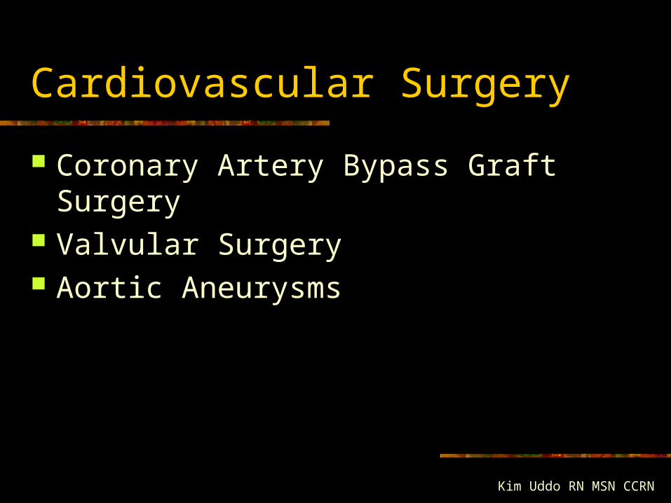
Cardiovascular Surgery
Coronary Artery Bypass Graft Surgery Valvular Surgery Aortic Aneurysms
CABG Indications
PTCA Failed Unstable Angina unresponsive to meds 3 vessels LAD > 75% stenosis Failed thrombolysis Ej fx < 35% Valve disease / vent. aneurysm
Diagnostic Studies
Cardiac Catheterization Echocardiogram Stress test CXR 12-lead ECG Blood: coags, CBC, CMP, Enzymes
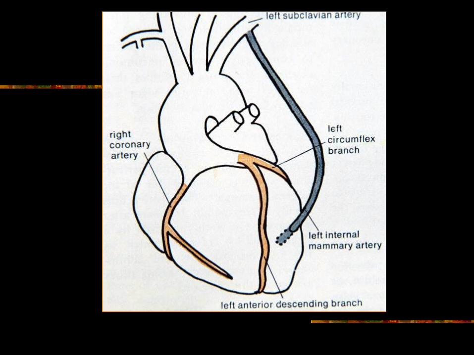
Conduits
Saphenous Vein Graft (SVG) Internal Mammary Artery (IMA)
LIMA RIMA
Right Gastroepiploic Radial

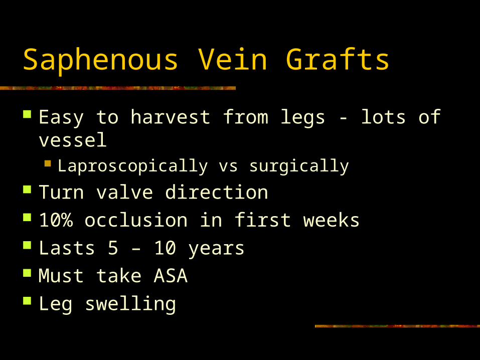
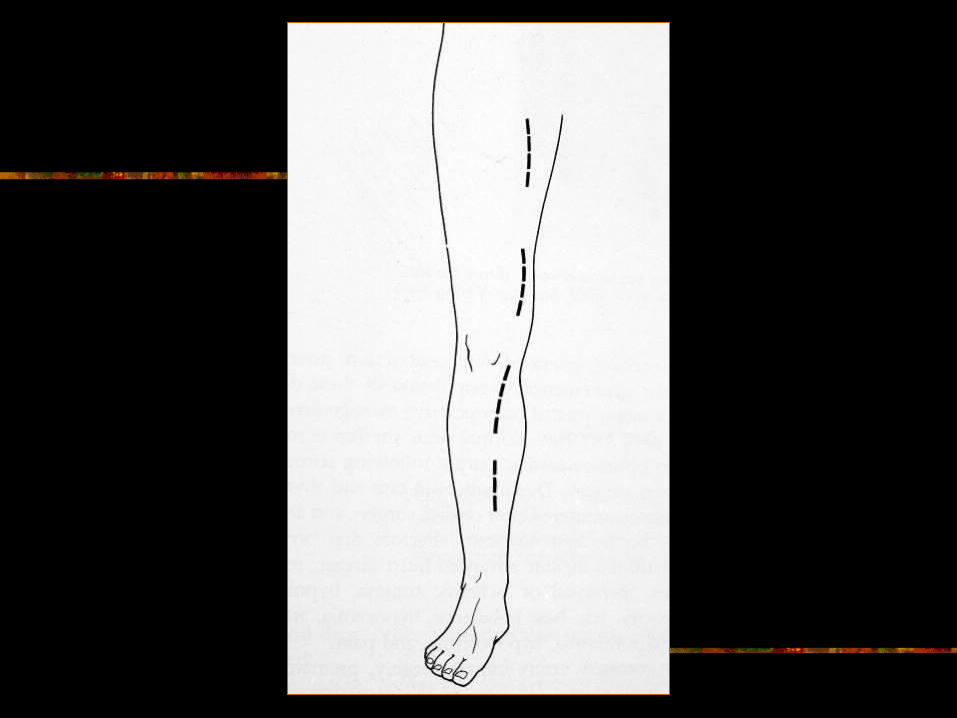
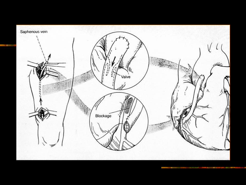
Saphenous Vein Grafts
Easy to harvest from legs - lots of vessel Laproscopically vs surgically
Turn valve direction 10% occlusion in first weeks Lasts 5 – 10 years Must take ASA Leg swelling
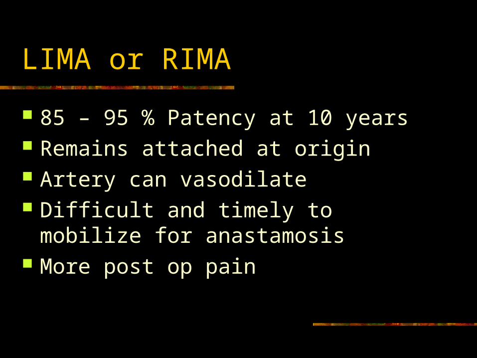
LIMA or RIMA
85 – 95 % Patency at 10 years Remains attached at origin Artery can vasodilate Difficult and timely to mobilize for
anastamosis More post op pain
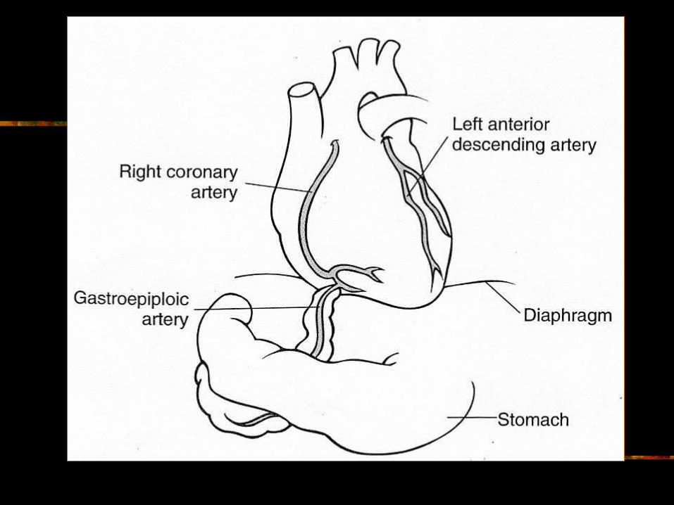
Right Gastroepiploic Artery
Branch of gastroduodenal artery Laporotomy Abdominal wound Timely and most difficult to harvest Pulled up to the pericardial cavity Origin intact and connected past blockage
RCA and Post. descending
Radial Artery
Artery dilates MUST have good ulnar flow Vasospasms (control with meds) Forearm swelling
Stabilize Pre-op Condition
Control dysrhythmias Treat CHF Relieve Angina Maintain cardiac output
CO=HR X SV (preload, afterload, contractility) Drugs Intra Aortic Balloon Pump (IABP)
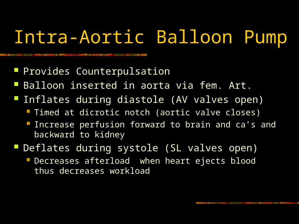
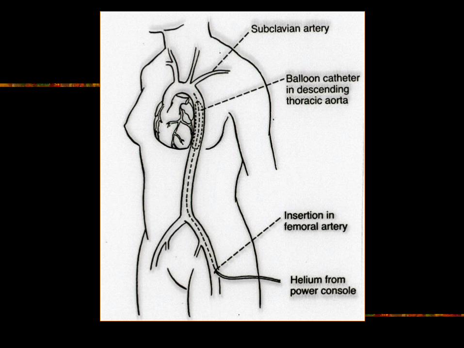
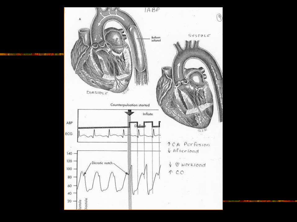
Intra-Aortic Balloon Pump
Provides Counterpulsation Balloon inserted in aorta via fem. Art. Inflates during diastole (AV valves open)
Timed at dicrotic notch (aortic valve closes) Increase perfusion forward to brain and ca’s and
backward to kidney Deflates during systole (SL valves open)
Decreases afterload when heart ejects blood thus decreases workload
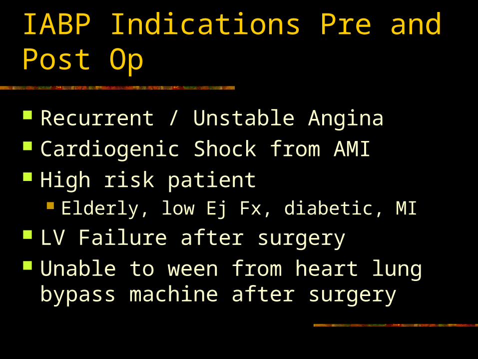
IABP Indications Pre and Post Op
Recurrent / Unstable Angina Cardiogenic Shock from AMI High risk patient
Elderly, low Ej Fx, diabetic, MI LV Failure after surgery Unable to ween from heart lung bypass
machine after surgery
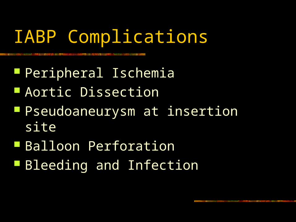
IABP Complications
Peripheral Ischemia Aortic Dissection Pseudoaneurysm at insertion site Balloon Perforation Bleeding and Infection
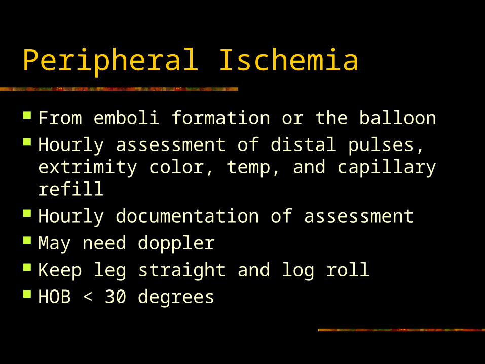
Peripheral Ischemia
From emboli formation or the balloon Hourly assessment of distal pulses,
extrimity color, temp, and capillary refill Hourly documentation of assessment May need doppler Keep leg straight and log roll HOB < 30 degrees
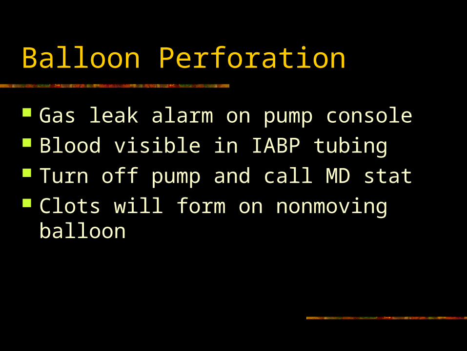
Balloon Perforation
Gas leak alarm on pump console Blood visible in IABP tubing Turn off pump and call MD stat Clots will form on nonmoving balloon
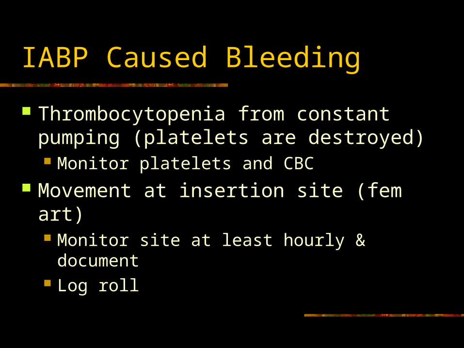
IABP Caused Bleeding
Thrombocytopenia from constant pumping (platelets are destroyed) Monitor platelets and CBC
Movement at insertion site (fem art) Monitor site at least hourly & document Log roll
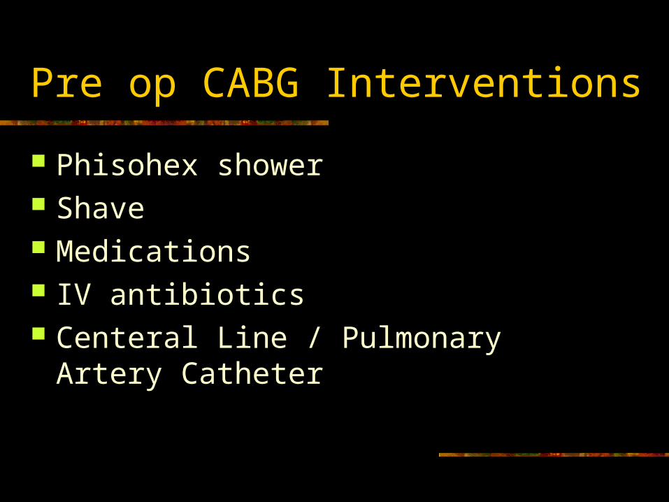
Pre op CABG Interventions
Phisohex shower Shave Medications IV antibiotics Centeral Line / Pulmonary Artery Catheter
CABG: The Operation Sequence
Patient to OR holding area Central line or PA Catheter (Swan)
insertion Harvest saph vein Median sternotomy Dissect vessels off of LIMA to mobilize Heparinize - bolus
CABG continued
Establish Cardioplegia Cold potassium aortic root injection regime Heart is motionless and bloodless
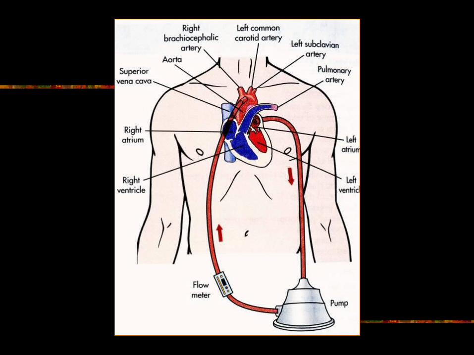
Cross clamp the aorta Insert Cardiopulmonary Bypass (CPB) Catheter
into Sup & Inf Vena Cava (directs venous return to pump oxygenator) and an aortic catheter (blood is returned to the body)
Establish extracorporeal circulation Diverts blood flow from heart and lungs
ABIOMEDS
BVS System 5000
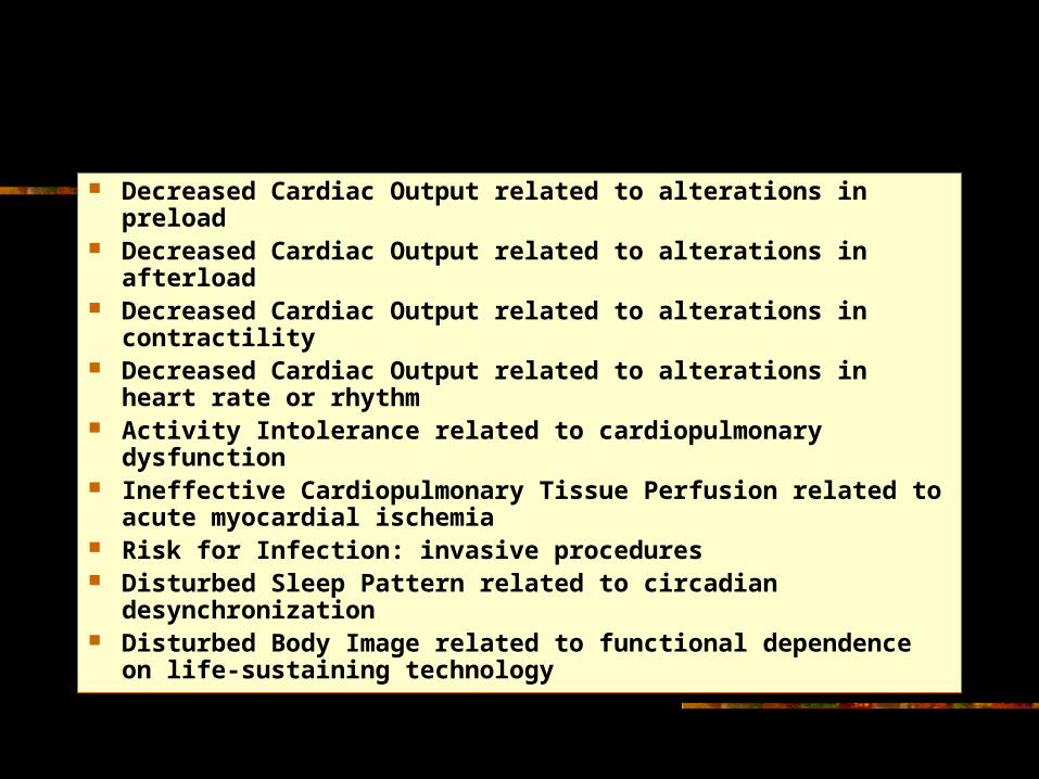
Decreased Cardiac Output related to alterations in preload Decreased Cardiac Output related to alterations in afterload Decreased Cardiac Output related to alterations in
contractility Decreased Cardiac Output related to alterations in heart
rate or rhythm Activity Intolerance related to cardiopulmonary dysfunction Ineffective Cardiopulmonary Tissue Perfusion related to
acute myocardial ischemia Risk for Infection: invasive procedures Disturbed Sleep Pattern related to circadian
desynchronization Disturbed Body Image related to functional dependence on
life-sustaining technology
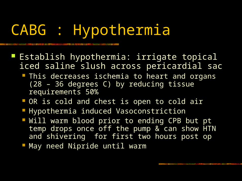
CABG : Hypothermia
Establish hypothermia: irrigate topical iced saline slush across pericardial sac This decreases ischemia to heart and organs (28 – 36
degrees C) by reducing tissue requirements 50% OR is cold and chest is open to cold air Hypothermia induced Vasoconstriction Will warm blood prior to ending CPB but pt temp
drops once off the pump & can show HTN and shivering for first two hours post op
May need Nipride until warm
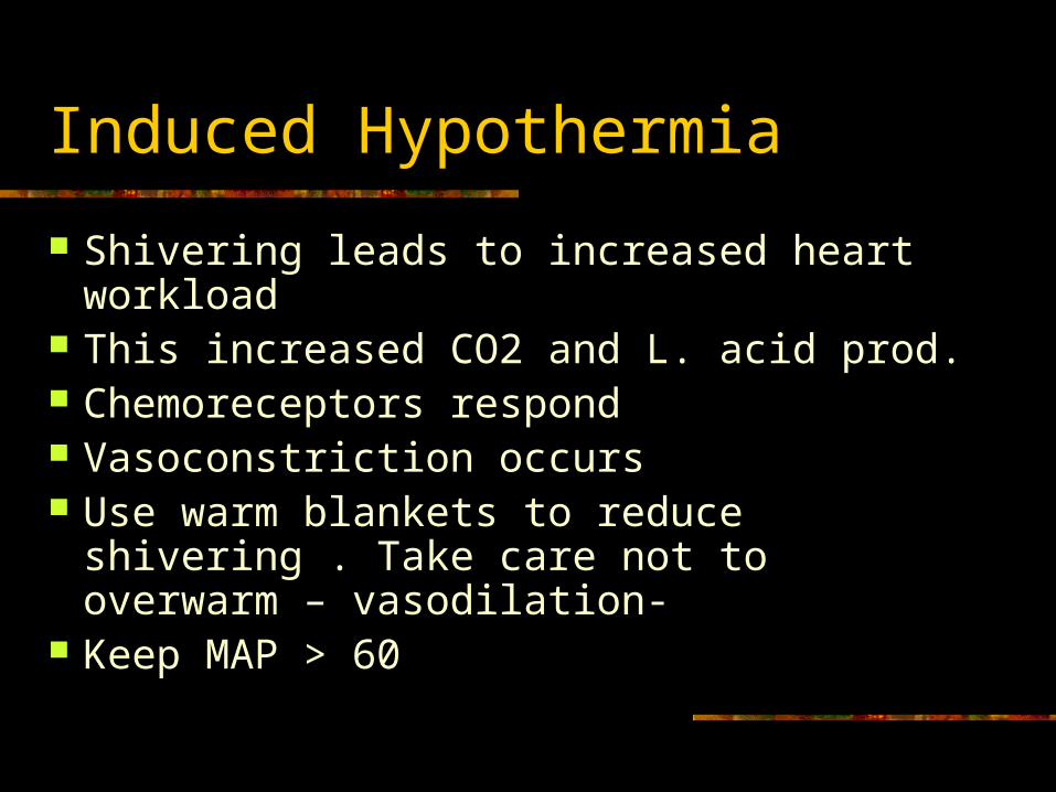
Induced Hypothermia
Shivering leads to increased heart workload
This increased CO2 and L. acid prod. Chemoreceptors respond Vasoconstriction occurs Use warm blankets to reduce shivering .
Take care not to overwarm – vasodilation- Keep MAP > 60
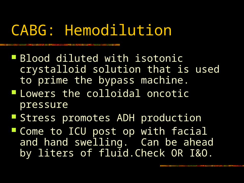
CABG: Hemodilution
Blood diluted with isotonic crystalloid solution that is used to prime the bypass machine.
Lowers the colloidal oncotic pressure Stress promotes ADH production Come to ICU post op with facial and hand
swelling. Can be ahead by liters of fluid.Check OR I&O.
CABG: Anticoagulation
Establish anticoagulation Heparin bolus Prevents coagulation in the bypass
machine once blood comes in contact with the machine’s surface
Stress promotes clotting
CABG continued
Perform graft anastamosis from aorta to past the obstructed coronary artery.
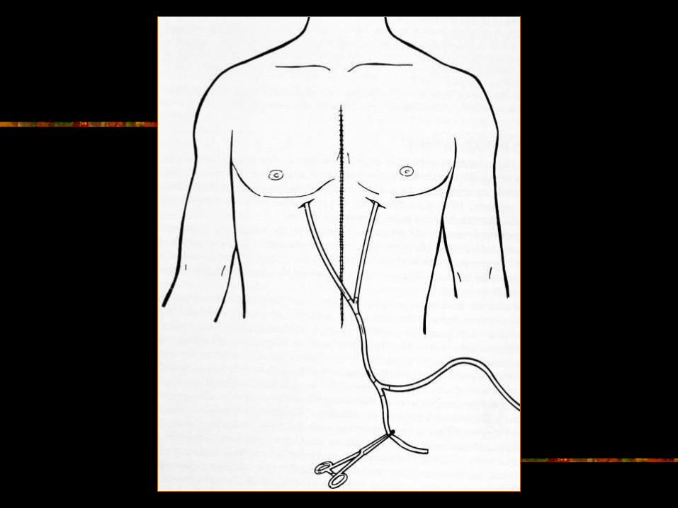
Place chest tubes in mediastinal cavity above and below the heart. Can place in the pericardial sac.
Place CT in intrapleural space if violated Loosely stitch on epicardial pacing
electrodes to atrial and ventricular wall.
CABG: continued
Jump start the heart: shock Disconnect from CPB Use IABP if difficulty coming off CPB Pull out pacing electrode wires through the
chest before closing sternum with wires. Skin stapled or sutured
Transport to ICU
CPB: Results / Complications
Hemodilution (interstitial / pulm edema) third spacing edema and wt gain due to plasma protein conc & capillary permeability.
Platelets damages & vasoactives subs released in blood (capillary permeability).
Alteration in fluid & electrolyte balance Alteration in cardiac function (dysrhythmias and
cardioplegia causes ischemia, acidosis, necrosis, and decreased cardiac output)
CPB cont
Coagulopathies (bleeding) damaged plts, heparin, hypothermia induced clotting factors in liver.
Catecholamine / renin release (HTN – watch out for bleeds at suture sites)
Supressed insulin release & epi stimulated glycogenolysis (hyperglycemia)
Alteration in central nervous system (cerebral ischemia, plaque, or embolic events)
CPB cont
Blood sitting in pulmonary capillaries and mesentary for length of time on CPB so capillary walls break down ( pulmonary edema, atelectasis, bowel ischemia, microemboli).
Impaired gas exchange from surfactant prod. Increased renin, angiotensin, aldosterone, and
ADH ( sodium & water retention, potassium excretion)
Serum dilution (Low K, Na, Cl, Mag) Hemolysis ( RBC’s damaged in CPB )
CPB cont
Hypothermia (increased SVR, decrease in contractility & HR & CO thus decreased perfusion pressure thus decreased urine output)
Hypothermia causes impaired release of insulin and low perfusion pressure, lytes are diluted so altered glucose transport (hyperglycemia)
Off Pump Normothermic Trend
Minimally invasive approach/ thoracotomy Less complications (no CPB) Higher technician difficulty suturing on
beating heart Poor visualization Some aspects of heart unreachable
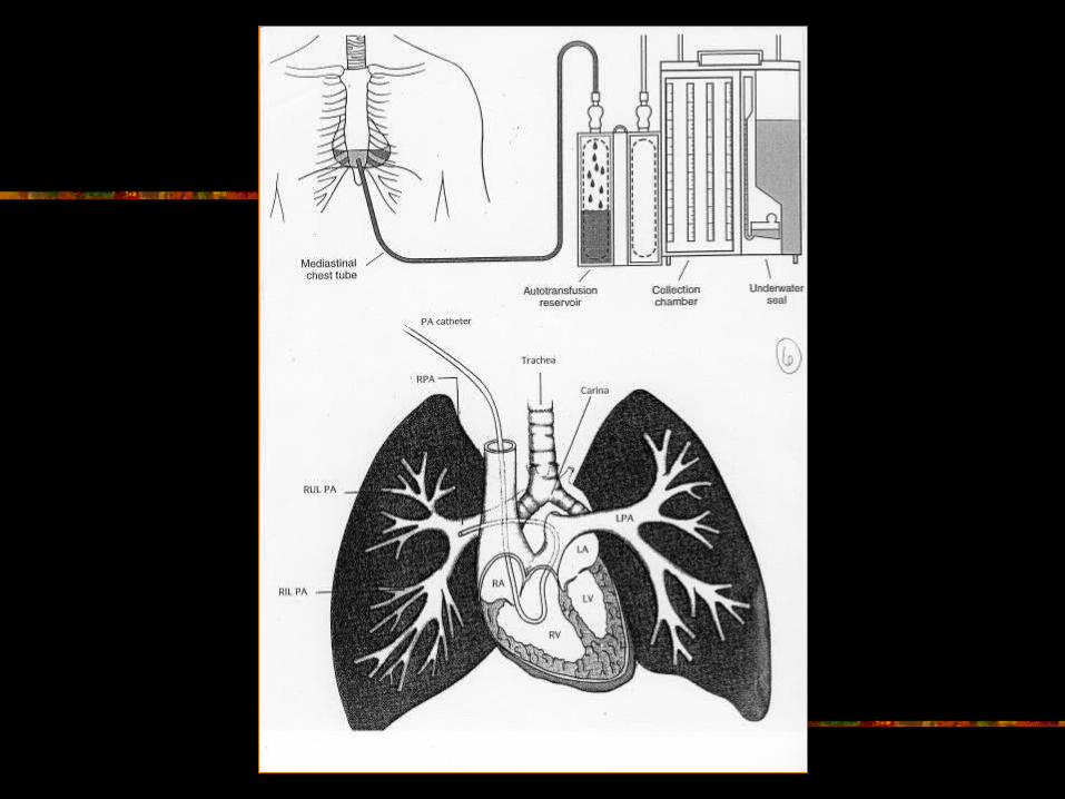
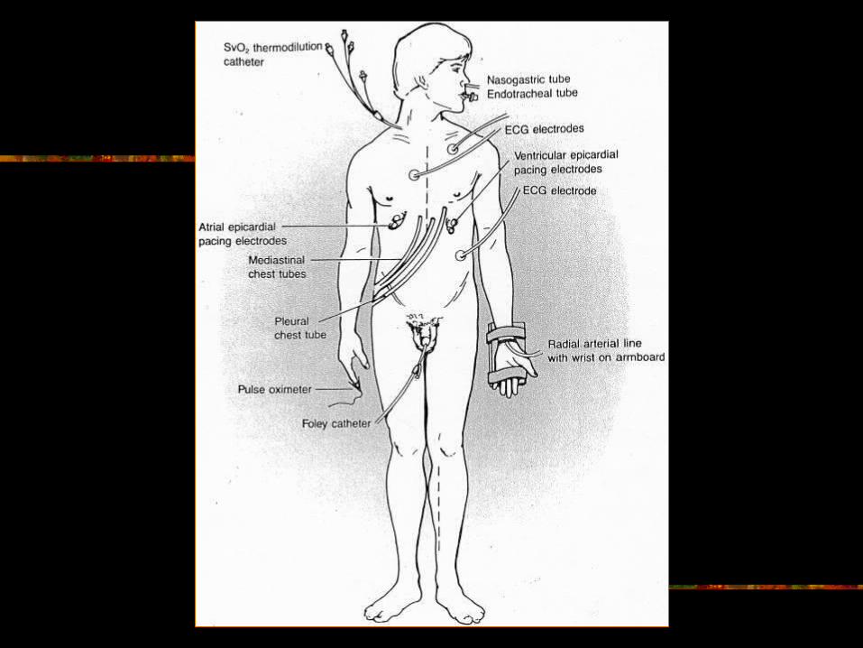
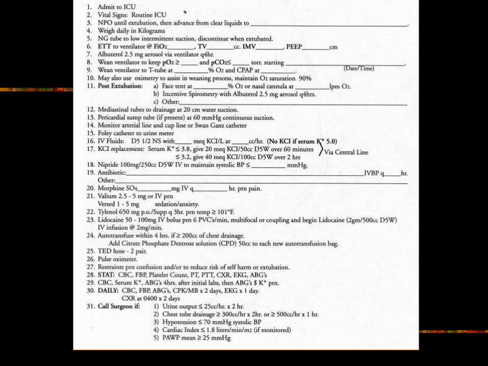
ICU : Equipment
Turn on ECG, vent settings, 12 lead, CXR 2 wall suctions (NG, ET) Brackets on IV poles, thermometer, doppler Multiple infusion pumps Autotransfusion set up with CPD External pacmaker generator with battery 60cc cath tip syringe
Admit into ICU
Hook to vent Hook to ECG, NIBP, Pulse Ox,
transducers Assess ET for placement (may dislodge) Connect pacer wires to generator Zero and calibrate pressure lines (Radial,
CVP, Pulmonary Artery)
Admit continued
Connect CT to autotransfusion Tape CT connections and check for clots Empty urometer every hour Record admit vitals and hemodynamics HOB 30 degrees Stat labs and ABG’s CMP, CBC, Plts,
PT/PTT, Enzymes, Troponin
Admit continued
12 lead ECG and CXR Hang necessary hemodynamic drugs and
do calculations Connect NG to suction Suction ETT Soft wrist restraints
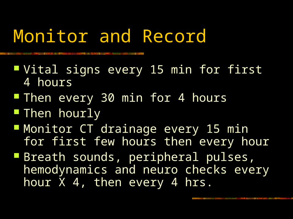
Monitor and Record
Vital signs every 15 min for first 4 hours Then every 30 min for 4 hours Then hourly Monitor CT drainage every 15 min for first
few hours then every hour Breath sounds, peripheral pulses,
hemodynamics and neuro checks every hour X 4, then every 4 hrs.
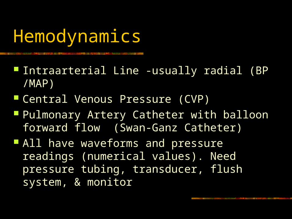
Hemodynamics
Intraarterial Line -usually radial (BP /MAP) Central Venous Pressure (CVP) Pulmonary Artery Catheter with balloon
forward flow (Swan-Ganz Catheter) All have waveforms and pressure readings
(numerical values). Need pressure tubing, transducer, flush system, & monitor
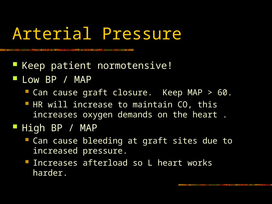
Arterial Pressure
Keep patient normotensive! Low BP / MAP
Can cause graft closure. Keep MAP > 60. HR will increase to maintain CO, this increases
oxygen demands on the heart . High BP / MAP
Can cause bleeding at graft sites due to increased pressure.
Increases afterload so L heart works harder.
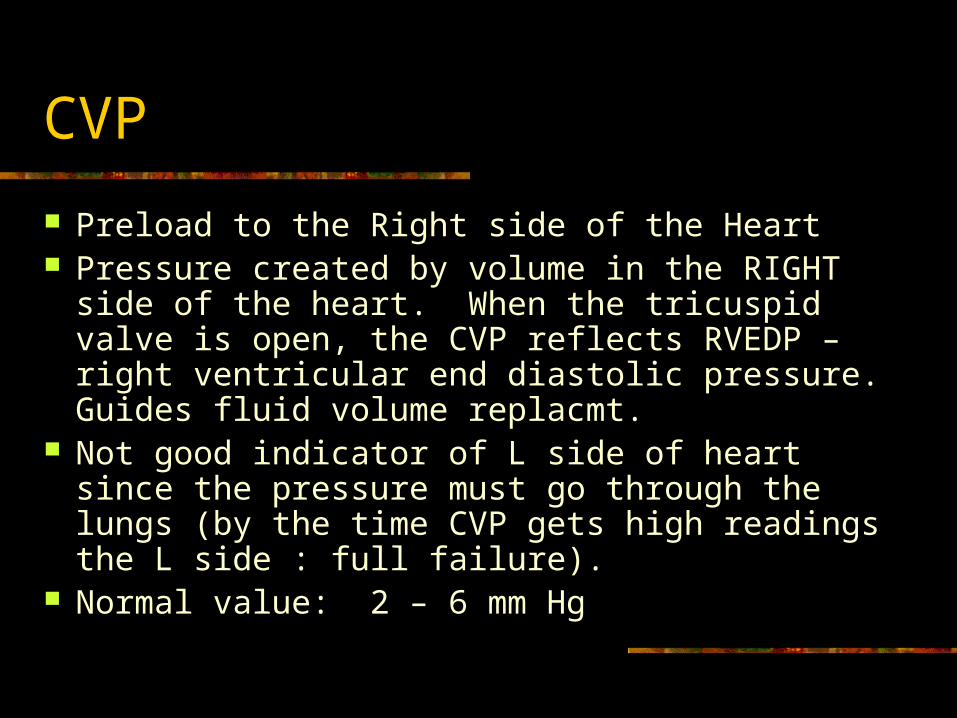
CVP
Preload to the Right side of the Heart Pressure created by volume in the RIGHT side
of the heart. When the tricuspid valve is open, the CVP reflects RVEDP – right ventricular end diastolic pressure. Guides fluid volume replacmt.
Not good indicator of L side of heart since the pressure must go through the lungs (by the time CVP gets high readings the L side : full failure).
Normal value: 2 – 6 mm Hg
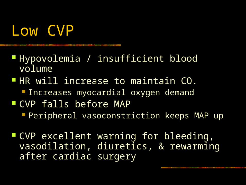
Low CVP
Hypovolemia / insufficient blood volume HR will increase to maintain CO.
Increases myocardial oxygen demand CVP falls before MAP
Peripheral vasoconstriction keeps MAP up CVP excellent warning for bleeding,
vasodilation, diuretics, & rewarming after cardiac surgery
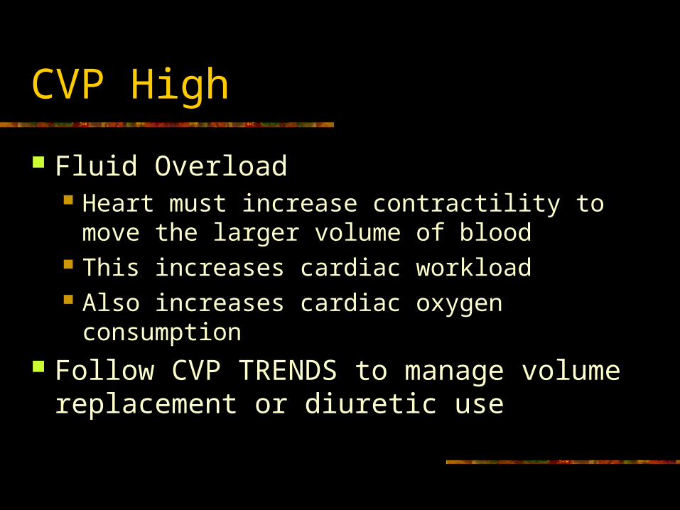
CVP High
Fluid Overload Heart must increase contractility to move the
larger volume of blood This increases cardiac workload Also increases cardiac oxygen consumption
Follow CVP TRENDS to manage volume replacement or diuretic use
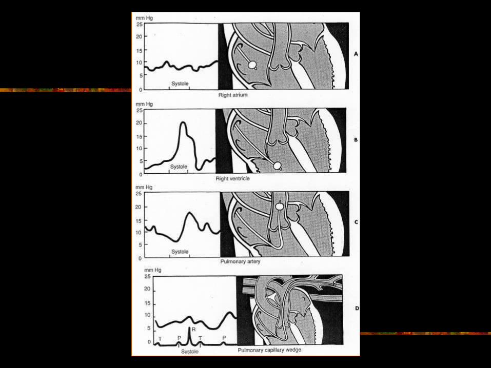
Pulmonary Artery Catheter
Similar to TLC but longer / has a balloon. Flow directional catheter/ 4-5 ports.
Advance to R Atrium (RAP : 4-6) Inflate Balloon 1 1/2 cc air into balloon port.
Advance to RV (20-30S) Advance to PA ( PAS: 20-30/ PAD: 8-15) Advance to wedge position into a small
pulmonary vessel (PCWP: 6-12). Deflate within 10 seconds. Floats back to PA.
PCWP: Preload to the L Heart
While balloon is inflated, only pressures on the Left side of the heart are seen.
Diastole: Mitral valve is open No valves obstructing between cath tip and
left ventricle. Pressure exerted by the volume in the LV is
reflected back to the pulmonary capillaries. “Preload” (L heart) = LVEDP = PCWP
PCWP Low
Volume Loss (bleeding or third spacing) Tachydysrhythmias (afib) Increased intrathoracic pressure (vent) Increased intracardiac pressures (cardiac
tamponade) Give volume replacement (autotransfusion
best) or control dysrhythmias or tx problem.
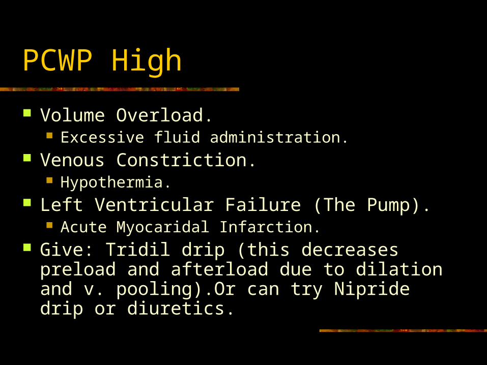
PCWP High
Volume Overload. Excessive fluid administration.
Venous Constriction. Hypothermia.
Left Ventricular Failure (The Pump). Acute Myocaridal Infarction.
Give: Tridil drip (this decreases preload and afterload due to dilation and v. pooling).Or can try Nipride drip or diuretics.
Afterload
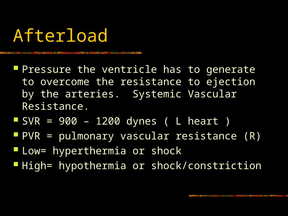
Pressure the ventricle has to generate to overcome the resistance to ejection by the arteries. Systemic Vascular Resistance.
SVR = 900 – 1200 dynes ( L heart ) PVR = pulmonary vascular resistance (R) Low= hyperthermia or shock High= hypothermia or shock/constriction
Afterload Drugs
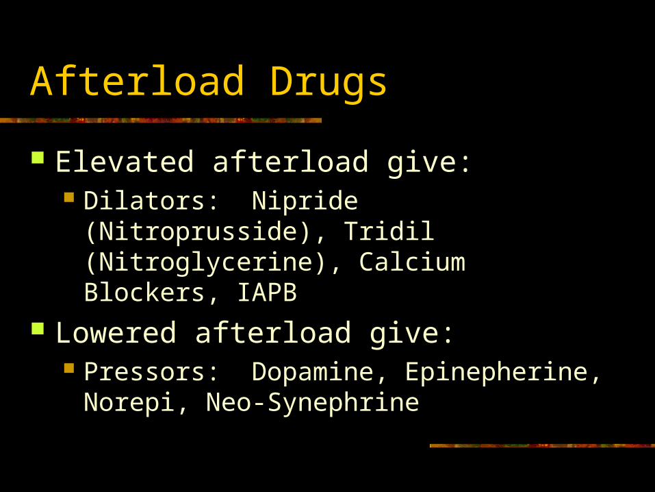
Elevated afterload give: Dilators: Nipride (Nitroprusside), Tridil
(Nitroglycerine), Calcium Blockers, IAPB Lowered afterload give:
Pressors: Dopamine, Epinepherine, Norepi, Neo-Synephrine
Decreased Contractility
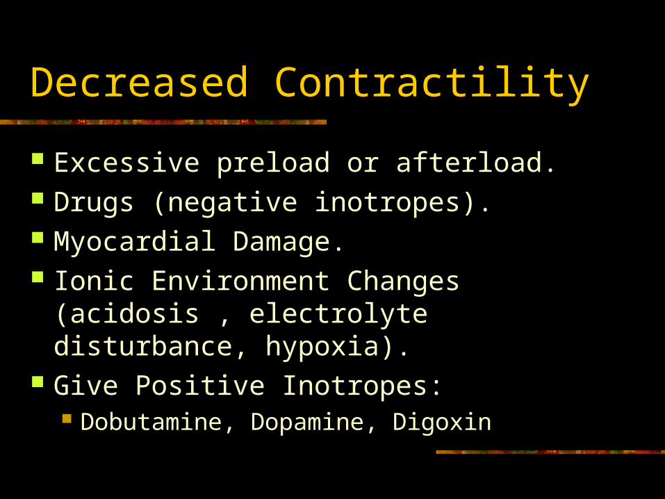
Excessive preload or afterload. Drugs (negative inotropes). Myocardial Damage. Ionic Environment Changes (acidosis ,
electrolyte disturbance, hypoxia). Give Positive Inotropes:
Dobutamine, Dopamine, Digoxin
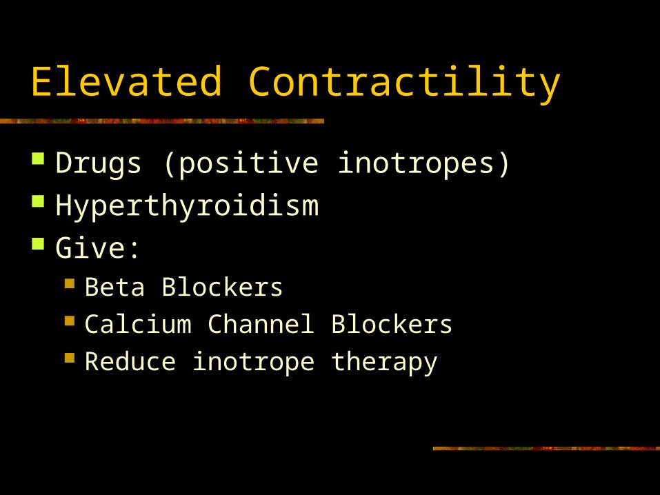
Elevated Contractility
Drugs (positive inotropes) Hyperthyroidism Give:
Beta Blockers Calcium Channel Blockers Reduce inotrope therapy
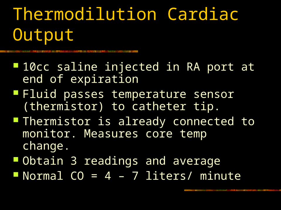
Thermodilution Cardiac Output
10cc saline injected in RA port at end of expiration
Fluid passes temperature sensor (thermistor) to catheter tip.
Thermistor is already connected to monitor. Measures core temp change.
Obtain 3 readings and average Normal CO = 4 – 7 liters/ minute
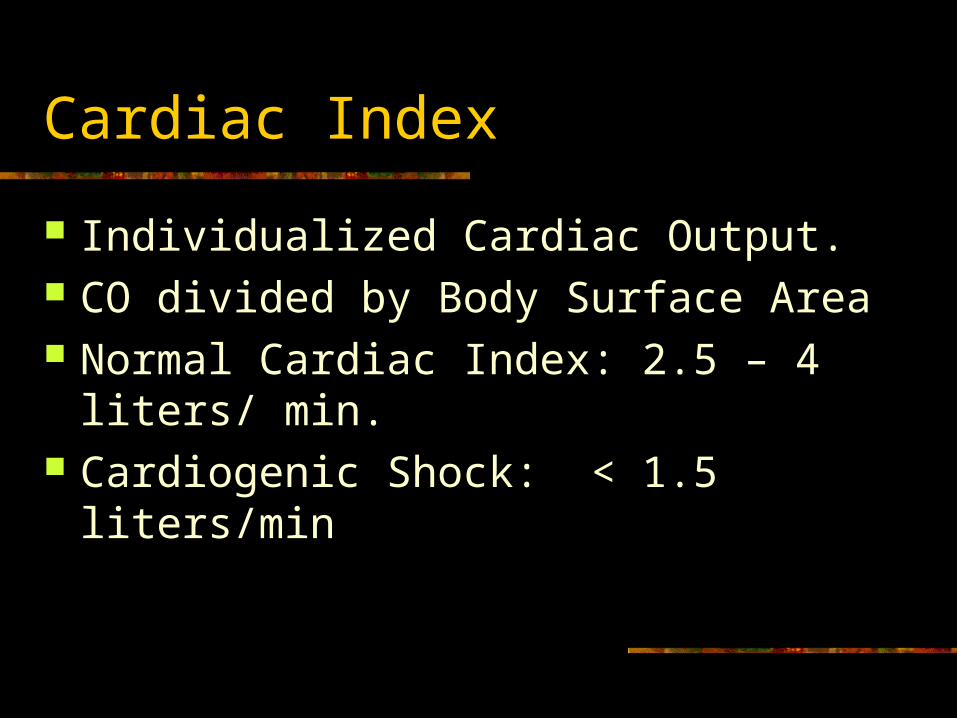
Cardiac Index
Individualized Cardiac Output. CO divided by Body Surface Area Normal Cardiac Index: 2.5 – 4 liters/ min. Cardiogenic Shock: < 1.5 liters/min
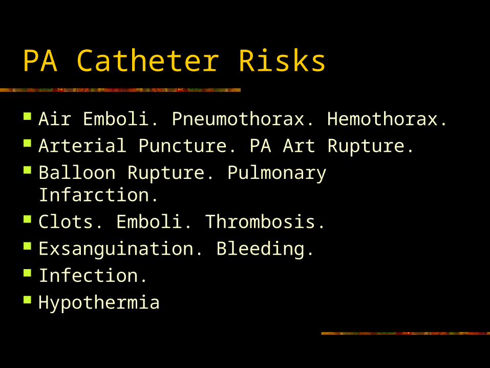
PA Catheter Risks
Air Emboli. Pneumothorax. Hemothorax. Arterial Puncture. PA Art Rupture. Balloon Rupture. Pulmonary Infarction. Clots. Emboli. Thrombosis. Exsanguination. Bleeding. Infection. Hypothermia
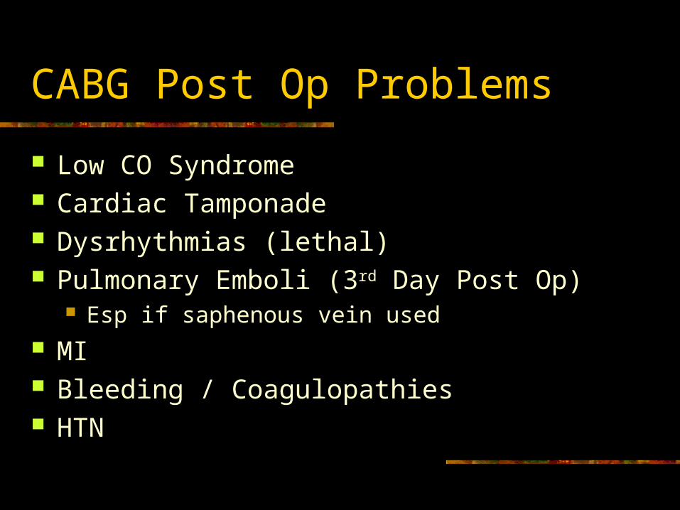
CABG Post Op Problems
Low CO Syndrome Cardiac Tamponade Dysrhythmias (lethal) Pulmonary Emboli (3rd Day Post Op)
Esp if saphenous vein used MI Bleeding / Coagulopathies HTN
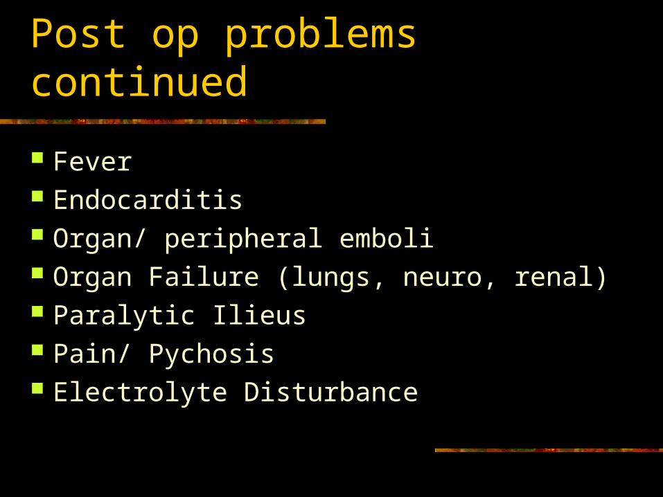
Post op problems continued
Fever Endocarditis Organ/ peripheral emboli Organ Failure (lungs, neuro, renal) Paralytic Ilieus Pain/ Pychosis Electrolyte Disturbance
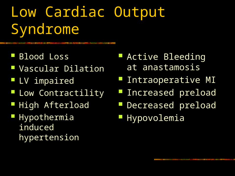
Low Cardiac Output Syndrome
Blood Loss Vascular Dilation LV impaired Low Contractility High Afterload Hypothermia induced
hypertension
Active Bleeding at anastamosis
Intraoperative MI Increased preload Decreased preload Hypovolemia
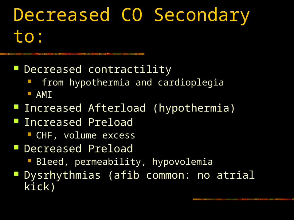
Decreased CO Secondary to:
Decreased contractility from hypothermia and cardioplegia AMI
Increased Afterload (hypothermia) Increased Preload
CHF, volume excess Decreased Preload
Bleed, permeability, hypovolemia Dysrhythmias (afib common: no atrial kick)
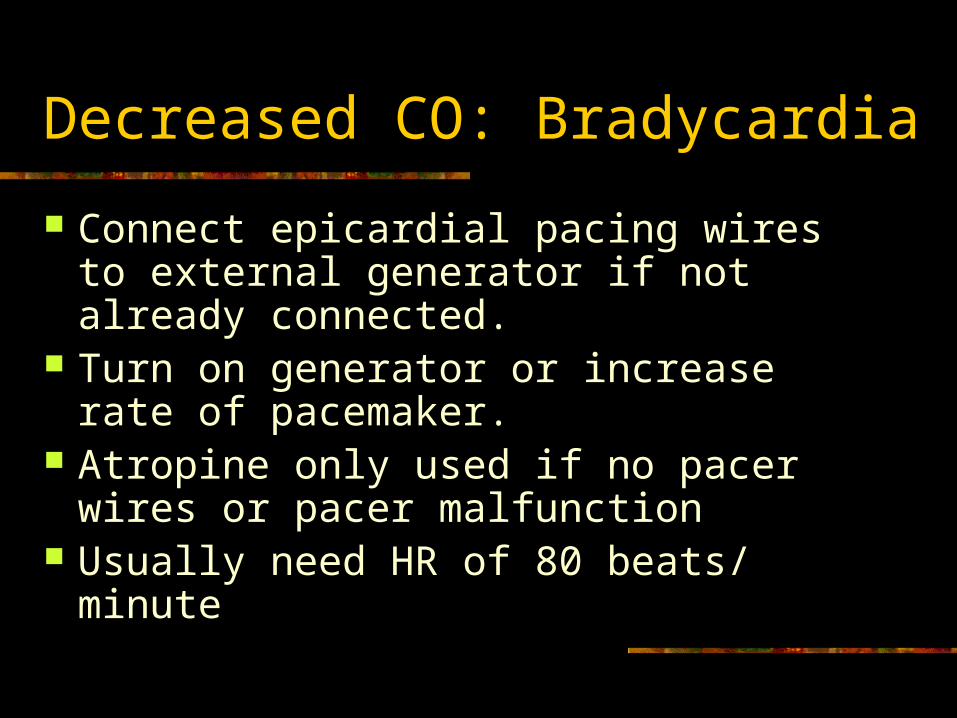
Decreased CO: Bradycardia
Connect epicardial pacing wires to external generator if not already connected.
Turn on generator or increase rate of pacemaker.
Atropine only used if no pacer wires or pacer malfunction
Usually need HR of 80 beats/ minute
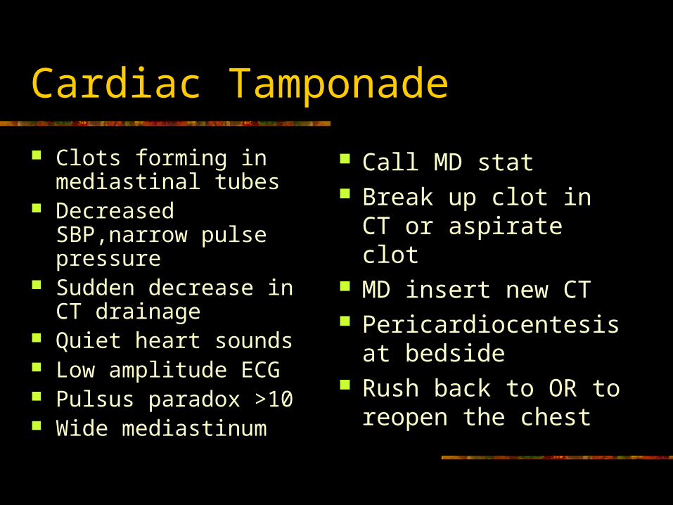
Cardiac Tamponade
Clots forming in mediastinal tubes
Decreased SBP,narrow pulse pressure
Sudden decrease in CT drainage
Quiet heart sounds Low amplitude ECG Pulsus paradox >10 Wide mediastinum
Call MD stat Break up clot in CT or
aspirate clot MD insert new CT Pericardiocentesis at
bedside Rush back to OR to
reopen the chest
Other Diagnoses
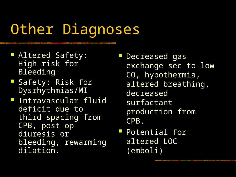
Altered Safety: High risk for Bleeding
Safety: Risk for Dysrhythmias/MI
Intravascular fluid deficit due to third spacing from CPB, post op diuresis or bleeding, rewarming dilation.
Decreased gas exchange sec to low CO, hypothermia, altered breathing, decreased surfactant production from CPB.
Potential for altered LOC (emboli)
Stress from Surgery
Activates ADH Hold onto water to
keep from going into shock
Cells swell and third spacing occurs from CPB
Edema noted
IV Fluid Post Op: D5 ½ Normal Saline Hypertonic Pulls water from
swollen cells into the intravascular space
Increases iv volume Increases urine
output
Progression
Extubated by next morning, hopefully sooner and up in chair. Lung exercises.
1 –2 nights in ICU if no complications. D/C PA and art lines, CT’s, pacer wires. Transfer to telemetry. Cardiac Rehab inpatient. Cardiac Rehab outpaitent.
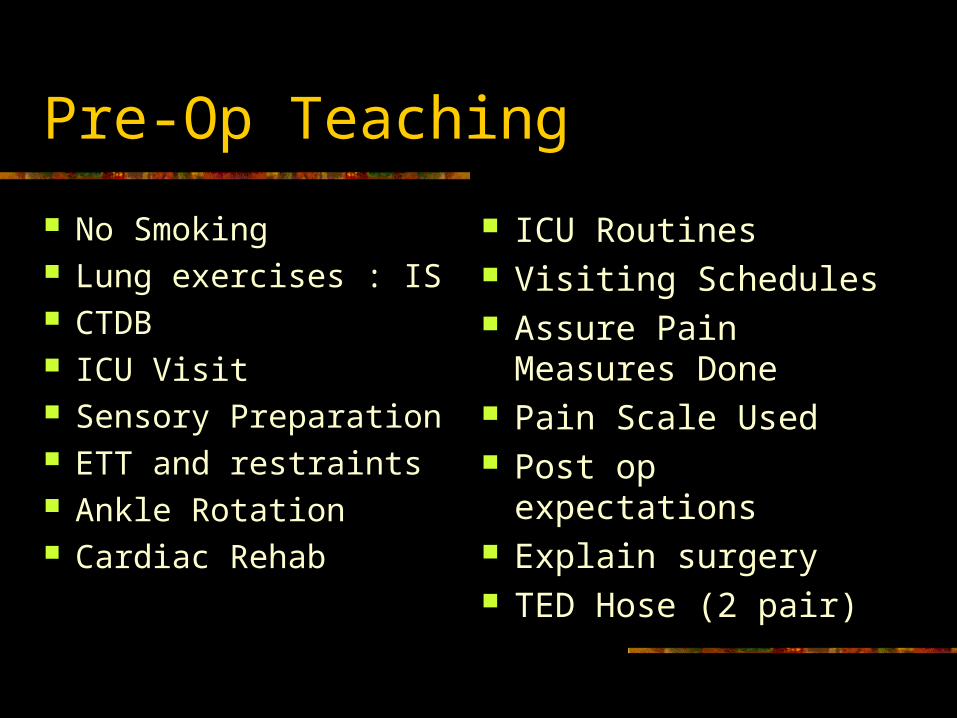
Pre-Op Teaching
No Smoking Lung exercises : IS CTDB ICU Visit Sensory Preparation ETT and restraints Ankle Rotation Cardiac Rehab
ICU Routines Visiting Schedules Assure Pain
Measures Done Pain Scale Used Post op expectations Explain surgery TED Hose (2 pair)
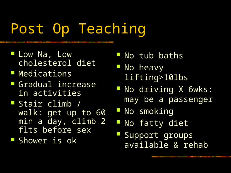
Post Op Teaching
Low Na, Low cholesterol diet
Medications Gradual increase in
activities Stair climb / walk: get
up to 60 min a day, climb 2 flts before sex
Shower is ok
No tub baths No heavy lifting>10lbs No driving X 6wks:
may be a passenger No smoking No fatty diet Support groups
available & rehab
Valvular Surgery
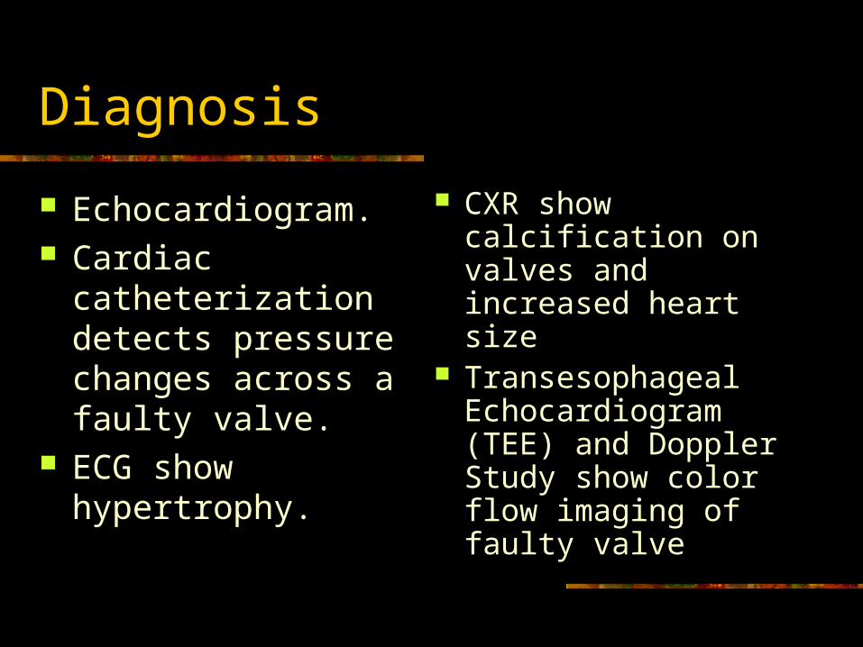
Diagnosis
Echocardiogram. Cardiac
catheterization detects pressure changes across a faulty valve.
ECG show hypertrophy.
CXR show calcification on valves and increased heart size
Transesophageal Echocardiogram (TEE) and Doppler Study show color flow imaging of faulty valve
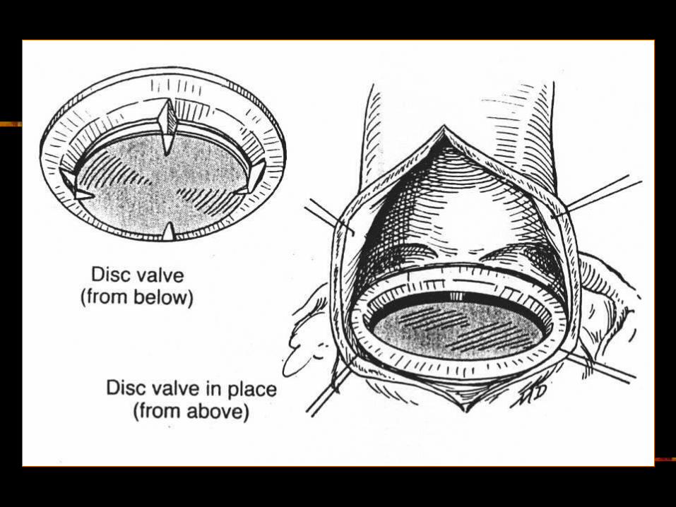
Three Methods of Valve REPAIR
Mitral commisurotomy Valvuloplasty Annuloplasty
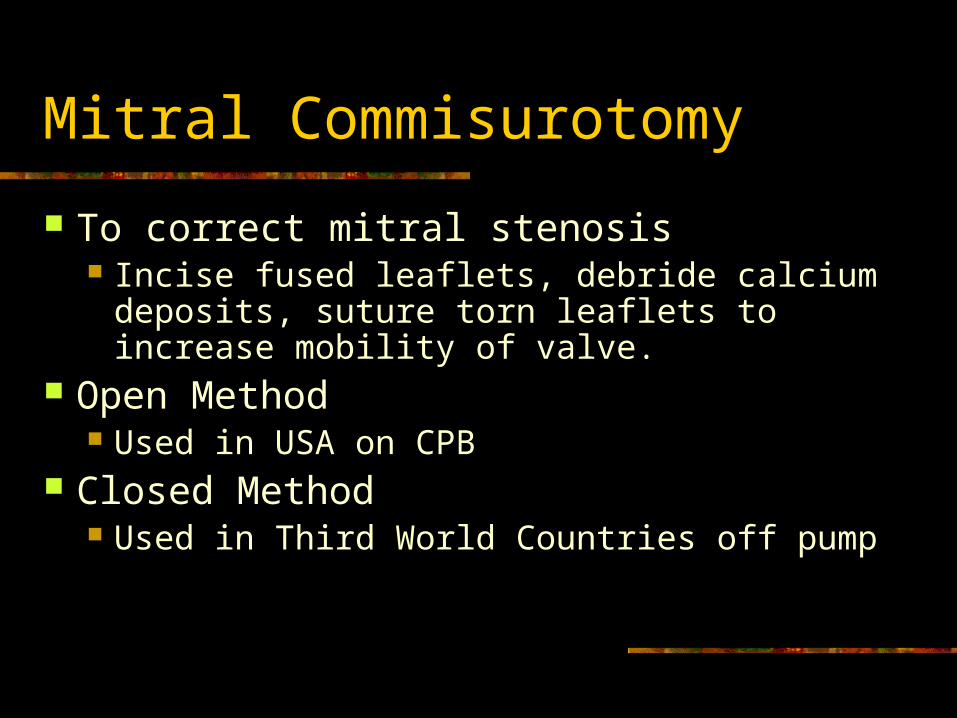
Mitral Commisurotomy
To correct mitral stenosis Incise fused leaflets, debride calcium
deposits, suture torn leaflets to increase mobility of valve.
Open Method Used in USA on CPB
Closed Method Used in Third World Countries off pump
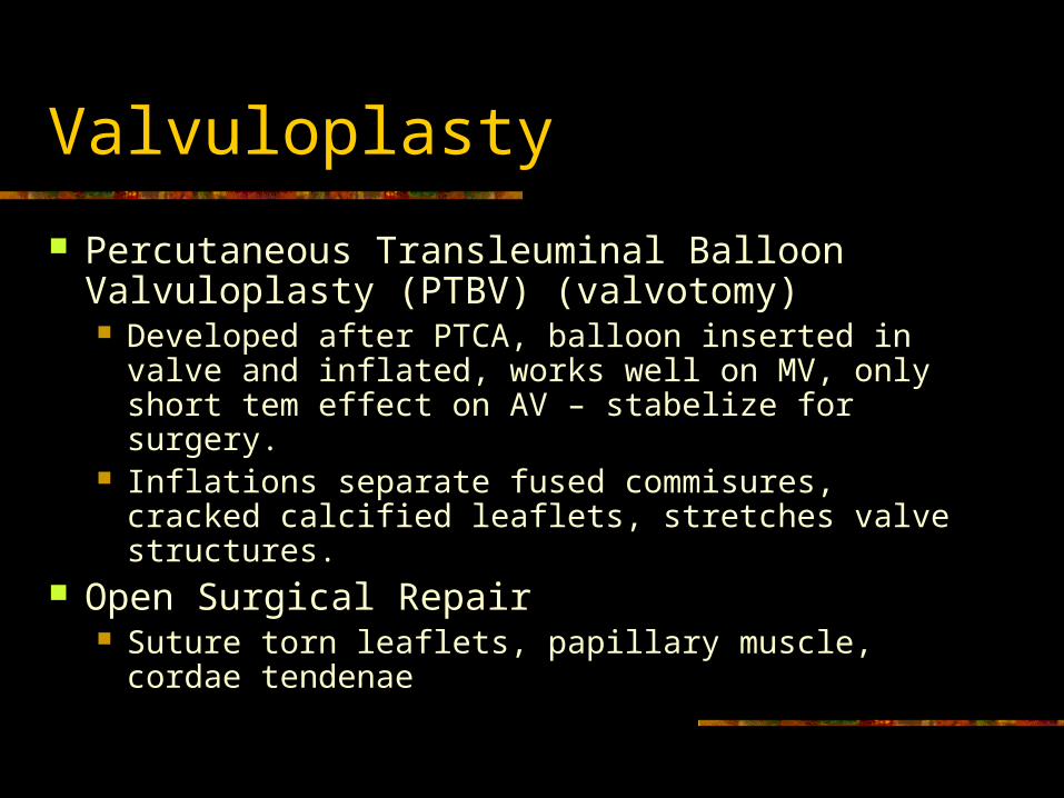
Valvuloplasty
Percutaneous Transleuminal Balloon Valvuloplasty (PTBV) (valvotomy) Developed after PTCA, balloon inserted in valve and
inflated, works well on MV, only short tem effect on AV – stabelize for surgery.
Inflations separate fused commisures, cracked calcified leaflets, stretches valve structures.
Open Surgical Repair Suture torn leaflets, papillary muscle, cordae
tendenae
Annuloplasty
Annulus Anatomical ring in which the valve sits.
Surgical Repair Good for Mitral Regurgitation Suture a prostetic ring to the dilated annulus
to increase leaflet coaption or reconstruct annulus without a ring
Valve Replacement
Most Common AVR for stenosis or regurgitation MVR for stenosis or regurgitation
Article: When to go to Surgery? Before LV dysfunctions or Ej Fx < 55% or Symptomatic even with good Ej Fx
True open heart surgery
Pre Op Care
Try to give 48 hours of antibiotics Begin on Nipride drip to decrease preload
and afterload Emergency AVR due to acute endocarditis
Body hasn’t compensated yet = emergency Give antibiotics on call to OR
Valves Used for Replacement
Biological: Tissue Valves Porcine (pig) Bovine (calf) Homograft (human cadaver)
Mechanical: Metal and Dacron Caged ball Tilting disc Bi-Leaflet: mechanical central flow disc/ most
Young Recipients
If a patient is young, they will probably receive a mechanical valve since they last much longer.
If conditions exist that prevent taking coumadin such as pregnancy, they will receive a biological valve.
Biological Valves
Chance of Rejection Virchow’s Triad activated Less Durable (lasts 7 years) Tendency to calcify Endocarditis: early and late (p 60 days) Low thrombogenicity (less emboli) Only 12 wks of anticoagulants if no a fib
Mechanical Valves
High chance of clot formation Long term anticoagulation therapy Mechanical malfunction Hemolytic anemia Early and late endocarditis Virchow’s triad Last longer (20 years)
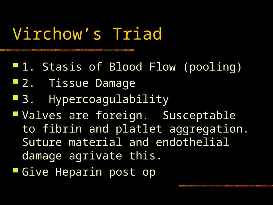
Virchow’s Triad
1. Stasis of Blood Flow (pooling) 2. Tissue Damage 3. Hypercoagulability Valves are foreign. Susceptable to fibrin
and platlet aggregation. Suture material and endothelial damage agrivate this.
Give Heparin post op
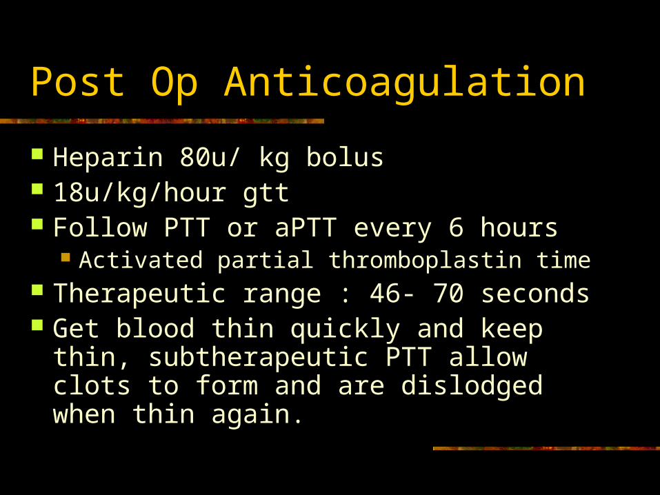
Post Op Anticoagulation
Heparin 80u/ kg bolus 18u/kg/hour gtt Follow PTT or aPTT every 6 hours
Activated partial thromboplastin time Therapeutic range : 46- 70 seconds Get blood thin quickly and keep thin,
subtherapeutic PTT allow clots to form and are dislodged when thin again.

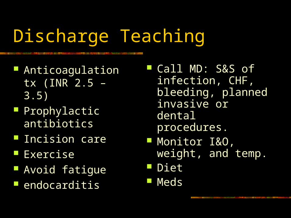
Discharge Teaching
Anticoagulation tx (INR 2.5 – 3.5)
Prophylactic antibiotics
Incision care Exercise Avoid fatigue endocarditis
Call MD: S&S of infection, CHF, bleeding, planned invasive or dental procedures.
Monitor I&O, weight, and temp.
Diet Meds
Aneurysms
Definition
An outpouching or sac (dilitation) of the arterial wall commonly occuring in the aorta at the weak spot.
½ of all aneurysms >6cm rupture in 1 yr Dangerous Thrombi like to deposit here
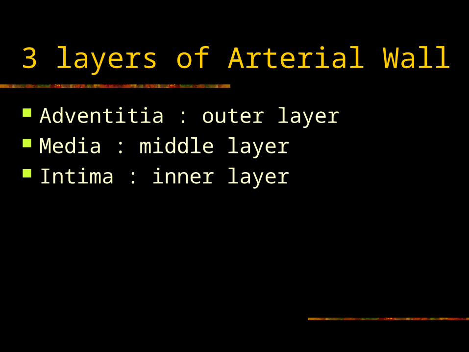
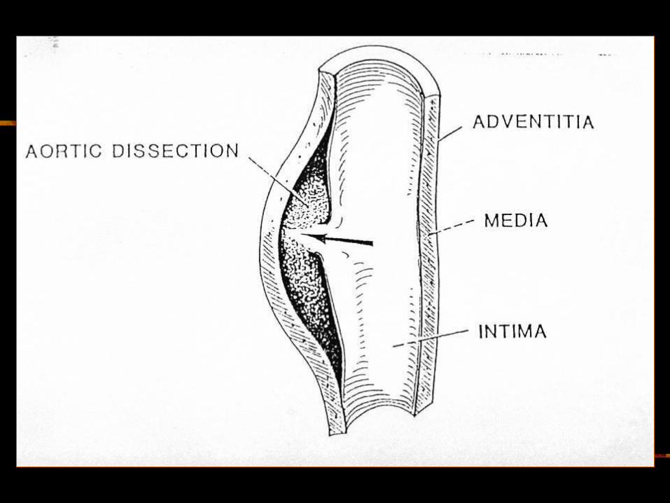
3 layers of Arterial Wall
Adventitia : outer layer Media : middle layer Intima : inner layer
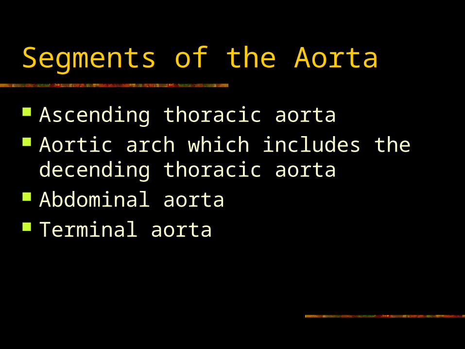
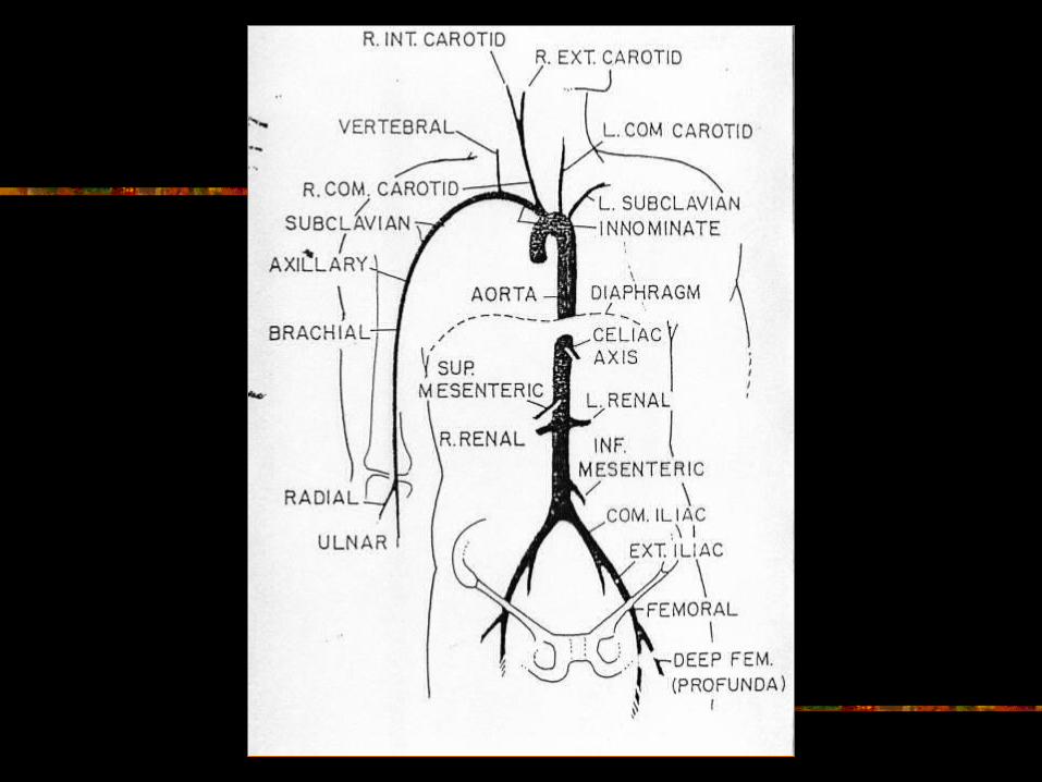
Segments of the Aorta
Ascending thoracic aorta Aortic arch which includes the decending
thoracic aorta Abdominal aorta Terminal aorta
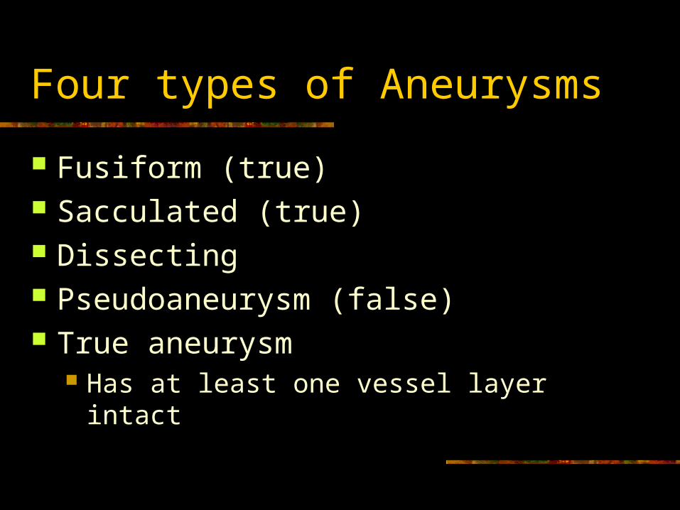
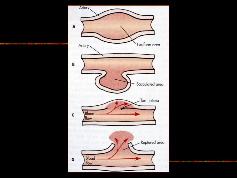
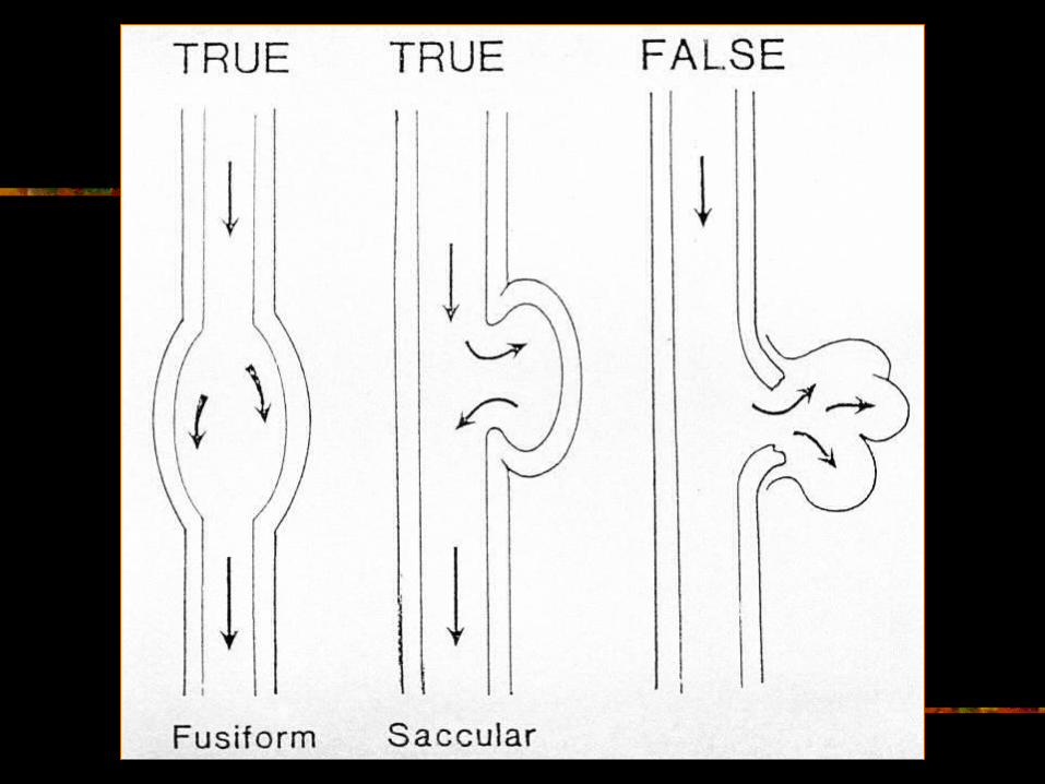
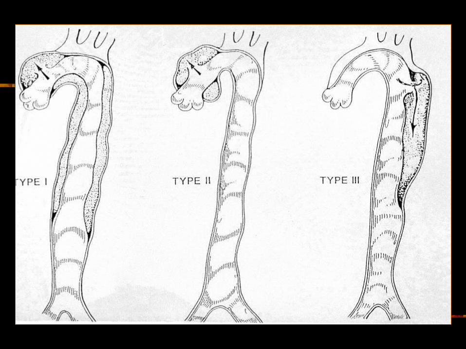
Four types of Aneurysms
Fusiform (true) Sacculated (true) Dissecting Pseudoaneurysm (false) True aneurysm
Has at least one vessel layer intact
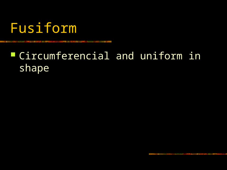
Fusiform
Circumferencial and uniform in shape
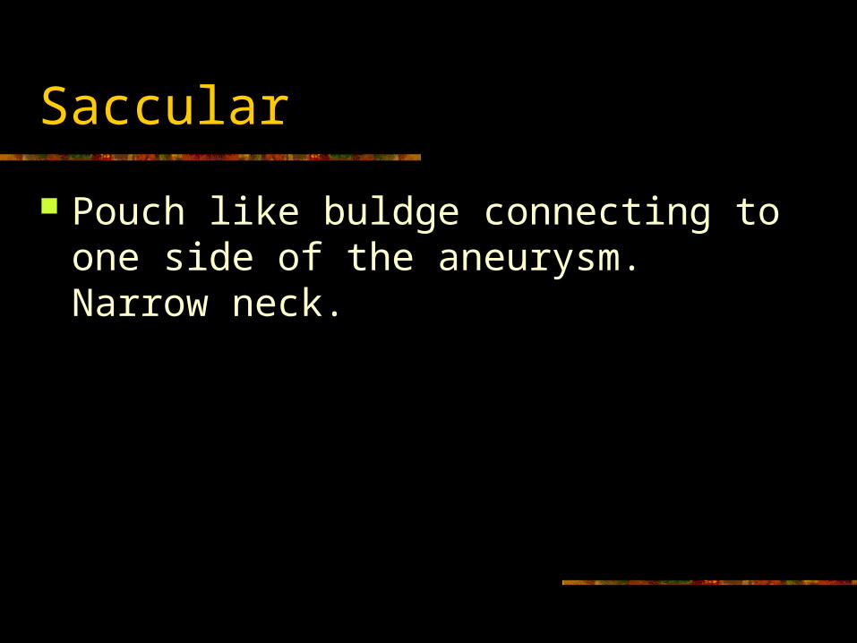
Saccular
Pouch like buldge connecting to one side of the aneurysm. Narrow neck.
Pseudoaneurysm
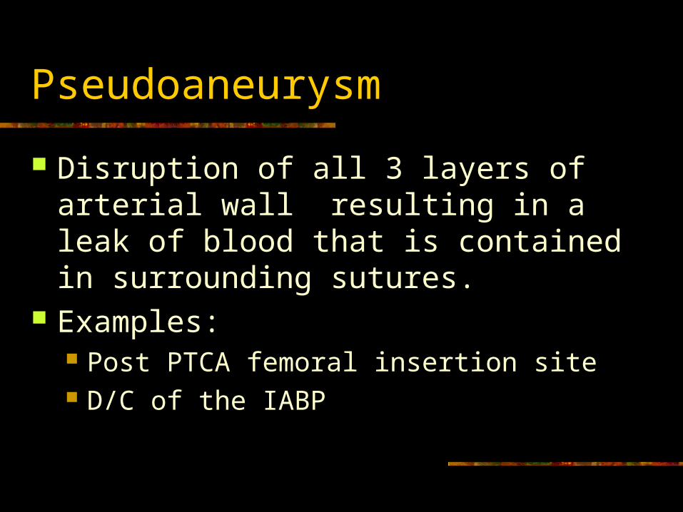
Disruption of all 3 layers of arterial wall resulting in a leak of blood that is contained in surrounding sutures.
Examples: Post PTCA femoral insertion site D/C of the IABP
Causes
Athlerosclerosis : plaques and fibrin deposits weaken wall, loose elasticity
Infection Congenital disorders Trauma HTN, men Smoking
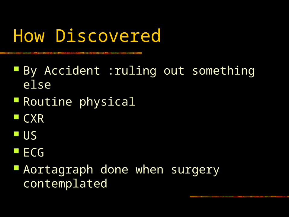
How Discovered
By Accident :ruling out something else Routine physical CXR US ECG Aortagraph done when surgery
contemplated
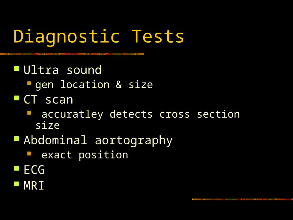
Diagnostic Tests
Ultra sound gen location & size
CT scan accuratley detects cross section size
Abdominal aortography exact position
ECG MRI
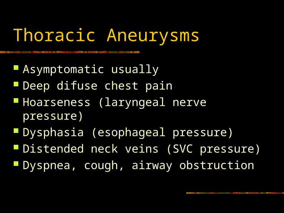
Thoracic Aneurysms
Asymptomatic usually Deep difuse chest pain Hoarseness (laryngeal nerve pressure) Dysphasia (esophageal pressure) Distended neck veins (SVC pressure) Dyspnea, cough, airway obstruction
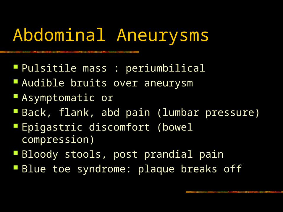
Abdominal Aneurysms
Pulsitile mass : periumbilical Audible bruits over aneurysm Asymptomatic or Back, flank, abd pain (lumbar pressure) Epigastric discomfort (bowel compression) Bloody stools, post prandial pain Blue toe syndrome: plaque breaks off
Repair criterion
Aneurysm > 6.0 cm (text says 6.5). If it is 2 times > normal artery diameter. Not repaired if small or poor surgical risk. Traditional Surgery vs. Endovascular Graft Surgery
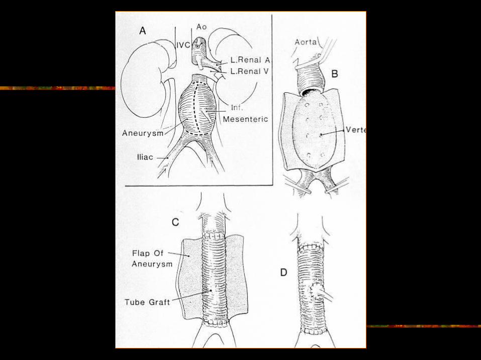
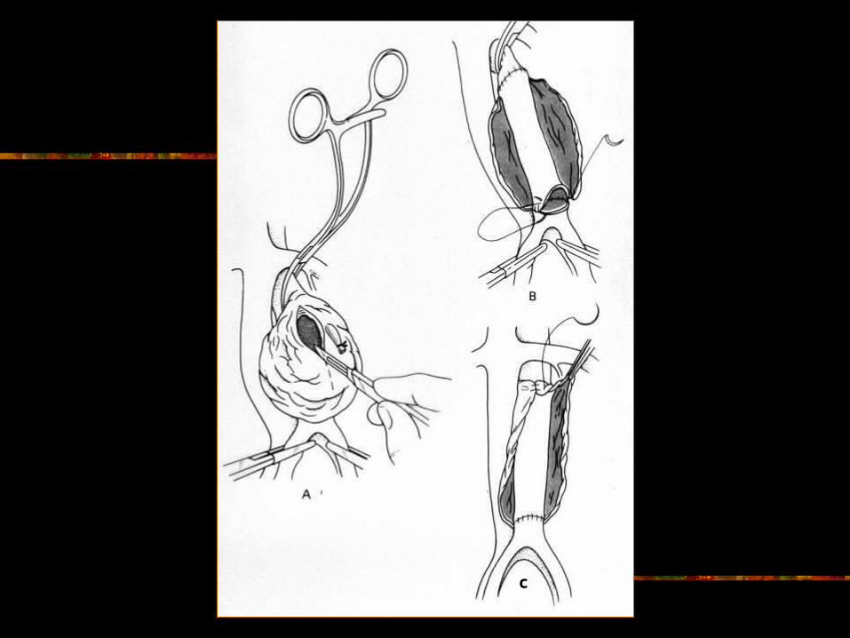
Traditional Surgery: Aneurysmectomy
Midline incision xyphoid to pubis Cross clamp aorta above and below site.
Heparinize first. If clamp above renals, check for acute tubular necrosis :ATN.
Incize diseased aorta. Insert synthetic graft Suture native aortic wall around graft.
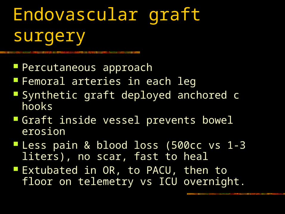
Endovascular graft surgery
Percutaneous approach Femoral arteries in each leg Synthetic graft deployed anchored c hooks Graft inside vessel prevents bowel erosion Less pain & blood loss (500cc vs 1-3
liters), no scar, fast to heal Extubated in OR, to PACU, then to floor
on telemetry vs ICU overnight.
Pre-op care
Fix carotid or coronary arteries first Get baseline vitals & mark pulses Abdominal girth Assess all systems Teaching Titrate Nipride or Tridil to Keep MAP 60-90
Most serious pre-op complication
Rupture, Bleeding , Shock High HR, Low BP, LOC, urine output, clammy
May have Turner’s Sign Severe back pain, flank eccymosis A clot could dislodge
Post op goals
Normal tissue perfusion Intact neurological / motor function Prevent post op complications
Tissue perfusion
Titrate Nipride or Tridil to keep MAP 60-90 May use lasix Keep systolic 95-160, warm if hypothermic Give fluids, blood, & meds to keep
adequate BP to keep graft open Prevent low BP to keep graft patent. Palpate pulses q 15 X4, q1hr x 4, q4 hrs.
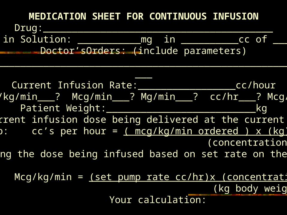
MEDICATION SHEET FOR CONTINUOUS INFUSIONDrug:________________________________________
Actual Drug in Solution: ___________mg in __________cc of ______________Doctor’sOrders: (include parameters) ______________________________________________________________________________________________________________________________________________________________________________________________________
Current Infusion Rate:_________________cc/hourHow is this drug normally infused? Check one. Mcg/kg/min___? Mcg/min___? Mg/min___? cc/hr___? Mcg/hour___? Mg/hr___? Units/min___? Units/hr___?
Patient Weight:__________________________kgCalculate the current infusion dose being delivered at the current rate. Show calc.Setting the pump: cc’s per hour = ( mcg/kg/min ordered ) x (kg) x (60 minutes)
(concentration of solution in mcg per cc) Calculating the dose being infused based on set rate on the infusion pump:
Mcg/kg/min = (set pump rate cc/hr)x (concentration mcg/cc) (kg body weight) x ( 60 minutes)
Your calculation:
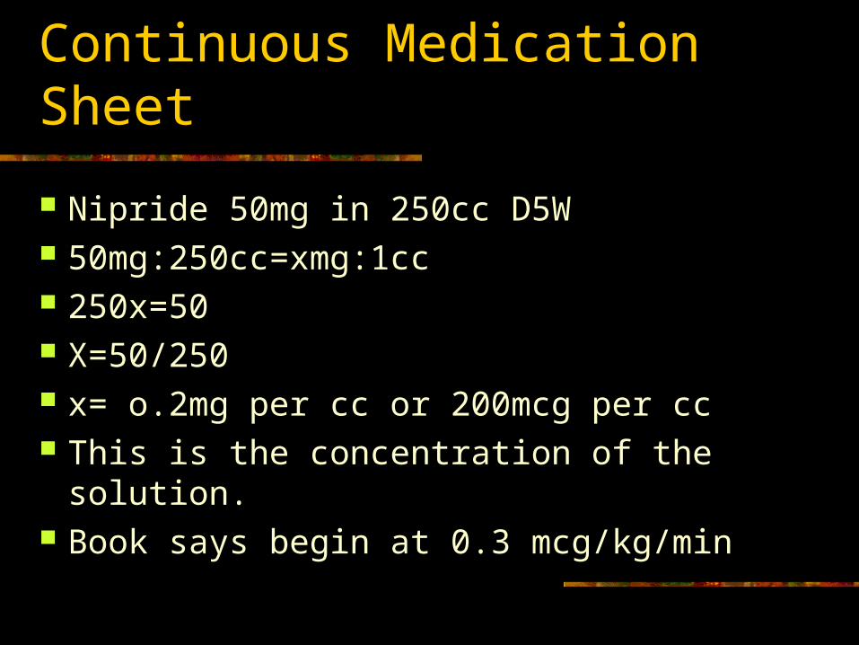
Continuous Medication Sheet
Nipride 50mg in 250cc D5W 50mg:250cc=xmg:1cc 250x=50 X=50/250 x= o.2mg per cc or 200mcg per cc This is the concentration of the solution. Book says begin at 0.3 mcg/kg/min
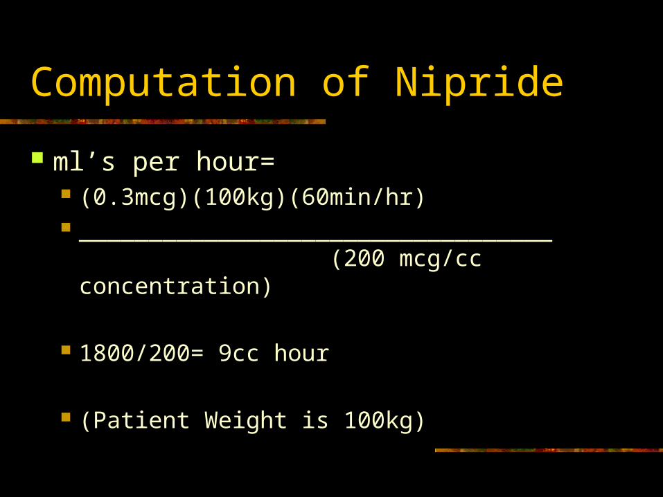
Computation of Nipride
ml’s per hour= (0.3mcg)(100kg)(60min/hr) __________________________________
(200 mcg/cc concentration)
1800/200= 9cc hour
(Patient Weight is 100kg)
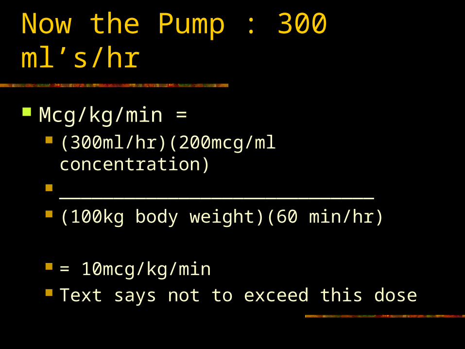
Now the Pump : 300 ml’s/hr
Mcg/kg/min = (300ml/hr)(200mcg/ml concentration) _____________________________ (100kg body weight)(60 min/hr)
= 10mcg/kg/min Text says not to exceed this dose
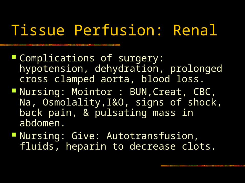
Tissue Perfusion: Renal
Complications of surgery: hypotension, dehydration, prolonged cross clamped aorta, blood loss.
Nursing: Mointor : BUN,Creat, CBC, Na, Osmolality,I&O, signs of shock, back pain, & pulsating mass in abdomen.
Nursing: Give: Autotransfusion, fluids, heparin to decrease clots.
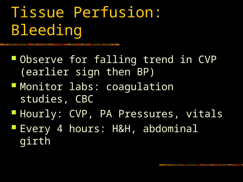
Tissue Perfusion: Bleeding
Observe for falling trend in CVP (earlier sign then BP)
Monitor labs: coagulation studies, CBC Hourly: CVP, PA Pressures, vitals Every 4 hours: H&H, abdominal girth
Tissue Perfusion: Mesentary
Paralytic ileus common. Intestines become swollen and bruised from manipulation.
Observe for: absent bowel sounds and passing of flatus, distended abdomen, N&V.
Encourage early ambulation. Connect NG to low cont. suction & irrigate for patency.
Tissue Perfusion: Fluids/ Lytes
Monitor: daily weights, I&O, wound & NG drainage, abnormal labs.
Give: autotransfusion, blood transfusion – PRBC’S, colloids, intravascular fluids.
Tissue Perfusion: Cerebral
Neuro checks hourly X 4, then every 4 hours.
Motor checks hourly X 4, then every 4 hours.
Other Post Op Complications
Impaired resp function from vent and abdominal pain. NGT to decr. gastric distention and aspiration
Dysrhythmias from hypothermia, hypoxia, lytes disturbed.
Infection: Wound and invasive lines HTN: graft bleed Hypotension: graft collapse Pain: scale, comfort, possible dissection
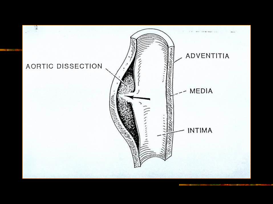
Aortic Dissection
Results from a small tear in the intimal lining of the artery, allowing blood to track between the intima and the media – creating a false leumen.
Most Common: thoracic aorta: 90%mort. Ascending aorta or aortic arch Longitudinal splitting of medial layer Each pulse=dissection continues=Emergency
Risks
Cystic medial necrosis Destruction of medial layer of elastic fibers
HTN Marfan’s Syndrome
Connective tissue disease Pregnancy
Symptoms
Sudden pain in back, chest, or abdomen Ripping or Tearing pain Dyspnea possibly Possible murmur
Ascending aortic dissection usually produces aortic valve insufficiency.
Complications of Dissection
Cardiac Tamponade Blood escapes form dissection into the
pericardial sac.
Pre Op Management
BP and contractility to pulsitile force. Vasodilators (Nipride) and beta blockers (olol)
ECG r/o MI, echocardiogram, aortography. CXR may show wide mediastinum. Manage CHF and Pain. Blood replacement. Stat Surgery: ascending aortic dissection. Descending a.: try meds first if possible.

Post op Care: Synthetic Graft Rpr
BP: semifowlers bedrest, quiet, pain control, antihypertensive meds.
Monitor: ECG, A-line BP, LOC (clots) Observe for: widening pulse pressure-
aortic valve insufficiency. Pulse checks . CPB and vent complications.
Discharge Teaching
Same as CABG No lifting of >5lbs for 4-6wks Sex dysfunction possible due to clamping
of aorta. This flow to penis. Observe feet for color, warmth, swelling. Antihypertensives and neg inotropes. Antibiotics prior to invasive procedures.
c