Embed Size (px)
Citation preview
RESEARCH ARTICLE Open Access
Kidney epithelium specific deletion ofkelch-like ECH-associated protein 1 (Keap1)causes hydronephrosis in miceSanjeev Noel1, Lois J. Arend2, Samatha Bandapalle1, Sekhar P. Reddy3 and Hamid Rabb1,4*
Abstract
Background: Transcription factor Nrf2 protects from experimental acute kidney injury (AKI) and is promising to limitprogression in human chronic kidney disease (CKD) by upregulating multiple antioxidant genes. We recentlydemonstrated that deletion of Keap1, the endogenous inhibitor of Nrf2, in T lymphocytes significantly protects fromAKI. In this study, we investigated the effect of Keap1 deletion on Nrf2 mediated antioxidant response in the renaltubular epithelial cells.
Methods: We deleted Keap1 exon 2 and 3 in the renal tubular epithelial cells by crossing Ksp-Cre mice with Keap1floxed (Keap1f/f) mice. Deletion of Keap1 gene in the kidney epithelial cells of Ksp-Keap1-/- mice and its effect onNrf2 target gene expression was performed using PCR and real-time PCR respectively. Histological evaluation wasperformed on H&E stained sections. Complete blood count, serum and urine analysis were performed to assesssystemic effects of defective kidney development. Student’s T test was used to determine statistical differencebetween the groups.
Results: Ksp-Cre resulted in the deletion of Keap1 exon 2 and 3 and subsequent upregulation of Nrf2 target genes,Nqo1, Gclm and Gclc in the kidney epithelial cells of Ksp-Keap1-/- mice at baseline. Renal epithelial cell specific deletionof Keap1 in Ksp-Keap1-/- mice caused marked renal pelvic expansion and significant compression of medullaryparenchyma consistent with hydronephrosis in both (3 month-old) males and females. Kidneys from 6 month-oldKsp-Keap1-/- mice showed progressive hydronephrosis. Hematological, biochemical and urinary analysis showedsignificantly higher red blood cell count (p = 0.04), hemoglobin (p = 0.01), hematocrit (p = 0.02), mean cell volume(p = 0.02) and mean cell hemoglobin concentration (p = 0.003) in Ksp-Keap1-/- mice in comparison to Keap1f/f mice.
Conclusions: These unexpected findings demonstrate that Keap1 deletion in renal tubular epithelial cells results in anabnormal kidney development consistent with hydronephrosis and reveals a novel Keap1 mediated signaling pathwayin renal development.
Keywords: Keap1-Nrf2 pathway, Hydronephrosis, Kidney epithelial cells, Kidney development
BackgroundThe Keap1-Nrf2 cytoprotective response is critical tocombat reactive oxygen species (ROS) and electrophilesgenerated during endogenous and exogenous stresses[1–3]. Keap1 (Kelch-like ECH-associated protein 1) is arepressor protein that regulates transcriptional activityof Nrf2 (nuclear factor erythroid 2-related factor 2) by
retaining it in the cytoplasm and maintaining its homeo-static level by directing it to proteasomal degradation[4–6]. However, during stress conditions, such as ische-mic and nephrotoxic injury, Nrf2 is released in to thenucleus to up regulate the transcription of cytoprotectivegenes. An insufficient Nrf2 activity has been shown toworsen ischemia induced kidney injury and acceleratedisease progression largely due to an attenuated antioxi-dant response [1, 7]. Nrf2 levels have also been shown todecrease with ageing and correlate with the progressionof many human diseases [8].
* Correspondence: [email protected] of Medicine, Johns Hopkins University, Baltimore, MD, USA4Division of Nephrology, Department of Medicine, Johns Hopkins University,Ross 965 720 Rutland Avenue, Baltimore, MD 21205, USAFull list of author information is available at the end of the article
© 2016 The Author(s). Open Access This article is distributed under the terms of the Creative Commons Attribution 4.0International License (http://creativecommons.org/licenses/by/4.0/), which permits unrestricted use, distribution, andreproduction in any medium, provided you give appropriate credit to the original author(s) and the source, provide a link tothe Creative Commons license, and indicate if changes were made. The Creative Commons Public Domain Dedication waiver(http://creativecommons.org/publicdomain/zero/1.0/) applies to the data made available in this article, unless otherwise stated.
Noel et al. BMC Nephrology (2016) 17:110 DOI 10.1186/s12882-016-0310-y
Attempts to up regulate global Nrf2 levels have beendifficult because homozygous knock out of Keap1 geneis lethal. Whole body Keap1-/- mice do not survive be-yond 21 days postnatal due to progressive asthenia as aresult of hyperkeratosis of esophagus and forestomach[9]. However, recent use of Cre-LoxP technology hasfacilitated researchers to up regulate Nrf2 activity in atissue specific manner [10]. We recently generatedmice with increased Nrf2 activity in T lymphocyte bygenetically deleting Keap1 and found these mice to besignificantly protected from ischemia reperfusion inducedAKI [11].In the present study we deleted Keap1 in renal epithe-
lial cells, specifically in the distal convoluted tubules andcollecting ducts, primarily to accomplish kidney epithelialcell specific up regulation of Nrf2 mediated antioxidantresponse and to study its effect on ischemic kidney injury.Surprisingly, renal epithelial cell specific deletion of Keap1
resulted in significant developmental defects in the col-lecting system. These anatomical defects were also ac-companied by polycythemia. In summary, these datademonstrate that Keap1 may be involved in normal kid-ney development and that a defective Keap1 results inhydronephrosis.
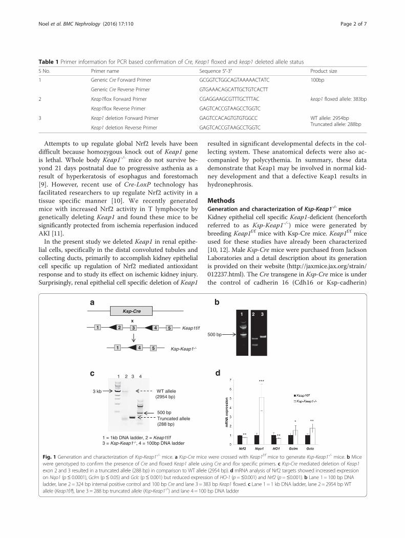
MethodsGeneration and characterization of Ksp-Keap1-/- miceKidney epithelial cell specific Keap1-deficient (henceforthreferred to as Ksp-Keap1-/-) mice were generated bybreeding Keap1f/f mice with Ksp-Cre mice. Keap1f/f miceused for these studies have already been characterized[10, 12]. Male Ksp-Cre mice were purchased from JacksonLaboratories and a detail description about its generationis provided on their website (http://jaxmice.jax.org/strain/012237.html). The Cre transgene in Ksp-Cre mice is underthe control of cadherin 16 (Cdh16 or Ksp-cadherin)
Table 1 Primer information for PCR based confirmation of Cre, Keap1 floxed and keap1 deleted allele status
S No. Primer name Sequence 5′-3′ Product size
1 Generic Cre Forward Primer GCGGTCTGGCAGTAAAAACTATC 100bp
Generic Cre Reverse Primer GTGAAACAGCATTGCTGTCACTT
2 Keap1flox Forward Primer CGAGGAAGCGTTTGCTTTAC keap1 floxed allele: 383bp
Keap1flox Reverse Primer GAGTCACCGTAAGCCTGGTC
3 Keap1 deletion Forward Primer GAGTCCACAGTGTGTGGCC WT allele: 2954bpTruncated allele: 288bp
Keap1 deletion Reverse Primer GAGTCACCGTAAGCCTGGTC
a b
Keap1f/f
Ksp-Keap1-/-
Ksp-Cre
2 3 51 4
4 51
x1 2 3
3 kb
1 2 3 4
1 = 1kb DNA ladder, 2 = Keap1f/f3 = Ksp-Keap1-/-, 4 = 100bp DNA ladder
500 bp
WT allele(2954 bp)
Truncated allele(288 bp)
c d
500 bp
** **
**
***
*
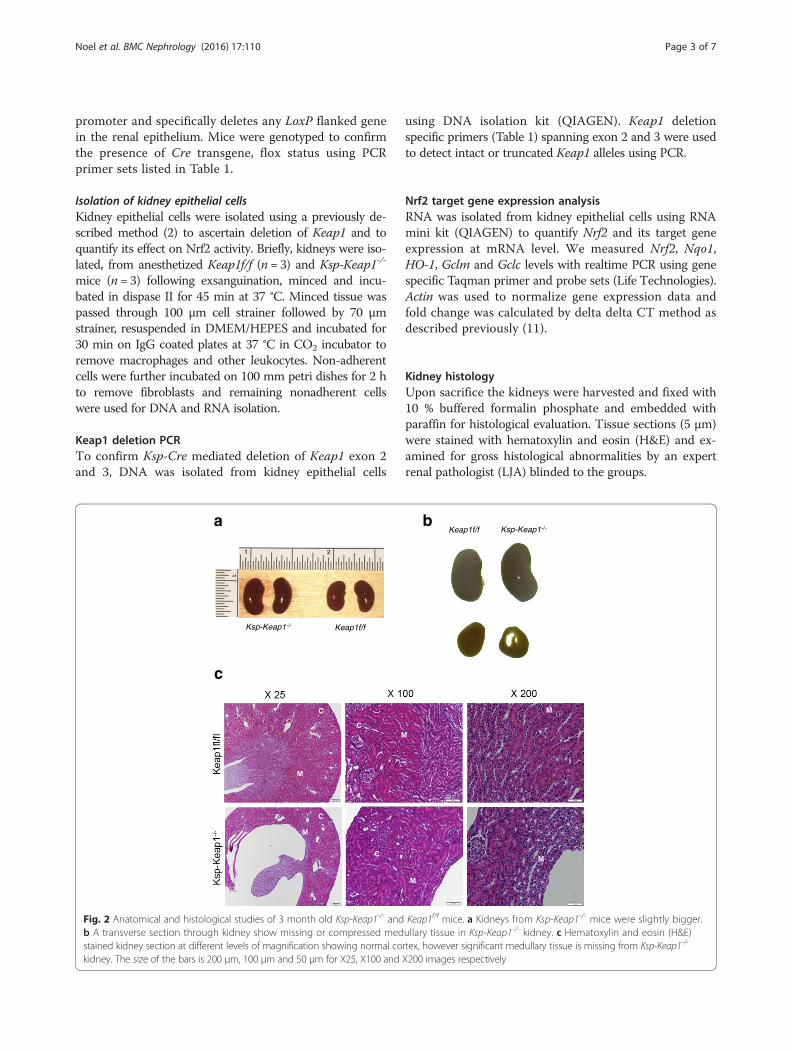
Fig. 1 Generation and characterization of Ksp-Keap1-/- mice. a Ksp-Cre mice were crossed with Keap1f/f mice to generate Ksp-Keap1-/- mice. b Micewere genotyped to confirm the presence of Cre and floxed Keap1 allele using Cre and flox specific primers. c Ksp-Cre mediated deletion of Keap1exon 2 and 3 resulted in a truncated allele (288 bp) in comparison to WT allele (2954 bp). d mRNA analysis of Nrf2 targets showed increased expressionon Nqo1 (p≤ 0.0001), Gclm (p≤ 0.05) and Gclc (p≤ 0.001) but reduced expression of HO-1 (p =≤0.001) and Nrf2 (p=≤0.001). b Lane 1 = 100 bp DNAladder, lane 2 = 324 bp internal positive control and 100 bp Cre and lane 3 = 383 bp Keap1 floxed. c Lane 1 = 1 kb DNA ladder, lane 2 = 2954 bp WTallele (Keap1f/f), lane 3 = 288 bp truncated allele (Ksp-Keap1-/-) and lane 4 = 100 bp DNA ladder
Noel et al. BMC Nephrology (2016) 17:110 Page 2 of 7
promoter and specifically deletes any LoxP flanked genein the renal epithelium. Mice were genotyped to confirmthe presence of Cre transgene, flox status using PCRprimer sets listed in Table 1.
Isolation of kidney epithelial cellsKidney epithelial cells were isolated using a previously de-scribed method (2) to ascertain deletion of Keap1 and toquantify its effect on Nrf2 activity. Briefly, kidneys were iso-lated, from anesthetized Keap1f/f (n = 3) and Ksp-Keap1-/-
mice (n = 3) following exsanguination, minced and incu-bated in dispase II for 45 min at 37 °C. Minced tissue waspassed through 100 μm cell strainer followed by 70 μmstrainer, resuspended in DMEM/HEPES and incubated for30 min on IgG coated plates at 37 °C in CO2 incubator toremove macrophages and other leukocytes. Non-adherentcells were further incubated on 100 mm petri dishes for 2 hto remove fibroblasts and remaining nonadherent cellswere used for DNA and RNA isolation.
Keap1 deletion PCRTo confirm Ksp-Cre mediated deletion of Keap1 exon 2and 3, DNA was isolated from kidney epithelial cells
using DNA isolation kit (QIAGEN). Keap1 deletionspecific primers (Table 1) spanning exon 2 and 3 were usedto detect intact or truncated Keap1 alleles using PCR.
Nrf2 target gene expression analysisRNA was isolated from kidney epithelial cells using RNAmini kit (QIAGEN) to quantify Nrf2 and its target geneexpression at mRNA level. We measured Nrf2, Nqo1,HO-1, Gclm and Gclc levels with realtime PCR using genespecific Taqman primer and probe sets (Life Technologies).Actin was used to normalize gene expression data andfold change was calculated by delta delta CT method asdescribed previously (11).
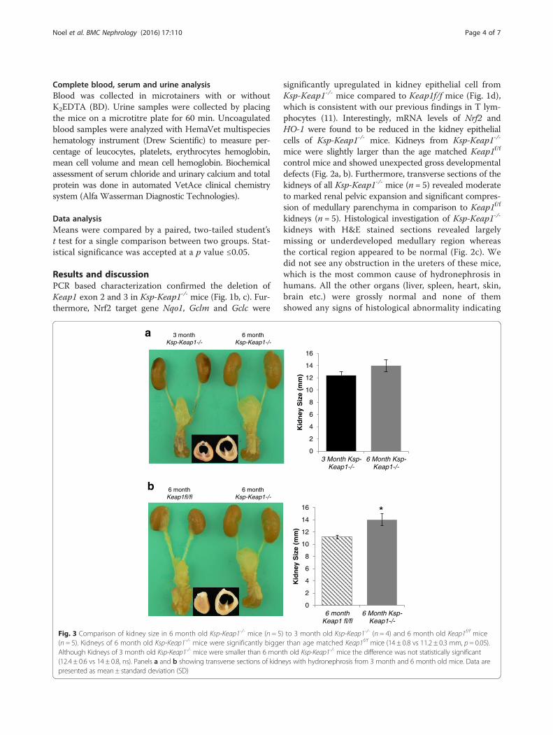
Kidney histologyUpon sacrifice the kidneys were harvested and fixed with10 % buffered formalin phosphate and embedded withparaffin for histological evaluation. Tissue sections (5 μm)were stained with hematoxylin and eosin (H&E) and ex-amined for gross histological abnormalities by an expertrenal pathologist (LJA) blinded to the groups.
Keap1f/f Ksp-Keap1-/-a b
c
Keap1f/fKsp-Keap1-/-
C
C
C
C
M
M
M
M
M
M
Fig. 2 Anatomical and histological studies of 3 month old Ksp-Keap1-/- and Keap1f/f mice. a Kidneys from Ksp-Keap1-/- mice were slightly bigger.b A transverse section through kidney show missing or compressed medullary tissue in Ksp-Keap1-/- kidney. c Hematoxylin and eosin (H&E)stained kidney section at different levels of magnification showing normal cortex, however significant medullary tissue is missing from Ksp-Keap1-/-
kidney. The size of the bars is 200 μm, 100 μm and 50 μm for X25, X100 and X200 images respectively
Noel et al. BMC Nephrology (2016) 17:110 Page 3 of 7
Complete blood, serum and urine analysisBlood was collected in microtainers with or withoutK2EDTA (BD). Urine samples were collected by placingthe mice on a microtitre plate for 60 min. Uncoagulatedblood samples were analyzed with HemaVet multispecieshematology instrument (Drew Scientific) to measure per-centage of leucocytes, platelets, erythrocytes hemoglobin,mean cell volume and mean cell hemoglobin. Biochemicalassessment of serum chloride and urinary calcium and totalprotein was done in automated VetAce clinical chemistrysystem (Alfa Wasserman Diagnostic Technologies).
Data analysisMeans were compared by a paired, two-tailed student’st test for a single comparison between two groups. Stat-istical significance was accepted at a p value ≤0.05.
Results and discussionPCR based characterization confirmed the deletion ofKeap1 exon 2 and 3 in Ksp-Keap1-/- mice (Fig. 1b, c). Fur-thermore, Nrf2 target gene Nqo1, Gclm and Gclc were
significantly upregulated in kidney epithelial cell fromKsp-Keap1-/- mice compared to Keap1f/f mice (Fig. 1d),which is consistent with our previous findings in T lym-phocytes (11). Interestingly, mRNA levels of Nrf2 andHO-1 were found to be reduced in the kidney epithelialcells of Ksp-Keap1-/- mice. Kidneys from Ksp-Keap1-/-
mice were slightly larger than the age matched Keap1f/f
control mice and showed unexpected gross developmentaldefects (Fig. 2a, b). Furthermore, transverse sections of thekidneys of all Ksp-Keap1-/- mice (n = 5) revealed moderateto marked renal pelvic expansion and significant compres-sion of medullary parenchyma in comparison to Keap1f/f
kidneys (n = 5). Histological investigation of Ksp-Keap1-/-
kidneys with H&E stained sections revealed largelymissing or underdeveloped medullary region whereasthe cortical region appeared to be normal (Fig. 2c). Wedid not see any obstruction in the ureters of these mice,which is the most common cause of hydronephrosis inhumans. All the other organs (liver, spleen, heart, skin,brain etc.) were grossly normal and none of themshowed any signs of histological abnormality indicating
0
2
4
6
8
10
12
14
16
6 monthKeap1 fl/fl
6 Month Ksp-Keap1-/-
Kid
ney
Siz
e (m
m)
3 month Ksp-Keap1-/-
a
b
6 month Ksp-Keap1-/-
6 month Ksp-Keap1-/-
6 month Keap1fl/fl
*
0
2
4
6
8
10
12
14
16
3 Month Ksp-Keap1-/-
6 Month Ksp-Keap1-/-
Kid
ney
Siz
e (m
m)
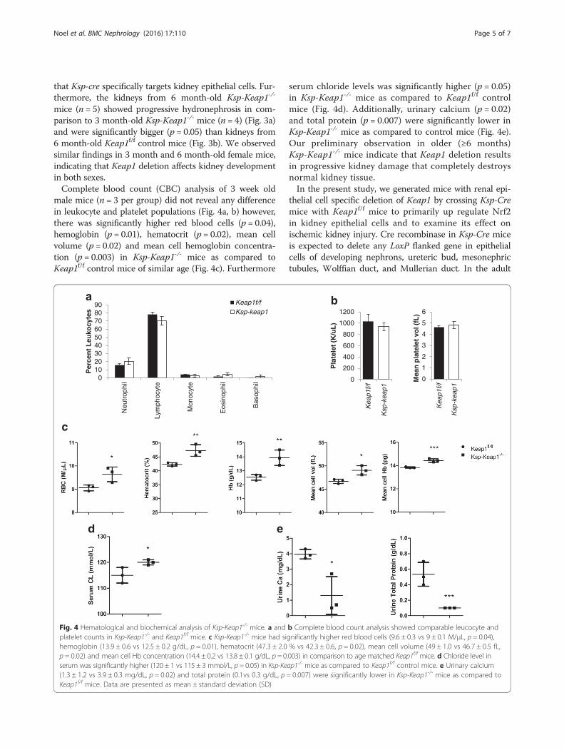
Fig. 3 Comparison of kidney size in 6 month old Ksp-Keap1-/- mice (n = 5) to 3 month old Ksp-Keap1-/- (n = 4) and 6 month old Keap1f/f mice(n = 5). Kidneys of 6 month old Ksp-Keap1-/- mice were significantly bigger than age matched Keap1f/f mice (14 ± 0.8 vs 11.2 ± 0.3 mm, p = 0.05).Although Kidneys of 3 month old Ksp-Keap1-/- mice were smaller than 6 month old Ksp-Keap1-/- mice the difference was not statistically significant(12.4 ± 0.6 vs 14 ± 0.8, ns). Panels a and b showing transverse sections of kidneys with hydronephrosis from 3 month and 6 month old mice. Data arepresented as mean ± standard deviation (SD)
Noel et al. BMC Nephrology (2016) 17:110 Page 4 of 7
that Ksp-cre specifically targets kidney epithelial cells. Fur-thermore, the kidneys from 6 month-old Ksp-Keap1-/-
mice (n = 5) showed progressive hydronephrosis in com-parison to 3 month-old Ksp-Keap1-/- mice (n = 4) (Fig. 3a)and were significantly bigger (p = 0.05) than kidneys from6 month-old Keap1f/f control mice (Fig. 3b). We observedsimilar findings in 3 month and 6 month-old female mice,indicating that Keap1 deletion affects kidney developmentin both sexes.Complete blood count (CBC) analysis of 3 week old
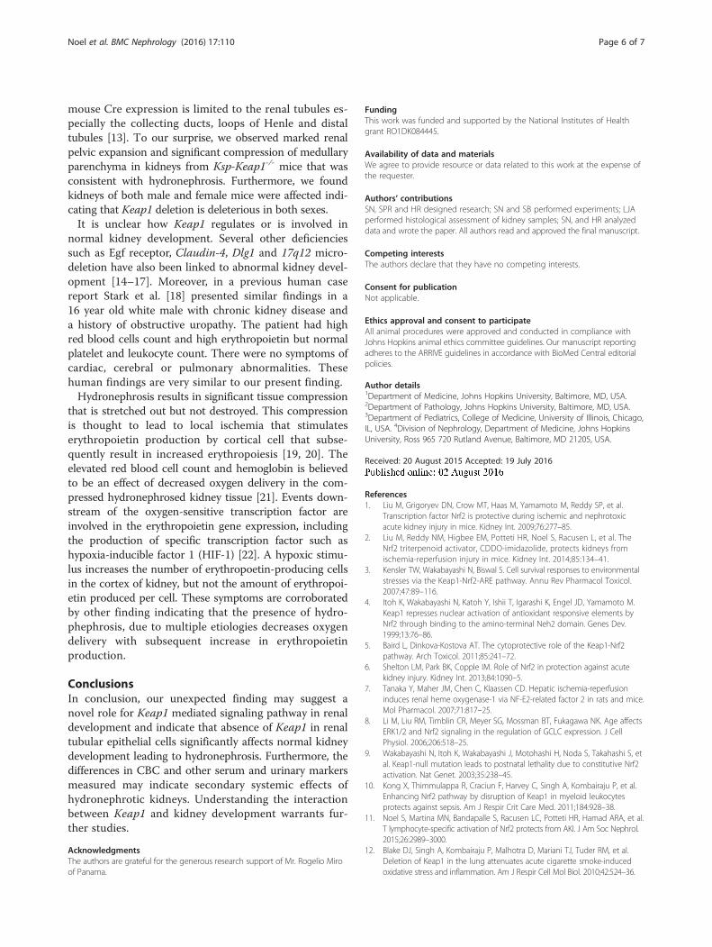
male mice (n = 3 per group) did not reveal any differencein leukocyte and platelet populations (Fig. 4a, b) however,there was significantly higher red blood cells (p = 0.04),hemoglobin (p = 0.01), hematocrit (p = 0.02), mean cellvolume (p = 0.02) and mean cell hemoglobin concentra-tion (p = 0.003) in Ksp-Keap1-/- mice as compared toKeap1f/f control mice of similar age (Fig. 4c). Furthermore
serum chloride levels was significantly higher (p = 0.05)in Ksp-Keap1-/- mice as compared to Keap1f/f controlmice (Fig. 4d). Additionally, urinary calcium (p = 0.02)and total protein (p = 0.007) were significantly lower inKsp-Keap1-/- mice as compared to control mice (Fig. 4e).Our preliminary observation in older (≥6 months)Ksp-Keap1-/- mice indicate that Keap1 deletion resultsin progressive kidney damage that completely destroysnormal kidney tissue.In the present study, we generated mice with renal epi-
thelial cell specific deletion of Keap1 by crossing Ksp-Cremice with Keap1f/f mice to primarily up regulate Nrf2in kidney epithelial cells and to examine its effect onischemic kidney injury. Cre recombinase in Ksp-Cre miceis expected to delete any LoxP flanked gene in epithelialcells of developing nephrons, ureteric bud, mesonephrictubules, Wolffian duct, and Mullerian duct. In the adult
0102030405060708090
Neu
trop
hil
Lym
phoc
yte
Mon
ocyt
e
Eos
inop
hil
Bas
ophi
l
Per
cen
t L
euko
cyte
s
Keap1f/fKsp-keap1
0
200
400
600
800
1000
1200
Kea
p1f/f
Ksp
-kea
p1
Pla
tele
t (K
/uL
)
0
1
2
3
4
5
6
Kea
p1f/f
Ksp
-kea
p1
Mea
n p
late
let
vol (
fL)
a b
c
d e
Fig. 4 Hematological and biochemical analysis of Ksp-Keap1-/- mice. a and b Complete blood count analysis showed comparable leucocyte andplatelet counts in Ksp-Keap1-/- and Keap1f/f mice. c Ksp-Keap1-/- mice had significantly higher red blood cells (9.6 ± 0.3 vs 9 ± 0.1 M/μL, p = 0.04),hemoglobin (13.9 ± 0.6 vs 12.5 ± 0.2 g/dL, p = 0.01), hematocrit (47.3 ± 2.0 % vs 42.3 ± 0.6, p = 0.02), mean cell volume (49 ± 1.0 vs 46.7 ± 0.5 fL,p = 0.02) and mean cell Hb concentration (14.4 ± 0.2 vs 13.8 ± 0.1 g/dL, p = 0.003) in comparison to age matched Keap1f/f mice. d Chloride level inserum was significantly higher (120 ± 1 vs 115 ± 3 mmol/L, p = 0.05) in Ksp-Keap1-/- mice as compared to Keap1f/f control mice. e Urinary calcium(1.3 ± 1.2 vs 3.9 ± 0.3 mg/dL, p = 0.02) and total protein (0.1vs 0.3 g/dL, p = 0.007) were significantly lower in Ksp-Keap1-/- mice as compared toKeap1f/f mice. Data are presented as mean ± standard deviation (SD)
Noel et al. BMC Nephrology (2016) 17:110 Page 5 of 7
mouse Cre expression is limited to the renal tubules es-pecially the collecting ducts, loops of Henle and distaltubules [13]. To our surprise, we observed marked renalpelvic expansion and significant compression of medullaryparenchyma in kidneys from Ksp-Keap1-/- mice that wasconsistent with hydronephrosis. Furthermore, we foundkidneys of both male and female mice were affected indi-cating that Keap1 deletion is deleterious in both sexes.It is unclear how Keap1 regulates or is involved in
normal kidney development. Several other deficienciessuch as Egf receptor, Claudin-4, Dlg1 and 17q12 micro-deletion have also been linked to abnormal kidney devel-opment [14–17]. Moreover, in a previous human casereport Stark et al. [18] presented similar findings in a16 year old white male with chronic kidney disease anda history of obstructive uropathy. The patient had highred blood cells count and high erythropoietin but normalplatelet and leukocyte count. There were no symptoms ofcardiac, cerebral or pulmonary abnormalities. Thesehuman findings are very similar to our present finding.Hydronephrosis results in significant tissue compression
that is stretched out but not destroyed. This compressionis thought to lead to local ischemia that stimulateserythropoietin production by cortical cell that subse-quently result in increased erythropoiesis [19, 20]. Theelevated red blood cell count and hemoglobin is believedto be an effect of decreased oxygen delivery in the com-pressed hydronephrosed kidney tissue [21]. Events down-stream of the oxygen-sensitive transcription factor areinvolved in the erythropoietin gene expression, includingthe production of specific transcription factor such ashypoxia-inducible factor 1 (HIF-1) [22]. A hypoxic stimu-lus increases the number of erythropoetin-producing cellsin the cortex of kidney, but not the amount of erythropoi-etin produced per cell. These symptoms are corroboratedby other finding indicating that the presence of hydro-phephrosis, due to multiple etiologies decreases oxygendelivery with subsequent increase in erythropoietinproduction.
ConclusionsIn conclusion, our unexpected finding may suggest anovel role for Keap1 mediated signaling pathway in renaldevelopment and indicate that absence of Keap1 in renaltubular epithelial cells significantly affects normal kidneydevelopment leading to hydronephrosis. Furthermore, thedifferences in CBC and other serum and urinary markersmeasured may indicate secondary systemic effects ofhydronephrotic kidneys. Understanding the interactionbetween Keap1 and kidney development warrants fur-ther studies.
AcknowledgmentsThe authors are grateful for the generous research support of Mr. Rogelio Miroof Panama.
FundingThis work was funded and supported by the National Institutes of Healthgrant RO1DK084445.
Availability of data and materialsWe agree to provide resource or data related to this work at the expense ofthe requester.
Authors’ contributionsSN, SPR and HR designed research; SN and SB performed experiments; LJAperformed histological assessment of kidney samples; SN, and HR analyzeddata and wrote the paper. All authors read and approved the final manuscript.
Competing interestsThe authors declare that they have no competing interests.
Consent for publicationNot applicable.
Ethics approval and consent to participateAll animal procedures were approved and conducted in compliance withJohns Hopkins animal ethics committee guidelines. Our manuscript reportingadheres to the ARRIVE guidelines in accordance with BioMed Central editorialpolicies.
Author details1Department of Medicine, Johns Hopkins University, Baltimore, MD, USA.2Department of Pathology, Johns Hopkins University, Baltimore, MD, USA.3Department of Pediatrics, College of Medicine, University of Illinois, Chicago,IL, USA. 4Division of Nephrology, Department of Medicine, Johns HopkinsUniversity, Ross 965 720 Rutland Avenue, Baltimore, MD 21205, USA.
Received: 20 August 2015 Accepted: 19 July 2016
References1. Liu M, Grigoryev DN, Crow MT, Haas M, Yamamoto M, Reddy SP, et al.
Transcription factor Nrf2 is protective during ischemic and nephrotoxicacute kidney injury in mice. Kidney Int. 2009;76:277–85.
2. Liu M, Reddy NM, Higbee EM, Potteti HR, Noel S, Racusen L, et al. TheNrf2 triterpenoid activator, CDDO-imidazolide, protects kidneys fromischemia-reperfusion injury in mice. Kidney Int. 2014;85:134–41.
3. Kensler TW, Wakabayashi N, Biswal S. Cell survival responses to environmentalstresses via the Keap1-Nrf2-ARE pathway. Annu Rev Pharmacol Toxicol.2007;47:89–116.
4. Itoh K, Wakabayashi N, Katoh Y, Ishii T, Igarashi K, Engel JD, Yamamoto M.Keap1 represses nuclear activation of antioxidant responsive elements byNrf2 through binding to the amino-terminal Neh2 domain. Genes Dev.1999;13:76–86.
5. Baird L, Dinkova-Kostova AT. The cytoprotective role of the Keap1-Nrf2pathway. Arch Toxicol. 2011;85:241–72.
6. Shelton LM, Park BK, Copple IM. Role of Nrf2 in protection against acutekidney injury. Kidney Int. 2013;84:1090–5.
7. Tanaka Y, Maher JM, Chen C, Klaassen CD. Hepatic ischemia-reperfusioninduces renal heme oxygenase-1 via NF-E2-related factor 2 in rats and mice.Mol Pharmacol. 2007;71:817–25.
8. Li M, Liu RM, Timblin CR, Meyer SG, Mossman BT, Fukagawa NK. Age affectsERK1/2 and Nrf2 signaling in the regulation of GCLC expression. J CellPhysiol. 2006;206:518–25.
9. Wakabayashi N, Itoh K, Wakabayashi J, Motohashi H, Noda S, Takahashi S, etal. Keap1-null mutation leads to postnatal lethality due to constitutive Nrf2activation. Nat Genet. 2003;35:238–45.
10. Kong X, Thimmulappa R, Craciun F, Harvey C, Singh A, Kombairaju P, et al.Enhancing Nrf2 pathway by disruption of Keap1 in myeloid leukocytesprotects against sepsis. Am J Respir Crit Care Med. 2011;184:928–38.
11. Noel S, Martina MN, Bandapalle S, Racusen LC, Potteti HR, Hamad ARA, et al.T lymphocyte-specific activation of Nrf2 protects from AKI. J Am Soc Nephrol.2015;26:2989–3000.
12. Blake DJ, Singh A, Kombairaju P, Malhotra D, Mariani TJ, Tuder RM, et al.Deletion of Keap1 in the lung attenuates acute cigarette smoke-inducedoxidative stress and inflammation. Am J Respir Cell Mol Biol. 2010;42:524–36.
Noel et al. BMC Nephrology (2016) 17:110 Page 6 of 7
13. Shao X, Somlo S, Igarashi P. Epithelial-specific Cre/lox recombination in thedeveloping kidney and genitourinary tract. J Am Soc Nephrol. 2002;13:1837–46.
14. Zhang Z, Pascuet E, Hueber PA, Chu L, Bichet DG, Lee TC, et al. Targetedinactivation of EGF receptor inhibits renal collecting duct development andfunction. J Am Soc Nephrol. 2010;21:573–8.
15. Fujita H, Hamazaki Y, Noda Y, Oshima M, Minato N. Claudin-4 deficiencyresults in urothelial hyperplasia and lethal hydronephrosis. PLoS One.2012;7(12):e52272.
16. Iizuka-Kogo A, Akiyama T, Senda T. Decreased apoptosis and persistence ofthe common nephric duct during the development of an aberrantvesicoureteral junction in Dlg1 genetargeted mice. Anat Rec (Hoboken).2013;296:1936–42.
17. Chen CP, Chang SD, Wang TH, Wang LK, Tsai JD, Liu YP, et al. Detection ofrecurrent transmission of 17q12 microdeletion by array comparative genomichybridization in a fetus with prenatally diagnosed hydronephrosis, hydroureter,and multicystic kidney, and variable clinical spectrum in the family. Taiwan JObstet Gynecol. 2013;52:551–7.
18. Stark S, Winkelmann B, Kluthe C, Roigas J, Querfeld U, Müller D. Polycythemiaand increased erythropoietin in a patient with chronic kidney disease. Nat ClinPract Nephrol. 2007;3:222–6.
19. Toyama K, Mitus WJ. Experimental renal erythrocytosis. 3. Relationshipbetween the degree of hydronephrotic pressure and the production oferythrocytosis. J Lab Clin Med. 1966;68:740–52.
20. Fisher JW, Schofield R, Porteous DD. Effects of renal hypoxia on erythropoietinproduction. Br J Haematol. 1965;11:382–8.
21. Eckardt KU, Kurtz A. Regulation of erythropoietin production. Eur J Clin Invest.2005;35 Suppl 3:13–9.
22. Maxwell PH, Wiesener MS, Chang GW, Clifford SC, Vaux EC, Cockman ME, et al.The tumour suppressor protein VHL targets hypoxia-inducible factors foroxygen-dependentproteolysis. Nature. 1999;399:271–5.
• We accept pre-submission inquiries
• Our selector tool helps you to find the most relevant journal
• We provide round the clock customer support
• Convenient online submission
• Thorough peer review
• Inclusion in PubMed and all major indexing services
• Maximum visibility for your research
Submit your manuscript atwww.biomedcentral.com/submit
Submit your next manuscript to BioMed Central and we will help you at every step:
Noel et al. BMC Nephrology (2016) 17:110 Page 7 of 7