Embed Size (px)
Citation preview

Ki-67 detects a nuclear matrix-associated proliferation-related antigen
I. Intracellular localization during interphase
R. VERHEIJEN1'*, H. J. H. KUIJPERS1, R. O. SCHLINGEMANN1, A. L. M. BOEHMER1,
R. van DRIEL2, G. J. BRAKENHOFF3 and F. C. S. RAMAEKERS1
1 Department of Pathology, University Hospital of Nijmegen, Geert Crooteplein Zmd 24, 6525 CA Nijmegen, The Netherlands2Department of Biochemistry, University of Amsterdam, Plantage Muidergracht 12, 1018 T\' Amsterdam, The Netherlands^Department of Electron Microscopy and Molecular Cytology, University of Amsterdam, Plantage Muidergracht 14, 1018 7V Amsterdam,The Netherlands
* Author for correspondence
Summary
Ki-67 is a commercially available mouse mono-clonal antibody, 'which reacts with a nuclear anti-gen in proliferating cells. The antibody can be usedto determine the growth fraction of human tumoursin situ and has been shown to be of prognosticimportance.
In this study it is shown that in interphase cellsKi-67 reacts with an antigen, mainly present in thenucleoli. Confocal scanning laser microscopy andimmunoelectron microscopy on human MR65monolayer cells revealed that this nucleolar antigenis predominantly localized in the nucleolar cortexand in the dense fibrillar components.
The Ki-67 antigen appeared to be preserved innuclear matrix preparations obtained after in situfractionation of MR65 cells. Despite many efforts,we could not identify the antigen in immunoblot-ting or immunoprecipitation assays.
Testing of cell cultures of different species bymeans of indirect immunofluorescence revealedthat the antibody reacted 'with human cells and withthe Rhesus monkey kidney-derived cell line LLC-MK2.
Key words: nucleolar antigen, confocal scanning lasermicroscopy, immunocytochemistry.
Introduction
In 1983 Gerdes and co-workers described the productionof a mouse monoclonal antibody, Ki-67, which recog-nized a nuclear antigen expressed in proliferating cells,but not in resting cells. Ki-67 was obtained from a fusionof mouse myelomas with lymphocytes from a mouseinjected with crude nuclear fractions of L428 cells, aHodgkin's disease-derived cell line (Gerdes et al. 1983).It was found that normal peripheral blood lymphocyteswere negative for Ki-67, while stimulation of these cellswith phytohaemagglutinin (PHA) resulted in a positivenuclear reaction pattern. On the contrary, when HL-60cells (acute promyelocytic leukaemia) were stimulated todifferentiate into mature resting macrophages by treat-ment with the phorbol ester TPA, the Ki-67 antigenexpression disappeared.
A more detailed analysis of the cell cycle performed byGerdes et al. (1984a) revealed that expression of thisantigen is consistently detectable throughout the S, G2and M phases of continuously cycling cells but not in Gocells. However, the results concerning antigen expression
Journal of Cell Science 92, 123-130 (1989)Printed in Great Britain © The Company of Biologists Limited 1989
in Gi phase varied. In these early studies it was alsonoted that Ki-67 reacted with an antigen associated withchromosomes (Gerdes et al. 1984«). Expression of theKi-67 antigen could not be demonstrated in peripheralblood leucocytes during the early events of PHA-trig-gered transition from Go to Gj , whereas continuouslycycling cells were permanently positive in Gi phase.These observations led to the conclusion that Ki-67 couldbe a useful tool in determining the growth fraction of agiven human cell population and especially in assessingthe proportion of proliferating cells in immunostainedtissue sections of neoplasms (Gerdes et al. 1983, 1984a).This latter aspect has been explicitly examined for non-Hodgkin's lymphomas (Gerdes et al. 19846), the colonicepithelium in ulcerative colitis (Franklin et al. 1985), andfor tumours of breast (Gerdes et al. 1986; Barnard et al.1987; Franklin et al. 1987), lung (Gatter et al. 1986) andbrain (Giangaspero et al. 1987). Barnard et al. (1987)have determined a Ki-67 score (positive cells/total tu-mour cells) for a number of primary breast carcinomasand investigated the possible relationship betweenthis proliferative index and a number of clinical and
123

pathological parameters. These investigators concludedthat the Ki-67 score may prove to be an objectiveindicator of biological behaviour of breast carcinomas andthus may be of clinical significance. Ki-67 has also beenshown to be of great value in assessing the proliferativecapacities of the lymphoid cells in cutaneous infiltrates(Ralfkiaer et al. 1986) and in determining the growthfraction of tumour cells in tissues affected by Hodgkin'sdisease (Gerdes et al. 1987).
Recently, Schwarting et al. (1986) have shown that thegrowth fraction of cell suspensions labelled with Ki-67can be determined flow cytometrically. Furthermore,Franklin et al. (1987) have shown the usefulness of animage analysis system in quantifying the immunoperoxi-dase Ki-67 labelling in tissue sections of breast carcino-mas.
In summary, the estimation of the Ki-67 positive cellfraction of tumours is being introduced more and moreinto routine pathology and in future may have importantprognostic and therapeutic implications. On the otherhand, virtually nothing is known about the nature andbiochemical characteristics of the antigen recognized byKi-67. The present study was performed to examine theintracellular localization of the Ki-67 antigen. In thisrespect it is shown that during interphase the strongestimmunohistochemical staining reactions with Ki-67 areobtained in the nucleoli and that the antigen is preservedin nuclear matrix preparations of MR65 cells.
Materials and methods
AntibodiesMonoclonal antibody Ki-67 (IgGl; trade-name DAKO-PC,code no. M722) was purchased from DAKOpatts (Glostrup,Denmark) as tissue culture supernatant that had been dialysedagainst 0-05 M-Tris-HCl, pH7-2 and 15mM-sodium azide.
Ascites fluid obtained after intraperitoneal injection of Ki-67hybridoma cells in Balb/c mice was kindly provided by Dr J.Gerdes (Borstel, FRG).
Other monoclonal antibodies used in this study include 2.73,41CC4 and RKSE 60. Antibody 2.73, directed against the 70K(K = 103iWr) Ul RNA-associated protein was provided by DrS. Hoch (La Jolla, USA) and has been described (Billings et al.1982; Verheijen et al. 1986a,6). Antibody 41CC4, directedagainst the nuclear lamins, was a kind gift from Dr G. Warren(Heidelberg, FRG) and has also been documented (Burke et al.1983; Verheijen et al. 1986a,fc). RKSE 60 is an antibodydirected against cytokeratin 10 (Ramaekers et al. 1983). Sincethis protein is not expressed in MR65 cells, RKSE 60 was usedas a negative control antibody in these cells.
Cell culturesTissue-culture media and calf sera were purchased from FlowLaboratories Ltd, Irvine, UK.
Monolayer cells were grown on coverslips in Eagle's modifiedMinimum Essential Medium supplemented with 10 % newborncalf serum until about 50% confluency was reached. Theseincluded: HeLa S3 (human cervix carcinoma), MR65 (humanlung carcinoma; Broers et al. 1987), T24 (human bladdertransitional-cell carcinoma), HEp 2 (human epidermoid larynxcarcinoma), Cloll (human melanoma cell culture), culturedhuman fibroblaats, PtK2 (Potorous tridactylus kidney), BHK-21 (baby Syrian hamster kidney cells), VERO (African
green monkey kidney cells), LLC-MK2 derivative (Rhesusmonkey kidney cells), a primary culture of dog endothelial cells,a hamster lens cell culture (Bloemendal et al. 1980) and a bovinelens cell culture (Ramaekers et al. 1980).
Molt-4 (human acute T lymphoblastic leukaemia) and mousemyeloma SP2/0-Agl4 cells were grown in suspension at 37°C atdensities of approximately 0-3 X10⁶ cells ml"1 in RPMI 1640(Dutch modification) supplemented with 15 % foetal calfserum. Drosophila melanogaster mei-218 cells were cultured at25 °C at about 75% confluency in Schneider's Dmsopliilamedium (Gibco, UK) supplemented with 15 % heat-inacti-vated foetal calf serum.
Immunohistochemical staining proceduresThe indirect immunofluorescence assay was performed essen-tially as described by Verheijen et al. (19866). Culture super-natant of the mouse monoclonal antibody Ki-67 was used in a1:25 (v/v) dilution. As second antibody FITC-conjugatedrabbit anti-mouse IgG (heavy and light chains; Nordic, Til-burg, The Netherlands) was used in a 1:25 (v/v) dilution. Alldilutions were made in phosphate-buffered saline (PBS). DNAwas then stained by incubating the cells for 15 min with Hoechst
in 22mM-citric acid, 56 mM-disodium hydro-y
33258 (0-1gen phosphate).
Immunoelectron microscopyFor immunoelectron microscopy, MR65 cells were grown onMelinex foil (ICI, Hertz, UK) until about 75 % confluency wasreached. The cells were washed in PBS and fixed in methanol(-20°C, 5 s), followed by dipping in acetone at room tempera-ture (3 times, 5 s). Subsequently, the cells were incubated with1:20 (v/v) diluted culture supernatant of Ki-67 for 60 min atroom temperature, washed with three changes of PBS for10 min each, incubated with 1:50 (v/v) diluted peroxidase-conjugated rabbit anti-mouse Ig antibodies (DAKOpatts, Glos-trup, Denmark) and again washed in PBS (3 times, 10min).After detection of the peroxidase activity with 3,3'-diaminoben-zidine (DAB; Sigma Chemical Co., Miinchen, FRG) the slideswere processed for electron microscopy as described by vanDuineneta/. (1984).
In situ preparation of nuclear matrices from MR65 cellsCell fractionations were performed in the presence of 0-5 mM-phenylmethylsulphonyl chloride (PMSC) and 5 mM-A'-ethyl-maleimide (MalNEt) to reduce proteolytic degradation anddisulphide bridge formation, respectively. These agents wereadded from freshly prepared stocks. Ribonuclease A (RNase A)(Sigma Chemical Co., Miinchen, FRG) was pre-incubated for5 min at 100°C to reduce possible protease activity.
MR65 cells were grown on coverslips under appropriateculture conditions (Verheijen et al. 19866) until about 75 %confluency. The entire fractionation procedure was carried outunder continuous shaking of the slides in six-well plates.
The procedure that we have established for the isolation ofnuclear matrices in situ, carried out at 0-4°C, was as follows:cells were first washed in PBS for 5 min followed by threewashes in NKM buffer (130mM-NaCl, 5mM-KCl, 1-SmM-MgCl2), 5 min each. Each of the following steps in theprocedure was preceded by washing the slides twice withreticulocyte suspension buffer (RSB) (lOmM-NaCl, 10mM-Tris-HCl, pH7-4, 1-5 mM-MgCl2). Subsequently, the slideswere subjected to a hypertonic buffer (RSB with 0-3 M-sucrose)and incubated for 10 min after addition of 0-05 vol. 10 % TritonX-100 in RSB. Thereafter, the slides were incubated for 10minin a freshly prepared mixture of 0-5 % sodium deoxycholate(DOC)/1-0% Tween-40 in RSB, followed by a nucleic acid
124 R. Verheijen et al.

digestion for 20min at 20°C in a mixture of lmgmldeoxyribonuclease I (DNase I) (DPFF quality; Cooper Bio-medical, Malvern, USA) and 50^gml~' RNase A (Sigma) inRSB110 (HOmM-NaCl, 10mM-Tris-HCl, pH7-4, 1-SraM-MgClz). During this digestion step MalNEt was omitted, butimmediately after the incubation it was added again to a finalconcentration of 5mM. The DNA-depleted nuclei wereextracted for lOmin with a high-salt buffer containing 0-4 M-(NH4)2SO4, SOmM-Tris-HCl, pH7-4, l-SmM-MgCl2. Theobtained nuclear matrices were prepared for immunofluor-escence microscopy as described (Verheijen et al. 19866).
Analysis of proteins and RNASDS-polyacrylamide gel electrophoresis and immunoblottingwere performed as described (Verheijen et al. 1986a), whileanalysis of immunoprecipitated proteins and RNA using cul-ture supernatant of Ki-67 as well as ascites fluid were done asdocumented by Mimori et al. (1984).
Confocal scanning laser microscopy (CSLM)The confocal scanning laser microscopy technique has beendescribed in detail by Brakenhoff et al. (1985, 1988) and Vander Voort et al. (1988). Usually, 16 optical sections of 256x256pixels each were made per three-dimensional image. Fluor-escein-stained specimens were excited at 476 nm using a kryp-ton ion laser. Typical optical resolutions were O2/tfn and0-8 fim, perpendicular and parallel to the optical axis, respect-ively. The signal-to-noise ratio in the images of the opticalsections was improved by using a two- or three-dimensionalmedian filter (Brakenhoff et al. 1988; Van der Voort et al.1988). Furthermore, the contrast range in each series of sectionswas optimized with respect to the sensitivity of the photo-graphic film.
Actinomycin D treatmentAfter reaching a confluency of about 50%, various coverslipswith MR65 cell monolayers were placed in fresh culturemedium containing 1-0, 2-5 and lO^grnl" actinomycin D(Sharpe and Dohme International, New Jersey, USA), respect-ively. Each culture was permitted to grow for another 1, 2 or4h, after which the cells were prepared for indirect immunoflu-orescence.
Results
Subcellular localization of the Ki-67 antigen in MR65cellsTo study the intracellular localization of the Ki-67antigen in human cells we have used MR65, a humanlung carcinoma monolayer culture in which the cells
contain large nuclei and remain relatively flat duringmitosis. In such cultures the Ki-67 antigen appeared tobe exclusively located in the nuclei of virtually allindividual cells (Fig. 1). However, a considerable varia-bility of the staining intensities between the severalinterphase cells was observed. Besides a weak staining ofthe nucleoplasm, the highest fluorescence intensity wasfound in the nucleoli. The nucleoplasmic reactivity wasrepresented by variable numbers of small discrete struc-tures, while the nucleolar staining patterns appeared tobe very heterogeneous. In some cells fluorescence couldbe seen to be more intense in certain regions at thenucleolar periphery.
The antibiotic actinomycin D preferentially blocksRNA synthesis, resulting in a gradual nucleolar segre-gation, depending on concentration and exposure time.In order to follow the Ki-67 antigen localization duringthis segregation process, we exposed MR65 cell mono-layers for several hours to 1-0, 2-5 and lOjUgml of thedrug. The nucleoli appeared to be completely fragmentedafter 2h at a concentration of 2-5 ^gml"1 actinomycin Dor after 1 h at a concentration of 10/igml ' as concludedfrom phase-contrast microscopic observations (notshown). The micrographs in Fig. 2 show the Ki-67antigen distribution patterns at several stages of actino-mycin D treatment. As the nucleoli gradually dispersedinto a great number of small fragments, the distributionof the Ki-67 antigen simultaneously changed from adistinct nucleolar localization (Fig. 2A) via a speckledpattern (Fig. 2B,C) to a diffuse distribution throughoutthe entire nucleoplasm (Fig. 2D). Fig. 2B represents thesituation in which the fibrillar and granular componentsof the nucleoli have been segregated and in which theantigen seems to be associated with one of these twodeveloped subcompartments.
Confocal scanning laser microscopy (CSLM) wasapplied on MR65 cells to extend our indirect immunoflu-orescence data and to obtain more information about theprecise, spatial localization of the Ki-67 antigen in thenucleolus. Fig. 3 shows the image obtained from aCSLM analysis after superposition of 16 optical planes(all 0-85 /xm apart)from two nuclei of MR65 cells inpseudocolour to extend the contrast range in the pictures.The colour sequence purple, blue, orange, red and whiterepresents regions of increasing Ki-67 fluorescence.These images emphasize the heterogeneous distributionof the Ki-67 antigen in the nucleolus. The antigenappears to be localized predominantly in small areas in
Fig. 1. A. Distribution ofthe Ki-67 antigen in MR65cells. B. DNA staining wasperformed with Hoechst33258. XI ISO.
Distribution of the Ki-67 antigen 125

Fig. 2. Immunofluorescence micrographs of actinomycin D-treated MR6S cells. After reaching a confluency of about 50%,various coverslips with MR6S cell monolayers were placed in fresh culture medium containing 1-0 (A), 2-5 (B) and lO^gml"(C,D) actinomycin D, respectively. Each culture was permitted to grow for another 1 (A,B,C), 2 or 4h (D), after which thecells were prepared for indirect immunofluorescence with Ki-67. X11S0.
the cortex of the nucleoli. Furthermore, most of thesestrong positive areas contain centres with a low fluor-escence intensity.
The results obtained with immunoelectron microscopyon MR65 cells confirmed our immunofluorescence data(Fig. 4). It should be noted that the periphery of manyintersected nucleoli was positive with Ki-67. Also aheterogeneous internal nucleolar staining pattern wasobserved, with some parts stained as strongly as thenucleolar rim, while other areas showed a significantlylower staining intensity. The nucleolar interstices, thefibrillar centres and the granular components seem to benegative, while the structures surrounding the fibrillarcentres, probably the dense fibrillar components, werestrongly stained. In nucleoli of a human stomach adeno-carcinoma a similar localization of the Ki-67 antigen wasseen at the ultrastractural level (Fig. 4F).
After in situ extraction of MR65 cells with TritonX-100, a DOC/Tween mixture, DNase I, RNase A andhigh-salt solution (0'4M-ammonium sulphate), the ob-tained nuclear matrices were essentially negative withHoechst 33258, consistent with the removal of most of thenuclear DNA in these structures (Fig. 5E'). When com-paring the staining patterns of the nuclear matrix (associ-ated) 70K protein and the lamins (Verheijen et al. 19866)in untreated and extracted cells, it became obvious thatthese proteins remained present in the nuclear matrixpreparations (see Fig. 5B,C). The Ki-67 antigenremained associated with the nucleolar residue underthese circumstances (Fig. SA). The micrographs inFig. 5 clearly indicate structural rearrangements of thevarious antigens during extraction and enzyme treat-ment. The Ki-67 fluorescence in the nuclear matricesappears to have accumulated more at the nucleolar cortexcompared with its staining pattern in untreated cells.
Distribution of the Ki-67 antigen in different cell linesThe species cross-reactivity of the antibody was tested on
126 R. Verheijen et al.
a series of cell cultures of different origins, ranging fromman to Drosophila.
First, epithelial as well as non-epithelial human cells inculture were examined for their presence of the Ki-67antigen (Table 1). In MR65, HEp 2 and Molt-4(Fig. 6C) cultures virtually all cells were stained, whilethe cultures of HeLa S3 (Fig. 6A), T24 (Fig. 6B), CloII(Fig. 6F) and human fibroblasts (Fig. 6D) were only
Table 1. Detection of the Ki-67 antigen in cell culturesof various species using the indirect immunofluorescence
technique
Species
HumanFibroblastMolt-4#
HEp 2MR65HeLa S3T24CloII
MonkeyLLC-MK2VERO
BovineLens cells
DogPrimary endothelium culture
HamsterBHK-21Lens cells
MouseMyeloma SP2/0-Agl4
Rat kangarooPtK2
Drosophilamei-218
Nuclear reaction with Ki-67
+ / - (30%)+/++ (100%)+/++ (100%)+/++ (100%)+ (90%)+ (30%)+ + (60%)
+ + (60%)—
—, All cells negative; + / —, weakly positive; +, positive;+ + , strongly positive. Values in parenthesis indicate percentages ofpositive cells.
•For cell line characteristics see Materials and methods.
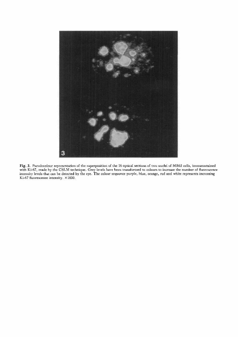
Fig. 3. Pseudocolour representation of the superposition of the 16 optical sections of two nuclei of MR6S cells, immunostainedwith Ki-67, made by the CSLM technique. Grey levels have been transformed to colours to increase the number of fluorescenceintensity levels that can be detected by the eye. The colour sequence purple, blue, orange, red and white represents increasingKi-67 fluorescence intensity. X1850.


4A — B
Fig. 4. Immunoelectron micrographs of MR65 cells in interphase, stained for the Ki-67 antigen using the immunoperoxidasetechnique. A-D. Staining of MR65 nucleoli with Ki-67. E. MR65 negative control cells. F. Staining pattern in an interphasenucleolus of a human stomach adenocarcinoma. Bars, 1-0 fim.
partly positive. In all interphase cells the fluorescencewas localized predominantly in the nucleoli and in somenucleoplasmic bodies (see Fig. 6). The presence of theantigen appeared not to be restricted to human cells, asabout 60 % of the cells of the Rhesus monkey kidney-derived cell line LLC-MK2 (Fig. 6H) gave a positivestaining reaction as well (Table 1). In contrast, VERO(African green monkey kidney) cells appeared to becompletely negative. Although the various positive celltypes all gave similar staining patterns with Ki-67 com-pared with MR65 cells, a considerable variability of thestaifiing intensities could be observed (see also Table 1).The highest fluorescence intensities were found in thepositive fractions of CloII and LLC-MK2 (Fig. 6F,H,respectively). In cells with a moderately high fluor-escence intensity such as HeLa S3, T24 and Molt-4(Fig. 6A,B,C, respectively), again the brighter periph-eral nucleolar fluorescence was observed. A weak stainingreaction was seen in about one third of the cells in human
fibroblast cultures. In none of the various cell culturesexamined could cytoplasmic staining be detected.
Discussion
Ki-67 is a mouse monoclonal antibody that reacts withproliferating cells and was recently introduced into histo-pathology to determine the growth fraction of humantumours in situ.
The present study was performed to examine theintracellular localization of the Ki-67 antigen in variouscell types. Using immunohistochemical techniques wehave shown that in cultured cells in interphase the mostintense staining reaction with Ki-67 is seen in nucleoli,particularly at its periphery and in the dense fibrillarcomponents. As several other anti-nucleolar antibodiesappeared to react strongly with antigens localized in theinterior of the nucleoli (data not shown; see Verheijen etal. 1986a), it is unlikely that the intense peripheral
Distribution of the Ki-67 antigen 127

Fig. 5. Comparison of the immunofluorescence stainingpatterns in untreated MR6S cells (A-E) and in nuclearmatrix-intermediate filament scaffolds of MR6S cells (A'-E')with monoclonal antibodies Ki-67 (A), 2.73 (B), directedagainst the Ul RNP-associated 70K protein, 41CC4 (C),directed against the nuclear lamins and RKSE 60 (D),directed against cytokeratin 10. E. DNA staining withHoechst 33258. XllSO.
staining of Ki-67 is due to an accessibility artefact. In thehuman cell types tested, the nucleoplasmic appearance ofthe Ki-67 antigen varied from a distinct number of dot-like structures to an almost diffuse staining reaction.
In addition we have tested Ki-67 on cell cultures fromvarious species. Our results indicate that expression ofthe antigen is not restricted to human cells and tissues,
but that positive staining reactions can also be obtained inproliferating cells from Rhesus monkey.
Experiments using actinomycin D also revealed that ininterphase cells the Ki-67 antigen behaved as a nucleolarcomponent. This is concluded from the observations thatduring segregation of the nucleoli the Ki-67 antigenseems to be mainly located in one of the two developedsubcompartments, while it shows a speckled distributionpattern in cells treated with high concentrations ofactinomycin D, resulting in a total fragmentation of theoriginal nucleoli.
For further identification of the nature of the Ki-67antigen we examined its association with the nuclearmatrix. This structure may be denned as the residualentity remaining after subsequent treatments of cells withdetergents, nucleases and high-salt solutions. In nuclearmatrix preparations of monolayer cell cultures, cytoskel-etal elements remain intact and for this reason we use theterm nuclear matrix—intermediate filament scaffold(NM-IF) according to Fey et al. (1984) for our MR65 insitu extraction preparations. It was demonstrated bymeans of immunofluorescence that the Ki-67 antigen waspreserved in such MR65 NM-IF scaffolds. However,high-salt treatment causes shrinkage of nuclei and anapparent increase in the fluorescence intensity (seeFig. 5). Therefore, one has to be cautious about con-clusions based on these fluorescence studies concerningthe amount of Ki-67 antigen preserved in these scaffolds.
Despite many efforts we have not succeeded in deter-mining the biochemical characteristics of the Ki-67antigen. In situ extraction experiments showed that theantigen sustained all treatments to obtain NM-IF scaf-folds (see Materials and methods) as the antibody stillreacted with such structures in the immunofluorescenceassay. However, the various Western blots on which theantibody was tested did not permit detection of any HeLaS3 nuclear protein specifically reacting with Ki-67. Alsothe zwittergent method (Mandrell & Zollinger, 1984),aiming at a partial renaturation of lost antigenic epitopes,gave no unambiguous results. From these data one couldconclude that the procedure followed to identify theantigen on SDS-containing gels apparently alters thestructure of the epitope in such a way that it is no longerrecognized by the antibody. However, immunoprecipi-tation experiments in which no SDS was initially usedalso failed in precipitating any specific [35S]methionine-labelled proteins or any 3*P-labelled RNA from HeLa S3total cell lysates. We have no clear indication how thesenegative results can be explained.
The authors gratefully thank Professor Dr J. Gerdes (Bor-stel, FRG) for his gift of the ascites fluid of Ki-67, Dr G.Mungyer (Nijmegen, The Netherlands) for her gift of the dogprimary endothelium culture and Dr W. Ferro (Leiden, TheNetherlands) for his gift of the Drosophila cell culture mei-218.We are indebted to Mr F. Rietveld (Nijmegen) and Mr B. J.Mauw (Leiden) for performing the immunoelectron mi-croscopy and Professor Dr D. Ruiter for useful discussions andcritical reading of the manuscript.
We also acknowledge the kind gifts of monoclonal antibodies41CC4 (from Dr G. Warren; Heidelberg, FRG) and 2.73 (fromDr S. Hoch; La Jolla, USA).
128 R. Verheijen et al.

Fig. 6. Immunofluorescence localization of the Ki-67 antigen in different cell types: A, HeLa S3; B, T24; C, Molt-4;D, human fibroblasts; F, CloII; H, LLC-MK2. E,G. DNA staining patterns with Hoechst 33258, of preparations D and F,respectively. Note the differences in the levels of Ki-67 fluorescence between the various cell cultures. X 1 ISO.
This study was supported by the Netherlands Cancer Foun-dation, grant no. NUKC 1984-11, the Foundation for Funda-mental Biological Research (BION), the Maurits and Anna deKock Foundation and the Nijbakker-Morra Foundation.
References
BARNARD, N. J., HALL, P. A., LEMOINE, N. R. & KADAR, N.(1987). Proliferative index in breast carcinoma determined in situby Ki-67 immunostaining and its relationship to clinical andpathological variables. 7. Path. 152, 287-295.
BILLINGS, P. B., ALLEN, R. W., JENSEN, F. C. & HOCH, S. O.(1982). Anti-RNP monoclonal antibodies derived from a mousestrain with lupus-like autoimmunity._7- Immun. 128, 1176-1180.
BLOEMENDAL, H., LENSTRA, J. H., DODEMONT, H., RAMAEKERS, F.C. S., GROENEVELD, A. A., DUNIA, I. & BENEDETTI, E. L. (1980).SV-4O transformed hamster lens epithelial cells: A novel system forthe isolation of cytoskeletal messenger RNAs and their translationproducts. Expl Eye Res. 31, 513-525.
BRAKENHOFF, G. J., VAN DER VOORT, H. T. M., VAN SPRONSEN, E.A., UNNEMANS, W. A. M. & NANNINGA, N. (1985). Three-dimensional chromatin distribution in neuroblastoma nuclei shownby confocal scanning microscopy. Nature, Land. 317, 748-749.
BRAKENHOFF, G. J., VAN DER VOORT, H. T. M., VAN SPRONSEN, E.A. & NANNINGA, N. (1988). Three-dimensional imaging influorescence by confocal scanning microscopy. J. Microsc. (inpress).
BROERS, J. L. V., GROPP, C , BEPLER, G., KLEIN ROT, M., BECK, J.,SCHAART, G., VOOIJS, G. P. & RAMAEKERS, F. C. S. (1987).Identification of cytoskeletal structures in hormone producing lungcancer cell cultures. Acta histoche/nica, Suppl.-Band XXXIV,s.57-75.
BURKE, B., TOOZE, J. & WARREN, G. (1983). A monoclonal antibodywhich recognizes each of the nuclear lamin polypeptides inmammalian cells. EMBOJ. 2, 361-367.
FEY, E. G., WAN, K. M. & PENMAN, S. (1984). Epithelialcytoskeletal framework and nuclear matnx-intermediate filamentscaffold: three-dimensional organization and protein composition.J. CellBiol. 98, 1973-1984.
FRANKLIN, W. A., BIBBO, M., DORIA, M. I., DYTCH, H. E., TOTH,J., DESOMBRE, E. & WIED, G. L. (1987). Quantitation of estrogenreceptor content and Ki-67 staining in breast carcinoma by themicro TICAS image analysis system. Ajialvt. quant. Cvtol. Histol.9, 279-286.
FRANKUN, W. A., MCDONALD, G. B., STEIN, H. O., GATTER, K.C , JEWELL, D. P., CLARKE, L. C. & MASON, D. Y. (1985).Immunohistologic demonstration of abnormal colonic crypt cellkinetics in ulcerative colitis. Human Path. 16, 1129-1132.
GATTER, K. C , DUNNILL, M. S., GERDES, J., STEIN, H. & MASON,
D. Y. (1986). New approach to assessing lung tumours in man.J. din. Pathol. 39, 590-593.
GERDES, J., DALLENBACH, F. & LENNERT, K. (19846). Growthfractions in malignant non-Hodgkin's lymphomas (NHL) asdetermined in situ with the monoclonal antibody Ki-67. Hemat.Oncol. 2, 365-371.
GERDES, J., LELLE, R. J., PICKARTZ, H., HEIDENREICH, W.,
SCHWARTING, R., KURTSIEFER, L. , STAUCH, G. & STEIN, H .(1986). Growth fractions in breast cancers determined in situ withmonoclonal antibody Ki-67.7. din. Path. 39, 977-980.
GERDES, J., LEMKE, H., BAISCH, H., WACKER, H.-H., SCHWAB, U.& STEIN, H. (1984a). Cell cycle analysis of a cell proliferation-associated human nuclear antigen denned by the monoclonalantibody Ki-67.J. Immun. 133, 1710-1715.
GERDES, J., SCHWAB, U., LEMKE, H. & STEIN, H. (1983).Production of a mouse monoclonal antibody reactive with a humannuclear antigen associated with cell proliferation. Int.J. Cancer 31,
Distribution of the Ki-67 antigen 129
13-20.GERDES, J., VAN BAARLEN, J., PILERI, S., SCHWARTING, R., VAN
UNNIK, J. A. M. & STEIN, H. (1987). Tumor cell growth fractionin Hodgkin's disease. Am. J. Path. 128, 390-393.
GlANGASPERO, F . , DOGUONI, C , RlVANO, M. T . , PlLERI, S.,
GERDES, J. & STEIN, H. (1987). Growth fraction in human braintumors defined by the monoclonal antibody Ki-67. Acta neuropath.74, 179-182.
MANDRELL, R. E. & ZOLUNGER, W. D. (1984). Use of zwitterionic
detergent for the restoration of the antibody-binding capacity ofelectroblotted Meningococcal outer membrane proteins. J. lmmun.Meth. 67, 1-11.
MlMORI, T . , HlNTERBERGER, M. , PETTERSSON, I. & STETTZ, J. A.
(1984). Autoantibodies to the U2 small nuclear ribonucleoproteinin a patient with scleroderma-polymyositis overlap syndrome.J. biol. Chem. 259, 560-565.
RALFHAER, E., STEIN, H., BOSQ, J., GATTER, K. C , RALFKIAER, N.,
LANGE WANTZIN, G. & MASON, D. Y. (1986). Expression of a
cell-cycle-associated nuclear antigen (Ki-67) in cutaneous lymphoidinfiltrates. Am. J. Dermatopath. 8, 37-43.
RAMAEKERS, F. C. S., OSBORN, M., SCHMID, E., WEBER, K.,
BLOEMENDAL, H. & FRANKE, W. W. (1980). Identification of the
cytoskeletal proteins in lens-forming cells, a special epithelioid celltype. Expl Cell Res. 127, 309-327.
RAMAEKERS, F. C. S., PUTS, J. J. G., MOESKER, O., KANT, A.,
HUYSMANS, A., HAAG, D., JAP, P. H. K., HERMAN, C. J. &
Voous, G. P. (1983). Antibodies to intermediate filament proteinsin the immunohistochemical identification of human tumours: anoverview. Histochem.J. 15, 691-713.
SCHWARTING, R., GERDES, J., NIEHUS, J., JAESCHKE, L. & STEIN,
H. (1986). Determination of the growth fraction in cell suspensionsby flow cytometry using the monoclonal antibody Ki-67. .7. lmmun.Meth. 90, 65-70.
VAN DER VOORT, H. T. M., BRAKENHOFF, G. J. & BAARSLAG, M. W.
(1988). Three-dimensional visualisation methods for confocalmicroscopy. J. Microsc. (in press).
VAN DUINEN, S. G., MAUW, B. J., DE GRAAFF-REITSMA, C. B. &
RUTTER, D. J. (1984). Immunoelectron microscopic methods fordemonstration of antigens on normal human melanocytes and otherepidermal cells. Lab. Invest. 50, 733-741.
VERHEIJEN, R., KUUPERS, H., VOOUS, P., VAN VENROOU, W. &
RAMAEKERS, F. (1986a). Protein composition of nuclear matrixpreparations from HeLa cells: an immunochemical approach.J. Cell Sci. 80, 103-122.
VERHEUEN, R., KUUPERS, H., VOOUS, P., VAN VENROOU, W. &
RAMAEKERS, F. (19866). Distribution of the 70K Ul RNA-associated protein during interphase and mitosis. J. Cell Sci. 86,173-190.
(Received 28 April 1988 - Accepted, in revised form, 3 October 1988)
130 R. Verheijen et al.