Embed Size (px)
Citation preview
Encourage regular weight bearing exercise ■
Encourage smoking cessation ■
Bisphosphonates can decrease the incidence of ■fracture in women with established osteoporosis
Prevention of Osteoporosis
Key concepts
For prevention of osteoporosis:
Recommend adequate dietary intake of calcium ■and use supplements if necessary
Advise on the role of vitamin D and consider ■sun exposure and the use of supplements if necessary
Key reviewers:
Professor Ian Reid, Faculty of Medical and Health Sciences, University of Auckland
Dr Rebecca Grainger, Rheumatologist and Clinical Research Fellow, Malaghan Institute of Medical Research, Wellington
6 | BPJ | Issue 17
www.bpac.org.nz keyword: osteoporosis
Osteoporosis is not just a result of ageing
Osteoporosis develops from a combination of the following factors:
Age ▪
Genetics ▪
Lifestyle ▪
Hormones ▪
Medications ▪
Medical conditions ▪
Age
Peak bone mass is achieved by around age 30–35 years and from then on starts to decline. The higher the peak bone mass achieved, the lower the impact of subsequent bone loss.
Genetics
Genes play a role in determining peak bone mass.
A person with a history of a hip fracture in a parent is at increased risk of osteoporosis. In addition, a study of hip fracture in New Zealand showed approximately 30% higher prevalence in people of European origin than for Māori, Pacific or Asian peoples. However, these may be due to differences in life expectancy and lifestyle factors relating to diet and body mass, as well as genetics.2
The relationship between body mass and osteoporosis is complex. Inherited muscular body mass appears to be protective whereas obesity may be a risk factor.3
Lifestyle
Lifestyle factors can have a direct effect on bone strength or alter calcium absorption. Factors associated with an increased risk of osteoporosis include, vitamin D deficiency, excess vitamin A, low calcium intake, smoking (both active and passive), high alcohol intake, lack of physical activity, immobilisation (paralysis, ill health), low body weight, exercise induced amenorrhoea and falls.
Hormones
Both men and women can develop osteoporosis but women are more at risk as their bones are smaller and there is an accelerated loss of bone density at menopause due to decreasing oestrogen. Early or surgical menopause or amenorrhoea increases the risk.
Any condition in men causing a decrease in testosterone can increase the risk.
Medications
Medications that can increase the risk of osteoporosis include:
Steroids (>5mg/day for more than three months) ▪
Lithium ▪
Anticonvulsants ▪
Cancer chemotherapy drugs ▪
Depo-medroxyprogesterone (see BPJ 12) ▪
Proton pump inhibitors (see ETC, page 55) ▪
Medical conditions
Many medical conditions are associated with osteoporosis either as a risk factor or consequence of (see Box 1).1
Prevention of osteoporosis
Prevention of osteoporosis in the whole population focuses on nutritional and lifestyle changes. The goals include:
Acquiring maximal peak skeletal bone mass ▪
Maintaining this bone mass for as long as possible ▪
Increasing awareness of the modifiable risk factors for osteoporosis through patient education is an important primary care role. A recent study of attitudes and knowledge about osteoporosis in a group of well-educated New Zealand women did not show a high level of knowledge. Although most demonstrated high levels of health motivation and most considered osteoporosis to be a serious disease, the women had low perceptions of personal susceptibility.4
BPJ | Issue 17 | 7
Adequate calcium intake
Adequate calcium intake is necessary for the acquisition of peak bone mass by the age of 35 and its subsequent maintenance. When exogenous supply is inadequate, bone tissue is resorbed to maintain serum calcium at a constant level. Calcium needs vary throughout life and between genders (see Table 1).
Table 1: New Zealand recommended daily intake of calcium (mg/day)5
Children age 1–3 500
Children age 4–8 700
Children age 9–11 1000
Adolescents age 12–18 1300
Men 19–70 1000
Men 70+ 1300
Women 19–54 1000
Pregnancy and Lactation 1000–1300
Post menopausal 1300
It is estimated that 20% of New Zealanders have an inadequate intake of calcium.6 Inadequate intake was
higher amongst women and 15 to 18 year olds. In these groups, low calcium intake puts
them at particular risk. Calcium intake was also inadequate in
low socioeconomic groups and Māori.
Box 1: Examples of medical conditions associated with osteoporosis1
Endocrine e.g. diabetes mellitus, Cushing’s syndrome, hyperparathyroidism, thyrotoxicosis
Gastrointestinal e.g. coeliac disease, inflammatory bowel disease, gastric bypass, pancreatic disease, malabsorption
Genetic e.g. cystic fibrosis, haemochromatosis, Marfan syndrome
Hypogonadal states e.g. premature ovarian failure
Haematologic e.g. multiple myeloma, haemophilia, leukaemia and lymphomas, thalassaemia
Rheumatic/autoimmune e.g rheumatoid arthritis, ankylosing spondylitis, lupus
Other e.g. alcoholism, emphysema, end stage renal disease, prior fracture, anorexia nervosa, bulimia
8 | BPJ | Issue 17
Practical food suggestions for maximising dietary
calcium
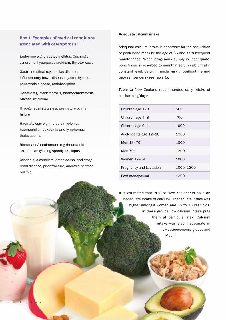
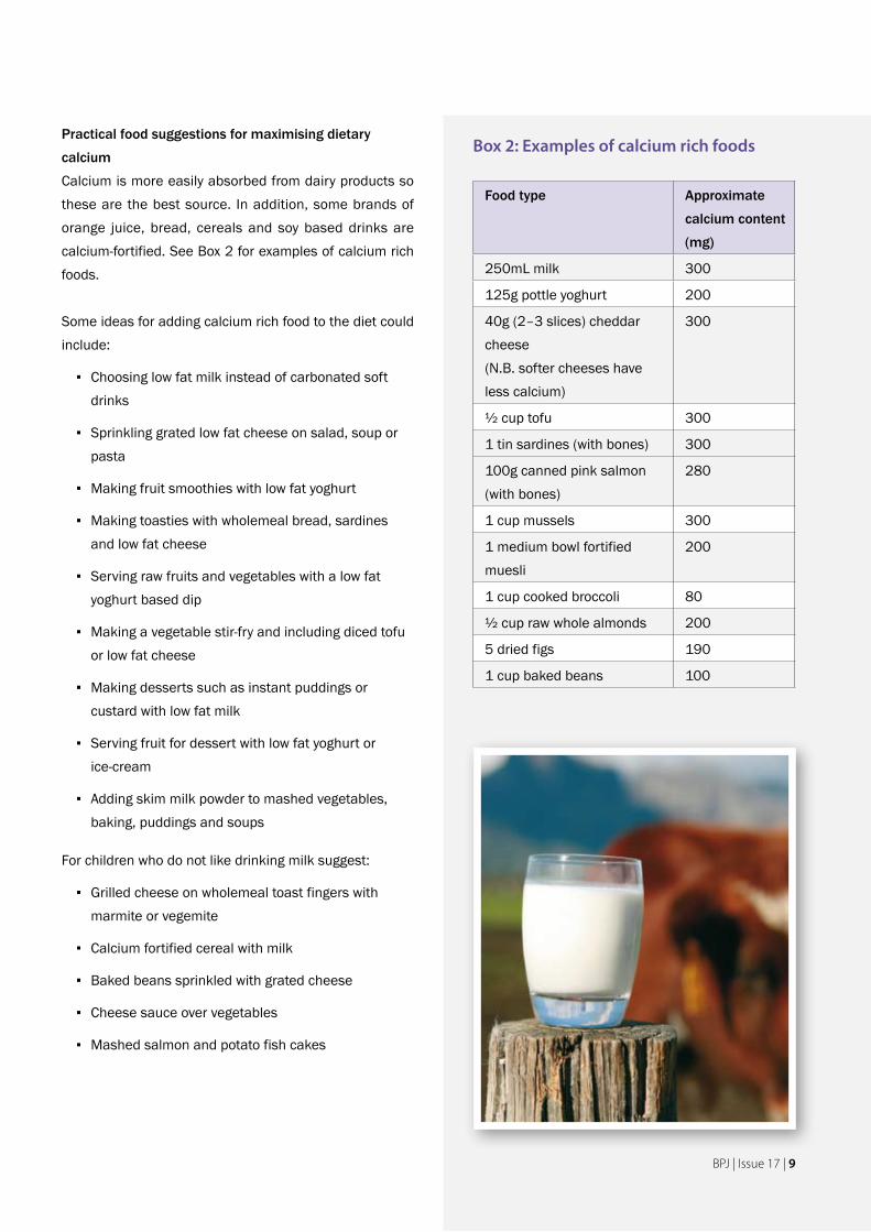
Calcium is more easily absorbed from dairy products so these are the best source. In addition, some brands of orange juice, bread, cereals and soy based drinks are calcium-fortified. See Box 2 for examples of calcium rich foods.
Some ideas for adding calcium rich food to the diet could include:
Choosing low fat milk instead of carbonated soft ▪drinks
Sprinkling grated low fat cheese on salad, soup or ▪pasta
Making fruit smoothies with low fat yoghurt ▪
Making toasties with wholemeal bread, sardines ▪and low fat cheese
Serving raw fruits and vegetables with a low fat ▪yoghurt based dip
Making a vegetable stir-fry and including diced tofu ▪or low fat cheese
Making desserts such as instant puddings or ▪custard with low fat milk
Serving fruit for dessert with low fat yoghurt or ▪ice-cream
Adding skim milk powder to mashed vegetables, ▪baking, puddings and soups
For children who do not like drinking milk suggest:
Grilled cheese on wholemeal toast fingers with ▪marmite or vegemite
Calcium fortified cereal with milk ▪
Baked beans sprinkled with grated cheese ▪
Cheese sauce over vegetables ▪
Mashed salmon and potato fish cakes ▪
Box 2: Examples of calcium rich foods
Food type Approximate
calcium content
(mg)
250mL milk 300
125g pottle yoghurt 200
40g (2–3 slices) cheddar cheese (N.B. softer cheeses have less calcium)
300
½ cup tofu 300
1 tin sardines (with bones) 300
100g canned pink salmon (with bones)
280
1 cup mussels 300
1 medium bowl fortified muesli
200
1 cup cooked broccoli 80
½ cup raw whole almonds 200
5 dried figs 190
1 cup baked beans 100
BPJ | Issue 17 | 9
Adequate Vitamin D
For more information see Best Tests – Vitamin D Testing in Primary Care, January 2007, www.bpac.org.nz keyword: vitaminD.
Vitamin D, which is produced in the skin, is essential for the acquisition and maintenance of bone mass.
Adequate exposure to sunlight is required to maintain vitamin D levels. This means about 15–20 minutes of sun exposure to the face and arms every day, avoiding sun exposure around midday in the summer. People with dark skin require approximately three to four times more exposure to gain the same benefit.
Although vitamin D is contained in some foods in small amounts such as oily fish (e.g. salmon, sardines, herring), adequate intake is not usually attained through diet alone.
Vitamin D deficiency is more prevalent in the following groups:
Older people in residential care ▪
Older people admitted to hospital ▪
People with hip fracture ▪
People with dark skin ▪
People unable to obtain regular sun exposure ▪
For these people, consider supplementation without testing. An appropriate dose is a single tablet of cholecalciferol 1.25mg monthly.
Regular exercise increases and maintains bone density
Regular weight bearing exercise should be recommended at all ages. This type of exercise can increase bone density and strength, particularly during childhood and adolescence. Exercise should be regular and ongoing as the beneficial effects on bone strength are lost when exercise is stopped.
Weight bearing exercise and muscle strengthening exercises may help prevent falls and fractures by improving agility, strength, co-ordination, posture and balance especially in older adults. Water exercise and cycling are regarded as non-weight bearing exercises but are still useful as muscle strength and fitness is maintained.
A person with established osteoporosis is at higher risk of fractures from high impact, jarring or twisting exercises such as running, jumping and aerobics.
Smoking is a significant risk factor for osteoporosis
There is some evidence that the significant risk of osteoporosis associated with smoking is a direct effect that is independent of confounding factors.7 Smoking cessation is recommended.
Avoid excessive alcohol
Current evidence about the role of alcohol and bone density is conflicting.8 Most publications report that an alcohol intake of three or more drinks per day is detrimental to bone density.1 However, the exact mechanism of action remains unclear.
Fracture prevention in people with osteoporosis
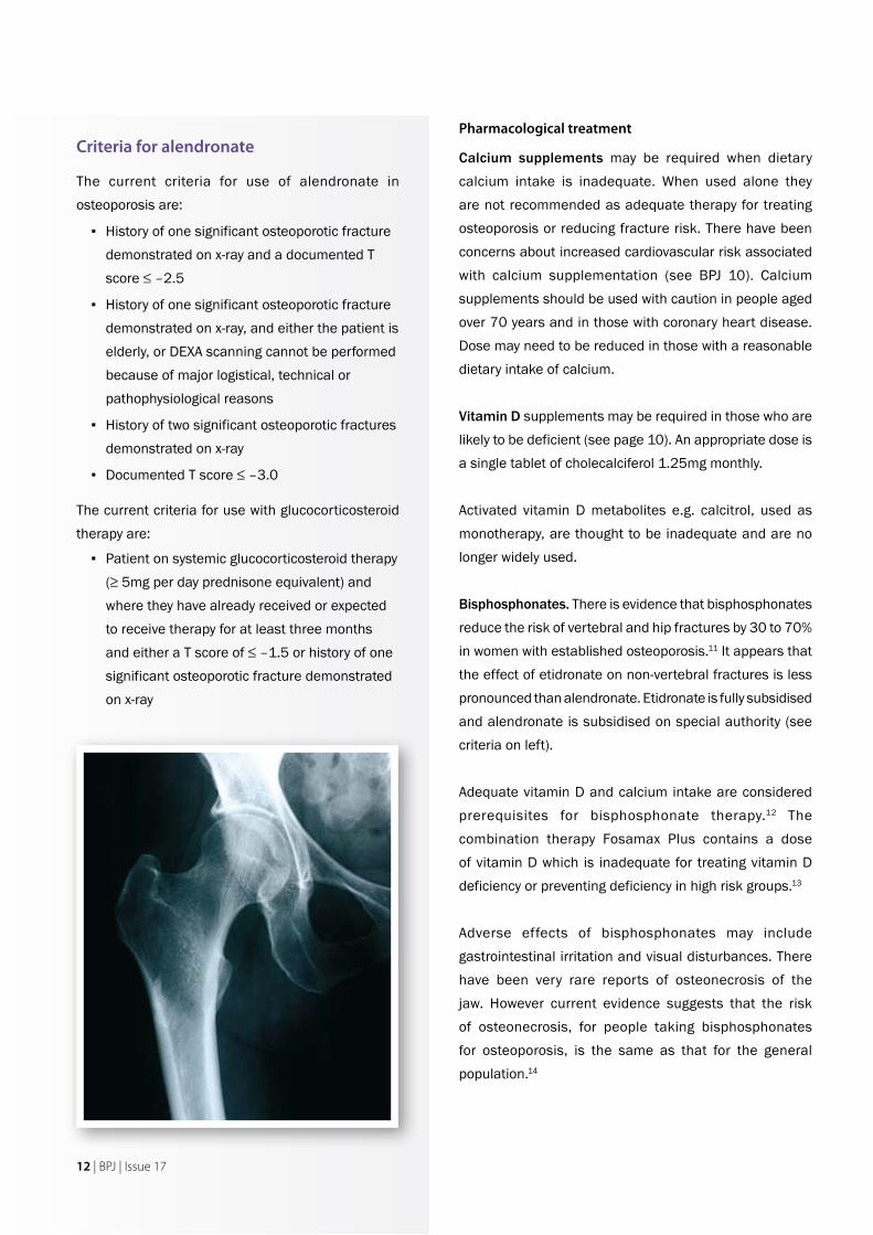
The most common osteoporotic fracture sites are the spine, hip and wrist. They can have major consequences, for example hip fracture causes 10–20% excess mortality within one year and two and a half times increased risk of future fractures. In addition, after a hip fracture 20% of patients require long term residential care and only 40% fully regain their pre-fracture independence.1
The consequences of spinal fracture can also be significant with chronic back pain, kyphosis, loss of height and limitation of activities that require bending, reaching and lifting. Multiple compression fractures result in an increasing curvature of the spine and
10 | BPJ | Issue 17
compression of thoracic and abdominal organs. This may then cause shortness of breath, stress incontinence, and gastrointestinal symptoms such as anorexia, constipation, distension and abdominal pain.
The focus for fracture prevention for people with osteoporosis is:
Preventing falls ▪
Detection of osteoporosis ▪
Pharmaceutical interventions ▪
Preventing falls
Preventing falls prevents fractures. Lowering the risk of falls may include checks on vision, hearing, adverse effects from medications, safety at home and promotion of exercise programmes.
Detection of osteoporosis
A clinical diagnosis of osteoporosis can be made if there is a low trauma fracture in an at-risk individual and may be suggested when an x-ray indicates low bone density. However the gold standard for diagnosis is bone densitometry (DEXA). Access to and funding of bone densitometry scanning varies throughout New Zealand.
Osteoporosis New Zealand Inc. recommends only measuring bone density when the result will impact on decision making.9
People who have had an osteoporotic fracture
A DEXA scan is not required for everyone with an osteoporotic fracture. Bisphosphonate treatment without the need for a prior DEXA scan can be considered for:
Women over the age of 75 years who have had an ▪osteoporotic fracture demonstrated on x-ray
People who have had two or more demonstrated ▪osteoporotic fractures
People who have had systemic glucocorticoid ▪steroid therapy (over 5mg per day prednisone equivalent for at least three months)
A DEXA scan is indicated for other people who have had an osteoporotic fracture.
People at high risk of osteoporosis
The contribution of individual risk factors towards the development of osteoporosis has not yet been quantified. Clinicians must make pragmatic decisions on who to refer for a DEXA scan based on major risk factors such as:10
Age ▪
Female gender ▪
Low BMI ▪
Untreated premature menopause ▪
Family history of maternal hip fracture before the ▪age of 75 years
Conditions affecting bone metabolism (primarily ▪inflammatory conditions, hyperthyroidism and prolonged immobility)
Chronic steroid use ▪
DEXA scanning
Bone densitometry (dual energy x-ray absorptiometry, DEXA) measures the bone mineral density usually at the hip and spine. Results may be given as a T score which is the result compared to “young normal” adults of the same sex or a Z score which is the result compared to that expected for the patient’s age and sex. The T score is most predictive of future fracture.
BPJ | Issue 17 | 11
Criteria for alendronate
The current criteria for use of alendronate in osteoporosis are:
History of one significant osteoporotic fracture ▪demonstrated on x-ray and a documented T score ≤ –2.5
History of one significant osteoporotic fracture ▪demonstrated on x-ray, and either the patient is elderly, or DEXA scanning cannot be performed because of major logistical, technical or pathophysiological reasons
History of two significant osteoporotic fractures ▪demonstrated on x-ray
Documented T score ≤ –3.0 ▪
The current criteria for use with glucocorticosteroid therapy are:
Patient on systemic glucocorticosteroid therapy ▪(≥ 5mg per day prednisone equivalent) and where they have already received or expected to receive therapy for at least three months and either a T score of ≤ –1.5 or history of one significant osteoporotic fracture demonstrated on x-ray
Pharmacological treatment
Calcium supplements may be required when dietary calcium intake is inadequate. When used alone they are not recommended as adequate therapy for treating osteoporosis or reducing fracture risk. There have been concerns about increased cardiovascular risk associated with calcium supplementation (see BPJ 10). Calcium supplements should be used with caution in people aged over 70 years and in those with coronary heart disease. Dose may need to be reduced in those with a reasonable dietary intake of calcium.
Vitamin D supplements may be required in those who are likely to be deficient (see page 10). An appropriate dose is a single tablet of cholecalciferol 1.25mg monthly.
Activated vitamin D metabolites e.g. calcitrol, used as monotherapy, are thought to be inadequate and are no longer widely used.
Bisphosphonates. There is evidence that bisphosphonates reduce the risk of vertebral and hip fractures by 30 to 70% in women with established osteoporosis.11 It appears that the effect of etidronate on non-vertebral fractures is less pronounced than alendronate. Etidronate is fully subsidised and alendronate is subsidised on special authority (see criteria on left).
Adequate vitamin D and calcium intake are considered prerequisites for bisphosphonate therapy.12 The combination therapy Fosamax Plus contains a dose of vitamin D which is inadequate for treating vitamin D deficiency or preventing deficiency in high risk groups.13
Adverse effects of bisphosphonates may include gastrointestinal irritation and visual disturbances. There have been very rare reports of osteonecrosis of the jaw. However current evidence suggests that the risk of osteonecrosis, for people taking bisphosphonates for osteoporosis, is the same as that for the general population.14
12 | BPJ | Issue 17
Hormone Replacement Therapy is known to decrease the risk of fractures in post menopausal women but it is no longer regarded as first line for prevention or treatment of osteoporosis due to increased risk of cardiovascular events, thromboembolic events and breast cancer.
Further reading:
Brown P, McNeill R, Radwan E, Willingale J. The Burden of Osteoporosis in New Zealand: 2007-2020.
Available from: www.bones.org.nz/downloads/BONZ%20
Report-final.pdf
References:
1. National Osteoporosis Foundation. Clinician’s guide to prevention and treatment of osteoporosis. 2008. Available from www.nof.org. (Accessed September 2008).
2. Brown P, McNeill R, Radwan E, Willingale J. The burden of osteoporosis in New Zealand: 2007-2020. Available from www.bones.org.nz/downloads/BONZ%20Report-final.pdf (Accessed September 2008).
3. Zhao LJ, Liu YJ, Liu PY et al. Relationship of obesity with osteoporosis. J Clin Endocrinol Metab 2007;92(5):1640-6.
4. Von Hurst PR, Wham CA. Attitudes and knowledge about osteoporosis risk prevention: a survey of New Zealand women. Public Health Nutr 2007;10(7):747-53.
5. Ministry of Health. Nutrient reference values for Australia and New Zealand. Available from www.nhmrc.gov.au/publications (Accessed September 2008).
6. Horwath C, Parnell WR, Wilson NC, Russel DG. Attaining optimal bone status: lessons from the 1997 National Nutrition Survey. N Z Med J 2001; 114:138-41.
7. Wong PKK, Christie JJ, Wark JD. The effects of smoking on bone health. Clin Sci 2007;113:233-41.
8. Berg KM, Kunins HV, Jackson JL et al. Association between alcohol consumption and both osteoporotic fracture and bone density. Am J Med 2008;121(5):406-18.
BPJ | Issue 17 | 13
9. Osteoporosis New Zealand Inc. Recommendations for the management of osteoporosis. Available from www.bones.org.nz. (Accessed September 2008).
10. National Institute for Clinical Excellence. Osteoposoris – secondary prevention. Bisphosphonates (alendronate, etidronate, risedronate), selective oestrogen receptor modulators (raloxifene) and parathyroid hormone (teriparatide) for the secondary prevention of osteoporotic fragility fractures in postmenopausal women. Technology Appraisal Guidance 87. January 2005. Available from www.nice.org.uk/Guidance/TA87 (Accessed September 2008).
11. Delmas PD. Treatment of postmenopausal osteoporosis. Lancet 2002;359:2018-26.
12. Endocrinology Writing Group. Therapeutic guidelines: Endocrinology: Version 3. Melbourne: Therapeutic Guidelines 2004.
13. Alendronate with cholecalciferol (vitamin D3) (Fosamax Plus) for osteoporosis. Available from www.nps.org.au/health_professionals/publications (Accessed October 2008).
14. Bolland M, Hay D, Grey A, et al. Prescriber Update Articles. Osteonecrosis of the jaw and bisphosphonates – putting the risk in perspective. 2007. Available from www.medsafe.govt.nz/Profs/PUArticles.asp (Accessed September 2008).
osteoarthritisSymptomatic management of
Key concepts
A core treatment for osteoarthritis is the provision ■of information and resources to assist patients in coping with both the physical and psychological aspects of this condition.
Exercise and weight loss are also core ■treatments.
Safe pharmacological options include regular ■paracetamol, topical NSAIDs and capsaicin.
If pain is not controlled, oral NSAIDs, opioids and ■steroid injections can be considered.
Joint replacement surgery can be considered ■when symptomatic control cannot be achieved with any other treatments.
Key reviewers:
Dr Andrew Harrison, Senior Lecturer, Rheumatology, Wellington School of Medicine, University of Otago, Wellington
Dr Rebecca Grainger, Rheumatologist and Clinical Research Fellow, Malaghan Institute of Medical Research, Wellington
14 | BPJ | Issue 17
www.bpac.org.nz keyword: osteoarthriris
Osteoarthritis is the most common form of arthritis and a leading cause of pain and disability around the world. It affects approximately 50% of people aged over 60 years and almost all people aged over 80 years. However osteoarthritis is not just caused by ageing. Factors which may lead to the development of osteoarthritis include:1
Genetic factors ▪
Age ▪
Joint damage by injury ▪
Joint damage by chronic obesity ▪
The joints most commonly affected are the knee, hip, spine and hand.
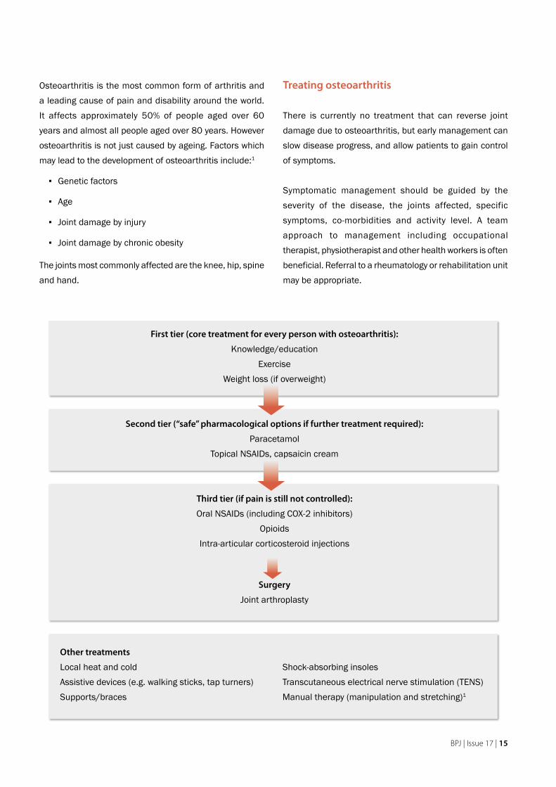
Treating osteoarthritis
There is currently no treatment that can reverse joint damage due to osteoarthritis, but early management can slow disease progress, and allow patients to gain control of symptoms.
Symptomatic management should be guided by the severity of the disease, the joints affected, specific symptoms, co-morbidities and activity level. A team approach to management including occupational therapist, physiotherapist and other health workers is often beneficial. Referral to a rheumatology or rehabilitation unit may be appropriate.
First tier (core treatment for every person with osteoarthritis):
Knowledge/educationExercise
Weight loss (if overweight)
Second tier (“safe” pharmacological options if further treatment required):
ParacetamolTopical NSAIDs, capsaicin cream
Third tier (if pain is still not controlled):
Oral NSAIDs (including COX-2 inhibitors)Opioids
Intra-articular corticosteroid injections
Surgery
Joint arthroplasty
Other treatments
Local heat and cold Shock-absorbing insolesAssistive devices (e.g. walking sticks, tap turners) Transcutaneous electrical nerve stimulation (TENS)Supports/braces Manual therapy (manipulation and stretching)1
BPJ | Issue 17 | 15
Core treatments for osteoarthritis
Providing accurate information helps patients cope with the ongoing nature of osteoarthritis and develop effective self-management strategies for both its physical and psychological aspects. This includes advice on lifestyle, exercise, activity and weight loss.2
Exercise is a core treatment for anyone with osteoarthritis, irrespective of age, co-morbidity, pain severity or disability.1
Exercise can help to manage pain, keep joints mobile and increase the ability to perform day to day tasks. A fitness programme should be individually tailored and may require modification at times depending on symptoms, but should always include cardiovascular (aerobic), muscle strength (especially around the damaged joint), muscle endurance and flexibility components. Joint pain can limit the intensity of exercise, but should not be a deterrent. Referral to a physiotherapist for a tailored exercise programme is appropriate.
General practitioners and practice nurses play an important role in encouraging and motivating patients to participate in and maintain exercise programmes e.g. “green prescription”.
Regular exercise is beneficial for weight loss, which is also an important component of symptomatic management. Patients who are overweight should be encouraged to reduce and maintain their weight at recommended levels.
Shock-absorbing footwear, joint supports and mobility aids, heat packs, warm baths or application of ice packs can all provide symptomatic relief.
Safe pharmacological options
Paracetamol
Regular paracetamol at a dose of up to 4g per day is effective as initial oral analgesia for treating mild to moderate pain associated with osteoarthritis.2 If pain is successfully managed, paracetamol can be maintained long-term.2
Topical NSAIDs
Topical NSAIDs are generally less effective than oral NSAIDs but they are considered to be safer and serious adverse effects are unlikely. Local reactions such as
16 | BPJ | Issue 17
itching or burning may occur.2 The “placebo effect” is said to play a role in the clinical effect of topical NSAIDs and they may only be effective in the first few weeks of treatment.2 Topical NSAIDs can be considered, but are not a key component of treatment for osteoarthritis.
Capsaicin cream
Topical capsaicin cream (0.025%) contains a chilli pepper extract and may cause burning pain at the site of application but is an effective analgesic. Other than the intended burning effect, capsaicin is not associated with other adverse effects.2
Capsaicin cream is applied by squeezing a bead of cream onto the finger and rubbing over each affected joint, four times per day. It should not be applied after a hot bath or shower or to broken skin. Hands should be washed after application to avoid inadvertent transfer to the eyes. Treatment should be continued for three months before assessing clinical effect.
Note that topical NSAIDs and capsaicin are not subsidised for the treatment of osteoarthritis in New Zealand.
Further treatment approaches if pain is still uncontrolled
NSAIDs
There is evidence that NSAIDs are superior to paracetamol for pain relief in patients with osteoarthritis,3 but they are also associated with more adverse effects. The major concern is serious gastrointestinal, renal and cardiovascular complications, with risk increasing with age, concurrent use of other medications and duration of therapy.2 NSAIDs help relieve pain, swelling and stiffness but they do not alter the progression of osteoarthritis.
Oral NSAIDs should only be considered when paracetamol or topical treatments are ineffective for pain relief. NSAIDs should be used at the lowest effective dose for the shortest possible time. Do not exceed maximum daily doses (Table
1). Long-term use of NSAIDs is not routinely recommended,2 however individual patient factors can be taken into account, such as risk of adverse events and the effect of NSAID treatment on functional ability. Paracetamol can be continued throughout NSAID treatment,1 however topical NSAIDs should be discontinued.
Cyclo-oxygenase-2 (COX-2) selective drugs are not recommended for routine use in osteoarthritis, except where the patient is at high risk of developing a serious GI adverse effect from other standard NSAIDs, or has GI intolerance of standard NSAIDs in spite of the use of a gastroprotective agent. COX-2 drugs are not subsidised in New Zealand.
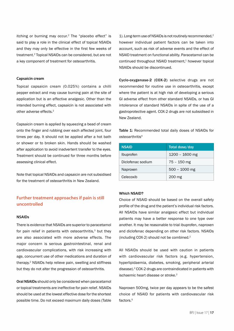
Table 1: Recommended total daily doses of NSAIDs for osteoarthritis4
NSAID Total dose/day
Ibuprofen 1200 – 1600 mg
Diclofenac sodium 75 – 150 mg
Naproxen 500 – 1000 mg
Celecoxib 200 mg
Which NSAID?
Choice of NSAID should be based on the overall safety profile of the drug and the patient’s individual risk factors. All NSAIDs have similar analgesic effect but individual patients may have a better response to one type over another. It may be reasonable to trial ibuprofen, naproxen and diclofenac depending on other risk factors. NSAIDs (including COX-2) should not be combined.2
All NSAIDs should be used with caution in patients with cardiovascular risk factors (e.g. hypertension, hyperlipidaemia, diabetes, smoking, peripheral arterial disease).2 COX-2 drugs are contraindicated in patients with ischaemic heart disease or stroke.2
Naproxen 500mg, twice per day appears to be the safest choice of NSAID for patients with cardiovascular risk factors.5
BPJ | Issue 17 | 17
All NSAIDs can increase blood pressure, especially in people with hypertension, and cause fluid retention and oedema. In rare cases, congestive heart failure and renal dysfunction may occur.5 Monitoring of blood pressure and for signs of fluid retention should occur within two to four weeks of initiating NSAID treatment.5
In patients with increased GI risk, prescribe a non-selective NSAID with a 20mg proton pump inhibitor (PPI) or a COX-2 selective agent.2 The GI protection associated with the use of a COX-2 drug is mostly lost when low-dose aspirin is concurrently administered.2 Aspirin also does not offset the increased cardiovascular risk associated with COX-2 drugs.
Opioids may be considered when other oral treatment
is unsuccessful
Weak opioids such as codeine can be considered for relieving pain where other pharmacological agents have been ineffective or are contraindicated. A recommended dose of codeine is one to two 30mg tablets, every four to six hours as required, to a maximum of 240 mg (60mg codeine is equivalent to 6mg morphine).4
Stronger opioids should only be used for severe pain in exceptional circumstances. Adverse effects including sedation, risk of falls and constipation are common and are of particular concern in older people.
Tramadol has no advantages over other opioids but is increasingly used for pain management (50mg tramadol is equivalent to 10mg morphine). A systematic review concluded that while tramadol is effective in relieving pain and improving function in people with osteoarthritis (compared to placebo) the benefits are small. Adverse effects may limit its use and include nausea, confusion and interaction with SSRIs.6
Intra-articular injections may be useful in severe pain
Intra-articular injections with corticosteroids can be considered when patients have moderate to severe pain
The role of glucosamine in osteoarthritis
A 2001 Cochrane systematic review reported that there was some evidence that glucosamine sulphate 1500mg/day provides pain relief and improved mobility in people with osteoarthritis. However an updated review that included newer and higher quality studies has concluded that glucosamine is not as effective in reducing pain and improving mobility as originally thought.7
Most guidelines do not recommend nutraceuticals for the treatment of osteoarthritis.1 If a patient wishes to purchase these supplements, ensure they select an adequate dose and if symptomatic benefit is not apparent within three months, treatment should be discontinued.2 Glucosamine is generally well tolerated and not associated with any significant adverse effects.
See BPJ 11 (February 2008) for more information on glucosamine and other alternative treatments for osteoarthritis.
18 | BPJ | Issue 17
that does not respond adequately to oral treatment and when there are physical signs of local inflammation or joint effusion.1, 2
Correct placement of the injection is crucial to maximise benefit and reduce the risk of adverse effects such as fat necrosis and peri-articular tissue atrophy. It is recommended that injections are administered no more than four times a year to the same joint without specialist review.2 Duration of symptomatic benefit varies between individuals.
Hyaluronic acid is a glycosaminoglycan which is a constituent of synovial fluid. Hyaluranate preparations are available in New Zealand but are not subsidised and are very costly. UK guidelines do not recommend hyaluronate injections due to their high cost in relation to their benefit.1 However, intra-articular hyaluronate injections can decrease pain and compared to corticosteroids, have a prolonged duration of symptomatic benefit (up to six months) but a delayed onset of action.2
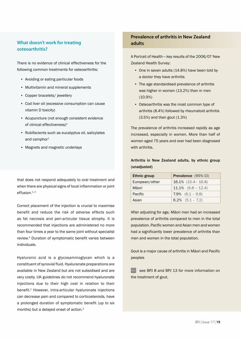
Prevalence of arthritis in New Zealand adults
A Portrait of Health — key results of the 2006/07 New Zealand Health Survey:
One in seven adults (14.8%) have been told by ▪a doctor they have arthritis
The age standardised prevalence of arthritis ▪was higher in women (13.2%) than in men (10.9%)
Osteoarthritis was the most common type of ▪arthritis (8.4%) followed by rheumatoid arthritis (3.5%) and then gout (1.3%)
The prevalence of arthritis increased rapidly as age increased, especially in women. More than half of women aged 75 years and over had been diagnosed with arthritis.
Arthritis in New Zealand adults, by ethnic group
(unadjusted)
Ethnic group Prevalence (95% CI)European/other 16.1% (15.4– 16.8)Māori 11.1% (9.8 – 12.4)Pacific 7.9% (6.1 – 9.8)Asian 6.2% (5.1 – 7.2)
After adjusting for age, Māori men had an increased prevalence of arthritis compared to men in the total population. Pacific women and Asian men and women had a significantly lower prevalence of arthritis than men and women in the total population.
Gout is a major cause of arthritis in Māori and Pacific peoples
see BPJ 8 and BPJ 13 for more information on the treatment of gout.
What doesn’t work for treating osteoarthritis?
There is no evidence of clinical effectiveness for the following common treatments for osteoarthritis:
Avoiding or eating particular foods ▪
Multivitamin and mineral supplements ▪
Copper bracelets/ jewellery ▪
Cod liver oil (excessive consumption can cause ▪vitamin D toxicity)
Acupuncture (not enough consistent evidence ▪of clinical effectiveness)1
Rubifacients such as eucalyptus oil, salicylates ▪and camphor1
Magnets and magnetic underlays ▪
BPJ | Issue 17 | 19
Surgical options
Patients who cannot obtain adequate pain relief and functional improvement from core treatments and are requiring strong opioids should be considered for joint replacement surgery.2 Patient specific factors such as age, gender, smoking, obesity and comorbidities should not be barriers to referral for surgery.1
Osteotomy and joint preserving surgical procedures can be considered in younger adults, to delay the need for joint replacement.2
Joint fusion may be used as first line surgical management of joints such as first metatarsophalangeal joints, subtalar and radiocarpal joints. Joint fusion can also be considered when joint replacement has failed.2
Joint lavage and arthroscopic debridement (removal of cartilage fragments and debris) are not always associated with a significant improvement in symptoms or mobility but may reduce locking.2 This procedure is not routinely recommended.1
References1. National Institute for Health and Clinical Excellence (NICE).
Osteoarthritis: The care and management of osteoarthritis in
adults. NICE clinical guideline 59. London, 2008.
2. Zhang W, Moskowitz R, Nuki M, et al. OARSI recommendations
for the management of hip and knee osteoarthritis, Part II: OARSI
evidence-based, expert consensus guidelines. Osteoarthritis
Cartilage 2008;16(2):137-62.
3. Towheed T, Maxwell L, Judd M, et al. Acetaminophen for
osteoarthritis. Cochrane Database Syst Rev 2006(1):CD004257.
4. Clinical Knowledge Summaries. Osteoarthritis management. UK:
National Library for Health, 2008.
5. Laine L, White W, Rostom A, Hochberg M. COX-2 selective
inhibitors in the treatment of osteoarthritis. Semin Arthritis Rheum
2008;[Epub ahead of print].
6. Cepeda M, Camargo F, Zea C, Valencia L. Tramadol for
osteoarthritis. Cochrane Database Syst Rev 2006(3):CD005522.
7. Towheed T, Maxwell L, Anastassiades T. Glucosamine therapy
for treating osteoarthritis. Cochrane Database Syst Rev
2005(2):CD002946.
20 | BPJ | Issue 17
www.bpac.org.nz keyword: DMARDS
Rheumatoid Arthritis – monitoring of DMARDs
Key concepts
Most people with rheumatoid arthritis require DMARD therapy to control symptoms and prevent joint ■damage. Treatment is initiated by a specialist as early as possible in the disease process.
Treatment usually begins with methotrexate and then other DMARDs such as sulphasalazine, ■hydroxychloroquine or leflunomide are added when inflammation is not controlled.
People prescribed DMARDs require close monitoring for adverse effects and drug interactions. ■
Key reviewers:
Professor John Highton, Head of Section, Department of Medical and Surgical Sciences, Dunedin School of Medicine, University of Otago.
Dr Andrew Harrison, Senior Lecturer, Rheumatology, Wellington School of Medicine, University of Otago, Wellington
Dr Rebecca Grainger, Rheumatologist and Clinical Research Fellow, Malaghan Institute of Medical Research, Wellington
22 | BPJ | Issue 17
Rheumatoid arthritis is a chronic autoimmune disease characterised by inflammation of the synovial tissue in joints causing swelling, pain, stiffness and joint destruction.1,2 Spontaneous remission is uncommon (<5%) and most affected individuals require long term disease modifying anti-rheumatic drug (DMARD) therapy to control symptoms and prevent joint damage.
A GPs role in the care of a patient with rheumatoid arthritis includes early referral for diagnosis and treatment, management of co-morbidities, co-ordination of secondary care and allied health care input and, in conjunction with the treating rheumatologist, monitoring of DMARD therapy.
This article covers important aspects of the care of patients taking DMARDs including monitoring requirements, adverse effects and drug interactions.
DMARDS are initiated in rheumatoid arthritis as soon as possible to prevent disease progression and reduce symptoms
The aim of treatment for rheumatoid arthritis is to achieve minimal joint inflammation (a therapeutic remission). Patients diagnosed with rheumatoid arthritis should start treatment with DMARDs as soon as possible, as early treatment has been shown to improve outcomes.3 Joint damage occurs early in the course of rheumatoid arthritis and is largely irreversible.1, 4 The degree of inflammation is closely related to joint damage therefore early control of inflammation should prevent joint damage. People with synovitis persisting for six weeks should be urgently referred to a rheumatologist.
Commonly used oral DMARDs include methotrexate, sulfasalazine, hydroxychloroquine, low-dose prednisone and a newer agent, leflunomide. Other less commonly used DMARDs include azathioprine, cyclosporin and sodium aurothiomalate (intramuscular gold). Biological DMARDS, tumour necrosis factor (TNF) inhibitors, are discussed on page 27.
Methotrexate is usually the first choice DMARD
Methotrexate is usually first line therapy in moderate to severe rheumatoid arthritis because it is effective, has a predictable adverse effect profile, is simple to administer and is inexpensive.3 In individuals with highly active disease methotrexate may be commenced in combination with other DMARDs. Mild disease may be treated with sulfasalazine or hydroxychloroquine monotherapy.
If methotrexate is not tolerated an alternative DMARD may be used as monotherapy.3
Add another DMARD when inflammation is not
controlled
If inflammation is not controlled, usually the next step is to add another DMARD such as sulfasalazine.
Triple therapy with methotrexate, sulfasalazine and hydroxychloroquine has been shown to be clinically effective and may also be used.1, 2
Other therapies include leflunomide, cyclosporin and
azathioprine
Leflunomide is available on special authority* for the treatment of rheumatoid arthritis that has not adequately responded to methotrexate and sulfasalazine. Leflunomide can be used alone or in combination with methotrexate. In clinical trials leflunomide had similar efficacy to methotrexate but may be less well tolerated, with additional adverse effects such as hair loss and hypertension.5
Cyclosporin and azathioprine are usually reserved for patients who are unresponsive to other DMARDs, due to the increased risk of adverse effects. Azathioprine is less well tolerated than methotrexate and cyclosporin is associated with nephrotoxicity. They can control inflammation but there is less evidence of their effect for long term treatment of rheumatoid arthritis.6
* Special Authority requirement is to be removed on 1st November 2008
BPJ | Issue 17 | 23
Biological DMARDs such as adalimumab and infliximab may be indicated if inflammation is uncontrolled by combination therapy (see page 27).2
Onset of action for DMARDs is between two to six
months
The onset of action for DMARDs varies. Response is seen with methotrexate within one to two months while hydroxychloroquine can take up to six months for a response.7
DMARDs require regular monitoring for toxicity
DMARDs require regular laboratory monitoring for adverse effects. A management plan should be agreed between the patient, GP and Rheumatologist. It should state which doctor is primarily responsible for arranging and reviewing the laboratory investigations.
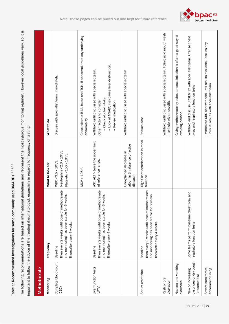
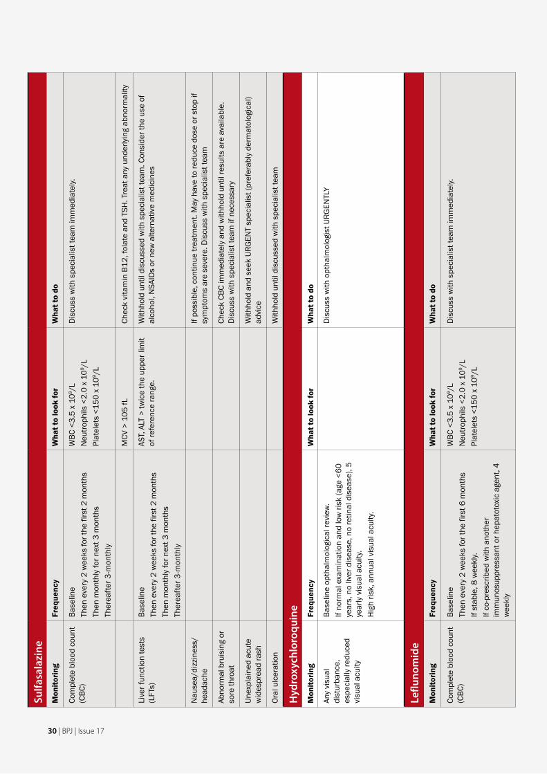
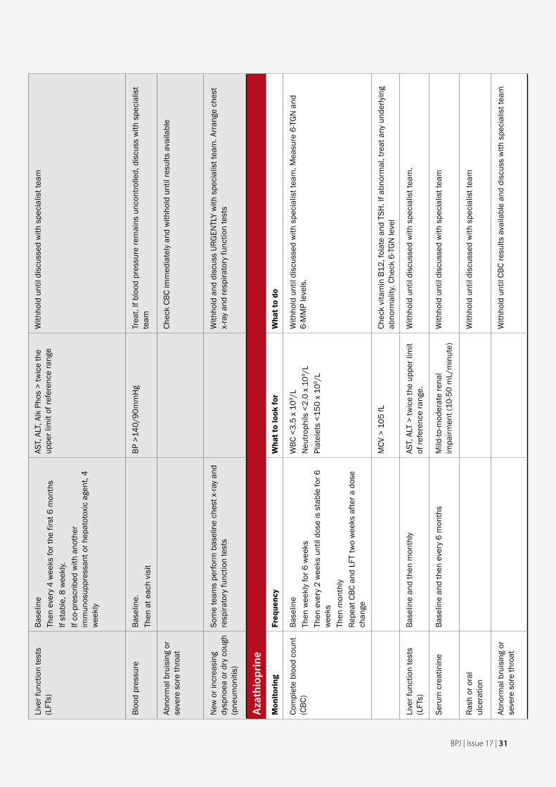
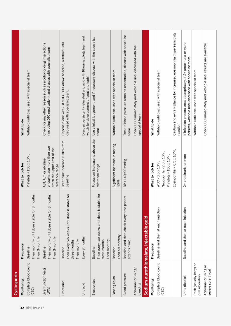
Recommended investigations for commonly used DMARDs are listed in Table 1 (see centre pages 29–32. Note: these pages can be pulled out for future reference). This includes recommended frequency of monitoring, what to look for and what to do about it. The frequency of monitoring varies between agents based on their likelihood of causing toxicity.
The table also includes clinical signs which may suggest toxicity. These clinical signs should be enquired about at consultations and patients should be advised to report them if they occur.
Set up a reminder on your PMS for monitoring.
Prescribing points for DMARDs
Methotrexate
Dosing — administered once a week orally or by ▪injection. Starting dose is 5–10 mg ONCE a week, increase by 2.5–10 mg every four to six weeks to a maximum of 25 mg ONCE a week.
Folic acid — 5mg folic acid, once per week (but not ▪on the same day as methotrexate), should always be prescribed.
Potential adverse effects — nausea, mouth ulcers, ▪hair loss, cytopaenias, elevated liver enzymes, rarely pneumonitis.3
Interactions — methotrexate accumulates in the ▪presence of renal impairment. Although this seldom has any clinical effect, patients with renal impairment, whether caused by NSAIDs, diuretics, dehydration or kidney disease should take lower doses and should be monitored carefully for any deterioration in renal function. Trimethoprim and cotrimoxazole interact with methotrexate and significantly increase the risk of marrow aplasia; the combination should be avoided.3
Alcohol — patients should be advised to limit alcohol ▪consumption to no more than one (females) or one and a half (males) standard drinks per day. This is roughly half of the level recommended for the general population. Liver function should be vigilantly monitored in patients who consume alcohol.
Flu vaccination — annual influenza vaccination ▪should be given but live vaccines should be avoided.8
Contraception — methotrexate is a known teratogen. ▪Effective contraception is required for women of child bearing potential taking methotrexate, or men taking methotrexate whose partner is of child bearing potential. Effective contraception needs to be continued for three months after stopping methotrexate.9
Sulfasalazine
Dosing — starting dose is 500 mg orally daily, ▪increase by 500 mg a week to a maximum of 40 mg/kg or 3g daily in divided doses.
Potential adverse effects — nausea, abdominal pain, ▪hair loss, cytopaenias, agranulocytosis, elevated liver enzymes, skin rashes.3
Interactions — potentially reduces the absorption of ▪digoxin, however the combination does not need to be avoided. Patients should be observed for signs
24 | BPJ | Issue 17
of under-digitalisation and digoxin levels should be measured if response is not adequate.10
Pregnancy — can be used in pregnancy but ▪doses should not exceed 2 g/day. Folic acid supplementation should be given during pregnancy and to women trying to conceive.8 Causes reversible oligospermia.6
Yellow discolouration — causes a yellow ▪discolouration of urine and tears; warn patients it may stain undergarments and soft contact lenses.9
Hydroxychloroquine
Dosing — starting dose is 400mg orally daily in ▪divided doses for one to three months (maximum 6mg/kg/day), then a maintenance dose of 200–400mg daily.
Potential adverse effects — blurred vision, skin rash, ▪photosensitivity, very rarely maculopathy. Blue-black discolouration of skin may occur with long-term use.3, 9
Photosensitivity and photophobia — may increase ▪the skin’s sensitivity to sunlight and also cause photophobia. Sunscreen is advised and patients should wear sunglasses in bright light.9
Leflunomide
Dosing — loading dose (optional) 100 mg orally once ▪daily for three days, then 10–20 mg once daily.
Potential adverse effects — GI disturbance, weight ▪loss, hair loss, rash or itch, mouth ulcers, headache, raised liver enzymes, cytopaenias, hypertension, rarely peripheral neuropathy and pneumonitis.3 There is an increased susceptibility to infections which should be treated promptly. With many of the adverse effects, discussion with the specialist team may result in dose reduction or trial of symptomatic treatment.
Interactions — concurrent use with other drugs that ▪have the potential to cause liver or marrow toxicity may increase the risk of these toxicities occurring.9 For example, the risk of pneumonitis is increased when leflunomide is combined with methotrexate.12
Alcohol — patients should be advised to limit alcohol ▪consumption8 to no more than one (females) or one and a half (males) standard drinks per day. This is roughly half of the level recommended for the general population. Liver function should be vigilantly monitored in patients who consume alcohol.
Flu vaccination — annual influenza vaccination is ▪recommended but live vaccines should be avoided.8
Contraception — leflunomide is very teratogenic. ▪Effective contraception is required for women for two years and men for three months after stopping leflunomide. Blood concentrations of its active metabolite should be measured before conception occurs.9
Washout — leflunomide has an extremely long ▪half life and can be retained in the body for up to two years. If toxicity occurs or for any other reason e.g. desire to conceive, a wash out procedure with cholestyramine may be considered.
BPJ | Issue 17 | 25
Additional prescribing points
Azathioprine. Dosing - 1 mg/kg orally daily, ▪increasing after four to six weeks to 2-3 mg/kg/day. Some rheumatology teams measure TPMT (Thiopurine Methyl Tranferase) at baseline. Low levels of this enzyme involved in the metabolism of azathioprine are an indication for reducing the dose. It is also possible to measure levels of azathioprine metabolites such as 6 Thioguanine (6 TGN). This may help guide treatment.
Gold injections (sodium aurothiomalate). Dosing ▪- 10 mg test dose (given in a clinic followed by 30 minutes observation), followed by weekly injections of 50 mg until significant response. Thereafter the interval between doses is increased in stages from 50 mg per week to 50 mg every four weeks. Nitritoid reactions (where the blood pressure falls) after injection of gold can occur. This should be checked after the first 10 mg dose of gold and thereafter. Concurrent use of ACE inhibitors may increase the incidence of nitritoid reactions. If a nitroid reaction occurs, gold injections should be withheld and discuss immediately with the Rheumatologist.
References:
1. National Prescribing Centre. Current issues in the drug treatment
of rheumatoid arthritis. MeRec Bulletin 2007; 17(5). Available
from: www.npc.co.uk/merec_index.htm (Accessed September
2008).
2. National Prescribing Service. Helping patients achieve remission
of rheumatoid arthritis. NPS News 2006; 48. Available from: www.
nps.org.au (Accessed September 2008).
3. Jones P. Medical management of rheumatoid arthritis. N Z Fam
Physician 2007; 34(6): 427-31.
4. O’Dell J. Therapeutic strategies for rheumatoid arthritis. N Engl J
Med 2004; 350: 2591-602.
5. Osiri M, Shea B, Robinson V, et al. Leflunomide for treating
rheumatoid arthritis. Cochrane Database Syst Rev 2003; 1:
CD002047.
6. Walker-Bone K, Fallow S. Rheumatoid arthritis. BMJ Clin Evid
2007;12: 1124.
7. Australian Medicines Handbook 2007.
8. Chakravarty K, McDonald H, Pullar T, et al. BSR/BHPR guideline
for disease-modifying anti-rheumatic drug (DMARD) therapy
in consultation with the British Association of Dermatologists.
Rheumatology 2008; 47(6): 924-5.
9. White CE, Cooper RG. Prescribing and monitoring of disease-
modifying anti-rheumatic drugs (DMARDs) for inflammatory
arthritis. Collected reports on the rheumatic diseases 2005.
Available from: www.arc.org.uk (Accessed September 2008).
10. Baxter K (ed). Stockley’s Drug Interactions. [online] London:
Pharmaceutical Press. Available from: www.medicinescomplete.
com (Accessed on September 2008).
11. Savage RL, Highton J, Boyd IW, Chapman P. Pneumonitis
associated with leflunomide: a profile of New Zealand and
Australian reports. Intern Med J 2006; 36(3):162-9.
References for Table 11. Chakravarty K, McDonald H, Pullar T, et al. BSR/BHPR guideline
for disease-modifying anti-rheumatic drug (DMARD) therapy
in consultation with the British Association of Dermatologists.
Rheumatology 2008; 47(6): 924-5
2. White CE, Cooper RG. Prescribing and monitoring of disease-
modifying anti-rheumatic drugs (DMARDs) for inflammatory
arthritis. Collected reports on the rheumatic diseases 2005.
Available from: www.arc.org.uk (Accessed September 2008).
3. Jones P. Medical management of rheumatoid arthritis. N Z Fam
Physician 2007; 34(6): 427-31.
4. Harrison A. Disease-modifying anti-rheumatic drugs (DMARDs)
for rheumatoid arthritis: benefits and risks. Medsafe Prescriber
Update 1999; 18: 4-12.
5. National Prescribing Service. Disease-modifying anti-rheumatic
drugs (DMARDs) for rheumatoid arthritis. Available from: www.nps.
org.au (Accessed September 2008).
6. Clinical Knowledge Summaries. DMARDs. Available from: http://
cks.library.nhs.uk/dmards# (Accessed September 2008).
26 | BPJ | Issue 17
What is their place in therapy?
In New Zealand TNF inhibitors are mainly used in individuals with rheumatoid arthritis which remains active despite optimal disease modifying anti-rheumatic drugs (DMARDs). Cochrane reviews have concluded that TNF inhibitors significantly reduce disease activity in rheumatoid arthritis compared to placebo.2, 3, 4
All three drugs are registered for use in rheumatoid arthritis, but only adalimumab is funded on special authority for this indication (see box below). Etanercept is funded on special authority for juvenile idiopathic arthritis. Patients who fail to respond to one TNF inhibitor, or who discontinue its use because of adverse effects, may respond to a second TNF inhibitor.3
Tumour necrosis factor inhibitors
Patients must have severe erosive rheumatoid arthritis that has not responded to a three month trial of each of the following; methotrexate, combination (triple) therapy, leflunomide or cyclosporin. They must also meet disease activity criteria (e.g. at least 20 joints affected). To continue to receive subsidy for
adalimumab patients must also show a 50% decrease in active joint count and a clinically significant response after four months of treatment.
For full details of special authority criteria see: www.pharmac.govt.nz/2008/10/01/SA0812.pdf
Adalimumab for rheumatoid arthritis – Pharmac criteria
What do they do?
Tumour necrosis factor (TNF) alpha, an inflammatory cytokine, is involved in the pathogenesis of rheumatoid arthritis.1 The three TNF inhibitors available in New Zealand (adalimumab, etanercept and infliximab) target this cytokine and block its effect. These parenteral agents are given by either self administered subcutaneous injection (adalimumab once a fortnight, etanercept weekly) or hospital administered intravenous infusion (infliximab every two months after induction).
BPJ | Issue 17 | 27
Safety concerns
The most common adverse effect with TNF inhibitors is injection site reactions. Reactions can be treated with the local application of ice or corticosteroid cream unless complicated by infection.5
Other more serious safety concerns are:
Reactivation of tuberculosis (TB) — most likely in ▪the first 12 months of treatment therefore extra vigilance is required during this time. British guidelines suggest screening all patients for TB prior to commencing treatment with a TNF inhibitor. Patients who are found to have latent or active TB should be treated.
Congestive heart failure — Infliximab has been ▪associated with an increase in mortality and hospitalisation due to cardiac failure. TNF inhibitors should not be started in people with Grade 3 or 4 congestive heart failure and used with caution in Grade 1 and 2. All patients on TNF inhibitors should be monitored for signs and symptoms of cardiac failure.6
Serious opportunistic infections — TNF inhibitors ▪should not be initiated in the presence of serious infections and extreme caution should be used in patients with increased risk of infection, e.g., bronchiectasis, history of chronic leg ulcers and history of septic arthritis. Patients should be advised of the increased risk of infection. Therapy should be discontinued if a serious infection develops but can be restarted once the infection has completely resolved.
Other contraindications to TNF inhibitors include a history of demyelinating disease, pregnancy and breastfeeding.6
Live vaccines should not be administered to individuals receiving TNF inhibitors.
Monitoring
No specific laboratory monitoring is required during TNF inhibitor therapy as haematological and liver test abnormalities are rarely caused by these agents. Most individuals will require ongoing laboratory monitoring for concomitant DMARD therapy (see Table 1, over page, for details on DMARD monitoring).
References:1. Australian Medicines Handbook 2006.
2. Navarro-Sarabia F, Ariza-Ariza R, Hernandez-Cruz B, Villanueva I.
Adalimumab for treating rheumatoid arthritis. Cochrane Database
Syst Rev 2005; 3: CD005113.
3. Blumenauer B, Judd M, Wells G, et al. Infliximab for the treatment
of rheumatoid arthritis. Cochrane Database Syst Rev 2002; 3:
CD003785.
4. Blumenauer B, Judd M, Cranney A, et al. Etanercept for the
treatment of rheumatoid arthritis. Cochrane Database Syst Rev
2003; 4: CD004525.
5. Jones P. Medical management of rheumatoid arthritis. N Z Fam
Physician 2007; 34(6): 427-31.
6. Ledingham J, Deighton C. Update on the British Society for
Rheumatology guidelines for prescribing TNFα blockers in adults
with rheumatoid arthritis (update of previous guidelines of April
2001). Rheumatology 2005; 44(2): 157-63.
28 | BPJ | Issue 17
Tabl
e 1:
Rec
omm
ende
d In
vest
igat
ions
for s
ome
com
mon
ly u
sed
DM
ARD
s1,2,
3,4,
5,6
The
follo
win
g re
com
men
datio
ns a
re b
ased
on
inte
rnat
iona
l gui
delin
es a
nd re
pres
ent t
he m
ost r
igor
ous
mon
itorin
g re
gim
en. H
owev
er lo
cal g
uide
lines
var
y, so
it is
im
port
ant t
o fo
llow
the
advi
ce o
f the
trea
ting
rheu
mat
olog
ist,
espe
cial
ly in
rega
rds
to fr
eque
ncy
of te
stin
g.
Met
hotr
exat
e
Mon
itorin
gFr
eque
ncy
Wha
t to
look
for
Wha
t to
do
Com
plet
e bl
ood
coun
t (C
BC)
Base
line
Then
eve
ry 2
wee
ks u
ntil
dose
of m
etho
trexa
te
and
mon
itorin
g ha
s be
en s
tabl
e fo
r 6 w
eeks
Ther
eafte
r eve
ry 4
wee
ks
WBC
<3.
5 x
109 /
LN
eutro
phils
<2.
0 x
109 /
LPl
atel
ets
<150
x 1
09 /L
Dis
cuss
with
spe
cial
ist t
eam
imm
edia
tely.
MCV
> 1
05 fL
Chec
k vi
tam
in B
12, f
olat
e an
d TS
H. I
f abn
orm
al, t
reat
any
und
erly
ing
abno
rmal
ity.
Live
r fun
ctio
n te
sts
(LFT
s)Ba
selin
e Th
en e
very
2 w
eeks
unt
il do
se o
f met
hotre
xate
an
d m
onito
ring
has
been
sta
ble
for 6
wee
ksTh
erea
fter e
very
4 w
eeks
AST,
ALT
> tw
ice
the
uppe
r lim
it of
refe
renc
e ra
nge.
With
hold
unt
il di
scus
sed
with
spe
cial
ist t
eam
. Ot
her f
acto
rs to
con
side
r:–
Chec
k al
coho
l int
ake.
–
Look
at N
SAID
; may
cau
se li
ver d
ysfu
nctio
n.
– Re
view
med
icat
ion
Unex
plai
ned
decr
ease
in
albu
min
(in
abse
nce
of a
ctiv
e di
seas
e)
With
hold
unt
il di
scus
sed
with
spe
cial
ist t
eam
Seru
m c
reat
inin
eBa
selin
e Th
en e
very
2 w
eeks
unt
il do
se o
f met
hotre
xate
an
d m
onito
ring
has
been
sta
ble
for 6
wee
ksTh
erea
fter e
very
4 w
eeks
Sign
ifica
nt d
eter
iora
tion
in re
nal
func
tion
Redu
ce d
ose
Rash
or o
ral
ulce
ratio
nW
ithho
ld u
ntil
disc
usse
d w
ith s
peci
alis
t tea
m. F
olin
ic a
cid
mou
th w
ash
may
hel
p w
ith m
ucos
itis.
Nau
sea
and
vom
iting
, di
arrh
oea
Giv
ing
met
hotre
xate
by
subc
utan
eous
inje
ctio
n is
ofte
n a
good
way
of
avoi
ding
nau
sea.
New
or i
ncre
asin
g dy
spno
ea o
r dry
cou
gh
(pne
umon
itis)
Som
e te
ams
perfo
rm b
asel
ine
ches
t x-ra
y an
d re
spira
tory
func
tion
test
sW
ithho
ld a
nd d
iscu
ss U
RGEN
TLY
with
spe
cial
ist t
eam
. Arr
ange
che
st
x-ra
y an
d re
spira
tory
func
tion
test
s
Seve
re s
ore
thro
at,
abno
rmal
bru
isin
gIm
med
iate
CBC
and
with
hold
unt
il re
sults
ava
ilabl
e. D
iscu
ss a
ny
unus
ual r
esul
ts w
ith s
peci
alis
t tea
m
nzbpacbetter edicin m eNote: These pages can be pulled out and kept for future reference.
BPJ | Issue 17 | 29
Sulfa
sala
zine
Mon
itorin
gFr
eque
ncy
Wha
t to
look
for
Wha
t to
do
Com
plet
e bl
ood
coun
t (C
BC)
Base
line
Then
eve
ry 2
wee
ks fo
r the
firs
t 2 m
onth
sTh
en m
onth
ly fo
r nex
t 3 m
onth
sTh
erea
fter 3
-mon
thly
WBC
<3.
5 x
109 /
LN
eutro
phils
<2.
0 x
109 /
LPl
atel
ets
<150
x 1
09 /L
Dis
cuss
with
spe
cial
ist t
eam
imm
edia
tely.
MCV
> 1
05 fL
Chec
k vi
tam
in B
12, f
olat
e an
d TS
H. T
reat
any
und
erly
ing
abno
rmal
ity
Live
r fun
ctio
n te
sts
(LFT
s)Ba
selin
eTh
en e
very
2 w
eeks
for t
he fi
rst 2
mon
ths
Then
mon
thly
for n
ext 3
mon
ths
Ther
eafte
r 3-m
onth
ly
AST,
ALT
> tw
ice
the
uppe
r lim
it of
refe
renc
e ra
nge.
With
hold
unt
il di
scus
sed
with
spe
cial
ist t
eam
. Con
side
r the
use
of
alco
hol,
NSA
IDs
or n
ew a
ltern
ativ
e m
edic
ines
Nau
sea/
dizz
ines
s/he
adac
heIf
poss
ible
, con
tinue
trea
tmen
t. M
ay h
ave
to re
duce
dos
e or
sto
p if
sym
ptom
s ar
e se
vere
. Dis
cuss
with
spe
cial
ist t
eam
Abno
rmal
bru
isin
g or
so
re th
roat
Chec
k CB
C im
med
iate
ly a
nd w
ithho
ld u
ntil
resu
lts a
re a
vaila
ble.
D
iscu
ss w
ith s
peci
alis
t tea
m if
nec
essa
ry
Unex
plai
ned
acut
e w
ides
prea
d ra
shW
ithho
ld a
nd s
eek
URG
ENT
spec
ialis
t (pr
efer
ably
der
mat
olog
ical
) ad
vice
Oral
ulc
erat
ion
With
hold
unt
il di
scus
sed
with
spe
cial
ist t
eam
Hyd
roxy
chlo
roqu
ine
Mon
itorin
gFr
eque
ncy
Wha
t to
look
for
Wha
t to
do
Any
visu
al
dist
urba
nce,
es
peci
ally
redu
ced
visu
al a
cuity
Base
line
opth
alm
olog
ical
revi
ew.
If no
rmal
exa
min
atio
n an
d lo
w ri
sk (a
ge <
60
year
s, n
o liv
er d
isea
se, n
o re
tinal
dis
ease
), 5
year
ly v
isua
l acu
ity.
Hig
h ris
k, a
nnua
l vis
ual a
cuity
.
Dis
cuss
with
opt
halm
olog
ist U
RGEN
TLY
Leflu
nom
ide
Mon
itorin
gFr
eque
ncy
Wha
t to
look
for
Wha
t to
do
Com
plet
e bl
ood
coun
t (C
BC)
Base
line
Then
eve
ry 2
wee
ks fo
r the
firs
t 6 m
onth
sIf
stab
le, 8
wee
kly.
If co
-pre
scrib
ed w
ith a
noth
er
imm
unos
uppr
essa
nt o
r hep
atot
oxic
age
nt, 4
w
eekl
y
WBC
<3.
5 x
109 /
LN
eutro
phils
<2.
0 x
109 /
LPl
atel
ets
<150
x 1
09 /L
Dis
cuss
with
spe
cial
ist t
eam
imm
edia
tely.
30 | BPJ | Issue 17
Live
r fun
ctio
n te
sts
(LFT
s)Ba
selin
e Th
en e
very
4 w
eeks
for t
he fi
rst 6
mon
ths
If st
able
, 8 w
eekl
y. If
co-p
resc
ribed
with
ano
ther
im
mun
osup
pres
sant
or h
epat
otox
ic a
gent
, 4
wee
kly
AST,
ALT
, Alk
Pho
s >
twic
e th
e up
per l
imit
of re
fere
nce
rang
eW
ithho
ld u
ntil
disc
usse
d w
ith s
peci
alis
t tea
m
Bloo
d pr
essu
reBa
selin
e.Th
en a
t eac
h vi
sit
BP >
140/
90m
mH
gTr
eat.
If bl
ood
pres
sure
rem
ains
unc
ontro
lled,
dis
cuss
with
spe
cial
ist
team
Abno
rmal
bru
isin
g or
se
vere
sor
e th
roat
Chec
k CB
C im
med
iate
ly a
nd w
ithho
ld u
ntil
resu
lts a
vaila
ble
New
or i
ncre
asin
g dy
spno
ea o
r dry
cou
gh
(pne
umon
itis)
Som
e te
ams
perfo
rm b
asel
ine
ches
t x-ra
y an
d re
spira
tory
func
tion
test
sW
ithho
ld a
nd d
iscu
ss U
RGEN
TLY
with
spe
cial
ist t
eam
. Arr
ange
che
st
x-ra
y an
d re
spira
tory
func
tion
test
s
Aza
thio
prin
eM
onito
ring
Freq
uenc
yW
hat t
o lo
ok fo
rW
hat t
o do
Com
plet
e bl
ood
coun
t (C
BC)
Base
line
Then
wee
kly
for 6
wee
ksTh
en e
very
2 w
eeks
unt
il do
se is
sta
ble
for 6
w
eeks
Th
en m
onth
lyRe
peat
CBC
and
LFT
two
wee
ks a
fter a
dos
e ch
ange
WBC
<3.
5 x
109 /
LN
eutro
phils
<2.
0 x
109 /
LPl
atel
ets
<150
x 1
09 /L
With
hold
unt
il di
scus
sed
with
spe
cial
ist t
eam
. Mea
sure
6-T
GN
and
6-
MM
P le
vels
.
MCV
> 1
05 fL
Chec
k vi
tam
in B
12, f
olat
e an
d TS
H. I
f abn
orm
al, t
reat
any
und
erly
ing
abno
rmal
ity. C
heck
6-T
GN
leve
l
Live
r fun
ctio
n te
sts
(LFT
s)Ba
selin
e an
d th
en m
onth
lyAS
T, A
LT >
twic
e th
e up
per l
imit
of re
fere
nce
rang
e.W
ithho
ld u
ntil
disc
usse
d w
ith s
peci
alis
t tea
m.
Seru
m c
reat
inin
eBa
selin
e an
d th
en e
very
6 m
onth
sM
ild-to
-mod
erat
e re
nal
impa
irmen
t (10
-50
mL/
min
ute)
With
hold
unt
il di
scus
sed
with
spe
cial
ist t
eam
Rash
or o
ral
ulce
ratio
nW
ithho
ld u
ntil
disc
usse
d w
ith s
peci
alis
t tea
m
Abno
rmal
bru
isin
g or
se
vere
sor
e th
roat
With
hold
unt
il CB
C re
sults
ava
ilabl
e an
d di
scus
s w
ith s
peci
alis
t tea
m
BPJ | Issue 17 | 31
Cycl
ospo
rin
Mon
itorin
gFr
eque
ncy
Wha
t to
look
for
Wha
t to
do
Com
plet
e bl
ood
coun
t (C
BC)
Base
line
Then
mon
thly
unt
il do
se s
tabl
e fo
r 3 m
onth
sTh
en 3
-mon
thly
Plat
elet
s <1
50 x
109 /
LW
ithho
ld u
ntil
disc
usse
d w
ith s
peci
alis
t tea
m
Live
r fun
ctio
n te
sts
(LFT
s)Ba
selin
eTh
en m
onth
ly u
ntil
dose
sta
ble
for 3
mon
ths
Then
3-m
onth
ly
AST,
ALT
, or a
lkal
ine
phos
phat
ase
mor
e th
an tw
o tim
es th
e up
per l
imit
of th
e re
fere
nce
rang
e
Chec
k fo
r any
oth
er re
ason
suc
h as
alc
ohol
or d
rug
inte
ract
ions
(in
clud
ing
OTC
med
icat
ion)
, and
dis
cuss
with
spe
cial
ist t
eam
Crea
tinin
eBa
selin
eTh
en e
very
two
wee
ks u
ntil
dose
is s
tabl
e fo
r th
ree
mon
ths
Then
mon
thly.
Crea
tinin
e in
crea
se >
30%
from
ba
selin
eRe
peat
in o
ne w
eek,
if s
till >
30%
abo
ve b
asel
ine,
with
hold
unt
il di
scus
sed
with
spe
cial
ist t
eam
.
Uric
aci
dEv
ery
3 m
onth
s.D
iscu
ss p
ersi
sten
tly e
leva
ted
uric
aci
d w
ith R
heum
atol
ogy
team
and
w
atch
for d
evel
opm
ent o
f gou
t and
toph
i.
Elec
troly
tes
Base
line
Then
eve
ry tw
o w
eeks
unt
il do
se is
sta
ble
for
thre
e m
onth
sTh
en m
onth
ly.
Pota
ssiu
m in
crea
se to
abo
ve th
e re
fere
nce
rang
eUs
e cl
inic
al ju
dgem
ent,
and
if ne
cess
ary
disc
uss
with
the
spec
ialis
t te
am
Fast
ing
lipid
sBa
selin
eTh
en s
ix m
onth
lySi
gnifi
cant
incr
ease
in fa
stin
g lip
ids
With
hold
unt
il di
scus
sed
with
spe
cial
ist t
eam
Bloo
d pr
essu
reBa
selin
e an
d th
en c
heck
eve
ry ti
me
patie
nt
atte
nds
clin
icBP
>14
0/90
mm
Hg
Trea
t. If
bloo
d pr
essu
re re
mai
ns u
ncon
trolle
d, d
iscu
ss w
ith s
peci
alis
t te
am
Abno
rmal
bru
isin
g/bl
eedi
ngCh
eck
CBC
imm
edia
tely
and
with
hold
unt
il di
scus
sed
with
the
spec
ialis
t tea
m
Sodi
um a
urot
hiom
alat
e, in
ject
able
gol
dM
onito
ring
Freq
uenc
yW
hat t
o lo
ok fo
rW
hat t
o do
Com
plet
e bl
ood
coun
t (C
BC)
Base
line
and
then
at e
ach
inje
ctio
nW
BC <
3.5
x 10
9 /L
Neu
troph
ils <
2.0
x 10
9 /L
Plat
elet
s <1
50 x
109 /
L
With
hold
unt
il di
scus
sed
with
spe
cial
ist t
eam
Eosi
noph
ilia
> 0.
5 x
109 /
LCa
utio
n an
d ex
tra v
igila
nce
for i
ncre
ased
eos
inop
hilia
(hyp
erse
nsiti
vity
re
actio
n)
Urin
e di
pstic
kBa
selin
e an
d th
en a
t eac
h in
ject
ion
2+ p
rote
inur
ia o
r mor
eIf
infe
ctio
n pr
esen
t tre
at a
ppro
pria
tely.
If 2
+ pr
otei
nuria
or m
ore
pers
ists
, with
hold
unt
il di
scus
sed
with
spe
cial
ist t
eam
.
Rash
(usu
ally
itch
y) o
r or
al u
lcer
atio
nW
ithho
ld u
ntil
disc
usse
d w
ith s
peci
alis
t tea
m
Abno
rmal
bru
isin
g or
se
vere
sor
e th
roat
Chec
k CB
C im
med
iate
ly a
nd w
ithho
ld u
ntil
resu
lts a
re a
vaila
ble
32 | BPJ | Issue 17