Embed Size (px)
Citation preview
BISP 194: Neural Prostheses Overview and Introduction
BISP 194 Special Topics in Modern Biology
Neural ProsthesesWinter 2008
Overview and Introduction
Gert [email protected]
BISP 194: Neural Prostheses Overview and Introduction
Neural Prostheses(aka Neural Prostheticsaka Neuroprosthetics)
(the art of designing) devices which restore or supplement function of the nervous system lost by disease or injury
• Highly interdisciplinary subject– at the interface between neuroscience and engineering
• analysis vs. synthesis• Emerging discipline
– rapid advances in technology– slow transition from neuroscience/engineering research to
medical practice• Several opportunities for learning
– learning about the latest advances in neurotechnology– learning to interpret and critically evaluate the literature
BISP 194: Neural Prostheses Overview and Introduction
Neural ProsthesesOverview
• Sensory prostheses– Cochlear implant for restored hearing
• The oldest, and the only widely used neural prosthesis– Retinal implant for restored vision
• Largest current R&D investment (Second Sight in USA, and severalgroups in Taiwan, Japan, and Germany)
• Motor prostheses– Functional neuromuscular stimulation
• Brain prostheses– Deep-brain stimulation
• Parkinson tremor remediation– Neuromorphic and biomorphic systems
BISP 194: Neural Prostheses Overview and Introduction
Sensory Prostheses
Cochlear ImplantRetinal Implant
BISP 194: Neural Prostheses Overview and Introduction
Cochlear Implant
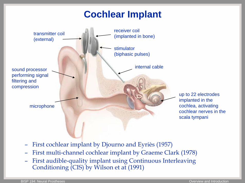
– First cochlear implant by Djourno and Eyriès (1957)– First multi-channel cochlear implant by Graeme Clark (1978) – First audible-quality implant using Continuous Interleaving
Conditioning (CIS) by Wilson et at (1991)
microphone
sound processor performing signal filtering and compression
transmitter coil (external)
receiver coil (implanted in bone)
stimulator (biphasic pulses)
internal cable
up to 22 electrodes implanted in the cochlea, activating cochlear nerves in the scala tympani
BISP 194: Neural Prostheses Overview and Introduction
Cochlea Cross Section
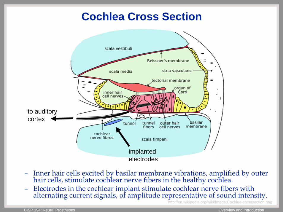
implanted electrodes
to auditory cortex
– Inner hair cells excited by basilar membrane vibrations, amplified by outer hair cells, stimulate cochlear nerve fibers in the healthy cochlea.
– Electrodes in the cochlear implant stimulate cochlear nerve fibers with alternating current signals, of amplitude representative of sound intensity.
http://en.wikipedia.org/wiki/Image:Cochlea-crosssection.png
BISP 194: Neural Prostheses Overview and Introduction
Silicon Cochlea and Auditory Periphery
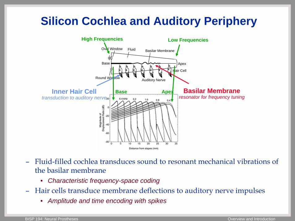
Basilar Membrane
Base Apex
Round Window
Oval Window Fluid
Hair Cell
Auditory Nerve
Inner Hair Celltransduction to auditory nerve
Basilar Membraneresonator for frequency tuning
High Frequencies Low Frequencies
ApexBase
– Fluid-filled cochlea transduces sound to resonant mechanical vibrations of the basilar membrane
• Characteristic frequency-space coding– Hair cells transduce membrane deflections to auditory nerve impulses
• Amplitude and time encoding with spikes
BISP 194: Neural Prostheses Overview and Introduction
Retinal Implant
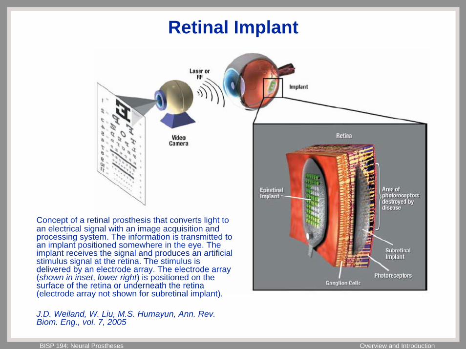
Concept of a retinal prosthesis that converts light to an electrical signal with an image acquisition and processing system. The information is transmitted to an implant positioned somewhere in the eye. The implant receives the signal and produces an artificial stimulus signal at the retina. The stimulus is delivered by an electrode array. The electrode array (shown in inset, lower right) is positioned on the surface of the retina or underneath the retina (electrode array not shown for subretinal implant).
J.D. Weiland, W. Liu, M.S. Humayun, Ann. Rev. Biom. Eng., vol. 7, 2005
BISP 194: Neural Prostheses Overview and Introduction
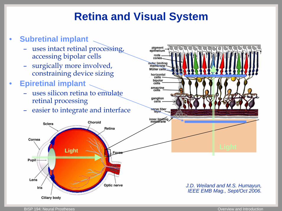
Retina and Visual System
• Subretinal implant– uses intact retinal processing,
accessing bipolar cells– surgically more involved,
constraining device sizing • Epiretinal implant
– uses silicon retina to emulate retinal processing
– easier to integrate and interface
LightLight
J.D. Weiland and M.S. Humayun, IEEE EMB Mag., Sept/Oct 2006.
BISP 194: Neural Prostheses Overview and Introduction
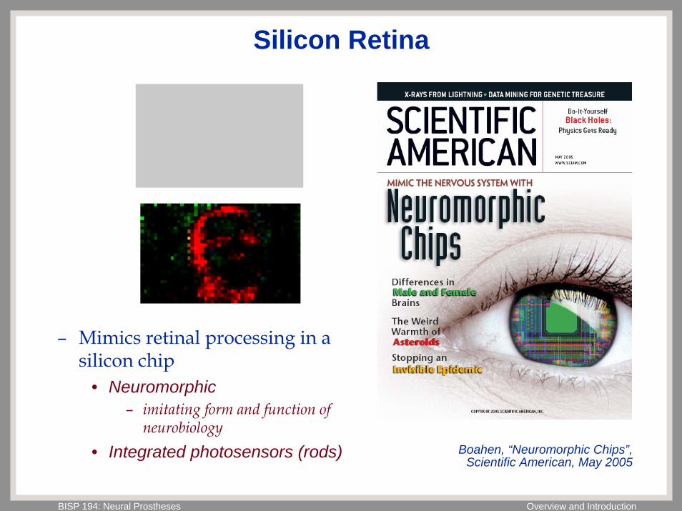
Silicon Retina
– Mimics retinal processing in a silicon chip
• Neuromorphic– imitating form and function of
neurobiology• Integrated photosensors (rods) Boahen, “Neuromorphic Chips”,
Scientific American, May 2005
BISP 194: Neural Prostheses Overview and Introduction
Brain Prostheses
Deep Brain StimulationCortical Vision Prostheses
Implantable Electrode Arrays
BISP 194: Neural Prostheses Overview and Introduction
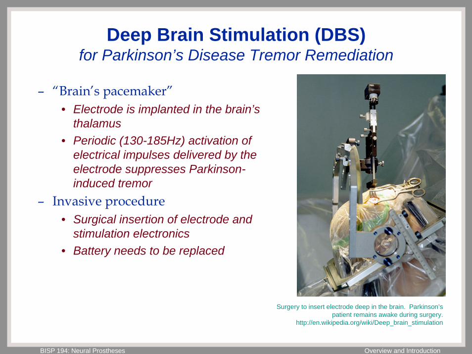
Deep Brain Stimulation (DBS)for Parkinson’s Disease Tremor Remediation
– “Brain’s pacemaker”• Electrode is implanted in the brain’s
thalamus• Periodic (130-185Hz) activation of
electrical impulses delivered by the electrode suppresses Parkinson-induced tremor
– Invasive procedure• Surgical insertion of electrode and
stimulation electronics• Battery needs to be replaced
Surgery to insert electrode deep in the brain. Parkinson’s patient remains awake during surgery.
http://en.wikipedia.org/wiki/Deep_brain_stimulation
BISP 194: Neural Prostheses Overview and Introduction
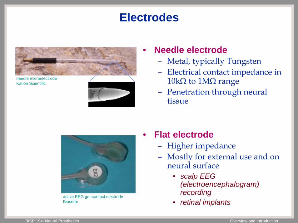
Electrodes
• Needle electrode– Metal, typically Tungsten– Electrical contact impedance in
10kΩ to 1MΩ range– Penetration through neural
tissue
• Flat electrode– Higher impedance– Mostly for external use and on
neural surface• scalp EEG
(electroencephalogram) recording
• retinal implants
needle microelectrodeKation Scientific
active EEG gel-contact electrodeBiosemi
BISP 194: Neural Prostheses Overview and Introduction
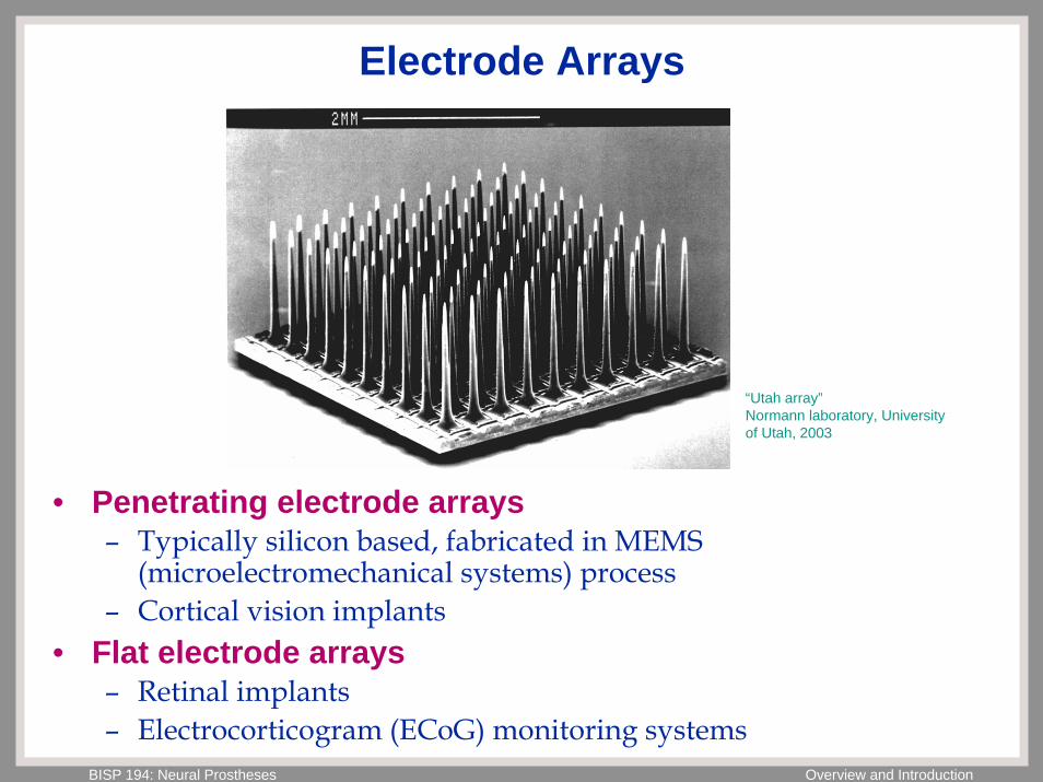
Electrode Arrays
“Utah array”Normann laboratory, University of Utah, 2003
• Penetrating electrode arrays– Typically silicon based, fabricated in MEMS
(microelectromechanical systems) process– Cortical vision implants
• Flat electrode arrays– Retinal implants– Electrocorticogram (ECoG) monitoring systems
BISP 194: Neural Prostheses Overview and Introduction
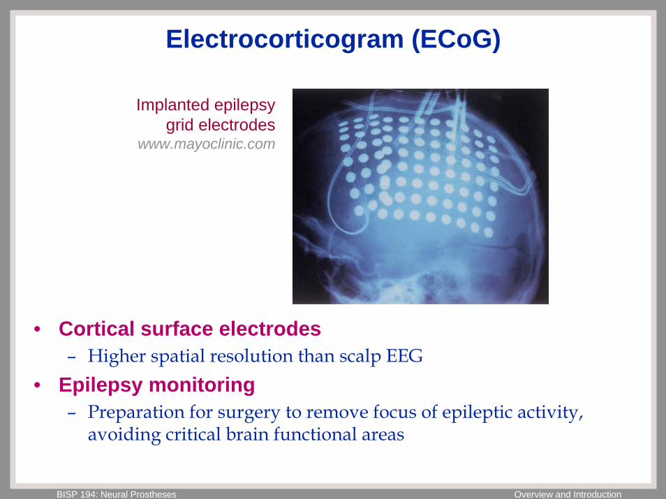
Electrocorticogram (ECoG)
Implanted epilepsy grid electrodes
www.mayoclinic.com
• Cortical surface electrodes– Higher spatial resolution than scalp EEG
• Epilepsy monitoring– Preparation for surgery to remove focus of epileptic activity,
avoiding critical brain functional areas
BISP 194: Neural Prostheses Overview and Introduction
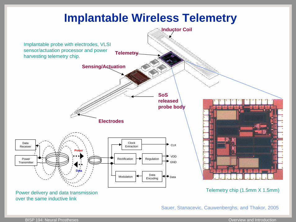
Implantable Wireless Telemetry
• Transcutaneous wires limit the application of implantable sensing/actuation technology to neural prostheses– Risk of infection
• Opening through the skin reduces the body’s natural defense against invading microorganisms
– Limited mobility• Tethered to power source and data logging instrumentation
• Wireless technology is widely available, however:– Frequency range of radio transmission is limited by the body’s
absorption spectra and safety considerations• Magnetic (inductive) coupling at low frequency, ~1-4 MHz• Very low transmitted power requires efficient low-power design
Sauer, Stanacevic, Cauwenberghs, and Thakor, 2005
BISP 194: Neural Prostheses Overview and Introduction
Implantable Wireless Telemetry
Regulation
Modulation Data Encoding
Clock Extraction CLK
VDD
GND
Data
Data
Power
Data Receiver
RectificationPower Transmitter
Sensing/Actuation
Telemetry
Inductor Coil
Electrodes
SoS released probe body
Implantable probe with electrodes, VLSI sensor/actuation processor and power harvesting telemetry chip.
Telemetry chip (1.5mm X 1.5mm)Power delivery and data transmission over the same inductive link
Sauer, Stanacevic, Cauwenberghs, and Thakor, 2005
BISP 194: Neural Prostheses Overview and Introduction
Motor Prostheses
Brain Machine (Computer) InterfacesImplantable Electrode Arrays
BISP 194: Neural Prostheses Overview and Introduction
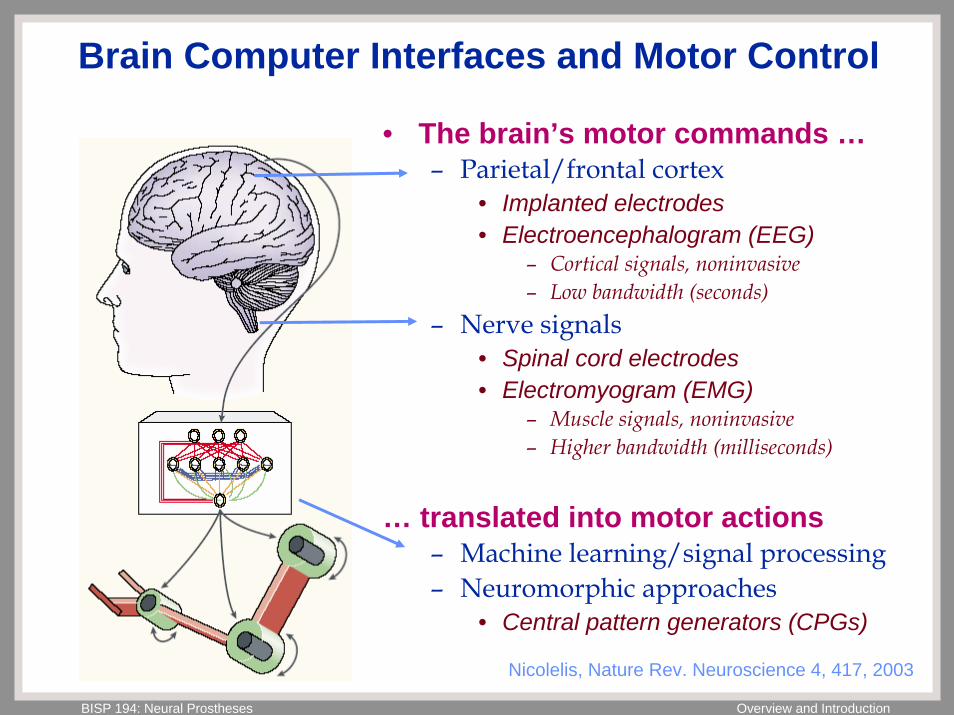
Brain Computer Interfaces and Motor Control
• The brain’s motor commands …– Parietal/frontal cortex
• Implanted electrodes• Electroencephalogram (EEG)
– Cortical signals, noninvasive– Low bandwidth (seconds)
– Nerve signals• Spinal cord electrodes• Electromyogram (EMG)
– Muscle signals, noninvasive– Higher bandwidth (milliseconds)
… translated into motor actions– Machine learning/signal processing– Neuromorphic approaches
• Central pattern generators (CPGs)
Nicolelis, Nature Rev. Neuroscience 4, 417, 2003
BISP 194: Neural Prostheses Overview and Introduction
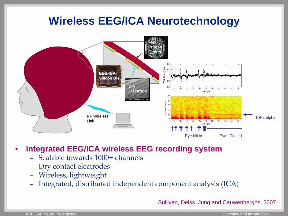
Wireless EEG/ICA Neurotechnology
10Hz alpha
Eye blinks Eyes Closed
RF Wireless Link
EEG/ICA Silicon Die
Dry Electrode
Flex Printed Circuit
• Integrated EEG/ICA wireless EEG recording system– Scalable towards 1000+ channels– Dry contact electrodes– Wireless, lightweight– Integrated, distributed independent component analysis (ICA)
Sullivan, Deiss, Jung and Cauwenberghs, 2007
BISP 194: Neural Prostheses Overview and Introduction
Emerging Technologies
Nanotechnology– Nanoparticles interacting with cells– Carbon nanofibers for enduring nanoelectrodes
Molecular optics– ChR2 optical activation of targeted neurons– NPhR optical inactivation of targeted neurons
Spinal cord regeneration
Others …
BISP 194: Neural Prostheses Overview and Introduction
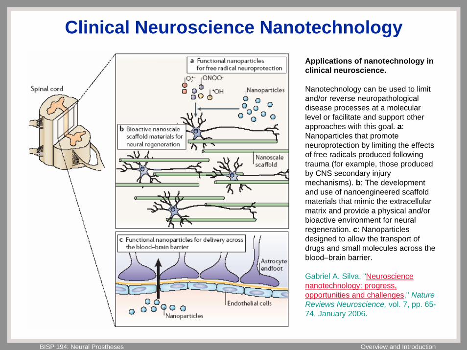
Clinical Neuroscience NanotechnologyApplications of nanotechnology in clinical neuroscience.
Nanotechnology can be used to limit and/or reverse neuropathologicaldisease processes at a molecular level or facilitate and support other approaches with this goal. a: Nanoparticles that promote neuroprotection by limiting the effects of free radicals produced following trauma (for example, those produced by CNS secondary injury mechanisms). b: The development and use of nanoengineered scaffoldmaterials that mimic the extracellularmatrix and provide a physical and/or bioactive environment for neural regeneration. c: Nanoparticlesdesigned to allow the transport of drugs and small molecules across the blood–brain barrier.
Gabriel A. Silva, "Neuroscience nanotechnology: progress, opportunities and challenges," Nature Reviews Neuroscience, vol. 7, pp. 65-74, January 2006.
BISP 194: Neural Prostheses Overview and Introduction
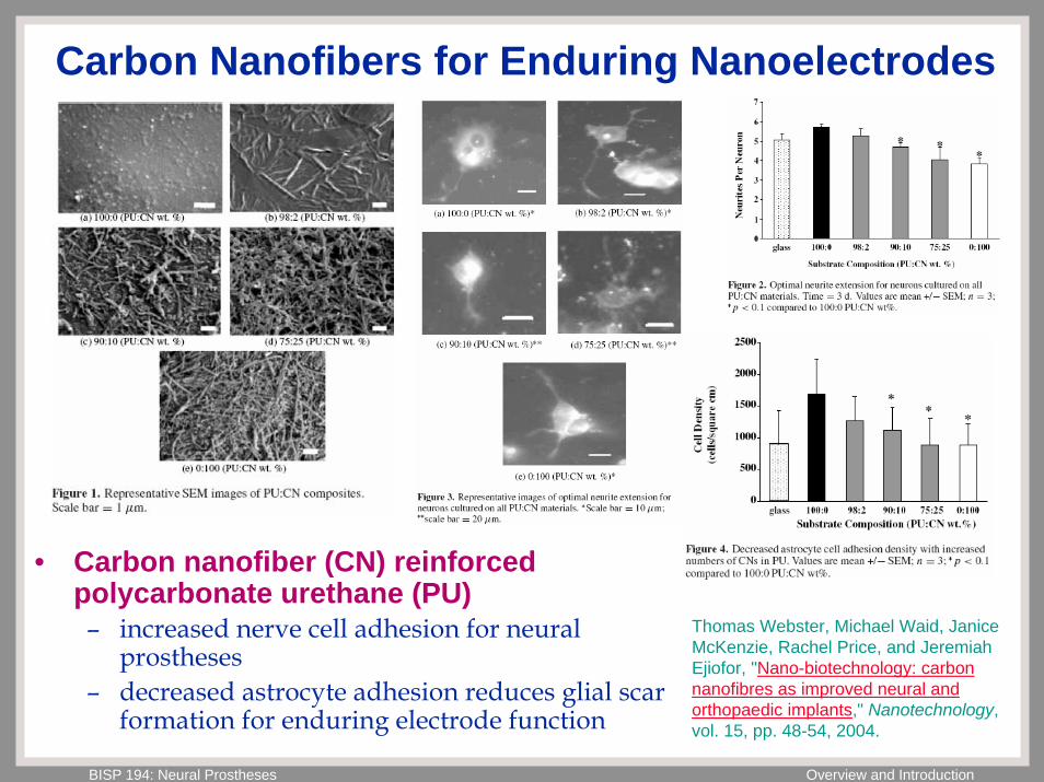
Carbon Nanofibers for Enduring Nanoelectrodes
• Carbon nanofiber (CN) reinforced polycarbonate urethane (PU)– increased nerve cell adhesion for neural
prostheses– decreased astrocyte adhesion reduces glial scar
formation for enduring electrode function
Thomas Webster, Michael Waid, Janice McKenzie, Rachel Price, and Jeremiah Ejiofor, "Nano-biotechnology: carbon nanofibres as improved neural and orthopaedic implants," Nanotechnology, vol. 15, pp. 48-54, 2004.
BISP 194: Neural Prostheses Overview and Introduction
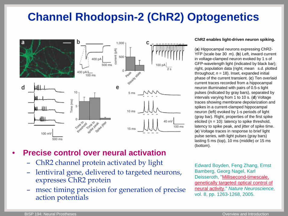
Channel Rhodopsin-2 (ChR2) Optogenetics
ChR2 enables light-driven neuron spiking.
(a) Hippocampal neurons expressing ChR2-YFP (scale bar 30 m). (b) Left, inward current in voltage-clamped neuron evoked by 1 s of GFP-wavelength light (indicated by black bar); right, population data (right; mean s.d. plotted throughout; n = 18). Inset, expanded initial phase of the current transient. (c) Ten overlaid current traces recorded from a hippocampalneuron illuminated with pairs of 0.5-s light pulses (indicated by gray bars), separated by intervals varying from 1 to 10 s. (d) Voltage traces showing membrane depolarization and spikes in a current-clamped hippocampalneuron (left) evoked by 1-s periods of light (gray bar). Right, properties of the first spike elicited (n = 10): latency to spike threshold, latency to spike peak, and jitter of spike time. (e) Voltage traces in response to brief light pulse series, with light pulses (gray bars) lasting 5 ms (top), 10 ms (middle) or 15 ms (bottom).
Edward Boyden, Feng Zhang, Ernst Bamberg, Georg Nagel, Karl Deisseroth, "Millisecond-timescale, genetically targeted optical control of neural activity," Nature Neuroscience,vol. 8, pp. 1263-1268, 2005.
• Precise control over neural activation– ChR2 channel protein activated by light– lentiviral gene, delivered to targeted neurons,
expresses ChR2 protein– msec timing precision for generation of precise
action potentials
BISP 194: Neural Prostheses Overview and Introduction
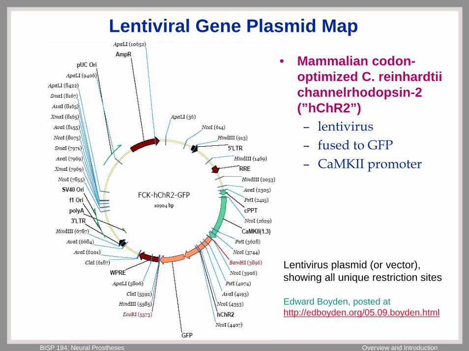
Lentiviral Gene Plasmid Map
Lentivirus plasmid (or vector), showing all unique restriction sites
Edward Boyden, posted at http://edboyden.org/05.09.boyden.html
• Mammalian codon-optimized C. reinhardtiichannelrhodopsin-2 (”hChR2”)– lentivirus– fused to GFP– CaMKII promoter
BISP 194: Neural Prostheses Overview and Introduction
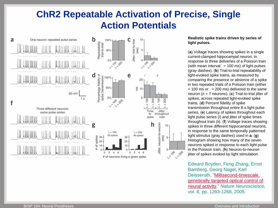
ChR2 Repeatable Activation of Precise, Single Action Potentials
Realistic spike trains driven by series of light pulses.
(a) Voltage traces showing spikes in a single current-clamped hippocampal neuron, in response to three deliveries of a Poisson train (with mean interval = 100 ms) of light pulses (gray dashes). (b) Trial-to-trial repeatability of light-evoked spike trains, as measured by comparing the presence or absence of a spike in two repeated trials of a Poisson train (either = 100 ms or = 200 ms) delivered to the same neuron (n = 7 neurons). (c) Trial-to-trial jitter of spikes, across repeated light-evoked spike trains. (d) Percent fidelity of spike transmission throughout entire 8-s light pulse series. (e) Latency of spikes throughout each light pulse series (i) and jitter of spike times throughout train (ii). (f) Voltage traces showing spikes in three different hippocampal neurons, in response to the same temporally patterned light stimulus (gray dashes) used in a. (g) Histogram showing how many of the seven neurons spiked in response to each light pulse in the Poisson train. (h) Neuron-to-neuron jitter of spikes evoked by light stimulation.
Edward Boyden, Feng Zhang, Ernst Bamberg, Georg Nagel, Karl Deisseroth, "Millisecond-timescale, genetically targeted optical control of neural activity," Nature Neuroscience,vol. 8, pp. 1263-1268, 2005.
BISP 194: Neural Prostheses Overview and Introduction
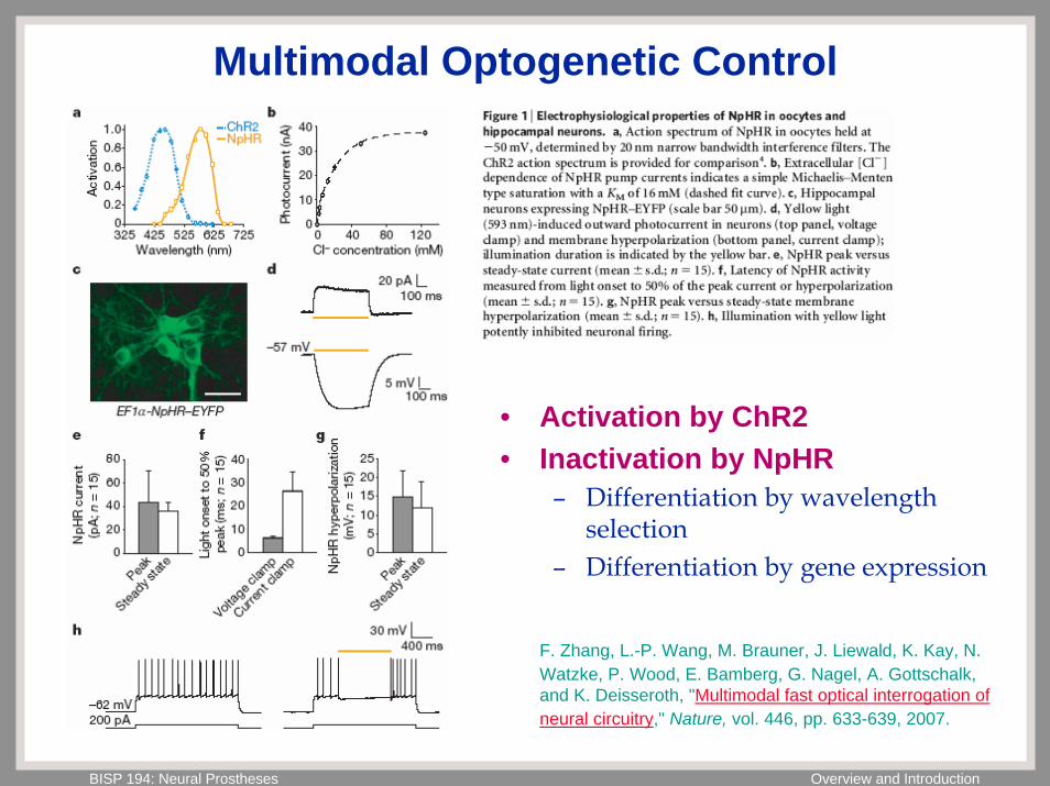
Multimodal Optogenetic Control
• Activation by ChR2• Inactivation by NpHR
– Differentiation by wavelength selection
– Differentiation by gene expression
F. Zhang, L.-P. Wang, M. Brauner, J. Liewald, K. Kay, N. Watzke, P. Wood, E. Bamberg, G. Nagel, A. Gottschalk, and K. Deisseroth, "Multimodal fast optical interrogation of neural circuitry," Nature, vol. 446, pp. 633-639, 2007.
BISP 194: Neural Prostheses Overview and Introduction
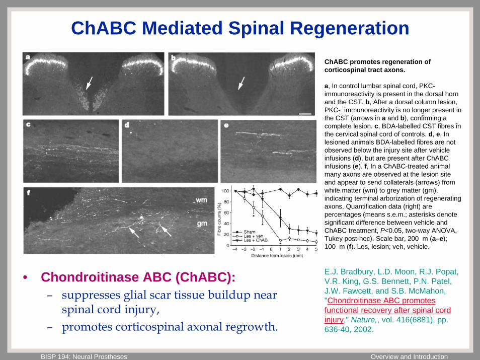
ChABC Mediated Spinal RegenerationChABC promotes regeneration of corticospinal tract axons.
a, In control lumbar spinal cord, PKC-immunoreactivity is present in the dorsal horn and the CST. b, After a dorsal column lesion, PKC- immunoreactivity is no longer present in the CST (arrows in a and b), confirming a complete lesion. c, BDA-labelled CST fibres in the cervical spinal cord of controls. d, e, In lesioned animals BDA-labelled fibres are not observed below the injury site after vehicle infusions (d), but are present after ChABCinfusions (e). f, In a ChABC-treated animal many axons are observed at the lesion site and appear to send collaterals (arrows) from white matter (wm) to grey matter (gm), indicating terminal arborization of regenerating axons. Quantification data (right) are percentages (means s.e.m.; asterisks denote significant difference between vehicle and ChABC treatment, P<0.05, two-way ANOVA, Tukey post-hoc). Scale bar, 200 m (a–e); 100 m (f). Les, lesion; veh, vehicle.
E.J. Bradbury, L.D. Moon, R.J. Popat, V.R. King, G.S. Bennett, P.N. Patel, J.W. Fawcett, and S.B. McMahon, "Chondroitinase ABC promotes functional recovery after spinal cord injury," Nature,, vol. 416(6881), pp. 636-40, 2002.
• Chondroitinase ABC (ChABC):– suppresses glial scar tissue buildup near
spinal cord injury,– promotes corticospinal axonal regrowth.